Impact of Helicobacter pylori infection on liver fibrosis in Egyptian patients with chronic hepatitis C
Bạn đang xem bản rút gọn của tài liệu. Xem và tải ngay bản đầy đủ của tài liệu tại đây (469.62 KB, 7 trang )
Journal of Advanced Research (2012) 3, 287–293
Cairo University
Journal of Advanced Research
ORIGINAL ARTICLE
Impact of Helicobacter pylori infection on liver fibrosis
in Egyptian patients with chronic hepatitis C
Mustafa M. Ragheb a, Mohamed M.E. Awad b, Loaa A. Tag Eldeen
Tamer M. Dosoki b
a
b
c
c,*
,
Infectious & Endemic Diseases Department, Suez Canal Faculty of Medicine, Ismailia, Egypt
Internal Medicine Department, Suez Canal Faculty of Medicine, Ismailia, Egypt
Medical Biochemistry and Molecular Biology Department, Suez Canal Faculty of Medicine, Ismailia, Egypt
Received 19 March 2011; revised 19 September 2011; accepted 25 September 2011
Available online 9 November 2011
KEYWORDS
H. pylori;
Chronic hepatitis C;
Liver fibrosis;
Helicobacter DNA
Abstract Both Helicobacter pylori (HP) and hepatitis C virus (HCV) infections are endemic in
Egypt. This work aimed to investigate the presence of HP in the liver of patients with chronic
hepatitis C (CHC) and explore the relation between HP infection, liver histopathology and HCV viral
load. The study included 60 patients with CHC. Virological, biochemical, liver biopsy and testing
for anti-Hp and anti-schistosomal antibodies in serum were done. Liver tissues were examined for
histopathological and presence of Hp by detection of HP 16S rRNA gene by PCR and sequence
analysis. Anti-schistosomal and anti HP antibody was found in 45% and 61.7%, respectively.
Low stages of fibrosis (F0–F3) were found in 73.3% and advanced fibrosis (F4–F6) in 26.7%.
HP DNA was found in 10% of the liver specimens. Although the frequency HP antibodies was
equally high in patients with advanced and low fibrosis (68.8% and 59.1%, P > 0.05), the HP
DNA in liver tissue was significantly more frequent in patients with advanced fibrosis (31.25%
vs. 2.7%, P = 0.004). Meanwhile, the median viral load of HCV was higher in patients with HP
DNA in liver tissue compared to patients with no HP DNA in liver tissue (337.000 vs. 165.000,
* Corresponding author. Tel.: +20 224097184; fax: +20 226960650.
E-mail address: (L.A. Tag Eldeen).
2090-1232 ª 2011 Cairo University. Production and hosting by
Elsevier B.V. All rights reserved.
Peer review under responsibility of Cairo University.
doi:10.1016/j.jare.2011.09.004
Production and hosting by Elsevier
288
M.M. Ragheb et al.
P = 0.3491). HCV RNA titer, fibrosis score and history of blood transfusion, are independent factors associated with HP DNA in liver tissue. In conclusion, the presence of HP in liver tissue of
patients with advanced fibrosis suggests a potential relation between HP infection and progression
of liver fibrosis due to HCV.
ª 2011 Cairo University. Production and hosting by Elsevier B.V. All rights reserved.
Introduction
Patients and methods
Hepatitis C virus (HCV) is the major agent in non-A non-B
hepatitis with serious complications ranging from chronic
inflammatory disease to hepatic cirrhosis and end-stage liver
failure or hepatocellular carcinoma (HCC) [1]. Egypt has high
prevalence of hepatitis C, resulting in high morbidity and mortality from liver disease. Approximately 12% of blood donors
are seropositive for HCV antibodies [2]. In a recent community-based study, El-Zanaty and Way, reported positive
HCV RNA in sera of 9.8% of 1126 representative Egyptian
citizens [2].
The course of HCV related hepatic disease varies markedly
from one patient to another. Several factors including age at
exposure, duration of infection, alcohol intake, male gender,
viral immune response and steatosis have been shown to be
associated with fibrosis progression [3].
However, even in the absence of these factors, disease progression may be observed in some patients, suggesting the role
of other factors. Host genetic factors or environmental factors,
such as a bacterial co-infection, could be involved [4]. It has
been observed that Helicobacter species were associated with
the pathogenesis of human enterohepatic diseases [5] The discovery of the presence of Helicobacter species DNA in liver
material from patients with liver disease has led to the challenging hypothesis that these bacteria may play a role in the
evolution of hepatic lesions from chronic viral hepatitis to cirrhosis and HCC. Determinants of this evolution are not yet
fully understood, including those occurring in HCV positive
patients [6].
Meyer-ter-Vehn et al. documented that several Helicobacter
spp. could secrete a liver specific toxin that causes hepatocyte
necrosis in cell culture, and might therefore also be involved in
damaging liver parenchyma in vivo [7].
Concerning HCV liver diseases, HP and H. pullorum DNA
have been detected in the liver tissue of patients with chronic
hepatitis C (CHC) and HCC, suggesting that these bacteria
could be implicated in the progression of CHC to cirrhosis
and HCC [8].
Infection with HP is common in Egypt and acquisition of
infection occurs at a very young age [9]. A study carried on
Egyptian patients found that HP antibodies were found in
55.6% of HCV-infected patients vs. 39.4% of the healthy controls. Moreover, the prevalence of HP infection was increased
significantly from chronic active hepatitis to cirrhosis [10].
The association between HP infection and severity of
chronic liver diseases in patients with hepatitis C virus has been
documented in different parts of the world. However, no conclusive data is available in Egypt till now. These observations
promoted us to seek out the possible occurrence and association of HP DNA with the pathological stages in liver among
CHC Egyptian patients.
This cross sectional descriptive study included 60 patients with
CHC, referred to the liver unit of Suez Canal University
Hospital to have a percutaneous liver biopsy, to evaluate suitability for antiviral therapy with pegylated interferon/ribavirin. Their ages ranged from 26 to 58 years.
Diagnosis of CHC was based on positivity to anti-HCV
antibodies, HCV RNA, either elevated or fluctuating ALT
for more than 6 months, and/or bright liver by abdominal
ultrasonography. The study excluded patients co-infected with
HBV or HIV and patients with clinical or ultrasonographic
evidence of cirrhosis.
Sera were collected from each individual and stored
immediately at À20 °C until use. Liver function tests,
alfa-fetoprotein (AFP), and anti-schistosomal antibodies were
measured using commercially available indirect haemagglutination assays (IHA) kits. The HCV RNA viral load was
quantified using Real Time PCR technique in an ABI
PRISMÒ 7000 thermocycler (Applied Biosystems, Foster
City, CA). The serological and biochemical tests were done
in clinical pathology department and the molecular analysis
was performed at oncology diagnostic unit of Suez Canal
University Hospital. The study was approved by the Research
Ethics Committee of the Faculty of Medicine and informed
consents were obtained from each participant.
Processing of liver tissues
Liver tissues were cut into two parts: one was formalin fixed
and paraffin embedded for histo-pathological examination
and the second was immediately stored at À20 °C until further molecular analysis. Histo-pathological examination was
performed by faculty staff of pathology. Hepatic fibrosis
staging was made according to Ishak scoring system [11].
Accordingly patients were divided a group of low fibrosis
including F0–F3 and a group of advanced fibrosis including
F4, F5 (incomplete cirrhosis) and F6 (complete cirrhosis).
According to the histological activity index patients were divided into a group of minimal to mild activity (grades from 1
to 8) and a group of moderate to severe activity (grades from
9 to18) [12].
Detection of anti-HP antibody
Plasma samples were tested for anti-HP IgG antibody using a
commercial test kit, AccuBindä ELISA Microwells (Monobind Inc., Lake Forest, USA) according to the manufacturer’s
instruction. Results were considered positive when higher than
20 U/ml.
Detection of Helicobacter DNA from liver biopsy.
H. pylori and HCV Liver fibrosis
DNA extraction
Genomic DNA was extracted from liver biopsy using WizardÒSV Genomic DNA Purification System (Promega Corporation, Madison, USA). DNA quantitation was performed
using the NanoDropÒ (ND)-1000 Spectrophotometer (NanoDrop Technologies Inc., Washington, USA). The extracted
DNA was stored in À20 °C until used.
PCR amplification
Nested PCR was performed with Helicobacter genus-specific
16S ribosomal RNA gene (16S rDNA) primers (Helinest-S &
R, Heli-S & R) which reported to amplify 26 species of Helicobacter genus [13].
First amplification
The amplification was carried out in a final volume of 50 ll
reaction mixture containing: 1 lg DNA, 25 ll DreamTaqäGreen PCR Master Mix (Fermentas, CA, USA), 50pM
Heli-nest-S primer: 50 -ATTAGTGGCGCACGGGTGAGTA
A-30 , 50pM Heli-nest-R primer: 50 -TTTAGCATCCCGACTT
AAGGC-30 .
The reaction mixture was initially denaturated at 94 °C for
2 min, then amplified for 35 cycles as follow: Denaturation at
94 °C for 30 s, annealing for 30 s at 55 °C, extension at 72 °C
for 11/2 min and final extension at 72 °C for 5 min in Robocycler Gradiant 96 Thermo cycler (STRATAGENÒ, LA, USA).
289
Table 1 Demographic and laboratory data of the studied
population (n = 60).
Age mean age ± SD (range)
Gender (male/female)
PLT mean ± SD (range)
ALT mean ± SD (range)
AST mean ± SD (range)
Total bilirubin mean ± SD (range)
direct bilirubin mean ± SD (range)
HCV PCR median (range)
AFP median (range)
Anti-Bilharzial Ab no (%)
Anti-HP Ab no (%)
42.98 ± 7.6 (26–58)
45/15
207 ± 175.48 (105–1480)
59.60 ± 36.922 (17–187)
51.64 ± 24.820 (18–159)
0.72 ± 0.26 (0.3–1.8)
0.27 ± 0.15 (0-.8)
165000 (327–7520,000)
2.3 (0.1–209)
27 (45%)
37 (61.7%)
Grade of chronic hepatitis C activity no (%)
Minimal to mild
49 (81.7%)
Moderate to severe
11 (18.3%).
Stage of fibrosis no (%)
Low stage
Advanced stage
44 (73.3%)
16 (26.7%).
Second amplification
5ll of the first amplification product, 25 ll DreamTaqäGreen
PCR Master Mix (Fermentas, CA, USA), 50pM Heli-S
primer: 50 -GAACCTTACCTAGGCTTGACATTG-30 , 50pM
Heli-R primer 50 -GGTGAGTACAAGACCCGGGAA-30 was
amplified by following the same PCR condition as first amplification step.
The amplified products were visualized on 2% ethidium
bromide stained agarose gel electrophoresis, the expected size
of product from second amplification step was approximately
480 bp.
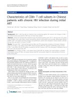
Fig. 1 Amplification of a 480-bp 16S rRNA DNA of Helicobacter. Lane M: molecular size marker (100–1000 bp); lane 2, 4, 5:
positive samples; lane 6: negative control (double-distilled water).
DNA sequencing
PCR products were sequenced as described [13]. The sequencing results were aligned and compared with known Helicobacter species using Basic Local Alignment Search tool (BLAST;
National Center for Biotechnology Information).
H. pylori DNA +ve
H. pylori DNA -ve
60
48
50
Results
40
Demographic and laboratory data of 60 patients included in
this study were summarized in Table 1. Their mean age was
42.98 ± 7.6 and 56.7% of patients were between 41 and
50 years. The majority of patients were males (45/60).
Anti-HP antibody was present in 61.7% of patients, being
equally high among male and females (62.3% and 60%,
respectively). Helicobacter DNA was present in liver tissue of
6 out of 60 (10%) of studied patients, using Helicobacter genus
specific 16S rRNA gene primers, Fig. 1. The PCR products
30
20
10
3
5
2
1
1
0
Non cirrhotic fibrosis
(F0-F4) (n=51)
Fig. 2
Incomplete cirrhosis Complete cirrhosis (F6)
(F5)
(n=7)
(n=2)
HP DNA in liver tissues according to severity of fibrosis.
290
50
45
40
35
30
25
20
15
10
5
0
M.M. Ragheb et al.
Table 3 Distribution of HP DNA in liver tissues according to
stage of fibrosis.
43
26
18
11
11
5
Stage of fibrosis
HP DNA +ve (no = 6)
Complete cirrhosis (F6)
Incomplete cirrhosis (F5)
Chronic hepatitis without cirrhosis
(F0–F4)
1 (50%)
2 (28%)
3 (6%)
5
1
Serum HP Ab
positive
Serum HP Ab
negative
Stage of fibrosis Low (n=44)
Liver HP DNA
positive
Liver HP DNA
negative
Stage of fibrosis Advanced (n=16)
Fig. 3 HP Sero-reactivity and HP DNA in liver tissue of 60
patients in relation to fibrosis staging.
and high HCV RNA viral titer (OR = 1.0, 95% C.I. = 1.0–
1.0, P = 0.044) Table 7.
Anti-schistosomal antibodies were found in 45% of
patients, 66.7% of them had anti-HP antibodies and only
3.7% had HP DNA in liver tissues Table 8.
Discussion
were sequenced and HP like organisms was identified. All HP
DNA positive cases were anti-HP antibodies positive. Most of
patients had low fibrosis (44 of 60, 73.7%) and minimal to mild
activity of necroinflammation (49/60, 81.9%). Cirrhosis was
found in nine patients; incomplete cirrhosis (F5) in seven
and complete cirrhosis (F6) in two Table 3.
Although anti-HP was equally high in patients with low
and advanced fibrosis with no statistically significant difference
(59.1% and 68.8%, P = 0.496) Fig. 3, HP DNA in liver tissue
was significantly more frequent in advanced fibrosis (5/16,
31.25%) compared to low fibrosis (1/44, 2.27%) (P = 0.004),
Tables 4 and 5 and Fig. 2.
Patients with HP DNA in liver tissue showed higher median
value of HCV RNA compared to patients with no HP DNA
(P = 0.3491), Table 2. Meanwhile the median viral load was
higher in patients with moderate to severe activity and patients
with advanced fibrosis (286,000 and 192,500, respectively) compared to patients with minimal to mild activity and patients with
low fibrosis (108,000 and 135,000, respectively), Table 6.
Although of no significance, higher values of ALT, AST and
AFP were found in patients with HP DNA in liver tissue compared to the other group, Table 2. Interestingly, independent
factors associated with positive HP DNA (as dependent factor)
in liver tissues included history of blood transfusion
(OR = 100.5, 95% C.I. = 1.6–6176.8, P = 0.028), advanced
fibrosis score (OR = 4.19, 95% C.I. = 1.4–12.52, P = 0.01)
Table 2
Previous studies showed that DNA from HP – and
H. pullorum-like organisms were present in the liver of cirrhotic patients with or without HCC due to HCV, suggesting
that Helicobacter species could be a co-morbid factor for disease progression [6]. In this study, we demonstrated the presence, Helicobacter DNA using genus-specific 16S rRNA gene
primers in liver tissue of 10% of the studied patients.
Interestingly, the gene sequence obtained from positive
Helicobacter species specific 16S rRNA PCR was analogous
to HP and not similar to H. hepaticus, found in mouse liver tumors [14], or to species previously found in the biliary tract of
humans, such as H. pullorum, H. bilis, and H. Rappini [15].
This finding encourages the speculation that the presence of
Helicobacter DNA in human liver tissue might reflect the
transport of HP of gastric origin or its DNA to the liver [13]
and that intestinal Helicobacter might be implicated in hepatobiliary disease [16].
In this study, although anti HP was equally high in patients
with low and advanced fibrosis with no significant difference,
HP DNA in liver tissue was significantly associated with HCV
related advanced hepatic fibrosis. It was present in 33.3% of patients with cirrhosis and 5.9% with no cirrhosis. This finding is
comparable to 41.6% and 17% as reported by Caste´ra et al. [4]
and 68% and 3.5% as reported by Rocha et al. [6] respectively in
cirrhotic and non cirrhotic patients. Other similar studies have
reported the association between HP DNA in liver tissue and
Comparison of laboratory data regarding presence of HP DNA in liver tissue.
Laboratory data
HCV RNA titre
Platelets
ALT
AST
Albumin
PT
T. Bilirubin
D. Bilirubin
AFP
Statistically significant (P < 0.05).
a
Kruskal-Wallis test.
HP DNA (in liver tissue)
P-value
Positive (no = 6)
Median (range)
Negative (no = 54)
Median (range)
337,000 (51,200–7520,000)
168,500 (11,000–236,000)
57 (17–143)
58 (22–90)
4.6 (4.1–5.1)
12.7 (12.1–15.2)
0.75 (0.5–1.8)
0.25 (0.1–0.4)
3.5 (0.5–32.9)
165,000 (327–7,060,000)
170,000 (105,000–336,000)
47 (22–187)
42 (18–159)
4.3 (3–5.1)
12.8 (11.2–15.5)
0.7 (0.3–1.3)
0.28 (0.02–0.8)
2.2 (0.1–33.7)
0.3491a
0.9803a
0.5962a
0.4159a
0.0979a
0.9409a
0.3974a
0.9388a
0.7115a
H. pylori and HCV Liver fibrosis
Table 4
HP sero-reactivity and HP DNA in liver tissue of patients with low and advanced stage of fibrosis.
HP infection
HP
HP
HP
HP
a
b
c
291
Stage of fibrosis
Ab sero-positive
Ab sero-negative
DNA positive in liver
DNA negative in liver
P-value
Low (no = 44)
No (%)
Advanced (no = 16)
No (%)
26 (59.1)
18 (40.9)
1 (2.3)
43 (97.7)
11
5
5
11
(0.496)c
(68.8)
(31.2)
(31.2)
(68.8)
(0.004)ab
Statistically significant (P < 0.05).
Fisher’s exact test.
Chi-square test.
Table 5 Relation between the grades of chronic hepatitis C
and HP DNA in liver tissue.
HP DNA
Positive
Negative
HAI scoring grade
P-value
Minimal/mild (49)
Moderate/severe (11)
No (%)
No (%)
3 (6.1%)
46 (93.9%)
3 (27.3)
8 (72.7)
0.068a
Statistically significant (P < 0.05).
a
Fisher’s exact test.
cirrhosis in patients with chronic liver disease related to HCV
[10–19]. This association might be explained by increased colonization of HP in the liver of patients with chronic hepatitis C
and advanced fibrosis. Otherwise, infection of the liver with
HP acts as a co-factor in promoting fibrogenesis [6] particularly
when the HCV RNA load is high. This hypothesis agreed with
that of Fagoonee et al. who supposed that co-infection
Table 6
with HP or Helicobacter species might amplify the chronic
inflammation of liver parenchyma, thereby leading to cirrhosis
and HCC [20].
Chronic hepatitis is an inflammatory disease, characterized
by increased levels of pro-inflammatory cytokines such as
interleukins 1, 6 (IL-1, IL-6), tumor necrosis factor (TNF)
and by the presence of lympho-mono cellular infiltrate and
lymphoid follicle formation [21]. Viruses such as HCV are only
capable of limited inflammation, due to shedding of IL-1
receptor in circulation, thereby limiting the possibility of
IL-1 binding to cellular receptors [22]. Helicobacters, on the
other hand, are strong inducers of the inflammatory cascade
[23] infection with them could lead to the accumulation of
extraordinary number of lymphocytes and polymorphonuclear
cells in the infected tissue [24].
It is worth noting that the lower prevalence of HP DNA in
liver specimens of cirrhotic patients in this study compared to
Rocha et al. [6] is possibly due to the difference in the severity
of liver disease in both studies. The cohort of Rocha and colleges included patients with chronic hepatitis and cirrhosis
HCV RNA viral load in the studied patients according to the stages of fibrosis and grades of chronic hepatitis C.
Min to mild activity (no = 49)
Mod to severe activity (no = 11)
Low fibrosis (no = 44)
Advanced fibrosis (no = 16)
HCV RNA range
Median
P-value
327–7520,000
51,200–2320,634
560–7520,000
327–2320,436
108,000
286,000
135,500
192,500
0.1906a
0.5418a
Statistically significant (P < 0.05).
a
Kruskal-Wallis test.
Table 7 Multiple logistic regression analysis for independent factors associated with detection of HP DNA in liver tissues of 60
patients with chronic hepatitis C.
Beta
Constant
Blood transfusion (reference: no)
HCV RNR titre (reference: low viral load < 400,000)
Stages of fibrosis (reference: low fibrosis)
0.237
4.61
0.006
1.434
Standard error
3.295
2.101
0.003
0.558
P-value
0.943
0.028a
0.044a
0.010a
Odds ratio
1.268
100.5
1.000
4.194
95.0% C.I. for odds ratio
Lower
Upper
1.6
1.000
1.404
6176.8
1.000
12.528
Dependent variable: (HP DNA +ve = 1, HP DNA Àve = 0).
Excluded variables: Gender, residence, surgery, dental extraction, smoking, DM, HTN, schistosomal titre, platelet count, ALT, prothrombin
time, T. Bilirubin, D. Bilirubin, AFP. HP antibody, HP PCR, degree of cirrhosis.
a
Statistically significant (P < 0.05).
292
Table 8
M.M. Ragheb et al.
HCV RNA viral load and stage of fibrosis in patients positive and negative for anti-schistosomal antibody.
Anti-schistosomal Ab
P value
Positive (no = 27)
Negative (no = 33)
HCV RNA (IU/ml)
Range
Median
377–7060,000
225,000
4320–7520,000
138,000
0.73a
Stage of fibrosis (F0–F6)
Range
Median
HP DNA positive no (%)
HP DNA negative no (%)
HP Ab positive no (%)
HP Ab negative no (%)
F0–F5
3
1 (3.7)
26 (96.3)
18 (66.7)
9 (33.3)
F1–F6
2
5 (15.2)
28 (84.8)
14 (42.4)
19 (57.6)
0.78a
0.15b
0.47c
Statistically significant (P < 0.05).
a
Kruskal-Wallis test.
b
Fisher’s exact test.
c
Chi-square test.
with and without hepatocellular carcinoma. In this study, all
the studied patients were diagnosed clinically as chronic hepatitis and only 9 of them had cirrhosis (incomplete in 7 and
complete in 2). In all there were no stigmata of portal hypertension, or decompensation.
The presence of HP in liver tissue could occur via a retrograde route from the duodenum or through the portal circulation. Rocha et al. suggested that the presence of Helicobacter
could be the consequence of structural changes in the liver
namely, intrahepatic shunts; when cirrhosis occurs [6]. However, this does not explain the existence of HP in liver specimens
in 3 of 51 patients with non cirrhotic fibrosis and representing
50% of all patients with HP in liver tissue. In this setting, a retrograde route, from the duodenum to the liver might be the
underlying mechanism for HP to colonize in liver tissue. It is
worth noting that all patients with HP in liver were seropositive
for anti-HP antibodies and patients negative for HP DNA in
liver tissue were also negative to anti-HP antibodies. This result
is similar to that reported by Petrenkiene¨ et al. [25].
In this study, patients with HP DNA in liver tissue showed
higher median value of HCV RNA compared to patients with
no HP DNA. Meanwhile the median viral load was higher in
patients with moderate to severe activity and patients with advanced fibrosis compared to patients with minimal to mild
activity and patients with low fibrosis.
Although the explanation of these findings is difficult, the
association of HP DNA in liver tissue with high serum HCV
RNA load could play a synergistic role in enhancing cytotoxic
immune response and promoting fibrosis in patients with
CHC. This hypothesis is opposed by the absence of association
between viral load and disease severity or progression in patients with chronic liver disease related to HCV in studies targeting the natural history of HCV infection [26–29].
It is worth noting that, history of blood transfusion, high
HCV RNA viral load and advanced stage of fibrosis were significantly associated as independent risk factors with presence
of HP DNA in liver tissues (P = 0.028, P = 0.044, P = 0.01,
respectively). Up to our knowledge, no data concerning these
factors and presence of HP DNA in liver tissues are available
in the literature.
This study revealed a higher median stage of fibrosis and
HCV viral load in patients positive for anti- schistosomal antibody compared to negative patients. However, the difference
was statistically not significant. Although non significant, anti
HP antibodies was more frequent in patients positive to antischistosomal Ab compared to negative patients (P = 0.47).
This results are consistent with the results of El-Masry et al.
study in which, In HCV-infected patients, the concurrent schistosoma infection was documented largely in anti-HP-positive
patients [10]. On the other hand, in the anti- schistosomal antibody positive group only 1/27 patient was positive for HP
DNA in liver tissue compared to 5/33 of the other group.
The higher viremia and stage of fibrosis found in patients positive to anti-schistosomal antibody was associated with a low
detection of HP DNA in liver tissue. Although the explanation
is difficult, it is suggested that patients concomitantly infected
with schistosomiasis and HCV may had an intense inflammatory reaction leading to less colonization of HP in liver tissue
[30].
Limitation of this study is the small number of the study
subjects and the inability to obtain normal liver tissue to examine for the presence of HP DNA. Therefore, further study on a
larger sample size to validate the impact of infection with HP
on the outcomes chronic hepatitis C and examine the possibility of finding HP DNA in normal liver tissues. Also to study
the molecular similarity between hepatic and gastric HP in
specimens from the gastric mucosa.
Acknowledgments
We acknowledged all members and staff of Oncology Diagnostic Unit, Suez Canal Faculty of Medicine, Ismailia, Egypt.
References
[1] Butel JS. Virology. In: Brooks GF, Carroll KC, Butel JS, Mores
SA, editors. Medical microbiology. New York: Mc Graw Hill;
2007. p. 367–620.
[2] El-Zanaty F, Way A. Egypt demographic and health survey 2008.
< />[accessed 06.01.11].
[3] Caste´ra L, Hezode C, Roudot-Thoraval F, Bastie A, Zafrani ES,
Pawlotsky JM, et al. Worsening of steatosis is an independent
factor of fibrosis progression in untreated patients with chronic
hepatitis C and paired liver biopsies. Gut 2003;52:288–92.
H. pylori and HCV Liver fibrosis
[4] Caste´ra L, Pedeboscq A, Rocha M, Le Bail B, Asencio C, de
Ledinghen V, et al. Relationship between the severity of
hepatitis C virus related liver disease and the presence of
Helicobacter species in the liver: a prospective study. World J
Gastroenterol 2006;12(45):7278–84.
[5] de Magalhaes Queiroz DM, Santos A. Isolation of a
Helicobacter strain from the human liver. Gastroenterology
2001;121:1023–4.
[6] Rocha M, Avenaud P, Menard A, Le Bail B, Balabaud C,
Bioulac-Sage P, et al. Association of Helicobacter species with
hepatitis C cirrhosis with or without hepatocellular carcinoma.
Gut 2005;54:396–401.
[7] Meyer-ter-Vehn T, Covacci A, Kist M, Pahl HL. Helicobacter
pylori activates mitogen-activated protein kinase cascades and
induces expression of the proto-oncogenes c-fos and c-jun. J Biol
Chem 2000;275(21):16064–72.
[8] Ponzetto A, Pellicano R, Leone N, Cutufia MA, Turrini F,
Grigioni WF, et al. Helicobacter infection and cirrhosis in
hepatitis C virus carriage: is it an innocent bystander or a
troublemaker? Med Hypotheses 2000;54:275–27711.
[9] Mohammad MA, Hussein L, Coward A, Jackson SJ. Prevalence
of Helicobacter pylori infection among Egyptian children:
impact of social background and effect on growth. Public
Health Nutr 2008;11(3):230–6.
[10] El-Masry S, El-Shahat M, Badra G, Aboel-Nour MF, Lotfy M.
Helicobacter pylori and Hepatitis C virus coinfection in Egyptian
patients. J Global Infect Dis 2010;2:4–9.
[11] Ishak K, Baptista A, Bianchi L, Callea F, De Groote J, Gudat
F, et al. Histological grading and staging of chronic hepatitis. J
Hepatol 1995;22:696–9.
[12] Knodell RG, Ishak KG, Black WC, Chen TS, Craig R,
Kaplowitz N, et al. Formulation and application of a
numerical scoring system for assessing histological activity in
asymptomatic
chronic
active
hepatitis.
Hepatology
1981;1:431–5.
[13] Pellicano R, Mazzaferro V, Grigioni WF, Cutufia MA,
Fagoonee S, Silengo L, et al. Helicobacter species sequences in
liver samples from patients with and without hepatocellular
carcinoma. World J Gastroenterol 2004;10(4):598–601.
[14] Fox JG, Li X, Yan L, Cahill RJ, Hurley R, Lewis R, et al.
Chronic proliferative hepatitis in A/Jcr mice associated with
persistent Helicobacter hepaticus infection; a model of
helicobacter-induced
carcinogenesis.
Infect
Immun
1996;64:1548–58.
[15] Fox JG, Li X, Yan L, Cahill RJ, Hurley R, Lewis R, et al. Hepatic
Helicobacter species identified in bile and gallbladder tissue from
Chileans with chronic cholecystitis. Gastroenterology
1998;14:755–63.
[16] Nilsson HO, Taneera J, Castedal M, Glatz E, Olsson R,
Wadstro MT. Identification of Helicobacter pylori, other
Helicobacter species by PCR, hybridization, and partial DNA
293
[17]
[18]
[19]
[20]
[21]
[22]
[23]
[24]
[25]
[26]
[27]
[28]
[29]
[30]
sequencing in human liver samples from patients with primary
sclerosing cholangitis or primary biliary cirrhosis. J Clin
Microbiol 2000;38:1072–6.
Omar MM, EL-Ansary M, Mostafa I, Akl M, El-Sherbini E, ElBadrawy N, et al. Helicobacter pylori among Egyptian patients
with chronic liver diseases. A comparative study. Giza, Egypt:
Theodore Bilharz Research Institute; 1997.
Pellicano R, Leone N, Berrutti M, Cutufia MA, Fiorentino M,
Rizzetto M, et al. Helicobacter pylori seroprevalence in hepatitis
C virus positive patients with cirrhosis. J Hepatol
2000;33:648–50.
Pellicano R, Me´nard A, Rizzetto M, Me´graud F. Helicobacter
species and liver diseases: association or causation? Lancet
Infect Dis 2008;8:254–60.
Fagoonee S, Pellicano R, Rizzetto M, Ponzetto A. The journey
from hepatitis to hepatocellular carcinoma: bridging role of
Helicobacter species. Panminerva Med 2001;43(4):279–82.
Balkwill F, Mantovani A. Inflammation and cancer: back to
Virchow? Lancet 2001;357:539–45.
Fagoonee S, Pellicano R, Rizzetto M, Ponzetto A. The journey
from hepatitis to hepatocellular carcinoma: bridging role of
Helicobacter species. Panminerva Med 2001;43:279–82.
Crabtree J. Cytokine responses to Helicobacter pylori-induced
infection. In: Riecken EO, Zeitz M, Stallmach A, Heise W,
editors. Malignancy and chronic inflammation in the gastrointestinal tract: new concepts. Lancaster: Kluwer Academic
Publishers; 1995. p. 25–36.
El-Omar EM, Carrington M, Chow WH, McColl KE, Bream
JH, Young HA, et al. Interleukin-1 polymorphisms associated
with
increased
risk
of
gastric
cancer.
Nature
2000;404:398–402.
Petrenkiene¨ V, Vitkauskiene¨ A, Jonaitis L, Kupe`inskas L,
Wadstro¨m T. Detection of Helicobacter spp. in liver biopsy
specimens. Acta Medica Lituanica 2004;11(3):31–5.
Bowden DS, Berzsenyi MD. Chronic hepatitis C virus infection:
genotyping and its clinical role. Future Microbiol
2006;1:103–12.
Webster G, Barnes E, Brown D, Dusheiko G. HCV genotypesrole in pathogenesis of disease and response to therapy.
Baillieres Best Pract Res Clin Gastroenterol 2000;14:229–40.
Murray KF, Finn LS, Taylor SL, Seidel KD, Larson AM. Liver
histology and alanine aminotransferase levels in children and
adults with chronic hepatitis C infection. J Pediatr GastroEnterol Nutr 2005;41:634–8.
Freeman AJ, Law MG, Kaldor JM, Dore GJ. Predicting
progression to cirrhosis in chronic hepatitis C virus infection. J
Viral Hepat 2003;10:285–93.
Kamal SM, Rasenack JW, Bianchi L, Al Tawil A, El Sayed
Khalifa K, Peter T, et al. Acute hepatitis C without and with
schistosomiasis: correlation with hepatitis C-specific CD4(+) Tcell and cytokine response. Gastroenterology 2001;121:646–56.