Evaluating the trends of bloodstream infections by nonfermenting gram negative bacilli among the patients in a tertiary care hospital of western part of India and its antibiogram
Bạn đang xem bản rút gọn của tài liệu. Xem và tải ngay bản đầy đủ của tài liệu tại đây (683.41 KB, 14 trang )
Int.J.Curr.Microbiol.App.Sci (2019) 8(1): 1149-1162
International Journal of Current Microbiology and Applied Sciences
ISSN: 2319-7706 Volume 8 Number 01 (2019)
Journal homepage:
Original Research Article
/>
Evaluating the Trends of Bloodstream Infections by Nonfermenting Gram
Negative Bacilli among the Patients in a Tertiary Care Hospital of
Western Part of India and its Antibiogram
Nabamita Chaudhury1, Retina Paul2, R.N. Misra3, Sankha Subhra Chaudhuri4*,
Shazad Mirza3 and Sukanta Sen5
1
Department of Microbiology, Burdwan Medical College and Hospital,
Purba Bardhaman, West Bengal, India
2
Department of Microbiology, College of Medicine and JNM Hospital, Nadia, West Bengal, India
3
Department of Microbiology, Dr. D.Y. Patil Medical College, Hospital and Research Centre,
Pune, Maharashtra, India
4
Department of Ophthalmology, Burdwan Medical College and Hospital, Purba Bardhaman,
West Bengal, India
5
Department of Pharmacology, ICARE Institute of Medical Sciences and Research,
Banbishnupur, Purba Medinipur, Haldia, West Bengal, India
*Corresponding author
ABSTRACT
Keywords
Gram-Negative
Non-Fermenting
Bacilli (NFGNB),
Blood Stream
Infections (BSIs),
Multi-drug
resistance
Article Info
Accepted:
10 December 2018
Available Online:
10 January 2019
Non-fermenting gram-negative bacilli (NFGNB) are an emerging problem in Blood stream
infections. A major concern is multi-drug resistance which severely limits treatment options.
Earlier it was believed to be non pathogenic, but recently they are more frequently isolated as
primary pathogen. Usually they cause hospital acquired infection (HAI). A prospective study
was conducted to isolate the NFGNB from blood samples, to identify the risk factors leading to
blood stream infections and to determine the antibiotic susceptibility pattern of them. The study
was conducted in a tertiary care hospital, over a period of 2 years. Identification of NFGNB
was done by biochemical tests and by VITEK 2. Antibiotic susceptibility was determined by
disc diffusion method. Extended-spectrum β-lactamases (ESBLs) and metallo-β-lactamases
(MBLs) production were detected by the combined disc diffusion test. Out of 2021 blood
samples, blood culture positive was in 32.7% of patients of whom the cause was NFGNB.
Acinetobacter boumannii was the most common organism, 27.69% followed by
Strenotrophomonas maltophilia, next to it was Pseudomonas aeruginosa Acinetobacter
lwoffiietc. The most common risk factors for colonization BSIs with NFGNB was comorbid
conditions, such as diabetes mellitus, cardiovascular diseases, hypertension, tuberculosis and
chronic renal disease patients on haemodialysis. In general, the isolates of NFGNB revealed
pretty much good sensitivity to carbapenem (imipenem, ertepenam), colistin and
aminoglycosides (amikacin, gentamicin), where as cephalosporin group revealed a low
susceptibility rate. ESBL and MBL producer NFGNB were identified and the isolation rate is
very alarming. The trend of increasing numbers of cases of NFGNB in Blood stream infections
compounded by MDR is of great concern. It is necessary to administer antibiotics judiciously,
strengthen surveillance and laboratory services in intensive care units, and re-evaluate treatment
guidelines for management of infection by these organisms.
1149
Int.J.Curr.Microbiol.App.Sci (2019) 8(1): 1149-1162
Introduction
The non-fermenting organisms are comprised
of gram negative rod shaped bacilli.1 The non
fermenting gram negative bacilli (NFGNB)
are taxonomically group of aerobic non spore
forming bacilli that either do not utilize
carbohydrates as the source of energy or
degrade them through metabolic pathways
other than fermentation.2 They are widely
distributed in nature as saprophytes, found in
soil, water, sewage or as commensals on
human skin or in the human gut and some of
them found in hospital environment.1, 3, 4
These nonfermenters are unfortunately the byproduct of medical and surgical advances in
health care system of serious ill patients.5
Recently, these NFGNB are emerging
problem in sepsis, which is associated with
significant mortality and morbidity. A major
concern is multi-drug resistance which
severely limits treatment options.
The predominant species of concern among
NFGNB are Pseudomonas aeruginosa,
Acinetobacter baumannii, Strenotrophomonas
maltophilia and, less so, members of the
Burkholderia cepacia group.3 Except P.
aeruginosa the NFGNB are most often cause
nosocomial
infections
in
immunecompromised patients like urinary tract
infections (UTI), Bloodstream infections
(BSIs), ventilator associated pneumonia
(VAP) and surgical site infections (SSI).1
Bloodstream infections (BSIs) are the
significant causes of morbidity and mortality
for many patients.6 BSIs are defined as the
presence of viable infectious microorganism
in the bloodstream causing clinical illness.7
The term bloodstream infection and
bacteremia are synonymously used, which
generally refer to the significant growth of a
microorganism in a blood culture obtained
from the patient with clinical signs of
infection.8 Bacteremia may range from self-
limiting infections to septicaemia which is life
threatening and needs rational antimicrobial
treatment.9 In the developing countries, like
India lack of standard antimicrobial
guidelines, emergence of antimicrobial
resistance, paucity of good diagnostic
facilities and poor hospital environment, poor
quality of hand hygiene are major
denominators for surge in BSI associated
morbidity and mortality.10
Materials and Methods
This was a prospective study. The study was
conducted in the Microbiology Department of
Dr. D.Y. Patil Medical College, Hospital and
Research Centre, over a period of 2 years (i.e.
July 2012 to September 2014). A total 2021
blood samples from the suspected patients of
sepsis were collected in the adult and
paediatric patients. Bloods were collected
aseptically in brain heart infusion broth (BHI)
or in BACT/ALERT 3D system. In case of
neonates 2 ml blood, children 3-5 ml blood
and for the adults 10 ml blood were taken.
The samples were taken from the suspected
patients, admitted to different wards and
various intensive care units (ICU) of this
hospital. The study was approved by the
Ethical Committee of our institute.
Blood samples were processed for culture by
standard conventional methods. Identification
of Nonfermenters were carried out by Gram
staining (gram negative bacilli/ gram negative
coccobacilli), cell and colony morphology,
pigment production, catalase test, p citrate
test, triple sugar iron (alkaline slant/ no
change butt), oxidase test and by motility test.
Further identification was done by Hugh and
Leifson oxidative-fermentative test (O-F) for
glucose, sucrose, lactose, mannitol; gelatin
liquefaction,
nitrate
reduction
test,
Decarboxylation of arginine, lysin and
ornithine and growth at 35⁰C and at 42⁰C for
18-24 hours on two tubes of trypticase soy
1150
Int.J.Curr.Microbiol.App.Sci (2019) 8(1): 1149-1162
agar (TSA). The final identification and
confirmation was done by the Vitek 2
system.2
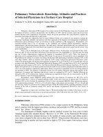
Identification of pigment production by
King’s A and King’s B medium11
King’s A medium11: Pyocyanin, a blue
phenazine derivative characteristic of P.
aeruginosa was diffusible and its production
was enhanced by growth in “King A (Fig.
1)”.11
King’s
B
medium11:
Fluorescent
Pseudomonas
were
characterised
by
production of water soluble pigment, which
diffused freely in the media and fluoresce
brightly under U.V ray. The organisms
produced this pigments were P. aeruginosa,
P. putida, P. fluorescens, P. chlororaphis etc.
and was manifested in low iron containing
media.6 “King B” medium was the universally
use medium for the production of fluorescent
pigment.11
Antibiotic susceptibility testing was
determined by Kirby - Bauer disc diffusion
method2, 12
Muller-Hinton agar media was used.
Commercially available Himedia discs were
used. The strength of the discs used and their
zone size interpretation were carried out by
National Committee for Clinical Laboratory
Studies (NCCLS) guideline. The antibiotics,
which were tested, Piperacillin (10mcg/disc),
Carbenicillin
(100mcg/disc),
Ampicillin
(10mcg/disc),
Cefotaxim
(30mcg/disc),
Ceftriaxone
(30mcg/disc),
Ceftazidime
(30mcg/disc), Cotrimaxazole (25 mcg/disc),
Ciprofloxacin (5 mcg/disc), Norfloxacin (10
mcg/disc)
Gentamicin
(10mcg/disc),
Amikacin
(30mcg/disc),
Imipenem
(10mcg/disc), Chloramphenicol (30 mcg/disc)
Tobramycin
(10mcg/disc),
Ofloxacin
(5mcg/disc), Amoxicillin/Clavulanic acid
(20/10mcg/disc),
Piperacillin/Tazobactam
(100/10mcg/disc), Tigecycline (15mcg/disc),
Colistin (10mcg/disc) and Ertepenem
(10mcg/disc).
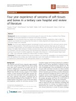
Detection of extended
lactamases production 12, 13
spectrum
β-
The Combine disk diffusion test (CDDT) was
used to determine the prevalence of extended
spectrum β-lactamases (ESBL) production.
Muller-Hinton agar media was used. One
Ceftazidime (CAZ) (30μg) disc was placed on
a lawn culture of test isolates and at the
distance of 15 mm on both side of CAZ disc,
a combination disc of Ceftazidime/
Tazobactam (30/10 μg) and Ceftazidime /
Clavulanic acid (30/10 μg) were placed. A≥ 5
mm increased in a zone diameter for either
antimicrobial agent tested in combination
with Clavulanic acid or Tazobactam versus
the zone diameter of the agent when tested
alone = ESBL producer (Fig. 2).10, 13
Detection
production
of
metallo
β-lactamases
Muller-Hinton agar media was used. One
Imipenem (10μg) disc was placed on a lawn
culture of isolates and at the distance of 15
mm a combination disc of 10μg of Imipenem
and 100μl of EDTA disc was placed. Then it
was incubated at 35⁰C for 18 - 24 hours. An
increase in zone size ≥ 7 mm around the
Imipenem -EDTA disc as compared to
Imipenem disc alone was recorded as positive
(Fig. 3).10, 13
Results and Discussion
In this study, out of 2021blood samples, total
number of culture positive isolates were 661
(32.7 %) among which 445 (67.32%) were
gram positive cocci (GPC) and 216 (32.68%)
were gram negative bacilli (GNB). Out of 216
GNB, 65 (30.1%) were non-fermenting gram
1151
Int.J.Curr.Microbiol.App.Sci (2019) 8(1): 1149-1162
negative bacilli (NFGNB). Out of the total 65
isolates, highest number of isolates (23%)
were obtained from male surgical ward,
followed by Medicine Intensive Care Unit
(MICU) (10.8%) next to it was male medicine
ward (9.6%) (Fig. 4). While discussing about
the gender distribution, in this study male
(69.23%) outnumbered the female (30.77%)
(Fig. 5). In our study the patients were
divided into ten age groups. The majority of
the patient belongs to 41 to 50 years,
accounting for 27%, followed by the age
group of 31 to 40 years comprises 18%, next
to this is the age group of 11 to 20 years
accounting for 9.23% (Fig. 6).
The highest number of isolates were
Acinetobacter boumannii, comprises 27.69%
followed by Strenotrophomonas maltophilia
(previous
designation:
Pseudomonas
maltophilia) 21.53%, next to it was
Pseudomonas
aeruginosa
(13.84%),
Acinetobacter lwoffii (6.15%), Pseudomonas
fluroscence
(4.61%),
Acinetobacter
boumannii
complex
(ABC)
(4.61%),
Burkhelderia cepacia (4.61%) (previous
designation:
Pseudomonas
cepacia),
Sphingomonas
paucimobilis
(3.07%)
(previous
designation:
Pseudomonas
paucimobilis), Pseudomonas stutzeri (3.07%),
Pseudomonas putida (3.07%) and each one
isolates of Acinetobacter radioresistance,
Acinetobacter calcoaceticus, Acinetobacter
haemolyticus, Burkholderia multivorans and
Moraxella oslonensis (Table 1).
In this study we have analyzed the risk factors
for colonization BSIs with NFGNB.
Prolonged
hospitalization,
mechanical
ventilation, indwelling foreign devices
(especially orthopedic implants, in-situcanula), unjudicial antimicrobial therapy and
comorbidities, have identified as risk factors
which are predisposing to acquisition BSIs by
NFGNB. In this study 29.23% isolates were
obtained from the patients who had comorbid
conditions, such as diabetes mellitus,
cardiovascular
diseases,
hypertension,
tuberculosis and chronic renal disease patients
on haemodialysis. Around 24.61% isolates
were obtained from the patients, who were on
indwelling
intravascular
catheters
or
orthopedics implants in situ, followed
by18.46% of isolates from those patients who
have admitted in this hospital for a long
tenure, next to it was 15.38% isolates from
those patients who were on mechanical
ventilators and 12.31% isolates were yield
from the patients who had prolonged history
of hospitalization (Fig. 7).
The isolates of Pseudomonas aeruginosa
revealed 100 % sensitivity to Colistin and also
revealed good susceptibility to Ertepenam
(90.8%) followed by Imipenem (86.77%),
Tobramycin (66.66%) next to it, was
Amikacin (64.02%) (Fig. 8). The isolates of
Acinetobacters showed 60% were sensitive to
Imipenem. In this study we have reported
52.2% susceptibility to chloramphenicol and
48.9% to gentamicin. Close to it, in this study
amikacin and norfloxacin each comprises of
47.8%. In this study Ceftazidime shows a bit
low sensitivity pattern, accounting for 37.8 %
(Fig. 9).
The
isolates
of
Strenotrophomonas
maltophilia showed 100 % sensitivity to
Colistin revealed good susceptibility to
Ertepenam (96.65%), Ofloxacin (94.12%),
Ceftazidime
(94.12%)
followed
by
Ciprofloxacin (88.23%) (Fig. 10). Among the
total 65 isolates of NFGNB, 20 isolates
(30.77%) were multidrug resistance (MDR).
However, amidst these 20 isolates 11 (55%)
were ESBL- producers and rest (45%) were
MBL- producers.
S. maltophilia showed a good sensitivity to
ertepenam (96.65%), ofloxacin (94.12%),
ceftazidime (94.12%) and ciprofloxacin
(88.23%)
1152
Int.J.Curr.Microbiol.App.Sci (2019) 8(1): 1149-1162
Table.1 Distribution of non-fermenting gram negative bacilli in different clinical samples (n=65)
Name of the organism
Pseudomonas aeruginosa
Pseudomonas fluroscence
Pseudomonas putida
Pseudomonas stutzeri
Acinetobacter boumannii
Acinetobacter boumanniicomplex(ABC)
Acinetobacter lwoffii
Acinetobacterradioresistance
Acinetobactercalcoaceticus
Acinetobacter haemolyticus
Burkholderiacepacia
Burkholderiamultivorans
Strenotrophomonasmaltophilia
Sphingomonaspaucimobilis
Moraxella oslonensis
Total
Number of isolates (%)
9 (13.84%)
3 (4.61%)
2(3.07%)
2(3.07%)
18(27.69%)
3(4.61%)
4(6.15%)
1(1.53%)
1(1.53%)
1(1.53%)
3(4.61%)
1(1.53%)
14(21.53%)
2(3.07%)
1(1.53%)
65
Fig.1 Kings B mediumunder U-V ray
Fig.2 ESBL producer
1153
Int.J.Curr.Microbiol.App.Sci (2019) 8(1): 1149-1162
Fig.3 MBL producer
Fig.4 Ward wise distribution of different clinical samples (n=65)
Fig.5 Gender distribution of the patients (n=65)
Female
Male
1154
Int.J.Curr.Microbiol.App.Sci (2019) 8(1): 1149-1162
Fig.6 Age distribution of the patients (n=65)
30.00%
25.00%
20.00%
15.00%
10.00%
5.00%
0.00%
27%
18%
4.61% 4.61% 6.15%
Neonates
11-20yrs
41-50yrs
>71 yrs
9.23%
12.11%
7.68%
6%
Infants
21-30yrs
51-60yrs
4.61%
13 months-10yrs
31-40yrs
61-70yrs
Fig.7 The incidence of infection due to gram negative nonfermenting organisms
29.23%
24.61%
30.00%
20.00%
18.46%
12.31%
15.38%
10.00%
0.00%
Fig.8 Antibiotic susceptibility pattern of Pseudomonas aeruginosa (n=9)
120
100
80
60
40
20
0
Piper
acillin
Cipro Gent
Carb Cefta Tobr
Amik Imipe Piper
Oflox Tigec
Colist Ertep
floxa amici
enicill zidim amyc
+Taz
acin nem acillin
acin ycline
in enam
obact
cin
n
in
e
in
am
RESISTANT 44.44 42.85 22.22 11.11 44.44 55.55 44.44 66.66 77.78 33.33 44.44
0
SENSITIVE 55.55 66.66 77.78 88.89 55.55 44.44 55.55 33.33 22.22 66.66 55.55 100
1155
0
100
Int.J.Curr.Microbiol.App.Sci (2019) 8(1): 1149-1162
Fig.9 Antibiotic susceptibility pattern of Acinetobacter species (n=28)
Fig.10 Antibiotic susceptibility pattern of Strenotrophomonas maltophilia (n=14)
Pipe Ami Gen Imip Ceft Cipr Cefo Oflo Ceft Pipe Tige Tobr Colis Erte
racill kaci tami ene riax oflo taxi xaci azidi racill cycli amy tin pen
in
n cin m one xaci m n me in+T ne cin
am
n
azob
acta
m
SENSITIVE 64.7172.3684.1279.7182.3588.2370.5894.1294.1276.3472.76 77.8 100 96.65
RESISTANT 35.2927.6415.8820.2917.6411.7629.42 5.88 5.88 23.6627.24 22.2
Bloodstream infections by NFGNB remained
a challenge for the clinician and
microbiologists due to the limited facilities in
the laboratories to identify NFGNB, changing
bacterial etiology and emergence of
antimicrobial resistance. Early detection of
NFGNB
and
determination
of
its
antimicrobial susceptibility can reduce the
occurrence of BSI and can also decrease the
rate of emergence of MDR isolates. Our study
evaluates the incidences of bloodstream
0
3.35
infections by NFGNB, risk factors underlying
and antimicrobial susceptibilities among the
paediatric and adult group of patients.
The non-fermenting gram negative bacilli are
found in nature as inhabitants of soil, water
and also the commensals of human and
animal mucous membranes. Recently these
organisms are gaining importance as the
frequently isolated primary pathogen in
patients with prolonged hospitalization.
1156
Int.J.Curr.Microbiol.App.Sci (2019) 8(1): 1149-1162
NFGNB have the ability to adapt well in
hospital environment as they can survive on
dry surfaces, in antiseptic solution and
distilled water for many days. They can easily
have transmitted to human body by sources
like indwelling intravascular catheters, drain
tubes from surgical site, surgical intervention
and from other inanimate objects like bed
rails, bedside tables, ventilators, air
humidifiers and sinks and from these the
NFGNB is transmitted to the patients.
In this study a total of 2021 blood samples
were processed. In this study, overall
incidence of bloodstream infection by
NFGNB was based on significant bacterial
growth in the blood cultures obtained from
suspected
patients
was
732.7%.
Comparatively, in 2013 a study done in
Eastern India had revealed 201 nonfermenters were isolated from 1650 clinical
samples, accounting for an isolation rate from
blood culture is 16.41%. 14Where as another
study in Gujrat by Patel et al., isolated 2397
(23.93%) NFGNB, out of total 20721 various
clinical samples, accounting for isolation rate
of blood culture is 6.96%.15
Infection due to NFGNB can occur at any
age. Bloodstream infections by NFGNB
varied significantly within age groups, where
the highest prevalence was recorded among
patients at the 41 to 50. Similarly, only few
studies suggest a correlation between the
infection due to NFGNB and age. A study,
done in Eastern part of India in 2013 revealed
that majority of the patients (45%) were
adults and above 45 years, which is similar to
this current study.14
The highest number of isolates were
Acinetobacter boumannii, comprises 27.69%.
Acinetobacter boumannii has emerged as an
important
opportunistic
pathogen
in
healthcare systems. As it hard to desiccate, so
difficult to eradicate and has numerous
intrinsic and acquired mechanisms of drug
resistance. Thus this organism possesses a
great threat to the clinician as well as to
microbiologists. These organisms found
extensively in nature and are able to alive in
environment. They can stay alive within
disinfectants and can create problem in health
care
facilities
spreading
by
cross
contamination and causing to blood stream
infections.16
Strenotrophomonas maltophilia was the
second
common
isolates
(21.53%).
Stenotrophomonas maltophilia is water borne
organisms and recently emerged as an
important
opportunistic
pathogen
in
debilitated host. They are enraging as a
known cause of infection in the nosocomial
settings.
The isolates of this emerging pathogen from
blood is quite difficult to interpret as primary
pathogen. However if this isolate yields from
a site which is supposed to be sterile, such as
from blood, drain tip or CVP tip, then this
isolate represents as true or primary pathogen.
Muder et al., report same kind of study where
he was reported a series of 91 patients with
Stenotrophomonas maltophilia bacteraemia,
among them 56% did not reveal any clinically
apparent portal of entry but 84 % of these
individuals had central venous catheter in
place.17 In 2007 Gautam et al., isolated 22
Stenotrophomonas maltophilia. Out of which
13 were from the blood samples of
bacteraemia patients and 9 were from
respiratory isolates.18
In this study, Pseudomonas spp was another
common
organism
causing
BSIs.
Pseudomonas are ubiquitous in nature as
saprophytes. Earlier it is believed to be non
pathogenic. But recently they have emerged
as primary opportunistic pathogens in
hospitalized
patients
as
well
as
immunocompromised
patients
and
1157
Int.J.Curr.Microbiol.App.Sci (2019) 8(1): 1149-1162
responsible for causing variant infections
including BSIs.They are very hard to
desiccate, difficult to eradicate and has
numerous intrinsic and acquired mechanisms
of drug resistance. They can stay alive within
disinfectants and can create problem in health
care
facilities
spreading
by
cross
contamination. The abuse and the unjudicial
practice of antibiotics are responsible for the
burgeoning resistance of commonly used
antibiotics towards Pseudomonas. More over
the multidrug resistance among these
organisms makes the treatment of this
infection difficult and expensive.19
Burkholderia cepacia complex (BCC) found
in many niches of both natural and clinical
environments BCC is emerging as an
important cause of morbidity and mortality in
hospitalized patients because of high intrinsic
antibiotic
resistance,
such
as
aminoglycosides,
chloramphenicol
and
polymyxins. An upsure of septicaemia due to
BCC is documented in various studies.18
In our study from 65 NFGNBs we have
isolated 3 isolates of B.cepacia and one
isolate of Burkholderia multivorans from the
blood taken in BACT/ALERT 3D SYSTEM
bottle. The patients was diagnosed with sepsis
and admitted in the ICU and the central
venous line was in situ. Similarly, in 20062007 Gautam et al., isolated 39 isolates of
BCC from various specimens. Out of these 39
total isolates, 30 isolates of BCC were
obtained from 8601 blood cultures,
accounting for 0.35%.18
In this current study we have yielded 2
isolates of Sphingomonas paucimobilis from
blood samples. These isolates were obtained
from the blood cultures of two young patients
who were admitted in ICU and female
medical ward for a long tenure with the
diagnosis of septicaemia. We have isolated
only one isolates of Moraxella group from the
the central venous tip of a young female,
admitted in ICU with the diagnosis of
septicaemia.
The risk factors associated with this pathogen
are intensive care admission, prolonged
hospitalization, on mechanical ventilation,
presence of central venous catheter, indwelling catheters, orthopaedic implants,
unjudicial use of broad spectrum, antibiotics
and comorbid conditions. These predisposing
factors accelerate the occurrence of the blood
stream infection due to these organisms.
These NFGNB are posing a great threat to
human race as they are resistant to routinely
used antibiotics. The abuse and the unjudicial
practice of antibiotics are responsible for the
burgeoning resistance of commonly used
antibiotics towards NFGNB. The resistance to
antimicrobials is increasing in recent years
and almost resistance to all commonly used
antibiotics. More over the multidrug
resistance among these organisms makes the
treatment of this infection caused by NFGNB
difficult and expensive.
Pseudomonas aeruginosa shows a good
sensitivity to Imipenem (86.77%) which is
almost similar to the study by Patel et al., who
reported 94% sensitivity to this drug.15 A
study by Rit et al., reported that P.aeruginosa
were highly susceptible to Colistin (100%),
Imipenem (91.8%) and Amikacin (69.3%). 14
In my study similarly Colisti (100%),
Imipenem (86.77%) and Amikacin (64.02%)
revealed the same findings. The isolates of P.
aeruginosa were sensitive to and Ciprofloacin
(57.67%), in comparison to this study another
study by Patel et al., revealed a very low
susceptibility rate to Amikacin (39.6%) and
Ciprofloacin (16.53%).15 Here we found a
good sensitivity to Gentamicin (57.14%)
unlike this current study, Rit et al., reported
only
23.76%
of
susceptibility
to
Gentamicin.14In this study 61.37% was
1158
Int.J.Curr.Microbiol.App.Sci (2019) 8(1): 1149-1162
susceptible to Piperacillin, similarly a study
by Juyal et al., revealed 52.13% sensitive to
this drug.20 Ciprofloacin and Ceftazidime both
accounting for 57.67%. Unlikely, a study by
Patel et al., who reported only 24.6%
susceptibility rate to Ceftazidime.15 Where as
Carbenicillin (44.44%) and Ceftazidimetazobactam (17.98%) reveals quite a low
sensitivity to this organism. In comparison to
my study by Juyal et al., revealed 69.15%
sensitivity to Piperacillin-tazobactam.20
Imipenem (88%) show the highest sensitivity
to Pseudomonas fluroscence, similarly a
study by Rit et al., reported 100 % sensitivity
to Imipenem.14 In this current study Amikacin
and Ceftazidime each of them show 66.7%
sensitivity to Pseudomonas fluroscence.
Similar to this study, Rit et al., revealed
66.66% sensitivity to Amikacin. However Rit
et al., revealed a low sensitivity rate to
Gentamicin (33.33%) and Ciprofloxacin
(33.33%)14, where as in this study Gentamicin
and Ciprofloxacin accounting for 71.4% and
61.9% susceptibility. Here Piperacillin
accounting for (71.4%).
Imipenem (89.65%) shows the highest
sensitivity for Pseudomonas putida, almost
similar to this study, a study by Patel et al.,
revealed 100% susceptibility to Imipenem.15
The other isolates of P.putida show a
moderate susceptibility pattern towards
Amikacin (68.96%) and Ciprofloxacin
(62.06%), where as in comparison to them
study by Patel et al., revealed 100%
sensitivity to Amikacine and Cefipime.15
Gentamicin show 58.62%, Ciprofloxacin,
Ceftazidime and Piperacillin an reveal a
susceptibility rate of, 62.1% 55.17% and
51.72% respectively.
Imipenem (95.23%) shows the highest
sensitivity
for
Pseudomonas
stutzeri,
Gentamicin reveals 90.47%. Ceftazidime
shows 85.71%, Ciprofloxacin and Amikacin
reveal 80.95% individually.
The isolates of A.boumannii showed 60%
were sensitive to Imipenem. Almost similar
susceptibility of Imipenem (68.06%) was
reported by Juyal et al.,20 In comparison to
this Rit reported a good sensitivity to
Imipenem (90%).14 Where as another study
by Parimal et al., revealed 72.9% sensitivity
to Imipenem.15 In this study we have reported
52.2% susceptibility to Chloramphenicol and
48.9% to Gentamicin. In contrast to this
study, another study by Rit revealed low
susceptibility to Chloramphenicol (28%) and
Gentamicin (24%).14 Where as Juyal et al.,
reported exactly similar sensitivity to
Gentamicin (48.61%).20 Close to it, in this
study Amikacin and Norfloxacin each
comprises of 47.8%, in comparison to this
study Rit et al., reported 62% susceptibility to
Amikacin.14 In comparison to this Parimal et
al., revealed a low sensitivity to Amikacin
(38.8%).15 However Juyal et al., reported a
good sensitivity to Amikacin, accounting for
73.61%.20 In this study Ceftazidime shows a
bit low sensitivity pattern, accounting for 37.8
%.Similarly a study by Rit K, reported 28 %
sensitivity to Ceftazidime.14
Strenotrophomonas maltophilia revealed
100% sensitivity to Colistin, followed by
Ertepenam 96.65%, next to it was
Ceftazidime and Ofloxacin, both accounting
for
94.12%,
Ciprofloxacin
(88.23%),
Gentamicin (84.12%). Whereas, a study by
Juyal et al., reported only 16.67%
susceptibility to Gentamicin and almost
resistant to Ceftazidime.20However another
study by Rit et al., reported a good
susceptibility to Ceftazidime (66.7%)14 In this
study 70.58% susceptibility rate was for
Cefotaxim. In this current study Amikacin
and Imipenem reveal a good sensitivity,
accounting for72.36% and 79.71% sensitivity
respectively, where as a study done by Juyal
et al., reported almost resistant to Imipenem
and Amikacin (33.33%).20
1159
Int.J.Curr.Microbiol.App.Sci (2019) 8(1): 1149-1162
Resistance to antimicrobials is common and
has increased over the years among NFGNBs.
Multidrug resistance among these organisms
makes the treatment of infections caused by
them, difficult and expensive. A large scale
use of the third- generation Cephalosporins
like
Cefotaxime,
Ceftriaxone,
and
Ceftazidime has led to the evolution of newer
betalactamases such as the ESBLs. ESBLs are
plasmid mediated enzymes that hydrolyze the
oxyimino 𝛽 lactams and the Monobactams
(Aztreonam) but have no effect on the
Cephamycins (Cefoxitin, Cefotetan) and the
Carbapenems (Imipenem).21, 22, 23 Being
plasmid mediated, they can be easily
transferred from one organism to another.23 A
rapid detection of ESBL and MBL positive
isolates is necessary to control infection and
to prevent their dissemination. In this study
we have performed Combined Disc Diffusion
Test (CDDT) to However in this study among
the total 65 isolates of NFGNB, 20 isolates
(30.77%) were multidrug resistance (MDR).
However, amidst these 20 isolates 11 (55%)
were ESBL- producers and rest (45%) were
MBL-producers. Among the total 65 isolates
of NFGNB, 20 isolates (30.77%) were
multidrug resistance (MDR). However,
amidst these 20 isolates 11 (55%) were ESBL
producers and rest (45%) were MBLproducers.
In conclusion a large number of NFGNB are
isolated as primary pathogen, which has a
potential to cause BSIs. The remarkable thing
about all these isolates is that these isolates
obtained from the typical cases of HAI. These
organisms have possibly come from
inanimate objects like ventilator, humidifier,
wash basin and from diluted disinfections.
Most of the patients had high risk factors, like
comorbid conditions (DM, hypertension, post
renal dialysis, cardiac disease, tuberculosis
etc) prolonged hospitalization, indwelling
catheters and orthopaedics implants in situ
and unjudicial use of antibiotics. Although
BSI rates declined over time, but BSI had
high mortality and these NFGNB pathogens
exhibited substantial antimicrobial resistance.
Most effective antibiotics are colistin,
imipenem,
ertepenam,
amikacin
and
gentamicin. There is a wide spread variability
of antibiotic profile in common hospital for
these pathogens. the antibiotic susceptibility
can change from hospital to hospital set up
and there may be a gross geographical
variation.So, it is imperative that every
hospital should monitor a proper antibiogram
profile for these isolates from time to time to
serve as a basic empirical therapy to prevent
the development of MDR cases. Treating
these pathogen should be based on the
laboratory data after identifying the proper
causative agents and antibiotic susceptibility
result. Minimized the use and abuse of
antimicrobial agents, proper surveillance of
antibiotic panel, strict infection control
measures and even simple yet proper hand
washing method and by using disinfections of
inanimate objects, can prevent the emergence
Acinetobacter and can reduce the rate of
MDR strains.
References
1.
Govan
J.R.W.
Pseudomonas,
Strenotrophomonas, Burkholderia. In:
Collee J.G., Fraser A.G., Marmion B.P.,
Simmons A, editors. Mackie &
McCartney.
Practical
Medical
th
Microbiology. 14
ed. Churchill
Livingstone; 1996. 413-23.
2. Winn W Jr, Allen S, Janda W, Koneman E,
Procop G, Schreckenberger P, et al., The
Nonfermentative Gram-Negative Bacilli.
Koneman’s Color Atlas and Textbook of
Diagnostic Microbiology, 2006; 305-91.
3. Steinberg JP, Rio DC, Gram negative and
Gram variable bacilli. Principles and
Practice of Infectious diseases, 2005; 2:
2751-68.
4. Gales AC, Jones RN, Forward KR, Linaes
1160
Int.J.Curr.Microbiol.App.Sci (2019) 8(1): 1149-1162
J, Sader HS, Verhoef J, Emerging
importance of multidrug -resistant
Acinetobacter
species
and
Stenotrophomonas
maltophilia
as
pathogens in seriously ill patients:
genetic
patterns,
Epidemiological
features and trends in the SENTRY
antimicrobial surveillance program.
Clinical Infectious Disease, 2001; 32:
104-13.
5. Gardner P, Griffin W.B, Smart MN, Kunz
L J, Non-fermentative gram negative
bacilli of nosocomial interest. American
Journal of Medicine, 1970; 48: 735-48.
6. Parajuli N.P, Parajuli H, Pandit R, Shakya
J, Khanal P.R, Evaluating the Trends of
Bloodstream Infections among Pediatric
and Adult Patients at a Teaching
Hospital of Kathmandu, Nepal: Role of
Drug Resistant Pathogens. Canadian
Journal of Infectious Diseases and
Medical Microbiology, volume 2017
7. C. Viscoli, “Bloodstream Infections: the
peak of the iceberg,” Virulence, vol. 7,
no. 3, pp. 248–251, 2016.
8. K.B. Laupland, “Incidence of bloodstream
infection: are view of population-based
studies,” Clinical Microbiology and
Infection, vol. 19, no. 6, pp. 492–500,
2013.
9. R. S. Munford and A. F. Su redini, “Sepsis,
severe sepsis, and septic shock,” in
Mandell, Douglas and Bennett’s
Principle and Practice of Infectious
Diseases. Volume 1, G. L. Mandell, J. E.
Ben- nett, and R. Dolin, Eds., pp. 987–
1010, Churchill Livingstone- Elsevier,
2010.
10. N. Obeng-Nkrumah, A.-K. Labi, N. O.
Addison, J. E. M. Labi, and G. AwuahMensah, “Trends in paediatric and adult
blood- stream infections at a Ghanaian
referral hospital: a retrospective study,”
Annals of Clinical Microbiology and
Antimicrobials, vol. 15, no. 1, article 49,
2016.
11. King E.O, Ward M.K, Raney DE. Two
simple media for the demonstration of
pyocyanin and fluorescein. Journal of
Laboratory
Clinical
Medicine,
1954;44:301-7.
12. National Committee for Clinical
Laboratory standards Methods for
dilution antimicrobial susceptibility tests
for bacterial that grow aerobically.
Informational supplement M100-S23.
National Committee for Clinical
Laboratory Standards 2013.Wayne, Pa.
13. Chong Y., Lee K., Shin H., Kim Y.,Yong
D., Yum J., Modified Hodge and EDTA
disk synergy test to screen metallo βlactamases
producing
strains
of
Pseudomonas and Acinetobacter species.
Society of Clinical Microbiology.
2001;7(2):88-91.
14. Rit K, Nag F, Rar, H.J, Maity PK,
Prevalence and Susceptibility Profiles of
Nonfermentative Gram-negative Bacilli
Infection in a Tertiary care hospital of
Eastern India. Indian Journal of Clinical
Practice.2013,451-5.
15. Patel P.H., Pethani J.D., Rathod S.D.,
Chauhan B., Shah P.D. Prevalence of
nonfermenting Gram negative bacilli
infection in tertiary care Hospital in
Ahmedabad, Gujarat. Indian Journal of
Basic and Applied Medical Research.
2013; 6(2):608-13.
16.Pathi B., Mishra S.N., Panigrahi K.,
Poddar N., Lenka P.R., Mallick B.,
Pattanik D., Jena J. Prevalence and
antibiogram pattern of Pseudomonas
aeruginosa in a tertiary care hospital
from Odisha, India. Transworld Medical
journal.2014; 1(3):77-80.
17. Muder R.R., HarrisA.P., Muller S.,
Edmond M., Chow J.W., Papadakis K.,
Wagener
M.W.,
Bodey
G.P.,
Steckelberg J.M. Bacteraemia due to
Stenotrophomonas
(Xanthomonas)
maltophilia: a prospective multicenter
study of 91 episodes. Journal of Clinical
1161
Int.J.Curr.Microbiol.App.Sci (2019) 8(1): 1149-1162
Infectious Disease.1996; 22: 508-12.
18. Gautam V,
Ray P, Vandamme P,
Chatterjee SS, DasA, Sharma K, Rana S,
Garg RK, Madhup, Mahajan M, Sharma
M, Identification of lysine positive nonfermenting gram negative bacilli
(Stenotrophomonas maltophilia and
Burkholderia cepacia complex). Indian
Journal of Medical Microbiology, 2009;
27(2): 128-33.
19. Edith B.H., Deborah A.H., Speert D.P.,
Pseudomonas. In: Baron EJ, Jorgensen
JH. Landry ML, Pfaller MA (editors).
Manual of Clinical Microbiology. 9th ed.
Washington DC: American Society for
Microbiology; 2007. p.734-48.
20. Juyal D., Prakask R., ShanakarmarayanS.
A., Sharma M., Negi V., Sharma N.
prevalence of nonfermenting gram
negative bacilli in vitro susceptibility
pattern in a tertiary care hospital of
Uttarakhand: A study from foothills of
Himalayas. Soudi journal of Heath
Sciences. 2013; 2(2):108-12.
21. Kaur A.,Singh S.Prevalence of Extended
Spectrum Betalactamase (ESBL) and
Metallobetalactamase (MBL) Producing
Pseudomonas
aeruginosa
and
Acinetobacter baumannii Isolated from
Various Clinical Samples. Hindawi
Journal of Pathogens. Volume 2018,
Article
ID
6845985,
7
pages
/>22 Z.A. Memish, A.M. Shibl, A.M. Kambal,
Y.A. Ohaly, A. Ishaq, and D. M.
Livermore, “Antimicrobial resistance
among non- fermenting gram-negative
bacteria in Saudi Arabia,” Journal of
Antimicrobial Chemotherapy, vol. 67,
no. 7, Article ID dks091, pp. 1701–1705,
2012.
23. A. O. Okesola and A. A. Oni,
“Occurrence of Extended-Spectrum
BetaLactamaseProducing
Pseudomonas aeruginosa Strains in
South-West Nigeria,” Research Journal
of Medical Sciences, vol. 6, no. 3, pp.
93–96, 2012
How to cite this article:
Nabamita Chaudhury, Retina Paul, R.N. Misra, Sankha Subhra Chaudhuri, Shazad Mirza and
Sukanta Sen. 2019. Evaluating the Trends of Bloodstream Infections by Nonfermenting Gram
Negative Bacilli among the Patients in a Tertiary Care Hospital of Western Part of India and its
Antibiogram. Int.J.Curr.Microbiol.App.Sci. 8(01): 1149-1162.
doi: />
1162