Assessment of clinical and subclinical response of patients with pituitary adenoma by gamma knife in choray hospital
Bạn đang xem bản rút gọn của tài liệu. Xem và tải ngay bản đầy đủ của tài liệu tại đây (252.15 KB, 9 trang )
Journal of military pharmaco-medicine no7-2019
ASSESSMENT OF CLINICAL AND SUBCLINICAL RESPONSE
OF PATIENTS WITH PITUITARY ADENOMA BY GAMMA KNIFE
IN CHORAY HOSPITAL
Nguyen Van Do1; Vu Van Hoe2; Nguyen Van Hung2; Nguyen Van Khoi3
SUMMARY
Objectives: To access the clinical and subclinical response of patients with pituitary
adenoma by Gamma knife in Choray Hospital. Subjects and methods: A clinical, intervention
study with no control group on 81 patients with definite diagnosis of recurrent or residual
pituitary adenoma at Gamma Knife Unit, Choray Hospital from January 2012 to December
2016. Results: 23 patients with functioning tumours and 58 patients with nonfunctioning
tumours. The average age was 43.35 ± 11.98 years, the youngest was 18, the oldest was 73
3
years old. Average volume of pituitary adenoma was 5,553.73 ± 2,991.15 mm . PLR increased
in 15 cases and GH increased in 10 cases. After radiotherapy, 52 cases (64.2%) responded to
radiotherapy. The time when the tumour started to decrease in response to radiotherapy from
th
the 12 month after radiotherapy was noted. There was an increase in the tumour size after the
follow-up period in 2 patients, the rate of tumour control was 79/81 (97.5%). GH concentrations
were normal in 13.3% of patients and 46.7% of patients at 36 and 40 months after radiotherapy,
respectively. The time of treatment response to GH concentration was from 12 months after
radiotherapy. The PLR levels were normal in 10% of patients and 20% of patients at 18 and 36
months after radiotherapy, respectively. The treatment response time of PRL concentration was
from 6 months after radiotherapy. Complications after radiotherapy accounted for 66.7%.
Conclusion: Radiotherapy for recurrent or residual pituitary adenomas had good results, the rate
of tumour control was very high after long follow-up period. Clinical symptoms and endocrine
blood levels responded appropriately to radiotherapy.
* Keywords: Pituitary tumours; Radiotherapy; Clinical, subclinical response.
INTRODUCTION
The pituitary adenomas are common
benign tumours, accounting for 10 - 15%
of the primary intracranial neoplasms. It
develops from pituitary tissue or from the
embryonic vestiges of Rathke's pouch
with an estimated disease rate of 15 18/100,000 people, which is the third
place after glioma and meningioma [2].
Due to the anatomical location and endocrine
function of the pituitary gland, pituitary
adenomas are only diagnosed in cases of
disturbances, two common kinds are
tumour syndrome and endocrine syndrome.
However, many tumours do not cause
any symptoms, therefore they are never
diagnosed throughout life. In recent years,
1. Choray Hospital
2. 103 Military Hospital
3. Vietnam Military Medical University
Corresponding author: Nguyen Van Do ()
Date received: 10/07/2019
Date accepted: 27/08/2019
142
Journal of military pharmaco-medicine no7-2019
thanks to the development of imaging
diagnostics, especially MRI, pituitary
adenomas have been early detected. The
main purpose of treatments was to remove
or control the tumour, but still ensure the
endocrine function of the pituitary gland,
inhibit or reduce the tumour-induced
hormone secretion, with the least invasion
[3, 4]. Radiotherapy for pituitary adenoma
has been carried out worldwide since the
1900s, its technique has been increasingly
improved and developed with satisfactory
results of treatment and long-term followup after radiotherapy [5]. In recent years,
in Vietnam radiotherapy has been applied
in some clinic cneters in treatment of
pituitary adenoma in combination with
surgery. However, there have been any
studies on Gamma knife radiotherapy for
patients with recurrent or residual pituitary
adenomas after surgery. Hence, this
research was conducted with aims:
Assessment of clinical and subclinical
response of patients with pituitary
adenoma by Gamma knife in Choray
Hospital.
SUBJECTS AND METHODS
1. Subjects.
81 patients with a recurrent or residual
pituitary adenoma after surgery.
All patients had examined, treated and
followed up at Gamma Knife Unit, Choray
Hospital from 01 - 2012 to 12 - 2016.
The patient was diagnosed with pituitary
adenoma and underwent surgery. The
result of pathology was pituitary adenoma.
The patient received a MRI to discover
the recurrent or residual pituitary tumour.
The patient was combined complementary
treatment with radiosurgery by Leksell
Gamma knife radiology system at Gamma
Knife Unit, Choray Hospital.
2. Methods.
A clincal intervention study with no control
group was carried out.
* The diagnostic criteria of residual
tumour:
The presence of the pituitary in the
procedure and its image on the MRI at
least 3 months after surgery.
* The diagnostic criteria for post-operative
recurrence tumours:
Pituitary tumours were removed
completely from surgery, there were
evidences of MRI that tumour increased
in size compared to the previous 6 months.
Diagnosis of pituitary tumours was
based on either American Association of
Brain Tumours (ABTA) criteria [2] and
histopathology or pituitary adenoma on
MRI. Diagnosis of pituitary tumour types
was based on hormones: Secreting tumour:
one or more hormones (PRL, ACTH, TSH,
FSH, GH, LH); nonfunctioning tumour: No
increase in pituitary hormones.
Patients were followed up after
radiotherapy with MRI and endocrine
tests at times of 3, 6, 12, 18, 24, 36, 40,
46 and 60 months after radiotherapy.
Criteria for normal hormone levels in the
adults are assessed according to Molina
[6] (American Clinical Endocrine Society).
Hypopituitarism was diagnosed when
one or more pituitary hormones decreased
below the threshold of lower limit in the
reference group, except for GH and
ACTH hormones. The diagnostic criteria
for hypopituitarism was based on Nemes
[7]. Dose radiation was accordance with
RTOG 90-05 (Radiotherapy oncology group)
143
Journal of military pharmaco-medicine no7-2019
guidelines [8]: The radiation dose was
based on the size and volume of the
tumour. Adoption of tumour size according
to RECIST standard.
Clinical and paraclinical features, tumour
images on MRI were collected during the
treatment. The data were processed by
SPSS 20.0.
RESUTLS
Table 1: Clinical and paraclinical characteristics of patients.
Functioning
tumour ( n = 23)
Nonfunctioning
tumour (n = 58)
Total
(n = 81)
p value
Male
11 (47.8)
27 (44.6)
38 (46.9)
Female
12 (52.2)
31 (53.4)
43 (53.1)
39.48 ± 12.12
44.88 ± 11.67
43.35 ± 11.98
9.39 ± 4.06
10.26 ± 5.66
10.01 ± 5.25
0.506
Dementia
1 (4.3)
12 (20.7)
13 (16)
0.071
Headache
18 (78.3)
50 (86.2)
68 (84)
0.380
Visual disorders
7 (30.4)
37 (63.8)
44 (54.3)
0.007
Galactorrhe
5 (21.7)
1 (1.7)
6 (7.4)
0.002
Menstrual irregularities
2 (8.7)
6 (10.8)
8 (9.9)
0.823
Decreased libido
3 (13)
8 (13.8)
11 (13.6)
0.929
14 (60.9)
0
14 (17.3)
< 0.001
4,835.26 ± 2,722.11
5,835.64 ± 3,066.98
5,553.73 ± 2,991.15
0.175
Degree 0
1 (4.3)
2 (3.4)
3 (3.7)
0.357
Degree 1
1 (4.3)
0
1 (1.2)
Degree 2
8 (34.8)
17 (29.3)
25 (30.9)
Degree 3
12 (52.2)
30 (51.7)
42 (51.9)
Degree 4
1 (4.3)
9 (15.5)
10 (12.3)
11 (47.8)
32 (53.1)
17.74 ± 2.28
15.55 ± 2.07
Gender ( n, %)
Age (year)
Duration from surgery to
radiation (weeks)
Compress syndrome (n; %)
Endocrine syndrome (n; %)
Acromeagaly
3
Tumour size (mm )
KNOSP classification
Hypopituitarism (n; %)
Radiation dose (Gy)
144
0.550
16,17 ± 2.33
< 0.001
Journal of military pharmaco-medicine no7-2019
Table 2: Post radiotherapy complications.
Functioning tumour
(n = 23)
Nonfunctioning tumour
(n = 58)
Total
(n = 81)
p value
Total complication
16 (69.9)
38 (65.5)
54 (66.7)
0.727
Headache
5 (21.7)
12 (20.7)
17 (21.0)
0.917
Nausea
5 (21.7)
10 (17.2)
15 (18.5)
0.638
Anorexia
6 (26.1)
16 (27.6)
22 (27.2)
0.891
Dry mouth
8 (34.8)
18 (31.0)
26 (32.1)
0.745
Insomnia
4 (17.4)
15 (25.9)
19 (23.5)
0.417
Hair loss
7 (30.4)
6 (10.3)
13 (16.0)
0.026
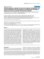
Chart 1: Clinical response to radiothepary in functioning pituitary adenomas (n = 23).
Chart 2: Clinical response to radiothepary in nonfunctioning pituitary adenomas (n = 58).
145
Journal of military pharmaco-medicine no7-2019
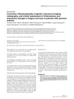
P<0.001
Time of followup (month)
Chart 3: Tumour size response to radiotherapy.
Chart 4: Tumour size response according to RECIST classification.
p < 0.01
Time of follow-up
(month)
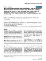
Chart 5: Treatment response of endocrine after radiotherapy (n = 23).
146
Journal of military pharmaco-medicine no7-2019
Time of follow-up
(month)
Chart 6: Hypopituitarism during the follow-up after radiotherapy.
(T0: Pre radiotherapy (n = 81); T1: 3rd month post radiotherapy (n = 81); T2: 6th
month post radiotherapy (n = 81); T3: 12th month post radiotherapy (n = 81); T4: 18th
month radiotherapy (n = 81); T5: 24th month post radiotherapy (n = 81); T6: 30th month
post radiotherapy (n = 81); T7: 36th month post radiotherapy (n = 69) T8: 42th month
post radiotherapy (n = 53); T9: 48th month post radiotherapy (n = 34); T10: 54th month
post radiotherapy (n = 18); T11: 60th month post radiotherapy (n = 4)
6 patients with galactorrhe and 8
patients with menstrual disorders were
followed up after treatment returned to
normal, 2 patients after radiotherapy had
pregnancy and gave normal birth.
14 cases had acromegaly, however,
there was no improvement in the course
of treatment. Clinical symptoms of memory
loss, headache, visual disturbances
decreased gradually compared to pre
treatment.
After radiotherapy, 52 cases (64.2%)
responded to radiotherapy with reduced
tumour size. The period when the tumour
size started to decrease in response to
radiotherapy from the 12th month after
radiotherapy. In the study, there was an
increase of the tumour size after the
follow-up period, the rate of tumour
control was 79/81 (97.5%).
GH concentrations were recorded at
normal levels in 13.3% of patients and
46.7% of patients at 36 and 40 months
after radiotherapy, respectively. The period
of treatment response to GH concentration
was from the 12th month after radiotherapy.
10% of patients and 20% of patients had
normal PLR levels in the 18th and the 36th
months after radiotherapy. The treatment
response time of PRL concentration was
from the 6th month after radiotherapy.
Complications after radiotherapy
accounted for 66.7% of the total study
subjects, of which the most symptoms
were dry mouth. Headache, nausea, loss
of appetite, dry mouth, insomnia were
147
Journal of military pharmaco-medicine no7-2019
similar in the two groups of patients,
particularly hair loss symptoms in the
functioning tumour group were higher than
in nonfunctioning tumour with statistically
significant difference.
DISCUSION
We studied 81 patients with recurrent
or residual pituitary adenomas. The
average volume of tumour was 5,553
mm3. Chui Bum Cho [9] reported that the
mean tumour volume was 2.6 cm3, they
also found that the volume of
nonfunctioning pituitary adenomas was
statistically significantly higher than the
secretory tumour (3.06 cm3 compared to
1.69 cm3). Bir [5] also showed that the
volume
of
nonfunctioning
pituitary
adenomas before radiotherapy was quite
large, an average volume of 3.7 cm3.
Guadalupe [10] also showed that pituitary
tumour volume before radiotherapy was
10,306 mm3. In addition, we found that
the volume of tumour > 4,500 mm3
accounted for a high rate (54.3%).
The mean radiation dose was 16.17 Gy,
which revealed that the secreting pituitary
adenomas had a higher radiation dose
than the non-functioning pituitary adenomas
(17.74 Gy compared to 15.55 Gy,
p < 0.001). Sheehan [11] studied 512
patients with nonfunctioning pituitary
adenomas, with an average tumour size
of 3.3 cm3, indicating an average radiation
dose of 16.4 Gy. The author also found
the relation between the dose radiation
and the free-disease survival rate over the
follow-up time, the patients who received
radiotherapy < 12 Gy or > 20 Gy would
have a lower rate of free-disease survival
than the group of 12 - 20 Gy.
148
Symptom of headache started to
decrease after 3 months of radiotherapy
(86.2% at radiotherapy and 81% at the 3rd
month) and then decreased sharply from
the 6th month (65.5%) until the 18th month
only 5.2% and was stable in the follow-up
months. This symptom reduction was
statistically significant. Bir [12] performed
radiotherapy for 57 patients with pituitary
tumours without increased secretion,
headache symptoms decreased from
49.1% before radiotherapy to 3.5% after
radiotherapy, with statistical significance,
p < 0.001. Chai Hong Rim [13] reported
60 patients with an average follow-up time
of 5.7 years, indicating a remarkable
decrease in headache symptoms (74%).
Nguyen Thi Minh Phuong [1] showed that
headache symptoms decreased slowly
within the first 12 months of follow-up
but by 24 months, headache symptoms
decreased significantly.
We recorded that pituitary adenomas
completely responded to radiotherapy
accounting for 13.6%, partially responding
accounted for 50.6%, stable disease made
up 33.3% and 2.5% of progressive disease,
tumour increased in size. The rate of
pituitary tumour control was 97.5%.
Nguyen Thi Minh Phuong [1] recorded
that tumour response with radiotherapy
according to RECIST criteria occured
in 44 patients with pituitary tumours:
Complete response accounted for 6.3%,
partial response presented in 41.7%,
stable disease explained for the highest
proportion (43.8%), progressive disease
was found in 8.3% of patients. Sallabanda
[14] gave the treatment for 30 patients
with pituitary tumours, 63% of patients,
whose tumours did not change in size
Journal of military pharmaco-medicine no7-2019
after radiotherapy, 30% of them decreased
in size and 7% of them increased the size
after radiotherapy. Yuan-Hao Chen [3]
treated 22 patients by radiotherapy with
an average followed up of 58.1 months
showed that 39.1% of patients had
reduced tumour size, 60.9% of them had
stabilized tumour size and none of them
had increased tumour size after follow-up
period.
The period when endocrine responded
to treatment returned to normal level was
the 18th month for PRL and the 30th month
for GH hormone after radiotherapy.
However, according to Nguyen Thi Minh
Phuong [1], this point of time was the 6th
month after radiotherapy. Grant et al [16]
reported that 31 patients with secreting
pituitary adenomas were treated with
radiotherapy with an everage follow-up of
40.2 months and found that 70% of
patients with endocrine concentrations
returned to normal level after everage
follow-up of 17.7 months. The author
revealed that mean time of endocrine
substances at normal level: ACTH was
11.7 months, GH was 18.4 months and
PLR was 57 months.
Visual complications were not obseved
in our study. In Sebastian’s et al research
[14] on 117 patients with pituitary adenoma,
visual complications after radiotherapy
were 5.3%. In multivariate analysis, the
author demontrated that risk factors for
visual complications after radiotherapy
were traditional radiotherapy (OR = 10.36,
p = 0.04). Gopalan [17] recorded that
visual complications after radiotherapy was
6.2% (3/48 patients), of which 2 patients
had visual disturbances before surgery,
2 out of 3 patients had progressive disease
after radiotherapy.
CONCLUSION
Radiotherapy for pituitary adenomas
has brought good results with high rate of
tumour control after long-term follow-up
period. Clinical symptoms and endocrine
levels respond to radiotherapy. Radiological
complications are transient and disappear
after a few days.
REFERENCES
1. Nguyễn Thị Minh Phương. Nghiên cứu
biến đổi triệu chứng lâm sàng, hình thái, chức
năng tuyến yên ở bệnh nhân u tuyến yên
trước và sau điều trị bằng dao Gamma quay.
Luận án Tiến sỹ Y học, Học viện Quân y.
2018, tr.62.
2. American Brain Tumour Association.
Pituitary tumours, ISBN 0-944093-90-6. 2015.
3. Chirag G, Hayden M, Katznelson L et al.
Non-surgical management of hormonesecreting pituitary tumours. Journal of Clinical
Neuroscience. 2009, 16, pp.985-993.
4. Camara Gomez R. Non-functioning
pituitary tumours: 2012 update. Endocrine
Nutrition. 2014, 61 (3), pp.160-170.
5. Wan H, Chihiro O, Yuan S. MASEP
Gamma knife radiosurgery for secretory pituitary
adenomas: Experience in 347 consecutive
cases. Journal of Experimental & Clinical
Cancer Research. 2009, 28 (1), p.36.
6. Molina P.E. Anterior pituitary gland.
th
Endocrine Physiology. 4 edition, McGraw-Hill
Companies, Inc, New York. 2013, 1, pp.49-72.
7. Nemes O. Hypopituitarism due to pituitary
adenomas, traumatic brain injury and stroke.
Clinical Medical Sciences. Hungary. 2016,
pp.10-13.
149
Journal of military pharmaco-medicine no7-2019
8. Moose B.D, Shaw E.G. Radiotherapy of
pituitary tumours. Diagnosis and Management
of Pituitary Tumours. Humana Press, Springer
Science. 2008, pp.269- 274.
13. Chai Hong Rim et al. Radiotherapy for
pituitary adenomas: Long-term outcome and
complication. Radial Oncol J. 2011, 29 (3),
pp.156-163.
9. Chul Bum Cho et al. Stereotactic
radiosurgery with the Cyber knife for pituitary
adenomas. J Korean Neurosurg Soc. 2009,
45, pp.157-163.
14. Sallabanda K et al. Stereotatic
radiosurgery in pituitary adenomas: Long-term
single institution experience and role of
the hypothalamic-pituitary axis. Journal of
Radiosurgery and SBRT. 2011, 1, pp.213-220.
10. Guadalupe V, Gonzalez B, Ramirez C
et al. Clinical characteristics and treatment
outcome of 485 patients with unfunctioning
pituitary macroadenomas. International Journal
of Endocrinology. 2015, pp.1-7.
15. Yuan-Hao Chen et al. Multisession
Cyber knife radiosurgery for post-surgical
residual and recurrent pituitary adenoma:
Preliminary result from one center. Journal of
Radiosurgery and SBRT. 2013, 2, pp.105-117.
11. Sheehan J.P et al. Gamma knife
radiosurgery for the management of
nonfunctioning pituitary adenomas: A
multicenter study. J Neurosurg. 2013, 119,
pp.446-456
16. Grant R.A et al. Efficacy and safety of
higher dose stereotatic radiosurgery for
functional pituitary adenoma: A preliminary
Report. World Neurosurg. 2014, 82 (1-2),
pp.195-201.
12. Bir S.C et al. Clinical and radiologic
outcome of Gamma knife radiosurgery on
nonfunctioning pituitary adenomas. J Neurol
Surg B. 2015, 76, pp.351-357.
17. Gopalan R et al. Long-term outcome
after Gamma knife radiosurgery for patients
with a nonfunctioning pituitary adenoma.
Nerosurgery. 2011, 69 (2), pp.284-293.
150