Gelsolin and progression of aortic arch calcification in chronic hemodialysis patients
Bạn đang xem bản rút gọn của tài liệu. Xem và tải ngay bản đầy đủ của tài liệu tại đây (763.67 KB, 7 trang )
Int. J. Med. Sci. 2016, Vol. 13
Ivyspring
International Publisher
92
International Journal of Medical Sciences
2016; 13(2): 92-98. doi: 10.7150/ijms.13785
Research Paper
Gelsolin and Progression of Aortic Arch Calcification in
Chronic Hemodialysis Patients
Terry Ting-Yu Chiou1, Shang-Chih Liao1, Yu-Yin Kao1, Wen-Chin Lee1, Yueh-Ting Lee1, Hwee-Yeong Ng1,
Po-Shun Lee2, Chien-Te Lee1
1.
2.
Division of Nephrology, Department of Internal Medicine, Kaohsiung Chang Gung Memorial Hospital and Chang-Gung University College of Medicine,
Taiwan.
Brigham and Women’s Hospital, Harvard Medical School, Boston, Massachusetts, USA.
Corresponding author: Chien-Te Lee M.D., Ph.D. Division of Nephrology, Department of Internal Medicine, Kaohsiung Chang-Gung Memorial Hospital,
Taiwan. 123 Ta-Pei Road, Niao Sung District, Kaohsiung 833, Taiwan. TEL: 886-7-7317123 EXT 8306 FAX: 886-7-7322402 E-mail:
© Ivyspring International Publisher. Reproduction is permitted for personal, noncommercial use, provided that the article is in whole, unmodified, and properly cited. See
for terms and conditions.
Received: 2015.09.07; Accepted: 2016.01.05; Published: 2016.01.29
Abstract
Background: Vascular calcification (VC) is a key process associated with cardiovascular mortality
in dialysis patients. Gelsolin is an actin-binding protein that can modulate inflammation, correlated
inversely with hemodialysis (HD) mortality and involved in bone calcification homeostasis. In this
report, we aim to characterize progression in aortic arch calcification (AAC) and investigate its
association with gelsolin.
Methods: 184 HD patients were enrolled and their annual posterior-anterior chest X-ray films
(CXR) in 2009 and 2013 were examined. The severity of AAC was classified as grade 0 to 3. Blood
levels of gelsolin were measured by ELISA kits. Biographic and biochemical data at baseline were
analyzed with status of AAC at baseline and changes after 4 years.
Results: At baseline, 60% of the patients had detectable AAC on CXR. After 4 years, 77% had
AAC. Patients with grade 1 and 2 AAC had increased risk of progression (Odds ratio [OR] 2~3,
P=0.001) compared to those with grade 0 at baseline. Compared to those with no AAC, patients
with AAC progression had older age, lower gelsolin, higher waist circumference and prevalence of
vascular disease. Regression analysis confirmed baseline gelsolin (odds ratio 0.845, 95% confidence
interval [0.734-0.974]) and waist circumference as the independent factors associated with AAC
progression. Gelsolin is positively correlated with serum albumin and negatively with tumor necrosis factor-alpha.
Conclusion: Our study demonstrated that HD patients with grades 1 or 2 baseline AAC are at
increased risk of further progression compared to those with grade 0. We also found lower blood
levels of gelsolin associated with progressive AAC. Further investigation into the mechanistic roles
of gelsolin in vascular calcification may provide new understanding of this key process.
Key words: Aortic arch calcification, gelsolin, hemodialysis.
Introduction
Cardiovascular disease is the most common
cause of death in patients on chronic dialysis [1].
Vascular calcification (VC) is increasingly recognized
as a key process contributing to the high cardiovascular mortality in dialysis patients [2-4]. Our previous
study on 712 prevalent hemodialysis (HD) patients
showed a strong correlation between aortic arch calcification (AAC) and 10-year mortality [5]. It also
substantiated the use of plain chest X-ray films (CXR)
as a simple tool to evaluate AAC.
Recent studies reported that 26~78% of dialysis
patients had various degrees of AAC on CXR, and
34~60% had progression after 1~5 years of follow-up
[6-10]. These variations in the prevalence of calcification and its progression may be related to the differences in ethnicity, comorbidity, length of observation,
Int. J. Med. Sci. 2016, Vol. 13
and methods of assessment. Progression in VC has
also been associated with mortality [6,7]. Factors contributing to VC include age, comorbidity (diabetes,
hypertension, metabolic syndrome, dyslipidemia),
dialysis vintage, medications (calcium, vitamin D,
coumadin), and uremia-related mineral bone disorder
(serum calcium [Ca], phosphorus [P], parathyroid
hormone [PTH]) [11-15]. Markers of inflammation
and oxidative stress have also been implicated [16,17].
Gelsolin, by regulating intracellular actin filaments, is important in cell morphology, migration and
phagocytosis [18]. Its extracellular isoform, plasma
gelsolin is secreted by different types of cells and
serves as a scavenging system for potentially toxic
actin filaments [19]. It can localize inflammation and
minimize tissue damage by binding excessive actins
released from tissue injury. The phenomenon of
plasma gelsolin depletion has been observed in different diseases. Specifically, in patients with acute
myocardial infarction and fulminant hepatic failure,
plasma gelsolin levels dropped dramatically and recovered if organ function improved from injury [20].
Further research found that in addition to actin,
plasma gelsolin may also modulate immune response
by binding to key inflammatory mediators, including
lipopolysaccharide, lysophospholipids, and platelet
activating factor [21,22].
Furthermore, gelsolin interacts with osteopontin
and both are involved in bone homeostasis [23]. In
HD patients, plasma gelsolin levels were reduced
nearly 50% compared to healthy volunteers and correlated with mortality and protein-energy wasting
[24,25]. However, whether gelsolin have independent
roles in vascular calcification is uncertain, and strategies to reduce inflammation and protect vascular integrity are poorly defined. In this report, we aim to
characterize the progression of aortic arch calcification
in HD patients and investigate its associations with
novel biomarker gelsolin.
Subjects and Methods
From January to December 2009, we enrolled 184
stable HD patients from Kaohsiung Chang Gung
Memorial Hospital. Inclusion criteria were over 20
years of age and regular 4-hour HD session three
times a week for at least 6 months. Exclusion criteria
were chronic viral hepatitis, malignancy (within 5
years), acute infection or hospitalization (within three
months). This study was approved by the Institutional Review Boards and Ethics Committee in Chang
Gung Memorial Hospital (IRB No. 98-2685B), and
informed consents were obtained from all participants.
93
Laboratory and Clinical Data
For every study participant, blood samples were
collected. The averages of biochemical data were obtained over the three months prior to study enrollment. The adequacy of dialysis was assessed by Kt/V
urea using the urea kinetic model of Gotch [26]. Gelsolin levels in blood were measured with ELISA kits
from Critical Biologics Corporation (Cambridge, MA.
USA). The immunoassay kits for interleukin-6 (IL-6)
and tumor necrosis factor-alpha (TNF-α) were from
R&D Systems, Minneapolis, MN. USA. All measurements were done in duplicate. All patients’ medical
records were carefully reviewed. Vascular disease
was identified by documented history of ischemic
heart disease, cerebrovascular disease or peripheral
vascular disease.
Evaluation of AAC on Chest Radiography
Our HD patients have chest X-ray examinations
(posterior-anterior approach, standing) annually. For
each study participant, the routine annual chest X-ray
film in 2009 was selected as the baseline. A second
follow-up chest X-ray film was selected in 2013 or the
year when the patient reached outcomes such as
death, kidney transplantation or transfer to other dialysis centers. A simple classification by Symeonidis
et al. [27] was used to evaluate AAC in the present
study. Briefly, the severity of calcification was classified as grade (Gr.) 0 (no calcification visible), Gr. 1
(single thin or small spots of calcification), Gr. 2 (one
or more areas of thick calcification, but ≤ 50% of the
circular area of the aortic knob) and Gr. 3 (circular
calcification with >50% of circular area of the aortic
knob).
Each posterior-anterior (P-A) chest X-ray (CXR)
of these HD patients was read and graded independently by two readers (one nephrologist and one
trained nurse practitioner) blinded to the patients’
clinical data. Eight X-rays were graded differently on
calcification score (±1). The discrepancies between the
two observers were resolved by a third independent
reader. The progression of aortic calcification was
determined by comparing the grades at baseline and
at follow-up. “Progressive AAC” was defined if the
second chest X-ray’s grade is greater than the first
one. “Stable AAC” was defined if the first CXR
showed calcification and the second CXR had the
same calcification grade. “No AAC” group was defined if both the first and second CXR’s were grade 0.
Statistical Analysis
Statistical analysis was performed using SPSS
version 12.0. Results were expressed as mean ±
standard deviation or median (interquartile range) for
nonparametric data. Comparisons between two
Int. J. Med. Sci. 2016, Vol. 13
groups were performed using Student t-test or
Mann-Whitney test for nonparametric data. We used
multiple logistic regressions to examine the independent relationships between AAC progression
(progressive vs. no AAC) and other variables. Regression adjusted for age, HD vintage, vascular disease,
BMI, waist circumference, Kt/V, parameters of mineral-bone disorder (serum calcium [Ca], phosphorus
[P] and intact parathyroid hormone [iPTH]), gelsolin
and IL-6. All results were considered significant if
P-value was less than 0.05.
Results
Aortic arch calcification and its progression
At baseline, the subjects with and without AAC
had similar dialysis adequacy (Kt/V 1.38 ± 0.22 vs.
1.45 ± 0.23, P>0.05) and biochemical control, particularly serum calcium (9.2 ± 0.8 vs. 9.4 ± 0.9 mg/dL,
P>0.05) and phosphate (4.8 ± 1.4 vs. 4.9 ± 1.4 mg/dL,
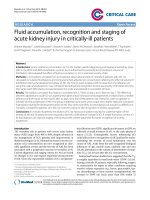
P>0.05). 75 (40%) patients had no detectable AAC
(grade 0) on plain chest X-ray. 52 (28%) patients had
grade 1, 43 (23%) grade 2, and 14 (8%) grade 3 (Figure
1A).
Figure 1. Distribution of different grades of aortic arch calcification
(AAC) at (A) baseline and (B) after 4 years of follow-up.
After 4 years of follow-up, only 43 (23%) patients
remained at grade 0. Thirty-one (17%) patients had
grade 1, 46 (25%) grade 2, and 64 (35%) grade 3 (Figure 1B). Moreover, 32 (43%) of those with grade 0 at
baseline progressed to higher grades of AAC over 4
years. 73% (38/52) of those with grade 1 at baseline
had progressive AAC, and 84% (36/43) of those with
grade 2 at baseline had progressive AAC. Fourteen
patients with grade 3 calcification at baseline re-
94
mained so after 4 years. Compared to those with
grade 0 (the reference group), patients with grade 1 at
baseline had increased risk of further progression in
AAC (Odds ratio [OR] 2.11, 95% confidence interval
[CI] 1.29-3.44, P=0.001, adjusted for age, waist circumference, gelsolin and IL-6). Patients with grade 2
at baseline also had higher risk of progression in AAC
(OR 3.49 [1.72-7.07], P<0.001).
Factors associated with aortic arch calcification and its progression
We compared the clinical and biochemical profiles between patients with and without AAC. At
baseline, patients with calcifications had significantly
lower serum albumin (3.9 ± 0.3 vs. 4.0 ± 0.2 g/dL,
P=0.015). They also tend to have older age (61 ± 10 vs.
58 ± 12, P=0.066) and lower gelsolin (824 ± 257 vs. 898
± 272 µU/ml, P=0.064). However, the two groups did
not differ significantly in serum Ca, P, iPTH, cholesterol or high-sensitivity C-reactive protein (hs-CRP).
At 4-year follow-up, patients with progressive
AAC had significantly older age, higher waist circumference, IL-6, and lower gelsolin than those with
no AAC (Table 1). They also had significantly higher
rate of vascular disease (51% vs. 31%, P<0.05). There
were no significant differences between patients with
stable and progressive AAC. Multiple logistic regression (adjusting for age, HD vintage, vascular disease,
BMI, waist circumference, Kt/V, serum calcium,
phosphorus, iPTH, gelsolin and IL-6) confirmed baseline gelsolin and waist circumference as the independent factors associated with progressive AAC
(Table 2). For every 10cm increase in waist circumference, the risk of AAC progression is increased by
47%. For every 100μU/ml increase in gelsolin level,
the risk of AAC progression is decreased by 15%.
We also analyzed the associations between gelsolin and important factors. Baseline gelsolin is positively correlated with serum albumin (ρ [Pearson’s
coefficient] 0.262, P<0.001) and creatinine (ρ 0.276,
P<0.001), and negatively correlated with age (ρ -0.212,
P=0.004), TNF-α (ρ -0.148, P=0.044) and white blood
cell counts (ρ -0.163, P=0.026).
Discussion
This study on 184 prevalent HD patients had two
major findings. First, the severity of AAC on plain
chest X-ray confers different risks for AAC progression and vascular disease. Patients with higher grade
AAC at baseline had higher risk of progression, and
subjects with AAC progression have higher rate of
vascular disease. Second, baseline gelsolin level was
independently associated with progression in AAC.
Other important factors of AAC and progression were
serum albumin and waist circumference.
Int. J. Med. Sci. 2016, Vol. 13
95
Table 1. Comparison between 3 groups of patients (no AAC, stable AAC and progressive AAC) after 4 years of follow-up.
Age, year
HD vintage, month
BMI, kg/m²
Waist circumference, cm
Vascular disease
Kt/V
nPCR, g/kg/day
Hemoglobin, g/dL
Serum albumin, g/dL
BUN, mmol/L
Serum creatinine, mg/dL
Calcium, mg/dL
Phosphorus, mg/dL
iPTH, pg/dL
Total cholesterol, mg/dL
Triglyceride, mg/dL
HDL, mg/dL
LDL, mg/dL
hsCRP, mg/L
Gelsolin, µU/ml
TNF-α, pg/mL
IL-6, pg/mL
No AAC (N = 43)
56 ± 12
52 (28-113)
22.1 ± 4.5
81.2 ± 12.1
31%
1.4 ± 0.2
1.22 ± 0.3
10.4 ± 1.0
4.0 ± 0.2
69.7 ± 10.2
10.8 ± 1.9
9.2 ± 0.8
4.8 ± 1.5
259 (98-687)
183.6 ± 40.7
164.0 ± 105.7
46.6 ± 12.9
101.0 ± 42.2
2.3 (1.0-5.1)
925.5 ± 277.4
5.7 (4.1-8.5)
2.0 (1.4-3.8)
Stable AAC (N = 35)
61 ± 8*
68 (37-121)
23.5 ± 4.1
86.7 ± 13.7
46%
1.4 ± 0.2
1.18 ± 0.31
10.5 ± 1.5
3.9 ± 0.2
70.8 ± 15.0
11.2 ± 2.1
9.1 ± 0.9
4.9 ± 1.3
231 (110-542)
189.7 ± 38.3
144.7 ± 94.8
54.2 ± 16.3
110.4 ± 23.3
3.1 (1.5-6.8)
846.9 ± 235.7
7.1 (4.1-9.7)
3.0 (1.7-6.4)*
Progressive AAC (N=106)
60 ± 10**
62.0 (36.0-102.0)
22.8 ± 3.5
85.7 ± 11.7**
51%**
1.4 ± 0.2
1.19 ± 0.28
10.6 ± 1.2
3.9 ± 0.3
68.6 ± 15.1
10.8 ± 2.1
9.4 ± 0.8
4.9 ± 1.4
238 (86-627)
188.2 ± 39.3
159.4 ± 106.5
45.1 ± 14.9
110.2 ± 34.2
2.9 (1.3-7.4)
827.6 ± 266.8**
6.7 (3.9-11.3)
2.7 (1.5-4.5)**
“No AAC” means no detectable aortic arch calcification on CXR at baseline and at follow-up. “Stable AAC” means same AAC on CXR at baseline and at follow-up.
“Progressive AAC” means AAC grades at follow-up higher than at baseline.
* P<0.05 Stable AAC vs. No AAC
** P<0.05 Progressive AAC vs. No AAC
Abbreviations BMI: body mass index; nPCR: normalized protein catabolic rate; iPTH: intact parathyroid hormone; HDL: high density lipoprotein cholesterol; LDL: low
density lipoprotein cholesterol; hsCRP: high-sensitivity C-reactive protein; TNF-α: tumor necrosis factor-alpha; IL-6: interleukin-6.
Table 2. Multiple logistic regression* analysis showing the independent factors associated with progressive aortic arch calcification after
4 years of follow-up.
Age (per 1-year increase)
HD vintage (per 1 year increase)
Vascular disease (yes vs. no)
BMI (per 1kg/m2 increase)
Waist circumference (per 10cm increase)
Kt/V (per 1 unit increase)
Calcium (per 1 mg/dL increase)
Phosphorus (per 1 mg/dL increase)
iPTH (per 1 pg/dL increase)
Gelsolin (per 100μU/ml increase)
IL-6 (per 1 pg/mL increase)
Odds Ratio (95%CI)
1.022 (0.985-1.062)
1.007 (0.999-1.015)
1.348 (0.586-3.100)
0.892 (0.756-1.008)
1.475 (1.050-2.074)
0.601 (0.127-3.452)
1.008 (0.621-1.636)
0.976 (0.973-1.238)
1.000 (0.999-1.001)
0.845 (0.734-0.974)
1.116 (0.986-1.262)
P-value
NS
NS
NS
NS
0.025
NS
NS
NS
NS
0.02
NS
*Regression (progressive vs. no AAC) adjusted for age, HD vintage, vascular disease, BMI, waist circumference, Kt/V, serum calcium, phosphorus, iPTH, gelsolin and IL-6.
Abbreviations: CI, Confidence Interval; BMI, body mass index; BMI: body mass index; iPTH: intact parathyroid hormone; IL-6: interleukin-6; NS, non-significant.
Our study demonstrated that AAC is common in
HD patients, 60% at baseline and 77% after 4 years of
follow-up. In addition, we showed that progression in
AAC is associated with higher prevalence of vascular
disease. Sigrist et al. [12], using multislice computed
tomography to quantify calcification in superficial
femoral artery reported a constant progression rate
over the first and second years of observation. Their
study examined a heterogeneous group of 101 patients with stage 4 CKD, HD and peritoneal dialysis.
However, from our cohort of HD patients, we found
that progression in AAC over a longer time period (4
years) is dependent on the initial calcification status.
Less than half of those with no evident AAC on baseline CXR developed calcification over 4 years of follow-up. However, 73% of those with baseline grade 1
AAC and 84% of those with grade 2 had more severe
calcification over the same period. This observation
suggests that when AAC becomes evident on CXR,
the risk of progression is much higher. This has several important clinical implications. First, the progression of AAC may not be the same for everyone
over 4 years, which makes risk stratification and timing of intervention extremely crucial: the later stage
Int. J. Med. Sci. 2016, Vol. 13
we intervene, the less time we may have to deliver an
effective therapy. Second, routine assessment of AAC
will help identify patients at increased risk of progression in vascular calcification. Third, plain chest
X-ray is an easily attainable yet powerful tool in the
assessment of vascular calcification and its risk of
progression.
The independent association between plasma
gelsolin and progression in aortic arch calcification
has not been reported in the literature before. Gelsolin
is involved in bone homeostasis through interaction
with osteopontin, an inhibitory protein in vascular
calcification. In osteoclast, osteopontin binds to integrin αvβ3 and stimulates gelsolin-associated polyphosphoinositides, leading to alterations in cytoskeleton and bone resorption [28]. Response to osteopontin stimulation was absent in gelsolin-deficient osteoclast [23]. In gelsolin-deficient adult mice, bone resorption was diminished, resulting in increased bone
mass. Gelsolin, together with osteopontin promotes
resorption, and modifies the process of calcification.
96
In patients with chronic kidney disease (CKD), the
diminishing gelsolin levels may contribute to the imbalance between the activators and inhibitors of vascular calcification. In normal bone homeostasis, osteoblasts promote the recruitment and activation of
osteoclasts, resulting in bone resorption and remodeling. However, in vascular calcification, the
trans-differentiated vascular smooth muscle cells with
an osteoblast phenotype do not promote resorption of
calcification by osteoclast-like cells [29]. In patients
with CKD, a gelsolin depleted state may promote the
propagation of calcification by exacerbating the osteoclast-like cells’ inability to resorb hydroxyapatite
crystals. It is tempting to speculate that correction of
gelsolin deficiency may reactivate osteoclasts, leading
to resorption of vascular calcification.
Our findings also complement previous work
demonstrating the connections between gelsolin, atherosclerosis, inflammation and oxidative stress, which
are also important mediators of vascular injury and
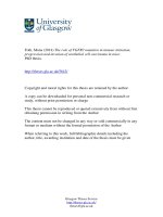
calcification (Figure 2).
Figure 2. Possible mechanisms for gelsolin depletion, vascular injury and calcification in chronic kidney disease. Gelsolin depletion may impair modulation
on lysophospholipids, macrophage receptor and nitric oxide system which are involved in the maintenance of vascular integrity, development of foam cells,
plaque and atherosclerosis. Blood levels of gelsolin have also been correlated inversely with oxidative stress and inflammatory cytokines (i.e. TNF-α). In
addition to an impaired resorption of calcification by osteoclast-like cells, gelsolin depletion may also exacerbate the imbalance between calcification
inhibitors and activators.
Int. J. Med. Sci. 2016, Vol. 13
Macrophage is a key player in the process of inflammation and atherosclerosis. Gelsolin can alter
macrophage function by triggering nitric oxide synthase, leading to reduced inflammation [30]. In animal
models of sepsis, supplementation with recombinant
human plasma gelsolin (rhu-pGSN) to restore its depleted level can provide favorable inflammatory
modulation and survival benefits [31]. Gelsolin can
also modulate cellular responses to lysophospholipids
which interact with macrophage receptors and mediate atherosclerosis [21,32]. Indeed, gelsolin expression
was reduced in the media layer of human atherosclerotic coronary arteries [33], and its secretion into the
ex vivo culture medium was also decreased [34]. This
depletion of gelsolin may have consequences in vascular functions. In atherosclerosis and CKD, depletion
of gelsolin may impair ligand interaction with macrophage receptors, the modulation on nitric oxide
system, and the buffering capacity for inflammatory
mediators. Ours and other studies also support the
connections between alterations in gelsolin levels and
inflammation, oxidative stress, and production of
reactive oxygen species [35-37]. In addition to its role
as a novel biomarker, gelsolin has therapeutic potential. In mouse models of sepsis and multiple sclerosis
where gelsolin levels were depleted, administration of
recombinant gelsolin proteins improved survival and
brain inflammation [31,40]. In a recent preliminary
clinical trial, the safety of rhu-pGSN was established
in humans [38].
In addition to the characterization of AAC and
its progression over 4 years, our study also explored
the important factors associated with these phenomena. In our study, markers of mineral bone metabolism (Ca, P, iPTH) were not associated with AAC and
its progression. This is likely because our patients
were in good control from this perspective. However,
despite keeping their serum Ca, P, PTH within the
guideline recommendations, these patients still have
high rate of AAC and progression. It should be emphasized that other important factors must be involved. Gelsolin, the novel protein may be one of
these key players in AAC. Our analysis also showed
that lower serum albumin is associated with baseline
AAC. Moreover, higher waist circumference is associated with AAC progression. Increased waist circumference suggests increased abdominal adiposity,
which is also associated with coronary artery calcification in CKD patients [39].
Our study has several limitations. First, using
plain CXR and 4-grade scoring method to evaluate the
severity of vascular calcification may underestimate
or miss some subtle calcification and changes. CT scan
would offer a much higher resolution, a finer differentiation of calcification, but more costly. Secondly,
97
our study does not have detailed accounts of patients’
medications because they may have various sources
of medications outside of our dialysis center. Lastly,
our study results may not be generalizable to patients
of different ethnicity or other modality of renal replacement therapy.
In conclusion, our study found that AAC based
on plain CXR was common in HD patients. Patients
with initially higher grades of AAC are at increased
risk of further progression, which is associated with
higher risk of vascular disease. Our identification of
plasma gelsolin as a novel protein associated with
progression in VC warrants further confirmation in
larger population and may offer new understanding
and therapeutic strategies toward vascular injuries
and calcification.
Acknowledgements
Grant support by Kaohsiung Chang Gung Memorial Hospital (CMRPG8B0491) and Minister of
Science and Technology, Republic of China (MOST
104-2314-B-182A- 143).
Competing Interests
None to declare.
References
1
2
3
4
5
6
7
8
9
10
11
12
13
14
Collins AJ, Li S, Ma JZ, Herzog C. Cardiovascular disease in end-stage renal
disease patients. Am J Kidney Dis. 2001; 38: S26-29.
McIntyre CW. The functional cardiovascular consequences of vascular calcification. Semin Dial. 2007; 20: 122-128.
Cannata-Andia JB, Rodriguez-Garcia M, Carrillo-Lopez N, Naves-Diaz M,
Diaz-Lopez B. Vascular calcifications: Pathogenesis, management, and impact
on clinical outcomes. J Am Soc Nephrol. 2006; 17: S267-273.
Rennenberg RJ, Kessels AG, Schurgers LJ, van Engelshoven JM, de Leeuw PW,
Kroon AA. Vascular calcifications as a marker of increased cardiovascular risk:
A meta-analysis. Vasc Health Risk Manag. 2009; 5: 185-197.
Lee CT, Huang CC, Hsu CY, Chiou TT, Ng HY, Wu CH, Kuo WH, Lee YT.
Calcification of the aortic arch predicts cardiovascular and all-cause mortality
in chronic hemodialysis patients. Cardiorenal Med. 2014; 4: 34-42.
Goldsmith DJ, Covic A, Sambrook PA, Ackrill P. Vascular calcification in
long-term haemodialysis patients in a single unit: A retrospective analysis.
Nephron. 1997; 77: 37-43.
Ammirati AL, Dalboni MA, Cendoroglo M, Draibe SA, Santos RD, Miname M,
Canziani ME. The progression and impact of vascular calcification in peritoneal dialysis patients. Perit Dial Int. 2007; 27: 340-346.
Kim HG, Song SW, Kim TY, Kim YO. Risk factors for progression of aortic
arch calcification in patients on maintenance hemodialysis and peritoneal dialysis. Hemodial Int. 2011; 15: 460-467.
Ogawa T, Ishida H, Akamatsu M, Matsuda N, Fujiu A, Ito K, Ando Y, Nitta K.
Relation of oral 1alpha-hydroxy vitamin d3 to the progression of aortic arch
calcification in hemodialysis patients. Heart Vessels. 2010; 25: 1-6.
Tamei N, Ogawa T, Ishida H, Ando Y, Nitta K. Serum fibroblast growth
factor-23 levels and progression of aortic arch calcification in non-diabetic patients on chronic hemodialysis. J Atheroscler Thromb. 2011; 18: 217-223.
Shroff R, Long DA, Shanahan C. Mechanistic insights into vascular calcification in CKD. J Am Soc Nephrol. 2013; 24: 179-189.
Sigrist MK, Taal MW, Bungay P, McIntyre CW. Progressive vascular calcification over 2 years is associated with arterial stiffening and increased mortality
in patients with stages 4 and 5 chronic kidney disease. Clin J Am Soc Nephrol.
2007; 2: 1241-1248.
Noordzij M, Cranenburg EM, Engelsman LF, Hermans MM, Boeschoten EW,
Brandenburg VM, Bos WJ, Kooman JP, Dekker FW, Ketteler M, Schurgers LJ,
Krediet RT, Korevaar JC, Group NS. Progression of aortic calcification is associated with disorders of mineral metabolism and mortality in chronic dialysis
patients. Nephrol Dial Transplant. 2011; 26: 1662-1669.
Iribarren C, Sidney S, Sternfeld B, Browner WS. Calcification of the aortic arch:
Risk factors and association with coronary heart disease, stroke, and peripheral vascular disease. JAMA. 2000; 283: 2810-2815.
Int. J. Med. Sci. 2016, Vol. 13
15
16
17
18
19
20
21
22
23
24
25
26
27
28
29
30
31
32
33
34
35
36
37
38
39
40
98
Blacher J, Guerin AP, Pannier B, Marchais SJ, London GM. Arterial calcifications, arterial stiffness, and cardiovascular risk in end-stage renal disease.
Hypertension. 2001; 38: 938-942.
Chen NX, Moe SM: Vascular calcification: Pathophysiology and risk factors.
Curr Hypertens Rep. 2012; 14: 228-237.
Lee CT, Chua S, Hsu CY, Tsai YC, Ng HY, Kuo CC, Wu CH, Chen TC, Chiu
TT, Lee YT. Biomarkers associated with vascular and valvular calcification in
chronic hemodialysis patients. Dis Markers 2013; 34: 229-235.
Bucki R, Levental I, Kulakowska A, Janmey PA. Plasma gelsolin: Function,
prognostic value, and potential therapeutic use. Curr Protein Pept Sci. 2008; 9:
541-551.
Lee WM, Galbraith RM. The extracellular actin-scavenger system and actin
toxicity. N Engl J Med. 1992; 326: 1335-1341.
Suhler E, Lin W, Yin HL, Lee WM. Decreased plasma gelsolin concentrations
in acute liver failure, myocardial infarction, septic shock, and myonecrosis.
Crit Care Med. 1997; 25: 594-598.
Osborn TM, Dahlgren C, Hartwig JH, Stossel TP. Modifications of cellular
responses to lysophosphatidic acid and platelet-activating factor by plasma
gelsolin. Am J Physiol Cell Physiol. 2007; 292: C1323-1330.
Bucki R, Kulakowska A, Byfield FJ, Zendzian-Piotrowska M, Baranowski M,
Marzec M, Winer JP, Ciccarelli NJ, Gorski J, Drozdowski W, Bittman R,
Janmey PA. Plasma gelsolin modulates cellular response to sphingosine
1-phosphate. Am J Physiol Cell Physiol. 2010; 299: C1516-1523.
Chellaiah M, Kizer N, Silva M, Alvarez U, Kwiatkowski D, Hruska KA. Gelsolin deficiency blocks podosome assembly and produces increased bone
mass and strength. J Cell Biol. 2000; 148: 665-678.
Lee PS, Sampath K, Karumanchi SA, Tamez H, Bhan I, Isakova T, Gutierrez
OM, Wolf M, Chang Y, Stossel TP, Thadhani R. Plasma gelsolin and circulating actin correlate with hemodialysis mortality. J Am Soc Nephrol. 2009; 20:
1140-1148.
Chiu TT, Liao SC, Lee WC, Lee PS, Ng HY, Chien YS, Lee CT. Gelsolin and
adipokines are associated with protein-energy wasting in hemodialysis patients. Artif Organs. 2015; 39: 150-155.
Gotch FA, Sargent JA. A mechanistic analysis of the national cooperative
dialysis study (NCDS). Kidney Int. 1985; 28: 526-534.
Symeonidis G, Papanas N, Giannakis I, Mavridis G, Lakasas G, Kyriakidis G,
Artopoulos I. Gravity of aortic arch calcification as evaluated in adult greek
patients. Int Angiol. 2002; 21: 233-236.
Chellaiah M, Hruska K. Osteopontin stimulates gelsolin-associated phosphoinositide levels and phosphatidylinositol triphosphate-hydroxyl kinase. Mol
Biol Cell. 1996; 7: 743-753.
Persy V, D'Haese P. Vascular calcification and bone disease: The calcification
paradox. Trends Mol Med. 2009; 15: 405-416.
Yang Z, Chiou TT, Stossel TP, Kobzik L. Plasma gelsolin improves lung host
defense against pneumonia by enhancing macrophage nos3 function. Am J
Physiol Lung Cell Mol Physiol. 2015; 309: L11-16.
Lee PS, Waxman AB, Cotich KL, Chung SW, Perrella MA, Stossel TP. Plasma
gelsolin is a marker and therapeutic agent in animal sepsis. Crit Care Med.
2007; 35: 849-855.
Nishikawa M, Kurano M, Ikeda H, Aoki J, Yatomi Y. Lysophosphatidylserine
has bilateral effects on macrophages in the pathogenesis of atherosclerosis. J
Atheroscler Thromb. 2015; 22: 518-526.
de la Cuesta F, Zubiri I, Maroto AS, Posada M, Padial LR, Vivanco F, Alvarez-Llamas G, Barderas MG. Deregulation of smooth muscle cell cytoskeleton
within the human atherosclerotic coronary media layer. J Proteomics. 2013; 82:
155-165.
de la Cuesta F, Barderas MG, Calvo E, Zubiri I, Maroto AS, Darde VM, Martin-Rojas T, Gil-Dones F, Posada-Ayala M, Tejerina T, Lopez JA, Vivanco F,
Alvarez-Llamas
G.
Secretome
analysis
of
atherosclerotic
and
non-atherosclerotic arteries reveals dynamic extracellular remodeling during
pathogenesis. J Proteomics. 2012; 75: 2960-2971.
Genre F, Lopez-Mejias R, Miranda-Filloy JA, Ubilla B, Carnero-Lopez B,
Gomez-Acebo I, Blanco R, Ochoa R, Rueda-Gotor J, Gonzalez-Juanatey C,
Llorca J, Gonzalez-Gay MA. Gelsolin levels are decreased in ankylosing
spondylitis patients undergoing anti-tnf-alpha therapy. Clin Exp Rheumatol.
2014; 32: 218-224.
Han C, Zhang L, Zhu X, Tang J, Jin X. Plasma gelsolin levels are decreased and
correlate with fibrosis in IgA nephropathy. Exp Biol Med. 2013; 238: 1318-1327.
Forsman H, Onnheim K, Andreasson E, Christenson K, Karlsson A, Bylund J,
Dahlgren C. Reactivation of desensitized formyl peptide receptors by platelet
activating factor: A novel receptor cross talk mechanism regulating neutrophil
superoxide anion production. PLoS One. 2013; 8: e60169.
Chan W, Lau L, Kwok K, Law W, Ho JCM, Chu K, Poon RTP. A randomized,
double-blind, placebo-controlled, ascending-dose trial of the pharmacokinetics and safety of intravenous infusion of recombinant human plasma gelsolin
in acutely ill patients with decreased plasma gelsolin levels. American Thoracic Society. 2011; 183: A5601.
Aoqui C, Cuppari L, Kamimura MA, Canziani ME. Increased visceral adiposity is associated with coronary artery calcification in male patients with
chronic kidney disease. Eur J Clin Nutr. 2013; 67: 610-614.
Kevin Li-Chun H, Schob S, Zeller MW, Pulli B, Ali M, Wang C, Chiou TT,
Tsang YM, Lee PS, Stossel TP, Chen JW. Gelsolin decreases actin toxicity and
inflammation in murine multiple sclerosis. J Neuroimmunol. 2015; 287: 36-42.