Differential response of non-cancerous and malignant breast cancer cells to conditioned medium of adipose tissue-derived stromal cells (ASCs)
Bạn đang xem bản rút gọn của tài liệu. Xem và tải ngay bản đầy đủ của tài liệu tại đây (1.23 MB, 9 trang )
Int. J. Med. Sci. 2019, Vol. 16
Ivyspring
International Publisher
893
International Journal of Medical Sciences
2019; 16(6): 893-901. doi: 10.7150/ijms.27125
Research Paper
Differential Response of Non-cancerous and Malignant
Breast Cancer Cells to Conditioned Medium of Adipose
tissue-derived Stromal Cells (ASCs)
Yi-Chia Wu1,2,3,4*, Wei-Ting Wang5*, Li-Ju Huang5, Ruo-You Cheng1, Yur-Ren Kuo1,3,6, Ming-Feng Hou3,6,
Chung-Sheng Lai1,3,6, and John Yu7,8
1.
2.
3.
4.
5.
6.
7.
8.
Division of Plastic Surgery, Department of Surgery, Kaohsiung Medical University Hospital, Kaohsiung, Taiwan
Department of Plastic Surgery, Kaohsiung Municipal Ta-Tung Hospital, Kaohsiung, Taiwan
Graduate Institute of Medicine, Kaohsiung Medical University, Kaohsiung, Taiwan
PhD. Programme in Translational Medicine, Kaohsiung Medical University, Kaohsiung, and Academia Sinica, Taipei, Taiwan
Center of Teaching and Research, Kaohsiung Municipal Ta-Tung Hospital, Kaohsiung, Taiwan
Faculty of Medicine, College of Medicine, Kaohsiung Medical University, Kaohsiung, Taiwan
Institute of Stem Cell and Translational Cancer Research, Chang Gung Memorial Hospital at Linko, Taoyuan, Taiwan
Institute of Cellular and Organismic Biology, Academia Sinica, Taipei, Taiwan
* These authors contributed equally to this work.
Corresponding author: John Yu, MD., PhD., No.5, Fuxing St., Guishan Dist., Taoyuan City 333, Taiwan. Tel.: 886-3-328-1200#5218; Fax: 886-3-328-1200#5214;
E-mail:
© Ivyspring International Publisher. This is an open access article distributed under the terms of the Creative Commons Attribution (CC BY-NC) license
( See for full terms and conditions.
Received: 2018.05.07; Accepted: 2019.04.03; Published: 2019.06.02
Abstract
Background: The application of adipose tissue-derived stromal cells (ASCs) in regenerative medicine has
become a growing trend due to its abundance and differentiation potentials. However, several breast cancer
studies indicated that ASCs promote tumor progression, therefore, the use of ASCs for reconstruction after
oncological surgery poses potential risks. In this study, we aimed to examine whether cancerous or
non-cancerous breast cells will exhibit different responses to ASC-derived CM.
Methods: ASCs were isolated from residuals of subcutaneous adipose tissue obtained from patients
undergoing surgery. Cancerous MCF-7, MDA-MB231, and MDA-MB468 cell lines and one non-cancerous
M10/H184B5F5 cell line were cultured with variant concentrations of ASC-derived conditioned medium (CM)
for analysis.
Results: ASC-derived CM significantly reduced cell viability by triggering apoptosis in MCF-7, MDA-MB231,
and MDA-MB468 cell lines. ATM-Chk2-dependent DNA damage response was activated early in cancer cells
when exposed to ASC-derived CM. By contrast, prompted cell proliferation instead of cell death was detected
in M10/H184B5F5 cells under the treatment of lower CM concentration. Even when exposed to the highest
concentration of CM, only cell cycle arrest accompanied by a weak DNA damage response were detected in
M10/H184B5F5 cells, no cell deaths were observed.
Conclusions: Overall, this study demonstrated that cancerous and non-cancerous breast cells respond
differently to ASC-derived CM. ASC-derived CM triggered significant cell death in breast cancer cell lines,
however non-cancerous breast cells exhibited dissimilar response to ASC-derived CM.
Key words: adipose tissue-derived stromal cell; ASCs; breast cancer cell line; apoptosis
Introduction
In recent medical practices, the use of bone
marrow-derived mesenchymal stem cells (MSCs),
particularly adipose tissue-derived stromal cells
(ASCs), has attracted growing attentions because of its
availability and differentiation potential of ASCs.
ASCs has the ability to differentiate into various
lineages, including adipogenic, chondrogenic,
osteogenic, and hepatocytic cell types as MSCs [1].
Paracrine secretion of cytokines and chemokines as
well as growth factors from ASCs have also been
reported to exhibit anti-apoptotic, anti-inflammatory,
immunomodulatory, and anti-scarring effects [2-3].
Int. J. Med. Sci. 2019, Vol. 16
These specific characteristics make ASCs a promising
candidate in a broad array of medical applications.
However, the effects of MSCs and ASCs on various
types of cancer remain controversial [4]. Several
experimental studies support the propensity of MSCs
and ASCs to promote growth, progression, and
metastasis in various cancer types, including breast
cancer, osteosarcoma, and brain tumor [2, 5-6]. Few
literature support progressions in other types of
cancer [7-9].
In breast surgery, compared with breast
prostheses, reconstruction with autologous tissue,
such as latissimus dorsi or rectus abdominis, and fat
grafting are commonly used, with a benefit of a more
natural
appearance
following
oncological
mastectomy. However, fat necrosis or apoptosis can
lead to transplanted graft resorption and loss of
volume, which can ultimately produce an
unsatisfactory
long-term
result
[10,11].
Supplementation with ASCs isolated from adipose
tissue has been proposed to improve the engraftment
and overcome this challenge [10,12].
Given its great potential, a thorough
understanding on the influence of ASCs on tumor
progression, particularly in breast cancer, may help in
exploring the safety of using ASCs in future
regenerative medicine and cell therapy. In this study,
we showed that non-tumor cells and cancerous breast
epithelial cells exhibited different responses to
ASC-derived
conditioned
medium
(CM).
ASC-derived CM yielded typical DNA damage
responses and eventually apoptosis in malignant
tumor cells, whereas non-cancerous M10/H184B5F5
cells showed enhanced proliferation in response to
lower concentration of ASC-derived CM. Even under
the highest concentration of ASC-derived CM, no
deaths were detected in M10/H184B5F5 cells.
Material and Methods
Human ASCs isolation and culture
ASC isolation in this study was approved by the
Institutional Review Board of Kaohsiung Medical
University
Hospital
(KMUH-IRB-F(I)-20170077).
Informed consent was obtained from individuals
prior to the surgery. Residual subcutaneous adipose
tissue was obtained from patients undergoing
liposuction surgery. ASCs were further isolated from
residuals of adipose tissue and cultured with Defined
Keratinocyte serum-free medium (Gibco 10744-019,
MA, USA)-based complete K-Medium by following a
previously described protocol [13]. ASC-derived CM
was collected from culture medium no later than five
passages of ASCs. Surface markers of isolated ASCs
were examined using an LSR II flow cytometer (BD
894
Biosciences, NJ, USA) with FlowJo software (FlowJo
LLC, OR, USA). The lineage differentiation ability of
ASCs was confirmed by adipogenic, osteogenic and
chondrogenic induction for 3 weeks followed by Oil
Red O (adipogenesis), Alizarin Red S (osteogenesis)
and
Alcian
blue
(chondrogenesis)
staining
(Supplementary Figure S1).
Cell lines
Four breast cell lines were analyzed: three
malignant MCF-7, MDA-MB231, and MDA-MB468
cell lines and one non-cancerous M10/H184B5F5
breast epithelial cell line. While MCF-7 is the most
common breast cancer cell line used to assess cancer
progression with a relatively low invasiveness,
MDA-MB231 is a triple negative (estrogen receptor,
progesterone receptor, and HER2 negative) breast
cancer cell line with a more malignant potency [2]. In
addition, another breast cancer cell lines,
MDA-MB468 and the non-cancerous M10/H184B5F5
breast epithelial cells in this study were used for
comparisons.
MCF-7,
MDA-MB231,
and
M10/H184B5F5 were purchased from Bioresource
Collection
and
Research
Center,
Taiwan;
MDA-MB468 was purchased from American Type
Culture Collection, USA. All cell lines in this study
were maintained with Dulbecco's modified Eagle's
medium (DMEM) supplemented with 10% FBS (Gibco
26140-079) and 1% antibiotic-antimycotic, 100× (Gibco
15240-062). Complete DMEM was used as the control
medium (ctrl) relative to ASC-derived CM.
Cell cycle analysis
Treated samples were prepared as previously
described [14], and the cell cycle distribution was
analyzed using an LSR II flow cytometer with FlowJo
software.
Immunofluorescence staining
Cells were grown on cover slips inside 6-well
plates. After treatment, cell-grown slips were fixed
with
3%
buffered
paraformaldehyde
for
immunostaining which were prepared as described
previously [14]. Fluorescence images were detected
and captured using a Zeiss AxioPlan 2 system (Carl
Zeiss, Oberkochen, Germany).
Cell viability assay
Cell viability after treatment was determined
using an MTT assay. Treated cells were washed with
PBS and incubated with MTT (10 µg/mL) in freshly
refed medium under 37°C, 5% CO2 for 2 hours.
Formazan crystal produced by living cells were
dissolved in dimethyl sulfoxide and quantified by
measuring absorbance at a wavelength of OD570.
Int. J. Med. Sci. 2019, Vol. 16
Western blot
Whole-cell protein lysates of treated cells were
extracted and fractionated using sodium dodecyl
sulfate–polyacrylamide gel electrophoresis and
blotted onto nitrocellulose as previously described
[14]. Protein expression was determined using the
Western Lightning Plus-ECL detection system
(NEL104001EA, PerkinElmer, MA, USA). Primary
antibodies used: γ-H2AX (ab2893, abcam), activated
caspase3 (ab32042, abcam), CHK2 pThr 68 (2661, Cell
Signaling), CHK2 (3440, Cell Signaling), ATM pSer
1981 (5883, Cell Signaling) and GAPDH (MAB374,
Millipore). Secondary antibodies used: anti-rabbit
IgG, HRP-linked antibody (7074, Cell Signaling) and
anti-mouse IgG, HRP-linked antibody (7076, Cell
Signaling).
Statistical analysis
SPSS 10.0 (one way analysis of variance followed
by post hoc analysis with Bonferroni correction; SPSS
Inc., Chicago, IL, USA) was used for statistical
analysis. Values are presented as means ± s.d. from
three independent experiments. P < 0.05 was
considered statistically significant between categories.
895
Results
ASC-derived CM reduced cell
proliferation/viability in MCF-7, MDA-MB231,
and MDA-MB468 tumor cells
To determine the influence caused by culture
medium alteration, we firstly examined the apoptotic
state of both the most malignant MDA-MB231 and the
non-cancerous M10/H184B5F5 cells under either
DMEM or fresh K-Medium (FM) which is the basal
medium of ASC-derived CM. 5×10⁵ MDA-MB231 or
M10/H184B5F5 cells were cultured with DMEM or
FM for 72 hours followed by flow cytometry analysis.
Both floating and attached cells were collected for
propidium iodide (PI) staining, commonly used for
detecting DNA content in cell cycle analysis. In this
study we used PI staining with flow cytometry to
examine the population of cell death (sub-G1
population) because small DNA fragments could be
observed during the late stage of apoptosis. As shown
in the presented results, these two different basal
medium triggered no significant difference of sub-G1
populations even in malignant MDA-MB231
(supplementary Figure S2). Furthermore, MCF-7,
MDA-MB231, and MDA-MB468 cell lines were
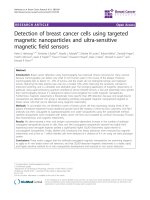
Figure 1. ASC-derived conditioned medium (CM) suppressed cell proliferation and viability in MCF-7, MDA-MB231 and MDA-MB468 breast cancer cells.
MCF7, MDA-MB231 and MDA-MB468 cells were cultured with control culture medium or conditioned K-medium (CM) for 72 hours. A) Cell morphology was observed and
recorded through microscopy analysis. Magnification: MCF-7 and MDA-MB231: ×200; MDA-MB468: ×400. B) Cell proliferation and viability were examined using the MTT assay.
1×10⁵ cells were cultured with control or ASC-derived CM as indicated concentrations for 72 hours. Presented values of bar graphs represent the mean of three independent
experiments ± s.d. *p < 0.05 compared with control cells.
Int. J. Med. Sci. 2019, Vol. 16
treated with CM derived from the incubation of 1×10⁶
ASCs for 24 h. The CM was filtered with 0.2 µm filter
to avoid contamination before using for cell culture.
All three breast cancer cell lines exhibited a significant
reduction in viability when cultured with CM. The
morphology of the attached cells became rounder,
and an increased number of floating cells was
observed, indicating cell death (Figure 1A). The
morphological observations were confirmed using an
MTT analysis. To avoid any false-positive effect
caused due to deficient nutrition in CM, we tested the
influence of ASC-derived CM following a
dose-dependent manner. ASC-derived CM was
mixed with fresh DMEM in proportion for the
experiments. As expected, all concentrations
(50-100%) of ASC-derived CM reduced cell viability
after 72 h exposure in both MCF-7 and MDA-MB231
cancer cells (Figure 1B). The similar results were also
obtained in MDA-MB468 (supplementary Figure
S3A).
ASC-derived CM triggered cell apoptosis in
MCF-7, MDA-MB231, and MDA-MB468 tumor
cells
To further examine the responses of these cells to
ASC-derived CM, cells were harvested after treatment
and DNA content was analyzed using flow
cytometry. In brief, 5 × 10⁵ cells were seeded into
6-well culture dishes overnight to allow attachment,
and treated with ASC-derived CM by following a
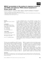
time-course protocol. As shown in Figure 2, the
quantification of cell cycle analysis indicated that the
sub-G1 population drastically increased and reached
a peak after 72 h of treatment with ASC-derived CM
in MCF-7, MDA-MB231 (Figure 2A), and
MDA-MB468 (Supplementary Figure S3B) breast
cancer cell lines. Consistent with the results obtained
from
flow
cytometry,
the
expression
of
cleaved-caspase 3, which is activated during cell
apoptosis was also demonstrated (Figure 2B, 2C).
896
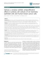
(Figure 3A). This DNA damage response was further
confirmed using a protein immunoblot. In both cells,
the protein immunoblot of γ-H2AX was more intense
than that of the control group in response to such
treatment (Figure 3B, 3C).
No cell death was identified in non-cancerous
breast cell lines under ASC-derived CM
treatment despite detection of little DNA
damage
Given our evidence that ASC-derived CM led to
DNA damage and cell apoptosis in MCF-7 and
MDA-MB231 cancer cell lines, we investigated
whether non-tumorous cells responded similarly. 1 ×
105 M10/H184B5F5 cells were seeded into 6-well
culture dishes overnight to allow attachment, and
treated with ASC-derived CM as a dose-dependent
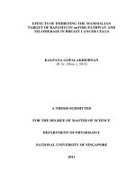
manner for 72 hours for MTT analysis. Even though
ASC-derived CM resulted in a significant decrease of
cell population in the cells after 72 hours of exposure,
the lower concentration (50 and 75%) of ASC-derived
CM dramatically enhanced cell proliferation (Figure
4A). Remarkably, no evidence of cell apoptosis was
detected in M10/H184B5F5 cells even exposed to the
highest concentration of ASC-derived CM. The cells
exhibited only an accumulation of cells with DNA
characteristic of the arrested progression in the G1
phase after treatment for the first 24 h. Few cells
exhibited sub-G1 DNA content characteristic of
apoptotic cells (Figure 4B). Consistent with this
finding, no activated- caspase 3 was evident, and only
weakly- expressed γ-H2AX was detected in
M10/H184B5F5 cells following the 72-h ASC-derived
CM incubation (Figure 4C). Taken together, these data
demonstrate entirely distinct effects of ASC-derived
CM on non-cancerous breast epitheliums. Even the
highest concentration of ASC-derived CM merely
resulted in delayed cell cycle progression in
M10/H184B5F5 rather than apoptosis.
DNA damage appeared in MCF-7 and
MDA-MB231 tumor cells when cultured with
ASC-derived CM
Fully- activated ATM-Chk2 signaling cascades
in malignant MCF-7 and MDA-MB231, but not
in non-cancerous M10/H184B5F5 cells when
exposed to ASC-derived CM
We next examined whether cell death triggered
by ASC-derived CM was accompanied by
unfavorable
DNA
damage.
We
performed
immunofluorescence assays for pSer139 histone
H2AX (γ-H2AX). γ-H2AX is recruited when DNA
damage occurs, particularly when double-strand
breaks occur. It is widely accepted as a marker for
DNA damage. MCF-7 and MDA-MB231 cells cultured
with ASC-derived CM for 72 hours exhibited the
expression of pan-nuclear γ-H2AX staining relative to
cells treated for 72 hours with control culture medium
Upon DNA damage, the ataxia telangiectasia
mutated (ATM)-Chk2 signal transduction cascade is
activated to phosphorylate a number of target
proteins to arrest cell cycle progression and trigger
DNA repair systems or cell apoptosis to ensure DNA
integrity [15-16].
To determine whether this checkpoint system
might be related to the induction of DNA damage and
cell death by ASC-derived CM, MCF-7, MDA-MB231,
and M10/H184B5F5 were cultured with ASC-derived
CM in a time-course manner and then harvested for
Int. J. Med. Sci. 2019, Vol. 16
Western blot analysis. The results revealed that
MCF-7 and MDA-MB231 cells exhibited typical DNA
damage checkpoint activation. Both pSer1981 ATM
and pThr68 Chk2 were phosphorylated within 24
hours following CM exposure. By contrast, this
pattern was greatly delayed in M10/H184B5F5 cells,
and pThr68 Chk2 activation was only weakly detected
after a 72-hours treatment with a slight increase in
pSer1981 ATM phosphorylation (Figure 5). Taken
together, these data revealed the distinct responses
between malignant tumor and non-cancerous cells
when exposed to ASC-derived CM.
897
Discussion
In the practice of plastic surgery, ASCs combined
with fat graft have been frequently used in
oncological breast reconstruction [17]. However,
many in vitro and in vivo studies have pointed out the
potential risk of ASCs in promoting breast cancer
progression [2,18]. In this regard, questions arise
whether the ASCs used in the breast reconstruction
may potentially interact with the remaining cancer
cells and promote its growth. In our study,
ASC-derived CM exhibited inhibitory effects on
breast cancer cell lines with increased DNA damage
and cell apoptosis.
Figure 2. ASC-derived conditioned medium (CM) enhanced apoptosis in MCF-7 and MDA-MB231 breast cancer cells. A) MCF7 and MDA-MB231 cells were
cultured with control culture medium or 100% conditioned K-medium (CM) for indicated time points before harvested, and the cell cycle distribution was analyzed using flow
cytometry. In each panel, cell cycle profiles are presented together with bar graphs, indicating the cell distribution in each phase of the cell cycle. Presented values represent the
mean of three independent experiments ± s.d. *p < 0.05 compared with control. B) Western blot analysis of activated-caspase-3 in MCF-7 or MDA-MB231 cells cultured with
100% ASC-derived CM for 72 hours. C) Western blot analysis of activated-caspase-3 in MCF-7 or MDA-MB231 cells cultured with 50% ASC-derived CM as indicated time points.
GAPDH levels are presented as loading controls.
Int. J. Med. Sci. 2019, Vol. 16
898
Figure 3. ASC-derived conditioned medium (CM) induced DNA damage in MCF-7 and MDA-MB231 breast cancer cells. A)
MCF-7 and MDA-MB231 cells
were cultured with 100% ASC-derived CM for 72 hours before being fixed and stained for DNA content and phosphorylated pSer139 H2AX (γ-H2AX). Images from the
immunofluorescence analysis of γ-H2AX positive cells in MCF-7 and MDA-MB231 are presented together with bar graphs, indicating the percentage of the mean of three
independent experiments ± s.d. *p < 0.05 compared with control. B) Western blot analysis of γ-H2AX in extracts prepared from MCF-7 and MDA-MB231 cells cultured with
100% ASC-derived CM for 72 h. C) Western blot analysis of γ-H2AX in extracts prepared from MCF-7 and MDA-MB231 cells cultured with 50% ASC-derived CM for indicated
time points. GAPDH levels are presented as loading controls.
The toxic metabolic waste products or the lack of
nutrition in the CM is not the reason for the inhibitory
effect observed in this study, since 50% or 75%
ASC-derived CM also suppressed cell viability on
breast cancer cell lines. While different research
groups demonstrated various responses in different
cancer cell types when interacting with ASC-derived
CM, the discrepancy between these studies may have
resulted from dissimilar ASC origins or different
culture conditions [2]. In addition to ASCs,
mesenchymal stromal cells in other studies also
support the inhibitory effects on breast cancer cell
lines with either direct co-culture or CM exposure
[19,20], even in highly malignant cell line such as
Int. J. Med. Sci. 2019, Vol. 16
MDA-MB231 [21]. Most importantly, no evidence of
increased cancer recurrence rate was noted in breast
reconstruction with fat grafts which contain ASCs
899
during long-term follow-ups [22-24]. In this study, we
provided evidences of the inhibitory effects of
ASC-derived CM on breast cancer cell lines.
Figure 4. ASC-derived conditioned medium (CM) failed to induce apoptosis in M10/H184B5F5 cells. A) M10/H184B5F5 cells were cultured with control culture
medium or conditioned K-medium (CM) as a dose-dependent manner for 72 hours before MTT analysis. Presented values represent the mean of three independent experiments
± s.d. *p < 0.05 compared with control. B) M10/H184B5F5 cells were cultured with control culture medium or 100% conditioned K-medium (CM) for indicated time points
before harvest, and the cell cycle distribution was analyzed using flow cytometry. In each panel, cell cycle profiles are presented together with bar graphs, indicating the cell
distribution in each phase of the cell cycle. Presented values represent the mean of three independent experiments ± s.d. C) Western blot analysis of γ-H2AX and activatedcaspase-3 in M10/H184B5F5 cells cultured with ASC-derived CM for indicated time points. GAPDH levels are presented as loading controls.
Int. J. Med. Sci. 2019, Vol. 16
900
Figure 5. ASC-derived conditioned medium (CM) induced fully activated ATM-Chk2 cascades in MCF-7 and MDA-MB231, but not in M10/H184B5F5 cells.
MCF-7, MDA-MB231, and M10/H184B5F5 cells were cultured with ASC-derived CM for up to 72 hours before the harvest and preparation of cell-free extracts. Phosphorylated
pSer1981 ATM and pThr68 Chk2 and the total protein level of Chk2 were analyzed using Western blot. GAPDH levels are presented as loading controls.
Notably, our data also revealed that the
ATM-Chk2 cascades were activated early by 24 hours
in both MCF-7 and MDA-MB231 breast cancer cell
lines when exposed to ASC-derived CM. This DNA
damage response and the inhibitory effects of
ASC-derived CM on tumor cell growth, cell cycle
progression, and apoptosis may be resulted from the
paracrine effect of ASCs. Some studies showed that
the inhibition of cancer cell line maybe related to the
increased level of transforming growth factor-beta
(TGF-β) [5,25] which is produced and released by
ASCs [26]. Furthermore, our results suggested that
there may be other undefined mechanisms that
protect non-cancerous M10/H184B5F5 cells against
stress caused by ASC-derived CM, because these cells
did not exhibit fully activated DNA damage signaling
and the treatment produced only minimal cell death.
In summary, our study evidently showed that
ASC-derived CM leads to DNA damage, signaling
activation of DNA damage, and eventually cell
apoptosis in breast cancer cell lines. By contrast, no
cell apoptosis was observed in the non-cancerous
breast cell lines when exposed to identical conditions.
This study provides additional information on the
ongoing debate on the potential risk of using ASCs in
breast reconstruction following oncological surgery,
however, additional data and further detailed
analysis such as the effect of cell-cell contact in ASCs
and breast cancer cells are warranted.
Supplementary Material
Supplementary figures.
/>
Acknowledgments
We would like to express our gratitude to the
Center for Research Resources and Development
(CCRD) of Kaohsiung Medical University for the
technical assistance. This study was partially funded
by grants from CGMH at Linko of Taiwan to Dr. John
Yu (OMRPG3C0041 to OMRPG3C0044); Ministry of
Science and Technology, Taiwan to Dr. Yi-Chia Wu
(MOST
103-2628-B-037-002-MY3);
Kaohsiung
Municipal Ta-Tung Hospital to Dr. Li-Ju Huang
(kmtth104-046) and Dr. Yi-Chia Wu (kmtth-105-011;
kmtth104-011); grants from Kaohsiung Medical
University Hospital to Dr. Yi-Chia Wu (kmuh98-8G42
and kmuh99-9M54); and grants from Academia
Sinica, Taiwan to Dr. Yi-Chia Wu (AS-TM-108-02-01).
This manuscript was edited by Wallace Academic
Editing. We also thank Dr. Tzu-Yu Lin for
constructive criticism of the manuscript.
Competing Interests
The authors have declared that no competing
interest exists.
References
1.
Zuk PA, Zhu M, Mizuno H, et al. Multilineage cells from human adipose
tissue: Implications for cell-based therapies. Tissue Eng. 2001; 7: 211-228.
2. Schweizer R, Tsuji W, Gorantla VS, et al. The role of adipose-derived stem cells
in breast cancer progression and metastasis. Stem Cells Int. 2015; 2015: 120949.
3. Zuk PA. The adipose-derived stem cell: Looking back and looking ahead. Mol
Biol Cell. 2010; 21: 1783-1787.
4. Klopp AH, Gupta A, Spaeth E, et al. Concise review: Dissecting a discrepancy
in the literature: do mesenchymal stem cells support or suppress tumor
growth? Stem Cells. 2011; 29: 11-19.
5. Xu WT, Bian ZY, Fan QM, et al. Human mesenchymal stem cells (hMSCs)
target osteosarcoma and promote its growth and pulmonary metastasis.
Cancer Lett. 2009; 281: 32-41.
6. Behnan J, Isakson P, Joel M, et al. Recruited brain tumor-derived mesenchymal
stem cells contribute to brain tumor progression. Stem Cells. 2014; 32:
1110-1123.
7. Cousin B, Ravet E, Poglio S, et al. Adult stromal cells derived from human
adipose tissue provoke pancreatic cancer cell death both in vitro and in vivo.
PLoS One. 2009; 4: e6278.
8. Takahara K, Ii M, Inamoto T, et al. Adipose-derived stromal cells inhibit
prostate cancer cell proliferation inducing apoptosis. Biochem Biophys Res
Commun. 2014; 446: 1102-1107.
9. Yu X, Su B, Ge P, et al. Human adipose derived stem cells induced cell
apoptosis and s phase arrest in bladder tumor. Stem Cells Int. 2015; 2015:
619290.
10. Kolle SF, Fischer-Nielsen A, Mathiasen AB, et al. Enrichment of autologous fat
grafts with ex-vivo expanded adipose tissue-derived stem cells for graft
survival: A randomised placebo-controlled trial. Lancet. 2013; 382: 1113-1120.
11. Zimmerlin L, Donnenberg AD, Rubin JP, et al. Regenerative therapy and
cancer: In vitro and in vivo studies of the interaction between adipose-derived
stem cells and breast cancer cells from clinical isolates. Tissue Eng Part A.
2011; 17: 93-106.
12. Yoshimura K, Sato K, Aoi N, et al. Cell-assisted lipotransfer for cosmetic breast
augmentation: Supportive use of adipose-derived stem/stromal cells. Aesthet
Plast Surg. 2008; 32: 48-55.
Int. J. Med. Sci. 2019, Vol. 16
901
13. Huang SH, Lin YN, Lee SS, et al. New adipose tissue formation by human
adipose-derived stem cells with hyaluronic acid gel in immunodeficient mice.
Int J Med Sci. 2015; 12: 154-162.
14. Wang WT, Catto JW, Meuth M. Differential response of normal and malignant
urothelial cells to CHK1 and ATM inhibitors. Oncogene. 2015; 34: 2887-2896.
15. Stracker TH, Usui T, Petrini JH. Taking the time to make important decisions:
The checkpoint effector kinases chk1 and chk2 and the DNA damage
response. DNA Repair (Amst). 2009; 8: 1047-1054.
16. Bartek J, Lukas J. Chk1 and chk2 kinases in checkpoint control and cancer.
Cancer Cell. 2003; 3: 421-429.
17. Arshad Z, Karmen L, Choudhary R, et al. Cell assisted lipotransfer in breast
augmentation and reconstruction: A systematic review of safety, efficacy,
use of patient reported outcomes and study quality. J Plast Reconstr Aesthetic
Surg Open. 2016; 10: 5-20.
18. Petit JY, Botteri E, Lohsiriwat V, et al. Locoregional recurrence risk after
lipofilling in breast cancer patients. Ann Oncol. 2012; 23: 582-588.
19. Kucerova L, Skolekova S, Matuskova M, et al. Altered features and increased
chemosensitivity of human breast cancer cells mediated by adipose
tissue-derived mesenchymal stromal cells. BMC Cancer. 2013; 13: 535.
20. Qiao L, Xu ZL, Zhao TJ, et al. Dkk-1 secreted by mesenchymal stem cells
inhibits growth of breast cancer cells via depression of wnt signalling. Cancer
Lett. 2008; 269: 67-77.
21. Sun B, Yu KR, Bhandari DR, et al. Human umbilical cord blood mesenchymal
stem cell-derived extracellular matrix prohibits metastatic cancer cell
mda-mb-231 proliferation. Cancer Lett. 2010; 296: 178-185.
22. Kronowitz SJ, Mandujano CC, Liu J, et al. Lipofilling of the Breast Does Not
Increase the Risk of Recurrence of Breast Cancer: A Matched Controlled Study.
Plast Reconstr Surg. 2016; 137: 385-393.
23. Petit JY, Rietjens M, Botteri E, et al. Evaluation of fat grafting safety in patients
with intraepithelial neoplasia: a matched-cohort study. Ann Oncol Off J Eur
Soc Med Oncol. 2013; 24: 1479-1484.
24. Charvet HJ, Orbay H, Wong MS, et al. The Oncologic Safety of Breast Fat
Grafting and Contradictions Between Basic Science and Clinical Studies: A
Systematic Review of the Recent Literature. Ann Plast Surg. 2015; 75: 471-479.
25. Hubackova S, Krejcikova K, Bartek J, et al. IL1- and TGFβ-nox4 signaling,
oxidative stress and DNA damage response are shared features of replicative,
oncogene-induced, and drug-induced paracrine 'bystander senescence'. Aging
(Albany NY). 2012; 4: 932-951.
26. Casiraghi F, Remuzzi G, Abbate M, et al. Multipotent mesenchymal stromal
cell therapy and risk of malignancies. Stem Cell Rev. 2013; 9: 65-79.