Rhododendron oldhamii leaf extract improves fatty liver syndrome by increasing lipid oxidation and decreasing the lipogenesis pathway in mice
Bạn đang xem bản rút gọn của tài liệu. Xem và tải ngay bản đầy đủ của tài liệu tại đây (1.76 MB, 9 trang )
Int. J. Med. Sci. 2017, Vol. 14
Ivyspring
International Publisher
862
International Journal of Medical Sciences
2017; 14(9): 862-870. doi: 10.7150/ijms.19553
Research Paper
Rhododendron oldhamii leaf extract improves fatty liver
syndrome by increasing lipid oxidation and decreasing
the lipogenesis pathway in mice
Ya-Ling Liu1*, Lei-Chen Lin2*, Yu-Tang Tung3, Shang-Tse Ho1, Yao-Li Chen4, Chi-Chen Lin5 and
Jyh-Horng Wu1
Department of Forestry, National Chung Hsing University, Taichung 402, Taiwan;
Department of Forestry and Natural Resources, National Chiayi University, Chiayi 600, Taiwan;
Graduate Institute of Metabolism and Obesity Sciences, Taipei Medical University, Taipei 110, Taiwan;
Division of General Surgery, Department of Surgery, Changhua Christian Hospital, Changhua 500, Taiwan;
Institute of Biomedical Sciences, National Chung Hsing University, Taichung 402, Taiwan.
*
Equal contributions to this paper.
Corresponding author: Tel.: +886 4 22840345-136. Fax: +886 4 22851308. E-mail: (J.-H. Wu); Tel.: +886 4 22840896-132. Fax: +886 4 22853469.
E-mail: (C.-C. Lin).
© Ivyspring International Publisher. This is an open access article distributed under the terms of the Creative Commons Attribution (CC BY-NC) license
( See for full terms and conditions.
Received: 2017.02.07; Accepted: 2017.05.21; Published: 2017.07.19
Abstract
Some members of Rhododendron genus are traditionally used as medicinal plants for arthritis, acute and
chronic bronchitis, asthma, pain, inflammation, rheumatism, hypertension and metabolic diseases. To
the best of our knowledge, there is no report on the protective effects of R. oldhamii leaf extract on
non-alcoholic fatty liver disease (NAFLD) in vivo and in vitro. In this study, the effects of R. oldhamii leaf
extract on inhibiting the free fatty acid (FFA)-induced accumulation of fat in HepG2 cells and on
improving fatty liver syndrome in mice with high fat diet (HFD)-induced NAFLD were investigated. For
the in vitro assay, HepG2 cells were treated with FFAs (oleate/palmitate = 2:1) with or without
treatment with R. oldhamii leaf ethyl acetate (EtOAc) fraction to observe lipid accumulation using Nile
red and oil red O stains. For the in vivo assay, C57BL/6 mice were randomly assigned to three groups (n
= 5), including the normal diet group, the HFD group and the HFD+EtOAc group. After 11 weeks, body
weight, serum biochemical indices and the mRNA expressions of the liver tissue, as well as the outward
appearance, weight and histopathological analysis of liver and adipose tissues were evaluated. Among
the fractions derived from R. oldhamii leaf, the EtOAc fraction exhibited a strong fat-accumulation
inhibitory activity. Following reverse-phase high-performance liquid chromatography (HPLC), four
specific phytochemicals, including (2R, 3R)-astilbin (AS), hyposide (HY), guaijaverin (GU) and quercitrin
(QU), were isolated and identified from the EtOAc fraction of R. oldhamii leaf extract. Among them, AS
and HY showed excellent fat-accumulation inhibitory activity. Thus, the EtOAc fraction of R. oldhamii
leaf and its derived phytochemicals have great potential in preventing FFA-induced fat accumulation. In
addition, the EtOAc fraction of R. oldhamii leaf significantly improved fatty liver syndrome and reduced
total cholesterol (TC) and triglyceride (TG) in HFD-induced NAFLD mice at a dosage of 200 mg/kg BW.
These results demonstrated that the methanolic extracts from R. oldhamii leaf have excellent inhibitory
activities against fat accumulation and anti-NAFLD activities and thus have great potential as a natural
health product.
Key words: Rhododendron oldhamii, free fatty acid (FFA), fat accumulation, high fat diet (HFD), non-alcoholic
fatty liver disease (NAFLD)
Introduction
In 2014, an estimated 600 million adults were
obesity according to the World Health Organization.
Obesity increases the risk of a number of health
problems, including coronary disease, particular
Int. J. Med. Sci. 2017, Vol. 14
types of carcinoma, respiratory system complications
and osteoarthritis of small and large joints [1].
Genetic, physiological and psychological factors, as
well as dietary habits, physical activity, lifestyle and
social and environmental factors are responsible for
the significant increase in the prevalence of obesity
and its consequences [1-4]. Obesity is a condition
when fat accumulation is excessive to the extent that it
produces adverse health consequences [1]. In the first
stage of the two-hit hypothesis, fat accumulation in
hepatocytes leads to steatosis, which is related to
obesity [5]. In addition, non-alcoholic fatty liver
disease (NAFLD) has been considered the 2-stage
process of the two-hit hypothesis.
The Rhododendron genus is widely distributed
throughout most of the world except for Africa and
South America [6]. In traditional medicine, some
members of the genus Rhododendron have been used to
treat diseases, including arthritis, acute and chronic
bronchitis, asthma, pain, inflammation, rheumatism,
hypertension and metabolic diseases [7, 8]. A variety
of phytochemicals with significant bioactivities,
including iridoids [9], diterpenoids [10], triterpenoids
[11], chromane derivatives, [12] and flavonoids [13],
have been discovered in this genus. R. groenlandicum
is a popular beverage to treat diabetes symptoms [14].
Ouchfoun et al. [15] showed that R. groenlandicum
alleviates insulin resistance in a high fat diet
(HFD)-induced obesity mice. R. arboreum has
hypolipidemic
activity
in
a
diet-induced
hypercholestermic rabbits [16, 17]. In addition, the
methanolic extract of R. arboretum also showed
significant in-vitro antidiabetic activity [18]. Therefore,
the present study was undertaken to investigate the
863
anti-NAFLD effect of R. oldhamii leaf extract.
The anti-NAFLD effects of flavonoids in vitro
and in vivo models have been reported in several
studies [19]. Flavonoids have been shown to help in
treating and reducing the risk of obesity [20-22]. In
previous studies, plant catechins and anthocyanins
reduced the weight of abdominal adipose tissues on
diet induced obesity animal models [23]. R. oldhamii is
rich in flavonoids, including (2R, 3R)-epicatechin, (2R,
3R)-taxifolin, (2R, 3R)-astilbin (AS), hyposide (HY),
guaijaverin (GU) and quercitrin (QU) [24]. Therefore,
R. oldhamii may be a good candidate for further
development as a remedy for treating fatty liver
syndrome. However, to the best of our knowledge,
there is no prior report on the improvement of R.
oldhamii leaf on fatty liver syndrome in HFD-induced
NAFLD mice. Thus, we used both lipid accumulation
induced by free fatty acid (FFA; oleate/palmitate =
2:1) in HepG2 cells and HFD-induced NAFLD mouse
model to investigate the anti-fatty liver effect of the
methanolic extract from R. oldhamii leaf.
Methods
Plant materials
The leaves of Rhododendron oldhamii Maxim. were
collected at the end of April 2011 from the Lion Head
Mountain of Taipei county in Taiwan. The species
were confirmed by Dr. Lei-Chen Lin of National
Chiayi University.
Extraction, fractionation, and isolation
Extraction, fractionation and isolation were
followed by the method of Tung et al. [24]. The leaves
were soaked in methanol at ambient temperature for 7
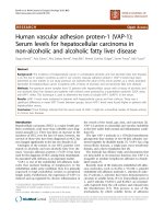
Figure 1. Chemical structures of four major phytocompounds isolated from the EtOAc fraction of R. oldhamii leaf: (2R, 3R)-astilbin (AS), hyposide (HY), guaijaverin
(GU) and quercitrin (QU).
Int. J. Med. Sci. 2017, Vol. 14
days to obtain extract. The crude extract was then
fractionated with n-hexane, ethyl acetate (EtOAc),
n-butanol (BuOH) and water to yield soluble hexane,
EtOAc, BuOH and water fractions. The 4 major
phytochemicals, AS, HY, GU and QU (Figure 1), from
the EtOAc fraction were isolated and characterized by
HPLC and NMR, respectively.
Cell culture
The HepG2 cell line purchased from ATCC was
cultured in Dulbecco’s modified Eagle’s medium
(DMEM) supplemented with 10% fetal bovine serum
(FBS). The cells were incubated in a 5% CO2 incubator
at 37°C.
Oil red O staining
HepG2 cells (2 × 10⁵ cells/well) were seeded into
a 6-well plate incubated for 24 h to allow cell
adherence. First, 2 mL of fresh medium containing
test samples was added into the cultures. After 1 h of
incubation
at
37°C,
0.25
mM
of
FFAs
(oleate/palmitate, 2:1) was added to the medium and
incubated at 37°C for 24 h. The cells were rinsed with
cold phosphate buffered saline (PBS) and fixed in 1%
paraformaldehyde for 30 min. After the cells were
washed with 75% EtOH, the cells were stained for 20
min in a 2 mg/mL oil red O solution to determine
hepatic lipid accumulation. After the stain was
removed, the cells were washed with PBS and then
counterstained with hematoxylin for 20 s.
Representative
photomicrographs
(400×
magnification) were conducted by a camera mounted
onto a microscope.
Nile red staining
Nile red staining was used to specifically stain
the intracellular fat. HepG2 cells (2 × 10⁵ cells/well)
were seeded into a 6-well plate and incubated for 24 h
to allow cell adherence. First, 1 mL of fresh medium
containing the test samples was added into the
cultures. After 1 h of incubation at 37°C, 1 mM of
FFAs (oleate/palmitate, 2:1) was added to the
medium and incubated at 37°C for 24 h. The cells
were collected using 0.05% Trypsin-EDTA and
incubated with Nile red (1 μg/mL) in PBS for 10 min.
After PBS washed, the cells were suspended in 1%
formaldehyde and then measured by flow cytometry
at a laser excitation wavelength of 488 nm.
Cell viability assay
To measure the cytotoxicity on the test samples,
HepG2 cells (1 × 10⁴ cells/well) were seeded into a
24-well plate and incubated for 24 h to allow cell
adherence. First, 1 mL of fresh medium containing the
test samples was added into the cultures and
incubated at 37°C for 24 h. Following the removal of
864
the medium, 1 mL of tetrazolium salt solutions (1 mL
3-(4,5-dimethylthiazol-2-yl)-2,5-diphenyl tetrazolium
bromide (MTT) in 10 mL DMEM) was added. After 3
h of incubation at 37°C, the medium was removed
and 600 μL of dimethyl sulfoxide (DMSO) was added
to dissolve the formazan crystals. Absorbance was
measured at a wavelength of 570 nm using an
enzyme-linked immunosorbent assay (ELISA) reader.
Animals
The C57BL/6 mice (6 weeks old) were given a
standard laboratory diet and distilled water ad libitum
and kept on a 12 h light/dark cycle at 24 ± 2°C. This
study was conducted according to institutional
guidelines and approved by the Institutional Animal
Care and Use Committee (IACUC) of National Chung
Hsing University and conformed to the guidelines of
the protocol IACUC-10393 approved by the IACUC
ethics committee. After 1 week of acclimatization, 15
mice were randomly divided into two groups: the
normal group (n = 5) was fed a standard chow diet
(ND) and the experimental group (n = 10) was fed a
HFD. The experimental mice were divided into two
groups (n = 5/group): 1) HFD receiving no treatment
(HFD) and 2) HFD receiving 50 mg/kg of the EtOAc
fraction from R. oldhamii leaf. Food intake and body
weight were recorded. Following 11 weeks of
treatment, mice were sacrificed at 18 weeks of age. At
the end of the experiment, each mouse was
anesthetized, and the liver and epididymal fat pad
tissues were collected.
Biochemical analysis of serum samples
Mouse blood samples were centrifuged at 1,400 g
at 4°C for 15 min, and the levels of serum glucose,
glutamate-pyruvate transaminase (GPT), triglyceride
(TG) and total cholesterol (TC) were measured using
an autoanalyzer (Hitachi 7060, Hitachi, Japan).
Pathological histology
Liver and epididymal fat pad tissues were fixed
in 10% buffered formaldehyde, and histologically
examined with hematoxylin and eosin (H&E)
staining.
RT-PCR
Total RNA of the liver tissue was extracted using
Trizol reagent (Invitrogen) following the protocol
specified by the manufacturer. RT-PCR was followed
by the method of Tung et al. [25]. The gene
expressions of 7 genes (SREBP1, ACC, FAS, CPT1α,
PPARα and PPARγ) using complementary DNA from
liver tissue was analyzed. GAPDH was used as an
internal control.
Int. J. Med. Sci. 2017, Vol. 14
865
Figure 2. Effects of R. oldhamii leaf soluble fractions on intracellular lipid accumulation in HepG2 cells. HepG2 cells were pretreated with 200 μg/kg of R. oldhamii leaf
crude and soluble fractions of hexane, EtOAc, BuOH and water in the presence of 0.25 mM of FFAs (oleate/palmitate, 2:1) for 24 h. The cells were stained with Nile
red and analyzed by flow cytometry, after which the percentage of lipid accumulation was quantitated (A). Cells were stained with oil red O and analyzed by
spectrophotometry (B). Cytotoxicity of R. oldhamii leaf soluble fractions in HepG2 cells was measured using the MTT assay (C). The results are presented as the mean
± SD (n = 3). The bars marked by different letters are significantly different at the level of p<0.05 according to Scheffe’s test.
Statistical analyses
The data for the in vitro and in vivo assays were
expressed as the mean ± SD (n = 3) and the mean ±
SEM (n = 5), respectively. Significant differences were
calculated by Scheffe’s test, and results with p<0.05
were considered statistically significant.
Results
Effects of R. oldhamii leaf extract soluble
fractions on FFA-induced lipid accumulation in
HepG2 cells
The imbalance between the hepatic uptake of
FFAs, TG synthesis and TG excretion leads to fatty
liver [26]. Recently, Gomez-Lechon et al. [27]
demonstrated that FFA (oleate/PA = 2:1)-induced
HepG2 cells mimic benign chronic steatosis. Wu et al.
[26] showed that FFA-overloaded HepG2 cells
reached maximal intracellular lipid accumulation that
was similar with human steatosis. First, we examined
whether R. oldhamii leaf crude and soluble fractions of
hexane, EtOAc, BuOH and water were able to reduce
lipid accumulation in HepG2 cells induced by FFA
using Nile red staining and oil red O staining. The
quantitative data from the Nile red staining displayed
that the crude, hexane and EtOAc fractions reduced
lipid accumulation by 21% (p<0.05), 32% (p<0.05) and
54% (p<0.05), respectively (Figure 2A). Among these,
the EtOAc fraction significantly reduced FFA-induced
Int. J. Med. Sci. 2017, Vol. 14
lipid accumulation (Figure 2A and 2B). In addition,
the MTT assay revealed no significant cytotoxic effects
866
on cells treated with 200 μg/mL of the EtOAc fraction
(Figure 2C).
Bioassay-guided
fractionation and
quantification of major
phytochemicals in R.
oldhamii leaf EtOAc fraction
R. oldhamii leaf EtOAc
fraction showed a stronger
inhibitory activity on lipid
accumulation than the other
fractions, indicating that R.
oldhamii leaf extract had an
inhibitory activity against lipid
accumulation, and efficiently
enriched in the EtOAc fraction.
Thus, the phytochemical characteristics of this fraction were
further investigated in this
study. The four major phytochemicals of R. oldhamii leaf
EtOAc fraction were found to be
AS, HY, GU and QU (Figure 1),
and
their
contents
were
determined to be 130.8 ± 10.9,
105.5 ± 8.5, 104.1 ± 4.7 and 108.6
± 4.0 mg per gram of EtOAc
fraction, respectively.
Effects of the
phytochemicals from R.
oldhamii leaf on
FFA-induced lipid
accumulation in HepG2 cells
Figure 3. Effects of the phytochemicals from R. oldhamii leaf EtOAc fraction on intracellular lipid accumulation in
HepG2 cells. HepG2 cells were pretreated with 200 μM of AS, HY, GU and QU in the presence of 0.25 mM of
FFAs (oleate/palmitate, 2:1) for 24 h. Cells were stained with Nile red by flow cytometry, after which the
percentage of lipid accumulation was quantitated (A). HepG2 cells were pretreated with 50 μM and 100 μM of
AS and HY in the presence of FFAs for 24 h. Cells were stained with Nile red by flow cytometry, after which the
percentage of lipid accumulation was quantitated (B). HepG2 cells were pretreated with 200 μM of AS, HY, GU
and QU in the presence of FFAs for 24 h. Cells were stained with oil red O by spectrophotometry (C).
Cytotoxicity of the phytochemicals in HepG2 cells was measured using the MTT assay. The results are presented
as the mean ± SD (n = 3). The bars marked by different letters are significantly different at the level of p<0.05
according to Scheffe’s test.
The inhibitory effects of the
phytochemicals from R. oldhamii
leaf on lipid accumulation are
shown in Figure 3A and 3C. To
understand the relationship
between the phytochemicals of
R. oldhamii leaf and its lipid
accumulation inhibitory effects
in HepG2 cells, 4 constituents,
namely AS, HY, GU and QU,
were
tested.
The
lipid
accumulation inhibitory activities of the four constituents ranked in the following order, from
highest to lowest: AS > HY > GU
> QU. In addition, the MTT
assay revealed no significant
cytotoxic effects on cells treated
with the 4 phytochemicals at the
200 μM dosage (Figure 3D).
Int. J. Med. Sci. 2017, Vol. 14
Among the 4 phytochemicals tested, AS and HY
exhibited the strongest activities. To examine further
the inhibition of lipid accumulation in FFA-stimulated
HepG2 cells, we selected 2 doses (50 μM and 100 μM)
of AS and HY. Figure 3B shows that HY inhibited
FFA-induced lipid accumulation in a concentrationdependent manner.
Effects of R. oldhamii leaf EtOAc fraction on
body weight and diet
The effects of R. oldhamii leaf EtOAc fraction on
final body weight and diet intake in HFD-fed mice are
shown in Figure 4A and 4B, respectively. The initial
body weight for the control and HFD rats were
similar. Mice fed the normal diet and HFD continued
to increase body weights until the end of the study.
After 2 weeks HFD-induced obesity, the body weight
was higher for the HFD mice than for the control mice
(p<0.05). After treatment with the EtOAc fraction in
the HFD mice, the body weight significantly
decreased compared with the HFD group (p<0.05).
867
The average final body weight of HFD+EtOAc group
decreased by 16.9% compared with the HFD group. In
addition, the HFD group and the HFD+EtOAc group
did not show diarrhea during the experiment. HFD
groups with or without treatment with R. oldhamii leaf
EtOAc fraction reduced their daily food intake
compared with the vehicle group because HFD had a
higher calorie intake than the normal diet.
Table 1. The Epididymal Fat Weights, Liver Weights and Serum
Levels of C57BL/6 Mice
Epididymal fat weight (g)
Liver weight (g)
Serum levels
Glucose (mmol/L)
GPT (U/L)
TG (mg/dl)
TC (mg/dl)
ND
0.14 ± 0.02
0.86 ± 0.09
HFD
1.08 ± 0.19##
1.01 ± 0.08#
HFD+EtOAc
0.48 ± 0.19**
0.81 ± 0.07**
63 ± 23
133 ± 100
81 ± 5
77 ± 7
112 ± 35#
163 ± 69
165 ± 65#
166 ± 10##
118 ± 18
127 ± 61
116 ± 17
156 ± 13
p<0.05; ##p<0.01 compared with the ND group. **p<0.01 compared with the HFD
group.
#
Effects of R. oldhamii leaf
EtOAc fraction on
epididymal fat pad and liver
The epididymal fat pad
(EFP) and liver tissue weights
at the end of the study are
shown in Table 1 and Figure
4C. The weights of the EFP and
liver were higher for HFD
alone than for the control mice
by 671% (p<0.01) and 17%
(p<0.05), respectively. However, the HFD+EtOAc group
decreased the weights of both
EFP and liver by 56% (p<0.05)
and 20% (p<0.01) compared
with HFD alone. Recent studies
showed that high-fat diet
induced obesity rats underwent
to several enzymatic changes
which
are
related
to
carbohydrate metabolism in
liver and adipose tissues [28,
29]. Thus, this supports the
possibility that R. oldhamii leaf
EtOAc fraction may reduce the
formation of adipose tissue
through
altering
several
enzymatic reactions.
Figure 4. Effects of R. oldhamii leaf EtOAc fraction on weekly body weight (A) and diet (B) over the course of 11
weeks in HFD-induced NAFLD mice. The results are presented as the mean ± SEM (n = 5). The markers by
different letters are significantly different at the level of p<0.05 according to Scheffe’s test. Outward appearances,
epididymal fats and perirenal fats of C57BL/6 mice at the end of the study (C).
Int. J. Med. Sci. 2017, Vol. 14
868
Effects of R. oldhamii leaf
EtOAc fraction on the
expression of genes
involved in lipid oxidation
and lipogenesis
The mRNA expression
patterns of genes encoding
enzymes
involved
in
lipogenesis (SREBP1, ACC and
FAS) and lipid oxidation
(CPT1α, PPARα and PPARγ)
were assessed using real-time
RT-PCR (Figure 6). SREBP1
was reduced by 21% (p<0.05),
and CPT1α, PPARα and PPARγ
were increased by 14%
(p<0.05), 30% (p<0.05) and 19%
(p<0.01), respectively, in the
Figure 5. Effects of R. oldhamii leaf EtOAc fraction on the hematoxylin and eosin (H&E) staining of histologically
HFD+EtOAc group compared
sectioned liver and epididymal fat tissues in HFD-induced NAFLD mice.
with the HFD group. Thus, the
HFD mice treated with R.
oldhamii leaf EtOAc fraction
exhibited mRNA levels markedly decreased for the
Effects of R. oldhamii leaf EtOAc fraction on
lipogenesis gene (SREBP) and increased for the lipid
serum biochemical parameters
oxidation genes (CPT1α, PPARα and PPARγ).
The effects of the EtOAc fraction from R. oldhamii
leaf on glucose, GPT, TG and TC are shown in Table 1.
Consumption of a HFD increased the serum levels of
glucose, GPT, TG and TC by 77.3% (p<0.05), 23%,
104% (p<0.05) and 116% (p<0.05), respectively. After
treatment with R. oldhamii leaf EtOAc fraction, the
HFD+EtOAc group exhibited a slight decrease in the
serum levels of GPT (22%), TG (30%) and TC (6%)
compared with the HFD group.
Effects of R. oldhamii leaf EtOAc fraction on
hepatomegaly
As shown in Figure 5A, histological analysis of
the liver tissues revealed abundant foamy cells in the
HFD group, and the HFD+EtOAc group showed
relatively normal cells compared with the HFD group.
R. oldhamii leaf EtOAc fraction had inhibitory effects
on the formation of foamy cells extending from the
hepatic portal vein to the central vein. In addition,
adipose tissues of the HFD group showed striking
morphological changes (Figure 5B). The EFPs of the
HFD+EtOAc group were smaller in size than those of
the HFD group, indicating that the reduced total fat
mass from treatment with R. oldhamii leaf EtOAc
fraction may result from decreased TG accumulation
rather than a reduced number of adipocytes.
Figure 6. Changes in mRNA expression levels of lipogenesis (SREBP1, ACC and
FAS) and lipid oxidation (CPT1α, PPARα and PPARγ) genes in the ND, HFD and
HFD+EtOAc groups. The transcription levels of SREBP1, ACC, FAS, CPT1α,
PPARα and PPARγ genes were normalized by an internal GAPDH mRNA control.
The data are expressed as the mean ± SE (n = 5). *p<0.05 and **p<0.01.
Int. J. Med. Sci. 2017, Vol. 14
Discussion
In recent studies, phytochemicals of plants have
shown potential as drug in several metabolic diseases
[30]. They have served as template molecules for the
development of new drugs [30]. FFA-induced
hepatocellular steatosis models have been used to
study in several studies on NAFLD pathogenesis and
anti-NAFLD drugs [31, 32]. Although primary human
hepatocytes represent the most relevant model for the
human liver, they are difficult to prepare. Thus, the
reproducibility of experimental results is often a large
problem [33, 34]. In contrast, the regulation of
metabolism of HepG2 cells is different from normal
hepatocytes, as HepG2 cells have acquired genetic
and epigenetic alterations [35]. In this study, the
HepG2 cells with FFA (oleate/palmitate =
2:1)-induced lipid accumulation were used to examine
the effects of R. oldhamii leaf methanolic extract and its
phytochemicals on fatty liver syndrome. Among the
fractions derived from R. oldhamii leaf, the EtOAc
fraction exhibited a strong inhibitory activity against
fat accumulation. Following reverse-phase HPLC,
four specific phytochemicals were isolated and
identified from the EtOAc fraction of R. oldhamii leaf
extract. In addition, AS and HY showed excellent
inhibitory activity against fat accumulation. Thus, R.
oldhamii leaf EtOAc fraction and its derived
phytochemicals have great potential in preventing
FFAs from causing fat accumulation. Next, we
examined whether R. oldhamii leaf EtOAc fraction
improved fatty liver syndrome in vivo.
The overconsumption of caloric food contributes
to visceral obesity. Therefore, the HFD-induced
model represents a valuable tool for investigating and
validating new therapeutic avenues for the treatment
of obesity and NAFLD [15]. The results of this study
showed that R. oldhamii leaf EtOAc fraction
significantly decreased body weight in HFD-induced
NAFLD mice at a dosage of 200 mg/kg (Figure 4A).
This attenuated body weight may be due to the
amount decrease of fatty tissue (Table 1) and the
adipocyte size (Figure 4C). R. oldhamii leaf EtOAc
fraction also significantly reduced the macrovesicular
fat quantity in liver tissues of HFD-induced NAFLD
mice. In addition, HFD alone elevated TC and TG
levels compared with the controls, and R. oldhamii leaf
EtOAc fraction significantly reduced the elevated
levels of TC and TG in HFD-induced NAFLD mice
(Table 1). DeAngelis et al. [36] found that the
accumulation of TC in liver parenchymal cells
(increase in fat or steatosis), a common liver
pathology, is a well-established effect of obesity.
Interesting, R. oldhamii leaf EtOAc fraction markedly
decreased SREBP1 gene expression and increased
CPT1α, PPARα and PPARγ in the livers of
869
HFD-induced NAFLD mice. These results indicate
that R. oldhamii leaf EtOAc fraction affected fat
deposition by stimulating lipid oxidation and
inhibiting the lipogenesis pathway.
Conclusions
This study demonstrated that consuming 200
mg/kg BW of the EtOAc fraction from R. oldhamii leaf
for 11 weeks attenuated body weight gain and serum
GPT in HFD-induced NAFLD mice. In addition, R.
oldhamii leaf EtOAc fraction significantly decreased
SREBP-1 mRNA; increased CPT1α, PPARα and PPARγ
mRNA in the liver tissues; and reduced the TG
content and TC accumulation in treated HFD-induced
NAFLD mice. Therefore, this study demonstrated that
the protective effect of R. oldhamii leaf in HFD-induced
NAFLD mice occurs by an increase in the hepatic lipid
oxidation and a decrease in the hepatic lipogenesis
pathways.
Acknowledgments
This work was financially supported by a
research grant from the National Chung Hsing
University (NCHU-CCH 10311).
Competing Interests
The authors declare that there is no conflict of
interests regarding the publication of this paper.
References
1.
2.
3.
4.
5.
6.
7.
8.
9.
10.
11.
12.
13.
14.
15.
Huang CC, Tseng TL, Huang WC, Chung YH, Chuang HL, Wu JH.
Whole-body vibration training effect on physical performance and obesity in
mice. International Journal of Medical Sciences. 2014; 11: 1218–1227.
Grundy SM. Multifactorial causation of obesity: implications for prevention.
American Journal of Clinical Nutrition. 1998; 67: 563S–572S.
Hill JO, Peters JC. Environmental contributions to the obesity epidemic.
Science. 1998; 280: 1371–1374.
Wickelgren I. Obesity: how big a problem? Science. 1998; 280: 1364–1367.
Salt WB. Nonalcoholic fatty liver disease (NAFLD): a comprehensive review.
Journal of insurance medicine. 2004; 36: 27–41.
Chung JD, Lin TP, Chen YL, Cheng YP, Hwang SY. Phylogeographic study
reveals the origin and evolutionary history of a Rhododendron species complex
in Taiwan. Molecular Phylogenetics and Evolution. 2007; 42: 14–24.
Popescu R, Kopp B. The genus Rhododendron: an ethnopharmacological and
toxicological review. Journal of Ethnopharmacology. 2013; 147: 42–62.
Iwata N, Wang N, Yao X, Kitanaka S. Structures and histamine release
inhibitory effects of prenylated orcinol derivatives from Rhododendron
dauricum. Journal of Natural Products. 2004; 67: 1106–1109.
Fan CQ, Zhao WM, Ding BY, Qin GW. Constituents from the leaves of
Rhododendron latoucheae. Fitoterapia. 2001; 72: 449−452.
Liu CC, Lei C, Zhong Y, Gao LX, Li JY, Yu MH, et al. Novel grayanane
diterpenoids from Rhododendron principis. Tetrahedron. 2014; 70: 4317−4322.
Choi YH, Zhou W, Oh J, Choe S, Kim DW, Lee SH, et al. Rhododendric acid A,
a new ursane-type PTP1B inhibitor from the endangered plant Rhododendron
brachycarpum G. Don. Bioorganic & Medicinal Chemistry Letters. 2012; 22:
6116−6119.
Iwata N, Kitanaka S. Tetracyclic chromane derivatives from Rhododendron
anthopogonoides. Journal of Natural Products. 2010; 73: 1203−1206.
Lin CY, Lin LC, Ho ST, Tung YT, Tseng YH, Wu JH. Antioxidant activities and
phytochemicals of leaf extracts from 10 native Rhododendron species in Taiwan.
Evidence-Based Complementary and Alternative Medicine. 2014; 2014:
283938.
Leduc C, Coonishish J, Haddad P, Cuerrier A. Plants used by the Cree Nation
of Eeyou Istchee (Quebec, Canada) for the treatment of diabetes: a novel
approach in quantitative ethnobotany. Journal of Ethnopharmacology. 2006;
105: 55−63.
Ouchfoun M, Eid HM, Musallam L, Brault A, Li S, Vallerand D, et al. Labrador
tea (Rhododendron groenlandicum) attenuates insulin resistance in a
Int. J. Med. Sci. 2017, Vol. 14
16.
17.
18.
19.
20.
21.
22.
23.
24.
25.
26.
27.
28.
29.
30.
31.
32.
33.
34.
35.
36.
870
diet-induced obesity mouse model. European Journal of Nutrition. 2015; 55:
941−954.
Murty D, Rajesh E, Raghava D, Raghavan TV, Surulivel MK. Hypolipidemic
effect of arborium plus in experimentally induced hypercholestermic rabbits.
Yakugaku Zasshi. 2010; 130: 841−846.
Verma N, Amresh G, Sahu PK, Rao ChV, Singh AP. Antihyperglycemic and
antihyperlipidemic activity of ethyl acetate fraction of Rhododendron arboreum
Smith flowers in streptozotocin induced diabetic rats and its role in regulating
carbohydrate metabolism. Asian Pacific Journal of Tropical Biomedicine. 2012,
2: 696−701.
Bhandary MR, Kawabata J. Antidiabetic activity of Laligurans (Rhododendron
arboreum Sm.) flower. Journal of Food Science and Technology. 2008, 4: 61−63.
Pisonero-Vaquero
S,
González-Gallego
J,
Sánchez-Campos
S,
García-Mediavilla MV. Flavonoids and related compounds in non-alcoholic
fatty liver disease therapy. Current Medicinal Chemistry. 2015, 22: 2991−3012.
Narkhede MB. Evaluation of alpha amylase inhibitory potential of four
traditional culinary leaves. Asian Journal of Pharmaceutical and Clinical
Research. 2012, 5: 75−76.
Tsuda T. Regulation of adipocyte functions by anthocyanins: possibility of
preventing the metabolic syndrome. Journal of Agricultural and Food
Chemistry. 2008, 56: 642−646.
Ghosh S, Ahire M, Patil S, Jabgunde A, Bhat Dusane M, Joshi BN, et al.
Antidiabetic activity of Gnidia glauca and Dioscorea bulbifera: potent amylase
and glucosidase inhibitors. Evidence-Based Complementary and Alternative
Medicine. 2012, 2012: 929051.
Murase T, Nagasawa A, Suzuki J, Hase T, Tokimitsu I. Beneficial effects of tea
catechins on diet induced obesity: stimulation of lipid metabolism in the liver.
International Journal of Obesity. 2002, 26: 1459−1464.
Tung YT, Lin LC, Liu YL, Ho ST, Lin CY, Chuang HL, et al. Antioxidative
phytochemicals from Rhododendron oldhamii Maxim. leaf extracts reduce serum
uric acid levels in potassium oxonate-induced hyperuricemic mice. BMC
Complementary and Alternative Medicine. 2015, 15: 423.
Tung YT, Chen HL, Lai CW, Shen CJ, Lai YW, Chen CM. Curcumin reduces
pulmonary tumorigenesis in vascular endothelial growth factor
(VEGF)-overexpressing transgenic mice. Molecular Nutrition & Food
Research. 2011, 55: 1036–1043.
Wu X, Zhang L, Gurley E, Studer E, Shang J, Wang T, et al. Prevention of free
fatty acid-induced hepatic lipotoxicity by 18β-glycyrrhetinic acid through
lysosomal and mitochondrial pathways. Hepatology. 2008, 47: 1905−1915.
Gómez-Lechón MJ, Donato MT, Martínez-Romero A, Jiménez N, Castell JV,
O'Connor JE. A human hepatocellular in vitro model to investigate steatosis.
Chemico-Biological Interactions. 2007, 165: 106–116.
Arias N, Macarulla MT, Aguirre L, Milton I, Portillo MP. The combination of
resveratrol and quercetin enhances the individual effects of these molecules on
triacylglycerol metabolism in white adipose tissue. European Journal of
Nutrition. 2016, 55: 341−348.
Yeh TS, Chan KH, Hsu MC, Liu JF. Supplementation with soybean peptides,
taurine, Pueraria isoflavone, and ginseng saponin complex improves
endurance exercise capacity in humans. Journal of Medicinal Food. 2011, 14:
219−225.
Kang OH, Kim SB, Seo YS, Joung DK, Mun SH, Choi JG, et al. Curcumin
decreases oleic acid-induced lipid accumulation via AMPK phosphorylation in
hepatocarcinoma cells. European Review for Medical and Pharmacological
Sciences. 2013, 17: 2578−2586.
Ricchi M, Odoardi MR, Carulli L, Anzivino C, Ballestri S, Pinetti A, et al.
Differential effect of oleic and palmitic acid on lipid accumulation and
apoptosis in cultured hepatocytes. Journal of Gastroenterology and
Hepatology. 2009, 24: 830−840.
Gómez-Lechón MJ, Donato MT, Martínez-Romero A, Jiménez N, Castell JV,
O'Connor JE. A human hepatocellular in vitro model to investigate steatosis.
Chemico-Biological Interactions. 2007, 165: 106−116.
Gómez-Lechón MJ, Donato MT, Castell JV, Jover R. Human hepatocytes as a
tool for studying toxicity and drug metabolism. Current Drug Metabolism.
2003, 4: 292−312.
Gómez-Lechón MJ, Donato MT, Castell JV, Jover R. Human hepatocytes in
primary culture: the choice to investigate drug metabolism in man. Current
Drug Metabolism. 2004, 5: 443−462.
De Gottardi A, Vinciguerra M, Sgroi A, Moukil M, Ravier-Dall'Antonia F,
Pazienza V, et al. Microarray analyses and molecular profiling of steatosis
induction in immortalized human hepatocytes. Laboratory Investigation.
2007, 87: 792−806.
DeAngelis RA, Markiewski MM, Taub R, Lambris JD. A high-fat diet impairs
liver regeneration in C57BL/6 mice through overexpression of the NF-κB
inhibitor, IκBα. Hepatology. 2005, 42: 1148−1157.