Evaluation of two rapid field level diagnostic tools for acute canine Leptospirosis in an endemic area
Bạn đang xem bản rút gọn của tài liệu. Xem và tải ngay bản đầy đủ của tài liệu tại đây (245.13 KB, 6 trang )
Int.J.Curr.Microbiol.App.Sci (2019) 8(2): 10-15
International Journal of Current Microbiology and Applied Sciences
ISSN: 2319-7706 Volume 8 Number 02 (2019)
Journal homepage:
Original Research Article
/>
Evaluation of Two Rapid Field Level Diagnostic Tools for Acute Canine
Leptospirosis in an Endemic Area
R. Ambily1*, M. Mini1, Siju Joseph1 and S.V. Krishna2
1
Department of Veterinary Microbiology, College of Veterinary and Animal Sciences,
Mannuthy -680 651, India
2
Department of Veterinary Microbiology, College of Veterinary Science,
Mamnoor Warangal Dist., India
*Corresponding author
ABSTRACT
Keywords
Acute canine
leptospirosis,
pPro.EX.HtC, IgM
dot ELISA, IgM
plate ELISA
Article Info
Accepted:
04 January 2018
Available Online:
10 February 2019
The present study aims at evaluating the efficacy of two rapid tools, IgM dot ELISA and
Latex Agglutination Test employing recombinant LipL32 as antigen in diagnosing acute
leptospirosis in dogs in Kerala, a disease endemic area. One hundred and five serum
samples were collected from dogs presented at Veterinary Hospitals attached to Kerala
Veterinary and Animal Sciences University during the period from September 2014 to
May 2016. Microscopic Agglutination test was carried out using the reference strains
maintained in the Dept. of Veterinary Microbiology, College of Veterinary and Animal
Sciences, Mannuthy. In MAT, samples with titre ≥ 1:800 were considered as positive. For
recombinant LipL32 protein production, the lipl32 gene of Leptospira interrogans was
ligated with prokaryotic expression vector pPro.EX.HtC and transformed into competent
E. coli DH5α cells. The clones were induced with Isopropyl β-D-1-thiogalactopyranoside
(IPTG) and the protein was purified using Nickel affinity chromatography and used as
antigen in IgM dot ELISA, plate ELISA and Latex Agglutination Test (LAT). All the 105
samples were subjected to the three tests. The relative sensitivity and specificity of IgM
dot ELISA, plate ELISA and LAT were evaluated in comparison with MAT. Among the
105 samples, 56 were found to have a titre of ≥1:800 in MAT. In IgM dot ELISA, 55
samples were found to be positive and one was negative. Latex Agglutination Test
detected 58 samples as positive. It was found to be a less specific (91.84 %) test than IgM
dot ELISA in detecting acute leptospirosis although it was found to be sensitive (96.43 %).
IgM dot ELISA was proved to be sensitive (96.43 %) and specific (97.96 %) and the
results were more satisfactory than IgM plate ELISA in detecting acute cases of
leptospirosis which is very relevant in an endemic area like Kerala.
non-specific
symptoms,
inadequate
surveillance system and lack of readily
available quick and simple diagnostic tests
(Picardeau, 2013). The infection is totally
amenable to treatment, if it is diagnosed at its
Introduction
Leptospirosis is highly endemic in Kerala
affecting human beings and animals. The
disease is often under-diagnosed because of
10
Int.J.Curr.Microbiol.App.Sci (2019) 8(2): 10-15
early acute phase (Toyokawa et al., 2011). A
wide range of domestic and wild animals are
known to be affected with the disease and the
dogs serve as immediate source of infection to
humans. Therefore, early diagnosis of canine
leptospirosis is of prime importance to prevent
its transmission to humans especially in areas
of high endemicity.
Materials and Methods
Sample collection
One hundred and five serum samples were
collected from dogs presented at Veterinary
Hospitals attached to Kerala Veterinary and
Animal Sciences University and from nearby
hospitals during the period from September
2014 to May 2016.
Routine diagnostic methods include dark field
microcopy, isolation of the bacteria and
Microscopic Agglutination Test (MAT) which
are either time consuming or cumbersome.
Polymerase Chain Reaction (PCR) provides
significantly faster results during the very
early stages of the infection, but cannot be
employed as a rapid diagnostic tool (Levett,
2003). So the diagnosis of leptospirosis is
focused on the detection of leptospiral
antibodies which appear within three to seven
days after infection. The accurate result of
serological test depends on the efficiency of
the antigen also. The protein profile of outer
membranes revealed the major band at
approximately 32 kDa molecular weight
(Haake et al., 2000; Abhinay et al., 2012),
which is immunogenic (Hauk et al., 2011).
The recombinant LipL32 (rLipL 32) protein is
an optimal antigen for serodiagnosis of
leptospirosis (Zhang et al., 2005) and ELISA
based on this protein is a good diagnostic tool
for leptospirosis (Dey et al., 2004), which is
easier to perform, can accommodate a large
number of samples and gives a less subjective
result than MAT. However, plate ELISA is
unfit for the routine field level diagnosis.
Therefore, a simplified version of ELISA such
as dot ELISA is commonly used (Sharma et
al., 2007). Latex Agglutination Test (LAT)
has been widely employed as a screening test
for leptospirosis (Senthilkumar, 2007). The
present study aims at evaluating rLipL32
based dot ELISA and LAT as rapid diagnostic
tools in comparison with the standard test
MAT in detecting leptospiral antibodies in
acute cases of leptospirosis in dogs in an
endemic area.
Microscopic agglutination test
The reference serovars used as antigens in
MAT were Leptospira interrogans serovars
Australis,
Autumnalis,
Canicola,
Grippotyphosa,
Icterohaemorrhagiae,
Javanica, Pomona, Pyrogenes and Bataviae.
The test was carried out as described by Faine
et al., (1999). In the first step, 1:800 serum
dilutions was prepared in PBS, 30 µL of
which is taken and mixed with 30µL of each
of the six day old live leptospiral serovars
separately. Antigen controls were set with 30
µL PBS and 30 µL of different live leptospiral
serovars and the plates were incubated at 37oC
for two hours. After incubation, the result was
read by examining a drop of serum-antigen
mixture from each well under low power of
DFM for agglutination of leptospires. The
combination at which 50 per cent or more
leptospiral organisms were seen agglutinated
was taken as positive. In MAT, samples with
titre ≥ 1:800 were considered as positive
(Ooteman et al., 2006).
Recombinant LipL32 protein production
For recombinant LipL32 protein production,
the lipl32 gene of Leptospira interrogans was
digested with restriction enzymes, Sal1 and
Pst1 (MBI, Fermentas) and ligated with
prokaryotic expression vector pPro. EX.HtC
and transformed into competent E. coli DH5α
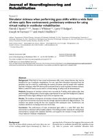
cells. The transformation was confirmed by
colony PCR (Fig. 1). The clones were induced
with Isopropyl β-D-1-thiogalactopyranoside
11
Int.J.Curr.Microbiol.App.Sci (2019) 8(2): 10-15
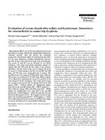
(IPTG) and were analyzed using SDS-PAGE
(Fig. 2). The protein was purified using Nickel
affinity chromatography, immunoblotted to
confirm the immunogenicity and used as
antigen in IgM dot ELISA, plate ELISA and
LAT.
1:50. Then, the cut off value was determined
using the formula Mean OD + 3 × Standard
deviation. The optimum concentration of
antigen was found to be 50 ng/ well and 150
ng/ well for rLipL32. The rabbit anti-canine
IgG HRP conjugate concentration estimated
was 1:2000. A 1:50 dilution of test serum was
found optimum working dilution. The relative
sensitivity, specificity and accuracy of IgM
dot ELISA, plate ELISA and LAT were
evaluated in comparison with MAT.
IgM ELISA
All the 105 samples were also subjected to
plate ELISA (Ooteman et al., 2006). The
optimum concentration of the antigen for
ELISA was found out employing the checker
board analysis. The recombinant LipL32
antigen was diluted in carbonate bicarbonate
buffer so as to incorporate the antigen
concentration ranging from 25 ng, 50 ng, 100
ng, 150 ng, 200 ng and 250 ng per well.
Similarly, serum samples were taken in the
dilutions ranging from 1: 50 to 1: 400. In IgM
ELISA, rabbit anti canine IgM peroxidase
conjugate was used. The cut-off value for the
interpretation of ELISA was determined as per
the report of Bomfim et al., (2005). The mean
OD with 40 negative sera was recorded by
performing ELISA. The negative sera used
were
those
collected
from
healthy
unvaccinated animals with MAT titre less than
Results and Discussion
The results of MAT, IgM dot ELISA, LAT
and IgM plate ELISA are given in table 1.
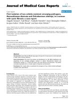
Among the 105 samples, 56 were found to
have a titre of ≥1:800 in MAT (Fig. 3). In IgM
dot ELISA, 55 samples were found to be
positive and one was negative. Latex
Agglutination Test could detect 58 samples as
positive. Comparison of the results with MAT
is presented in table 2. The diagnosis of acute
leptospirosis in dogs is a dilemma in endemic
areas like Kerala, where routine vaccination is
practiced, because the vaccinal antibodies and
past infections always interfere with the
results.
Table.1 Results of MAT, IgM dot ELISA and IgM plate ELISA
MAT
+VE
-VE
56
49
IgM dot ELISA
+VE
-VE
54
2
1
48
LAT
+VE
54
4
-VE
2
45
IgM plate ELISA
+VE
-VE
52
4
2
47
Table.2 Comparison of ELISAs and LAT in comparison with MAT
IgM dot ELISA
96.43%
Sensitivity
97.96%
Specificity
Positive Likelihood Ratio 47.05
Negative Likelihood Ratio 0.04
12
LAT
96.43%
91.84 %
11.81
0.04
IgM plate ELISA
92.86%
95.92 %
22.75
0.07
Int.J.Curr.Microbiol.App.Sci (2019) 8(2): 10-15
Fig.1 Confirmation of clones by colony PCR
Fig.2 Induction of recombinant LipL 32 protein
1
2
3
116 kDa
66 kDa
45 kDa
35 kDa
25 kDa
18 kDa
14 kDa
Lane 1- control (uninduced)
Lane 2. rLipL32 expression
Lane 3. Protein marker
13
Int.J.Curr.Microbiol.App.Sci (2019) 8(2): 10-15
Fig.3 Microscopic agglutination test (Representation)
1 in 400 dilution of test serum
1 in 800 dilution of test serum
As leptospirosis in dogs as well as human
beings is completely amenable to treatment,
prompt detection is quintessential for
effective therapy and management of the
disease. The conventional diagnostic tests
cannot be resorted to as they are time
consuming and proved to be limited to well
equipped laboratories. Hence, impetus is
being given to rapid field level diagnostic
methods. In this study, recombinant LipL32
based IgM dot ELISA and LAT were
evaluated as diagnostic tools for canine
leptospirosis. In MAT, a titre of ≥1:800 was
taken as positive (Ooteman et al., 2006).
Among the 105 samples, 56 (53.33 percent)
were found to have a titre of ≥1:800 in MAT,
indicating acute leptospirosis. IgM dot ELISA
revealed 55 samples as positive and one was
negative. Latex Agglutination Test could
detect 58 samples as positive. This test
detected four MAT negative samples as
positive, three of which had a MAT titre of 1:
200. This is insignificant in an endemic area.
This low antibody titre may be of
vaccinations or past infection which cannot be
differentiated in LAT. In one LAT positive
sample, the MAT titre was < 1:50. Two MAT
positive samples were detected as negative in
LAT. IgM dot ELISA was found to be
sensitive and specific in detecting acute cases
of leptospirosis which is very relevant in an
endemic area like Kerala. The tests could not
achieve 100 per cent sensitivity since some of
the MAT positive samples were found to be
negative to IgM antibodies. The antibodies
detected in MAT could be IgG antibodies
which were not detected in dot ELISA. The
presence of IgM antibodies is the indication
of immediate infection whereas IgG
antibodies may be due to past infections. The
LAT proved to be a less specific test than IgM
dot ELISA in detecting acute leptospirosis
although it was found to be sensitive (96.43
per cent). Moreover, LAT could not
differentiate past infections and vaccinates.
From the present study, it can be concluded
that rLipL32 based IgM dot ELISA was found
to be the specific test in rapid field level
diagnosis of acute canine leptospirosis in an
endemic area like Kerala.
References
Abhinay G, Ambily R and Joseph S. (2012).
IgM immunoprofile of leptospiral outer
membrane proteins in acute canine
leptospirosis. Indian Vet. J. 89(1): 0910.
Dey S, Mohan M, Senthilkumar T M A,
Ramadass P, Nainar M A and
Nachimuthu K. (2004). Recombinant
LipL32 antigen-based single serum
dilution ELISA for detection of canine
14
Int.J.Curr.Microbiol.App.Sci (2019) 8(2): 10-15
leptospirosis. Vet. Microbiol.103: 99106.
Faine S, Adler B, Bolin C A and Perolat P
(1999). Leptospira and leptospirosis,
(2nd Ed.) Medi Sci, Melbourne,
Australia. 272 pp.
Haake D A, Chao G, Zuerner R L, Barnett J
K, Barnett D, Mazel M, Matsunaga L,
Levett P N and Bolin C A. (2000). The
leptospiral major outer membrane
protein LipL32 is a lipoprotein
expressed during mammalian infection.
Infect. Immun. 68: 2276-2285.
Hauk, P., Carvalho, E. and Ho, P.L.
2011.Expression and purification of the
non-tagged LipL32 of pathogenic
Leptospira. Braz. J. Med. Biol. Res.44:
297-302.
Levett P.N. 2003. Leptospira and leptonema.
In: Murray, P.R., Baron, E.J.,
Jorgensen, J.H., Pfaller, M.A. and
Yolken, R.H. (eds.), Manual of clinical
microbiology, American Society for
Microbiology, Washington, D.C. 936 pp
Ooteman M C, Vago A R and Koury M C
(2006). Evaluation of MAT, IgM
ELISA and PCR methods for diagnosis
of human leptospirosis. J. Microbiol.
Meth. 65: 247-257
Picardeau M. (2013). Diagnosis and
epidemiology of leptospirosis. Med.
Mal. Infect. 43(1):1-9.
Sariprabha P. (2010). Evaluation of whole
cell antigen and outer membrane protein
based latex agglutination test for
serodiagnosis of canine leptospirosis.
M. V. Sc. Thesis, Kerala Agricultural
University, Thrissur, 75p.
Sharma R, Tuteja U, Shukla R K J and Batra
H V (2007). Application of rapid dotELISA for antibody detection of
leptospirosis. J. Med. Microbiol. 56:
873-874
Toyokawa T, Ohnishi M and Koizumi N.
(2011). Diagnosis of acute leptospirosis.
Expert Rev. Anti Infect. Ther. 9(1):11121.
Zhang XY, Yu Y, He P, Zhang YX, Hu
BY, Yang Y, Nie YX, Jiang XG, Zhao
GP, Guo XK. (2005). Expression and
comparative analysis of genes encoding
outer membrane proteins LipL21,
LipL32 and OmpL1 in epidemic
leptospires. Acta Biochem. Biophys. Sin.
(Shanghai). 37(10):649-656.
How to cite this article:
Ambily, R., M. Mini, Siju Joseph and Krishna, S.V. 2019. Evaluation of Two Rapid Field
Level Diagnostic Tools for Acute Canine Leptospirosis in an Endemic Area.
Int.J.Curr.Microbiol.App.Sci. 8(02): 10-15. doi: />
15