Mild hypothermia during the reperfusion phase protects mitochondrial bioenergetics against ischemia-reperfusion injury in an animal model of ex-vivo liver transplantation - an experimental
Bạn đang xem bản rút gọn của tài liệu. Xem và tải ngay bản đầy đủ của tài liệu tại đây (1.22 MB, 9 trang )
Int. J. Med. Sci. 2019, Vol. 16
Ivyspring
International Publisher
1304
International Journal of Medical Sciences
2019; 16(9): 1304-1312. doi: 10.7150/ijms.34617
Research Paper
Mild hypothermia during the reperfusion phase protects
mitochondrial bioenergetics against
ischemia-reperfusion injury in an animal model of
ex-vivo liver transplantation—an experimental study
Rui Miguel Martins1,, João Soeiro Teodoro2, Emanuel Furtado3, Rui Caetano Oliveira4, José Guilherme
Tralhão5, Anabela Pinto Rolo2, Carlos Marques Palmeira2
1.
2.
3.
4.
5.
Department of Surgery, Instituto Português de Oncologia de Coimbra, Coimbra, Portugal
Department of Life Sciences, Faculty of Sciences and Technology, University of Coimbra; and Center of Neurosciences and Cell Biology, University of
Coimbra, Coimbra, Portugal
Unidade de Transplantação Hepática de Crianças e Adultos, Hospitais da Universidade de Coimbra, Centro Hospitalar e Universitário de Coimbra,
Coimbra, Portugal
Department of Pathology, Centro Hospitalar e Universitário de Coimbra, Coimbra, Portugal
Department of Surgery, Hospitais da Universidade de Coimbra, Centro Hospitalar e Universitário de Coimbra, Coimbra, Portugal; Clínica Universitária de
Cirurgia III, Faculty of Medicine, University of Coimbra, Coimbra, Portugal; and Center for Investigation on Environment, Genetics and Oncobiology
(CIMAGO), Faculty of Medicine, University of Coimbra, Coimbra, Portugal
Corresponding author: Rui Miguel Martins, PhD, MD, Department of Surgery, Instituto Português de Oncologia de Coimbra, Av. Bissaya Barreto 98,
3000-075 Coimbra, Portugal. Email address: ; Telephone number: +351-239400200
© The author(s). This is an open access article distributed under the terms of the Creative Commons Attribution License ( />See for full terms and conditions.
Received: 2019.03.04; Accepted: 2019.08.23; Published: 2019.09.07
Abstract
The organ preservation paradigm has changed following the development of new ways to preserve
organs. The use of machine perfusion to preserve organs appears to have several advantages
compared with conventional static cold storage. For liver transplants, the temperature control
provided by machine perfusion improves organ preservation. In this experimental study, we
measured the effects of different temperatures on mitochondrial bioenergetics during the
reperfusion phase. An experimental model of ex-vivo liver transplantation was developed in Wistar
rats (Rattus norvegicus). After total hepatectomy, cold static preservation occurred at 4ºC and
reperfusion was performed at 37ºC and 32ºC using a Langendorff system. We measured parameters
associated with mitochondrial bioenergetics in the livers. Compared with the livers that underwent
normothermic reperfusion, mild hypothermia during reperfusion caused significant increases in the
mitochondrial membrane potential, the adenosine triphosphate content, and mitochondrial
respiration, and a significant reduction in the lag phase (all P < 0.001). Mild hypothermia during
reperfusion reduced the effect of ischemia-reperfusion injury on mitochondrial activity in liver tissue
and promoted an increase in bioenergetic availability compared with normothermic reperfusion.
Key words: hypothermia, mitochondria, bioenergetics, adenosine triphosphate, liver transplantation
Introduction
The lack of available organs is the principal
limitation associated with liver transplantation. To
increase the quantity of donor organs, marginal
organs have been used, including those from elderly
donors and patients with hepatic steatosis, those that
have experienced prolonged cold ischemia, and those
obtained after cardiac death [1, 2]. The use of these
poor-quality organs affects the clinical outcomes of
liver transplantation, which has led to the
development of new ways to preserve organs [3, 4].
Ex-vivo machine perfusion of the liver is an
alternative to conventional static cold storage, but
Int. J. Med. Sci. 2019, Vol. 16
there is no agreement about the most beneficial
temperature [5, 6]. Another issue that requires
resolution is whether or not these liver preservation
methods can be combined [7].
Machine perfusion is associated with declines in
primary non-function, graft failure, and biliary
complications. For liver ex-vivo preservation the
standard of organ preservation has not established,
contrary to the kidney ex-vivo preservation where the
hypothermic perfusion has become the standard
[8-10].
The process of cold and warm ischemia followed
by a reperfusion period is specific to liver
transplantation, and is the primary cause of cellular
damage [11]. Ischemia-reperfusion (I/R) injury
compromises
mitochondrial
function
and
bioenergetics, particularly during reperfusion when
the readmission of oxygen increases the production of
reactive oxygen species [12, 13]. We aimed to
investigate mitochondrial function and cellular
bioenergetics at different temperatures in an
experimental model of ex-vivo liver transplantation,
with a particular focus on the reperfusion phase.
Materials and Methods
The materials and methods used in this study
have been described in detail previously [13].
Animals
Twelve-week-old male Wistar rats (Rattus
norvegicus) weighing 320–350 g were purchased from
Charles River (Charles River, Lyon, France). Upon
arrival, the animals acclimatized for 1 week, and they
were housed in an environment comprising
controlled temperature and humidity and 12-h
light-dark cycles, and given unlimited access to
standard rodent food and acidified water. The study’s
protocol was approved by the Animal Ethics
Committee at the University of Coimbra’s Faculty of
Medicine (ORBEA 150 2016/04112016, April 11, 2016).
1305
All of the studies were conducted in accordance with
the principles and procedures in the EU
(1986/609/EEC and 2010/63/EU), Federation of
European Laboratory Animal Science Associations,
and Animal Research: Reporting of In Vivo
Experiments (ARRIVE) guidelines, and they were
approved by the Animal Care Committee at the
Center for Neurosciences and Cell Biology, University
of Coimbra. We also applied the principles of the
ARRIVE guidelines to data management and
interpretation, and we minimized the number of
animals used and their suffering.
Chemicals and reagents
Except when noted, all of the chemicals and
reagents were purchased from Sigma-Aldrich
Corporation (St. Louis, MO, USA). All of the reagents
and chemicals used were of the highest commercially
available purity.
Surgical protocol
The surgical procedures were performed under
anesthesia induced by ketamine (50 mg/kg) and
chlorpromazine (50 mg/kg), provided by the same
operator. A median laparotomy was performed, and
the liver was mobilized by dividing the hepatic
ligaments. The experimental model of ex-vivo liver
transplantation comprised the introduction of a
cannula into the portal vein and hepatic perfusion
with an organ preservation solution (Celsior®) at 4ºC
for 10 min. Then, we performed a total hepatectomy
while keeping the cannula inside the portal vein.
Adequate inflows and outflows were confirmed. Cold
static preservation at 4ºC was performed over 12 h.
Reperfusion was performed using a Langendorff
system at 32ºC or 37ºC for 1 h with a mixture
comprising 50% Plasma-Lyte 148 and 50% Krebs
solution at pH 7.2 that was supplemented with
oxygen by a pressurized membrane oxygenator (pO2,
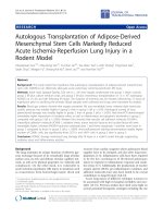
400–500 mm Hg) [14] (Fig. 1).
Figure 1. Schematic representation of reperfusion under hypothermic and normothermic conditions. Biopsies were taken at the end of the reperfusion time (A). The control
group is not represented. Ten animals were analyzed per group.
Int. J. Med. Sci. 2019, Vol. 16
1306
The animals (n = 30) were divided into three
groups. The control group (n = 10) underwent a sham
laparotomy, isolation of the hepatic pedicle,
cannulation of the portal vein, perfusion with the
organ preservation solution at 4ºC for 10 min, and
total hepatectomy. Group A (n = 10) underwent a
sham laparotomy, isolation of the hepatic pedicle,
cannulation of the portal vein, perfusion with the
organ preservation solution at 4ºC for 10 min, total
hepatectomy, cold static preservation at 4ºC for 12 h,
and reperfusion at 32ºC with the Plasma-Lyte/Krebs
solution (pH 7.2) supplemented with oxygen for 1 h.
Group B (n = 10) underwent a sham laparotomy,
isolation of the hepatic pedicle, cannulation of the
portal vein, perfusion with the organ preservation
solution at 4ºC for 10 min, total hepatectomy, cold
static preservation at 4ºC for 12 h, and reperfusion at
37ºC with the Plasma-Lyte/Krebs solution (pH 7.2)
supplemented with oxygen for 1 h.
lag phase (s), and repolarization (mV) were measured,
and the readings were recorded in triplicate.
Mitochondrial isolation
Adenosine triphosphate measurements
The mitochondria were isolated in a
homogenization medium comprising 250 mM
sucrose, 10 mM 4-(2-hydroxyethyl)-1-piperazineethanesulfonic acid (HEPES) (pH 7.4), 0.5 mM
ethylene glycol-bis(β-aminoethyl ether)-N,N,N′,N′tetraacetic acid (EGTA), and 0.1% fat-free bovine
serum albumin (BSA) [15, 16]. After homogenization
of the minced blood-free hepatic tissue, the
homogenates were centrifuged at 800 g for 10 min at
4°C. The supernatants were spun at 10 000 g for 10
min at 4°C to pellet the mitochondria that were then
resuspended in a final washing medium from which
EGTA and BSA were omitted, and it was adjusted to
pH 7.4. The protein content was determined using the
biuret method calibrated with BSA.
Mitochondrial membrane potential
measurements
The mitochondrial membrane potential was
estimated using an ion-selective electrode to measure
the distribution of tetraphenylphosphonium (TPP+).
The voltage response of the TPP+ electrode to log
(TPP+) was linear with a slope of 59 ± 1, and it
conformed to the Nernst equation. The mitochondria
(1 mg) were suspended in standard medium (1 mL),
comprising 130 mM sucrose, 50 mM potassium
chloride, 5 mM magnesium chloride, 5 mM
monopotassium phosphate, 50 mM EDTA, 5 mM
HEPES (pH 7.4), and 2 µM rotenone, supplemented
with 3 µL TPP+. A matrix volume of 1.1 µL/mg
protein was assumed. The reactions were carried out
at 25°C in a temperature-controlled chamber
surrounded by a water jacket with magnetic stirring.
The membrane potential (mV), depolarization (mV),
Oxygen consumption measurements
The oxygen consumption of the isolated
mitochondria was determined using a Clark-type
polarographic
oxygen
electrode
(Oxygraph;
Hansatech Instruments Ltd., King’s Lynn, Norfolk,
United Kingdom) [17]. Mitochondria (1 mg) were
suspended in the standard medium (1.4 mL) with
constant stirring at 25°C, as described previously. The
mitochondria were energized with succinate (5 mM)
and state 3 respiration was induced by adding
adenosine diphosphate (ADP) (200 nmol). Oxygen
consumption was also measured in the presence of 1
µM carbonyl cyanide-p-trifluoromethoxyphenylhydrazone. State 3 respiration and the respiratory
control ratio were calculated according to Chance and
Williams [18].
Liver adenosine triphosphate (ATP) was
extracted using an alkaline extraction procedure [19].
The tissue ATP levels were measured using a
luciferase/luciferin assay kit and a PerkinElmer
Victor 3™ plate-reader fluorometer (PerkinElmer,
Waltham, MA, USA), according to the manufacturers’
instructions.
Histological analysis
The tissue samples were grossly inspected and
divided, fixed in 4% formaldehyde, embedded in
paraffin wax, cut into 4-µm sections, and stained with
hematoxylin and eosin (Polysciences Inc., Warrington,
PA, USA) using a Sakura Autostainer-Prisma 81D
(Sakura Finetek Europe B.V., Alphen aan den Rijn,
The Netherlands). An experienced pathologist who
was blinded to the experimental groups, examined
the tissue sections using a light microscope (Nikon
Eclipse 50i; Nikon Corporation, Tokyo, Japan), and
images were obtained using a Nikon-Digital Sight
DS-Fi1 camera (Nikon Corporation).
Statistical analysis
The continuous variables are presented as the
means and standard errors of the means, unless
otherwise specified. The normality of the data
distributions
was
confirmed
using
the
Kolmogorov-Smirnov and Shapiro-Wilk tests when
indicated.
Between-group
comparisons
were
performed using Student’s t-test, and differences
among three or more groups were analyzed using a
one-way analysis of variance for post hoc multiple
comparisons. The statistical analyses were performed
using IBM®SPSS® software, version 22.0 (IBM
Int. J. Med. Sci. 2019, Vol. 16
Corporation, Armonk, NY, USA). A value of P < 0.05
was considered statistically significant.
Results
Reperfusion under hypothermic conditions was
performed to evaluate its effects on mitochondrial
function and bioenergetics. In this study, the cold
ischemia (12 h) and reperfusion (1 h) times were
maintained. Reperfusion occurred at 32°C in group A
and at 37°C in group B.
Mitochondrial membrane potential
The
mitochondrial
membrane
potential
estimates the phosphorylative capacity of isolated
liver mitochondria. In this study, succinate was used
to obtain the membrane potential data. A statistically
significant difference in the mitochondrial membrane
potential was evident between the groups (Table 1).
Compared with the group subjected to normothermic
1307
reperfusion, hypothermic reperfusion significantly
improved
the
parameters
associated
with
mitochondrial function (P < 0.001). The lag phase
declined in the hypothermic reperfusion group
compared with that in the normothermic reperfusion
group, thereby validating the measurement of the
membrane potential data (Figs. 2 and 3).
Table 1. The membrane potentials and lag phases in the control
group, group A (hypothermic reperfusion), and group B
(normothermic reperfusion).
Initial membrane potential (-mV)
Depolarization (mV)
Lag phase (s)
Repolarization (mV)
Succinate
Control
207.4 ± 5.0
24.0 ± 1.0
54.6 ± 2.8
194.7 ± 7.7
Group A
199.6 ± 1.5
21.7 ± 1.1
60.8 ± 1.0
189.8 ± 5.1
Group B
176.4 ± 2.3**
16.9 ± 0.8**
104.4 ± 4.1**
172.6 ± 2.1**
The data presented are the means and standard errors of the means. Statistically
significant differences were found between groups A (hypothermic reperfusion)
and B (normothermic reperfusion). ** P < 0.01.
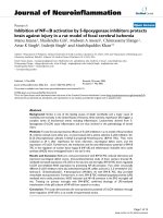
Figure 2. Initial membrane potentials (Δψ) in the control group, group A (hypothermic reperfusion), and group B (normothermic reperfusion). The membrane potentials were
determined in the presence of succinate as a respiratory substrate. Phosphorylation was induced by adding adenosine diphosphate (100 nmol). A statistically significant difference
was found between groups A (hypothermic reperfusion) and B (normothermic reperfusion). **P < 0.01.
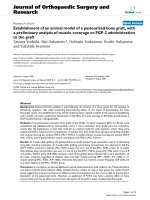
Figure 3. Lag phases in the control group, group A (hypothermic reperfusion), and group B (normothermic reperfusion) in the presence of succinate as a respiratory substrate.
Phosphorylation was induced by adding adenosine diphosphate (100 nmol). A statistically significant difference was found between groups A (hypothermic reperfusion) and B
(normothermic reperfusion). **P < 0.01.
Int. J. Med. Sci. 2019, Vol. 16
Mitochondrial respiration
The mitochondrial respiration measurements
evaluated oxygen consumption after respiration was
induced with succinate. Figures 4 and 5 summarize
the results.
Adenosine triphosphate content
Figure 6 illustrates the ATP levels in the hepatic
tissue subjected to hypothermic and normothermic
reperfusion. Lower ATP levels were present in the
tissues subjected to normothermic reperfusion
compared with those in the tissues subjected to
hypothermic reperfusion.
1308
Histological evaluation
The histological evaluation of the hepatic tissue
from the control group showed normal liver
architecture. In Group A, the hepatic tissue was
preserved, and there was no evidence of an
inflammatory infiltrate, steatosis, or fibrosis. The
structural integrity of the nuclei and organelles within
the hepatocytes was maintained, and there was no
evidence of necrosis or apoptosis. In Group B, the
structure of the hepatic parenchyma was preserved,
but the hepatocytes showed moderate-to-severe
disassociation and some ballonization. The structural
integrity of the organelles and nuclei within the
hepatocytes was maintained, and neither apoptosis
nor necrosis was visible (Figs. 7 and 8).
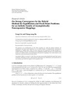
Figure 4. The respiratory state 3 values for the control group, group A (hypothermal reperfusion), and group B (normothermic reperfusion). The respiratory status was
determined in the presence of succinate. A statistically significant difference was found between groups A (hypothermic reperfusion) and B (normothermic reperfusion). **P <
0.01.
Figure 5. The respiratory control ratios in the control group, group A (hypothermal reperfusion), and group B (normothermic reperfusion). The respiratory control index was
determined in the presence of succinate. A statistically significant difference was found between groups A (hypothermic reperfusion) and B (normothermic reperfusion). **P <
0.01.
Int. J. Med. Sci. 2019, Vol. 16
1309
Figure 6. Representative plot of the adenosine triphosphate (ATP) levels in the hepatic tissue of the control group, group A (hypothermic reperfusion), and group B
(normothermic reperfusion). A statistically significant difference was found between groups A (hypothermic reperfusion) and B (normothermic reperfusion). **P < 0.01. ATP,
adenosine triphosphate.
Figure 7. Hematoxylin and eosin (H&E)-stained sections of hepatic tissue from the hypothermic reperfusion group. The hepatic sinusoids do not show endothelial injury, and
the hepatocytes contained normal intracellular organelles and nuclei, with no signs of apoptotic or necrosis (A: H&E 40×; B: H&E 400×).
Figure 8. Hematoxylin and eosin (H&E)-stained sections of hepatic tissue from the normothermic reperfusion group. The hepatic parenchyma architecture is preserved without
lesions. There is moderate-to-severe disassociation of the hepatocytes. The hepatocytes contain normal nuclei and organelles, with no signs of necrosis or apoptosis (A: H&E
40×; B: H&E 400×).
Discussion
Animal models of hepatic transplantation are
fundamental tools that have enhanced our
understanding of the biological and immunological
mechanisms involved in transplantation, and, thus,
they have helped to answer some clinically relevant
questions.
Int. J. Med. Sci. 2019, Vol. 16
The mouse is the most commonly used animal,
and several mouse models have been developed [20].
In 1973, Lee et al. reported the first orthotopic liver
transplantation in the rat, which consisted of
performing an extracorporeal shunt between the
portal and jugular vein at the recipient and posterior
anastomosis of the implant to the hepatic vein, the
portal vein and the recipient's aorta [21]. This very
complex technique was abandoned, and 18 years
later, following the development of vascular
microsurgery techniques, Qian et al. developed a
complex animal model of orthotopic liver
transplantation. However, the investigations based on
this model are very limited, because it requires a high
level of microsurgical expertise and specific technical
conditions. In addition, the high mortality rate caused
by disruptions to hepatocellular function has limited
the use of this animal model [22, 23].
Oscar Langendorff evaluated physiological and
pathophysiological events within ex-vivo heart tissue,
and, consequently, other animal models were
developed that enabled ex-vivo evaluations of the
liver, with an emphasis on I/R studies [24-26].
Previous ex-vivo liver transplantation studies are
invaluable, because they have paved the way for the
physiological and pathophysiological studies that are
essential for the development of new ways to preserve
the liver using dynamic preservation machines. These
studies contribute to the development of dynamic
preservation has altered the ways in which organs are
perfused, preserved, and transported [5, 8, 27-30].
The animal models of ex-vivo liver
transplantation are highly reproducible, and the
results are not influenced by the complex surgical
procedures of other models such as orthotopic liver
transplant model. Despite these, the main limitation
to these animal models of liver transplantation is
related to the impossibility to evaluate pos-operative
biomarkers of the liver function and the non-use of
blood in the reperfusion phase [31].
Functional evaluations of mitochondrial activity
in rodents have demonstrated that, like human
beings, I/R clearly affects mitochondrial function,
which has implications for bioenergetics, and
translates into lower energy production efficiency [32,
33]. This ATP deficiency is sufficient to trigger
changes in cellular metabolism; therefore, I/R injury
in liver transplants interferes with the cellular
bioenergetic balance.
Bigelow et al. introduced the concept of
hypothermia to clinical practice in the early 1950s, and
they demonstrated its neuroprotective effect during
cardiac surgery [34]. The benefits of hypothermia
include the preservation of hepatic metabolism, and
reductions in the inflammatory response and
1310
apoptosis during ischemia [35]. Recent experimental
studies have shown that mild hypothermia at 32–34°C
exerts a protective effect against warm I/R injury, but
the mechanisms underlying this effect remain unclear
[36]. Azoulay et al. studied patients who underwent
complex liver surgery as a consequence of central
hepatic tumors involving the inferior vena cava or the
confluence of the hepatic veins with the vena cava,
and they demonstrated the protective effect of
hypothermic in-situ hepatic perfusion compared with
total vascular exclusion for >60 min. This study’s
findings showed that patients who underwent
hypothermic perfusion had a better I/R-induced
injury tolerance, which translated into improved
postoperative liver and kidney function and reduced
morbidity [37].
In this study, we undertook a laboratory
evaluation of the concept of hypothermia applied to
reperfusion during hepatic transplantation; this
involved a reperfusion temperature of 32°C, which,
according to experimental studies, provides more
effective protection [38, 39]. Our study’s findings
showed statistically significant differences between
the hypothermic reperfusion group and the
normothermic reperfusion group regarding the
mitochondrial membrane potential and respiration
parameters, which were preserved to higher degrees
in the hypothermic reperfusion group. In addition,
the amount of ATP produced in the hepatic tissue
from the hypothermic reperfusion group was higher
than that recorded in the hepatic tissue from the
normothermic reperfusion group.
Compared with normothermic reperfusion,
hypothermic reperfusion reduced the effect of I/R on
mitochondrial activity, thereby increasing the
bioenergetic availability (42%). Hence, applying
hypothermic reperfusion to liver transplantation may
be beneficial from a bioenergetic perspective, because
mitochondrial function is preserved.
One of the main limitations regarding the use of
hypothermia in clinical practice is the potential for
coagulopathy. This seems to be associated with
platelet dysfunction and damage to the enzymes in
the coagulation cascade [40]. The risk of bleeding and
the subsequent need for transfusions increase by
approximately 20% for each degree Celsius decline in
the core temperature. Hypothermia reduces the
metabolic rate by 8% for each degree Celsius decline
[41], which, for this study, would imply a 34%
reduction in metabolic activity. In humans, the only
clinical applications of hypothermia that have led to
improved outcomes are extra-hospital cardiac arrest
and neonatal asphyxia [42, 43]. To integrate the
concept of hypothermic reperfusion into clinical
practice and apply it to hepatic transplantation,
Int. J. Med. Sci. 2019, Vol. 16
1311
further functional and technical studies will be
necessary.
7.
Acknowledgments
8.
We are grateful for the support provided by the
Sociedade Portuguesa de Transplantação (SPT),
Astellas Pharma and Centro de Investigação do Meio
Ambiente, Genética e Oncobiologia (CIMAGO), and
Groupe IGL (Institut Georges Lopez).
Funding Sources
This work was supported by the Sociedade
Portuguesa de Transplantação (SPT), Astellas
Pharma, Centro de Investigação do Meio Ambiente,
Genética e Oncobiologia (CIMAGO), Groupe IGL
(Institut Georges Lopez). JST is the recipient of a
postdoctoral scholarship from the Portuguese
Fundação para a Ciência e a Tecnologia
(SFRH/BPD/94036/2013).
9.
10.
11.
12.
13.
Authorship
Rui Miguel Martins, José Guilherme Tralhão,
Anabela Pinto Rolo and Carlos Marques Palmeira
designed the research. Rui Miguel Martins, João
Soeiro Teodoro, Anabela Pinto Rolo and Carlos
Marques Palmeira performed the research. Rui
Miguel Martins, João Soeiro Teodoro, Anabela Pinto
Rolo and Carlos Marques Palmeira collected and
analyzed the data. Emanuel Furtado and José
Guilherme Tralhão contributed to data interpretation.
Rui Caetano Oliveira performed the histologic
analysis. Rui Miguel Martins wrote the manuscript.
Competing Interests
The authors have declared that no competing
interest exists.
14.
15.
16.
17.
18.
19.
20.
References
1.
2.
3.
4.
5.
6.
Bertuzzo VR, Cescon M, Odaldi F, Di Laudo M, Cucchetti A, Ravaioli M,
et al. Actual Risk of Using Very Aged Donors for Unselected Liver
Transplant Candidates: A European Single-center Experience in the
MELD Era. Annals of surgery. 2017; 265: 388-96.
Flores A, Asrani SK. The donor risk index: A decade of experience. Liver
transplantation : official publication of the American Association for the
Study of Liver Diseases and the International Liver Transplantation
Society. 2017; 23: 1216-25.
Minambres E, Suberviola B, Dominguez-Gil B, Rodrigo E, Ruiz-San
Millan JC, Rodriguez-San Juan JC, et al. Improving the Outcomes of
Organs Obtained From Controlled Donation After Circulatory Death
Donors Using Abdominal Normothermic Regional Perfusion. American
journal of transplantation : official journal of the American Society of
Transplantation and the American Society of Transplant Surgeons. 2017;
17: 2165-72.
Ruiz P, Gastaca M, Bustamante FJ, Ventoso A, Palomares I, Prieto M, et
al. Favorable Outcomes After Liver Transplantation With Normothermic
Regional Perfusion From Donors After Circulatory Death: A
Single-Center Experience. Transplantation. 2018.
Barbas AS, Goldaracena N, Dib MJ, Selzner M. Ex-vivo liver perfusion
for organ preservation: Recent advances in the field. Transplantation
reviews. 2016; 30: 154-60.
Goldaracena N, Barbas AS, Selzner M. Normothermic and
subnormothermic ex-vivo liver perfusion in liver transplantation.
Current opinion in organ transplantation. 2016; 21: 315-21.
21.
22.
23.
24.
25.
26.
27.
28.
Hosgood SA, Mohamed IH, Bagul A, Nicholson ML. Hypothermic
machine perfusion after static cold storage does not improve the
preservation condition in an experimental porcine kidney model. The
British journal of surgery. 2011; 98: 943-50.
Marecki H, Bozorgzadeh A, Porte RJ, Leuvenink HG, Uygun K, Martins
PN. Liver ex situ machine perfusion preservation: A review of the
methodology and results of large animal studies and clinical trials. Liver
transplantation : official publication of the American Association for the
Study of Liver Diseases and the International Liver Transplantation
Society. 2017; 23: 679-95.
Jayant K, Reccia I, Virdis F, Shapiro AMJ. The Role of Normothermic
Perfusion in Liver Transplantation (TRaNsIT Study): A Systematic
Review of Preliminary Studies. HPB surgery : a world journal of hepatic,
pancreatic and biliary surgery. 2018; 2018: 6360423.
Schlegel A, Dutkowski P. Impact of Machine Perfusion on Biliary
Complications after Liver Transplantation. International journal of
molecular sciences. 2018; 19.
Robertson FP, Fuller BJ, Davidson BR. An Evaluation of Ischaemic
Preconditioning as a Method of Reducing Ischaemia Reperfusion Injury
in Liver Surgery and Transplantation. Journal of clinical medicine. 2017;
6.
Ma Z, Xin Z, Di W, Yan X, Li X, Reiter RJ, et al. Melatonin and
mitochondrial function during ischemia/reperfusion injury. Cellular
and molecular life sciences : CMLS. 2017; 74: 3989-98.
Martins RM, Pinto Rolo A, Soeiro Teodoro J, Furtado E, Caetano Oliveira
R, Tralhao JG, et al. Addition of Berberine to Preservation Solution in an
Animal Model of Ex Vivo Liver Transplant Preserves Mitochondrial
Function and Bioenergetics from the Damage Induced by
Ischemia/Reperfusion. International journal of molecular sciences. 2018;
19.
Gores GJ, Kost LJ, LaRusso NF. The isolated perfused rat liver:
conceptual and practical considerations. Hepatology. 1986; 6: 511-7.
Varela AT, Simoes AM, Teodoro JS, Duarte FV, Gomes AP, Palmeira
CM, et al. Indirubin-3'-oxime prevents hepatic I/R damage by inhibiting
GSK-3beta and mitochondrial permeability transition. Mitochondrion.
2010; 10: 456-63.
Palmeira CM, Moreno AJ, Madeira VM. Interactions of herbicides 2,4-D
and dinoseb with liver mitochondrial bioenergetics. Toxicology and
applied pharmacology. 1994; 127: 50-7.
Rolo AP, Oliveira PJ, Moreno AJ, Palmeira CM. Bile acids affect liver
mitochondrial bioenergetics: possible relevance for cholestasis therapy.
Toxicological sciences : an official journal of the Society of Toxicology.
2000; 57: 177-85.
Chance B, Williams GR. Respiratory enzymes in oxidative
phosphorylation. VI. The effects of adenosine diphosphate on
azide-treated mitochondria. The Journal of biological chemistry. 1956;
221: 477-89.
Stocchi V, Cucchiarini L, Magnani M, Chiarantini L, Palma P, Crescentini
G. Simultaneous extraction and reverse-phase high-performance liquid
chromatographic determination of adenine and pyridine nucleotides in
human red blood cells. Analytical biochemistry. 1985; 146: 118-24.
Czigany Z, Iwasaki J, Yagi S, Nagai K, Szijarto A, Uemoto S, et al.
Improving Research Practice in Rat Orthotopic and Partial Orthotopic
Liver Transplantation: A Review, Recommendation, and Publication
Guide. European surgical research Europaische chirurgische Forschung
Recherches chirurgicales europeennes. 2015; 55: 119-38.
Lee S, Charters AC, Chandler JG, Orloff MJ. A technique for orthotopic
liver transplantation in the rat. Transplantation. 1973; 16: 664-9.
Chen J, Gong W, Ge F, Huang T, Wu D, Liang T. A review of various
techniques of mouse liver transplantation. Transplantation proceedings.
2013; 45: 2517-21.
Qian SG, Fung JJ, Demetris AV, Ildstad ST, Starzl TE. Orthotopic liver
transplantation in the mouse. Transplantation. 1991; 52: 562-4.
Varela AT, Rolo AP, Palmeira CM. Fatty liver and ischemia/reperfusion:
are there drugs able to mitigate injury? Current medicinal chemistry.
2011; 18: 4987-5002.
Bell RM, Mocanu MM, Yellon DM. Retrograde heart perfusion: the
Langendorff technique of isolated heart perfusion. Journal of molecular
and cellular cardiology. 2011; 50: 940-50.
Ferreira FM, Palmeira CM, Seica R, Santos MS. Alterations of liver
mitochondrial bioenergetics in diabetic Goto-Kakizaki rats. Metabolism:
clinical and experimental. 1999; 48: 1115-9.
Karimian N, Matton AP, Westerkamp AC, Burlage LC, Op den Dries S,
Leuvenink HG, et al. Ex Situ Normothermic Machine Perfusion of Donor
Livers. Journal of visualized experiments : JoVE. 2015: e52688.
Burlage LC, Karimian N, Westerkamp AC, Visser N, Matton APM, van
Rijn R, et al. Oxygenated hypothermic machine perfusion after static cold
storage improves endothelial function of extended criteria donor livers.
Int. J. Med. Sci. 2019, Vol. 16
29.
30.
31.
32.
33.
34.
35.
36.
37.
38.
39.
40.
41.
42.
43.
1312
HPB : the official journal of the International Hepato Pancreato Biliary
Association. 2017; 19: 538-46.
Dutkowski P, Linecker M, DeOliveira ML, Mullhaupt B, Clavien PA.
Challenges to liver transplantation and strategies to improve outcomes.
Gastroenterology. 2015; 148: 307-23.
Schlegel A, Kron P, Graf R, Dutkowski P, Clavien PA. Warm vs. cold
perfusion techniques to rescue rodent liver grafts. Journal of hepatology.
2014; 61: 1267-75.
Beal EW, Dumond C, Kim JL, Akateh C, Eren E, Maynard K, et al. A
Small Animal Model of Ex Vivo Normothermic Liver Perfusion. Journal
of visualized experiments : JoVE. 2018.
Budai A, Horvath G, Tretter L, Radak Z, Koltai E, Bori Z, et al.
Mitochondrial function after associating liver partition and portal vein
ligation for staged hepatectomy in an experimental model. The British
journal of surgery. 2019; 106: 120-31.
Go KL, Lee S, Zendejas I, Behrns KE, Kim JS. Mitochondrial Dysfunction
and Autophagy in Hepatic Ischemia/Reperfusion Injury. BioMed
research international. 2015; 2015: 183469.
Bigelow WG, Callaghan JC, Hopps JA. General hypothermia for
experimental intracardiac surgery; the use of electrophrenic respirations,
an artificial pacemaker for cardiac standstill and radio-frequency
rewarming in general hypothermia. Annals of surgery. 1950; 132: 531-9.
Miao YF, Wu H, Yang SF, Dai J, Qiu YM, Tao ZY, et al. 5'-adenosine
monophosphate-induced
hypothermia
attenuates
brain
ischemia/reperfusion injury in a rat model by inhibiting the
inflammatory response. Mediators of inflammation. 2015; 2015: 520745.
Xiao Q, Ye Q, Wang W, Xiao J, Fu B, Xia Z, et al. Mild hypothermia
pretreatment protects against liver ischemia reperfusion injury via the
PI3K/AKT/FOXO3a pathway. Molecular medicine reports. 2017; 16:
7520-6.
Azoulay D, Eshkenazy R, Andreani P, Castaing D, Adam R, Ichai P, et al.
In situ hypothermic perfusion of the liver versus standard total vascular
exclusion for complex liver resection. Annals of surgery. 2005; 241:
277-85.
Behrends M, Hirose R, Serkova NJ, Coatney JL, Bedolli M, Yardi J, et al.
Mild hypothermia reduces the inflammatory response and hepatic
ischemia/reperfusion injury in rats. Liver international : official journal
of the International Association for the Study of the Liver. 2006; 26:
734-41.
Niemann CU, Choi S, Behrends M, Hirose R, Noh J, Coatney JL, et al.
Mild hypothermia protects obese rats from fulminant hepatic necrosis
induced by ischemia-reperfusion. Surgery. 2006; 140: 404-12.
Michelson AD, MacGregor H, Barnard MR, Kestin AS, Rohrer MJ, Valeri
CR. Reversible inhibition of human platelet activation by hypothermia in
vivo and in vitro. Thrombosis and haemostasis. 1994; 71: 633-40.
Suga H, Goto Y, Igarashi Y, Yasumura Y, Nozawa T, Futaki S, et al.
Cardiac cooling increases Emax without affecting relation between O2
consumption and systolic pressure-volume area in dog left ventricle.
Circulation research. 1988; 63: 61-71.
Arrich J, European Resuscitation Council Hypothermia After Cardiac
Arrest Registry Study G. Clinical application of mild therapeutic
hypothermia after cardiac arrest. Critical care medicine. 2007; 35: 1041-7.
Kracer B, Hintz SR, Van Meurs KP, Lee HC. Hypothermia therapy for
neonatal hypoxic ischemic encephalopathy in the state of California. The
Journal of pediatrics. 2014; 165: 267-73.