High mobility group box 1 protein (HMGB1) as biomarker in hypoxia-induced persistent pulmonary hypertension of the newborn: A clinical and in vivo pilot study
Bạn đang xem bản rút gọn của tài liệu. Xem và tải ngay bản đầy đủ của tài liệu tại đây (996.77 KB, 9 trang )
Int. J. Med. Sci. 2019, Vol. 16
Ivyspring
International Publisher
1123
International Journal of Medical Sciences
2019; 16(8): 1123-1131. doi: 10.7150/ijms.34344
Research Paper
High mobility group box 1 protein (HMGB1) as
biomarker in hypoxia-induced persistent pulmonary
hypertension of the newborn: a clinical and in vivo pilot
study
Zhen Tang1, Min Jiang2, Zhicui Ou-yang1, Hailan Wu2, Shixiao Dong2, Mingyan Hei2
1.
2.
Department of Pediatrics, the Third Xiangya Hospital of Central South University, Changsha, Hunan, 410013 China
Neonatal Center, Beijing Children’s Hospital, Capital Medical University, Beijing, 100045 China
Corresponding author: Prof. Mingyan Hei, Neonatal Center, Beijing Children’s Hospital, Capital Medical University.Tel: +86-10-59616745; FAX:
+86-10-59616745; Email:
© The author(s). This is an open access article distributed under the terms of the Creative Commons Attribution License ( />See for full terms and conditions.
Received: 2019.02.23; Accepted: 2019.07.17; Published: 2019.08.06
Abstract
Background: Inflammation plays an important role in neonatal hypoxia-induced organ damage.
Newborns with perinatal asphyxia often develop persistent pulmonary hypertension of the newborn
(PPHN). The objective of this study was to explore changes in the pro-inflammatory high mobility group
box-l (HMGB1) protein during hypoxia-induced PPHN clinically and in vivo.
Methods: Serum samples were collected from full-term newborns at PPHN onset and remission. As
controls, blood serum samples were collected from the umbilical arteries of healthy full-term newborns
born in our hospital during the same period. Clinical data for neonates were collected and serum levels of
HMGB1, IL-6, and TNF-α were detected by enzyme-linked immunosorbent assay (ELISA). An animal
study compared a PPHN Sprague–Dawley rat model to healthy newborn control rats. Histopathology
was used to evaluate changes in the pulmonary artery wall. ELISA and western blot analyses were used to
examine HMGB1 levels in the serum and lungs.
Results: Serum HMGB1 levels were significantly elevated in newborns with PPHN, compared to those in
healthy controls, and decreased dramatically after PPHN resolution. HMGB1 changes were positively
correlated with serum tumor necrosis factor-α (TNF-α) and interleukin-6 (IL-6) levels. Histopathological
analysis demonstrated that the median wall thickness of pulmonary arterioles accounting for the
percentage of pulmonary arteriole diameter (MT%) was not significantly different between PPHN and
control groups 3 d after PPHN, although thickness of the small pulmonary arterial wall middle membrane
and stenosis of the small pulmonary arteries. ELISA and western blot analyses showed similar trends
between serum HMGB1 levels and HMGB1 protein expression in the lungs. Serum and lung HMGB1
levels were significantly elevated soon after PPHN onset, peaked after 24 h, and then decreased after 3 d,
although they remained elevated compared to those in the control group.
Conclusions: This study indicates that HMGB1 is related to hypoxia-induced PPHN pathogenesis.
HMGB1 changes might thus be used as an early indicator to diagnose hypoxia-induced PPHN and evaluate
its improvement. We also provide important evidence for the involvement of inflammation in the
progression of hypoxia-induced PPHN.
Key words: Hypoxia; Newborn, infants; Persistent pulmonary hypertension of the newborn (PPHN); High
mobility group box-l (HMGB1); Rat
Introduction
In addition to hypoxic-ischemic brain damage,
persistent pulmonary hypertension of the newborn
(PPHN) is not uncommon in newborns with perinatal
asphyxia. PPHN is a syndrome characterized by the
Int. J. Med. Sci. 2019, Vol. 16
sustained elevation of pulmonary vascular resistance,
which results in poor lung perfusion and further
aggravates systematic and brain tissue hypoxia,
resulting in deterioration of the patient’s condition
and increased mortality and morbidity [1, 2]. As a
systemic syndrome, PPHN has a variety of causes, but
lacks an early and effective diagnostic method [3, 4].
PPHN diagnosis is currently performed by
determining the pulmonary artery pressure based on
cardiac catheterization and echocardiography [5, 6],
which are not practical for newborns. Inflammation
plays an important role in neonatal hypoxia-induced
organ damage. One clinical study indicated that
serum levels of a variety of inflammatory factors are
significantly increased during adult pulmonary
arterial hypertension (idiopathic pulmonary arterial
hypertension, IPAH) mediated by chronic hypoxia,
including tumor necrosis factor-alpha (TNF-α) and
interleukin-6 (IL-6). Further, IPAH prognosis is
closely linked to increases in these inflammatory
factors [7, 8], illustrating the important role
inflammation plays in the occurrence and
development of IPAH. Recently, high-mobility group
box-1 (HMGB1), an inflammatory mediator, has
received increasing attention for its role in the
pathogenesis of pulmonary arterial hypertension.
Clinical studies confirmed that high levels of HMGB1
could be detected around the pulmonary artery walls
of plexiform lesions in patients with IPAH, and that
serum and alveolar lavage fluid HMGB1 levels in
patients with IPAH were significantly increased [9].
Animal experiments have also reported significant
increases in serum HMGB1 levels in mouse models of
chronic hypoxia, and demonstrated that exogenous
recombinant HMGB1 can exacerbate pulmonary
arterial hypertension, whereas the administration of
HMGB1-neutralizing antibodies can slow its
progression [10]. Elevated serum levels of HMGB1 in
a rat model of pulmonary arterial hypertension were
found to mainly result from alveolar macrophages
and smooth muscle cells [11]. However, the effects of
HMGB1 on neonatal PPHN remain unknown.
To determine whether similar changes in
HMGB1 levels occur in PPHN, we first examined
HMGB1 levels in the serum of full-term newborns
admitted for perinatal hypoxia that rapidly
progressed to PPHN. HMGB1 changes were also
investigated using an animal model of PPHN to
understand the role of HMGB1 in neonatal
hypoxia-induced PPHN pathogenesis.
Materials and Methods
Research design
The study included both clinical studies and
1124
animal experiments. Clinical studies included a case
group comprising newborns with no evidence of
infection, who were admitted to the hospital with
perinatal asphyxia and diagnosed with PPHN shortly
thereafter, and a control group consisting of newborns
born in the same hospital during the same period.
With the parents’ informed consent, umbilical cord
blood samples and laboratory residual blood samples
were collected from patients in the case group. After
centrifugation, 100 μL of serum was collected and
stored at −80 °C for further testing. Regarding ethical
considerations, only healthy full-term neonatal cord
blood samples were taken for the control group,
following the same process as that performed for the
case group. The animals were randomly divided into
PPHN and normal control groups. At each designated
time point, newborn rats were sacrificed by
decapitation and 200 μL blood samples were taken.
After centrifugation, 30 μL serum samples were
collected and stored at −80°C. At the same time, the
lungs of the animals were collected, and proteins were
extracted and stored at −80 °C.
Clinical research objectives
Inclusion criteria for newborns with PPHN
The clinical component of this study was
approved by the medical ethics committee of the
Third Xiangya Hospital of Central South University
and Beijing Children’s Hospital, Capital Medical
University. PPHN newborns with complete clinical
data and an initial diagnosis of perinatal asphyxia
who were diagnosed with PPHN within 3 d of birth
were enrolled at the neonatal intensive care unit of the
Third Xiangya Hospital of Central South University
and Beijing Children’s Hospital, Capital Medical
University from January 2016 to December 2017.
Formal written consent was signed by the parents of
each infant enrolled in this study. PPHN diagnosis
included perinatal history, clinical manifestations,
and laboratory examinations, and was confirmed by
color Doppler echocardiography [12]. When the
patient’s oxygen index was < 20 and Doppler
echocardiography showed a pulmonary artery
systolic pressure ≤ 2/3 the systolic pressure, they
were considered to be in PPHN remission [13, 14].
Exclusion criteria for newborns with PPHN
Newborns were excluded based on any of the
following criteria: (1) their mothers were diagnosed
with chronic hepatitis B, acute and chronic
pancreatitis, diabetes, autoimmune diseases, thyroid
dysfunction, or cancer; (2) there was a serious
infection during pregnancy, including sepsis or
significant prenatal infection; (3) their mothers had a
special medication history such as taking
Int. J. Med. Sci. 2019, Vol. 16
non-steroidal anti-inflammatory drugs or selective
serotonin re-uptake inhibitors during pregnancy; (4)
the newborns suffered from severe congenital
cardiovascular disease, diaphragmatic hernia, severe
pathological jaundice, or abnormal thyroid function.
Newborns who died within 72 h or did not achieve
remission allowing the cessation of treatment were
excluded. According to the inclusion and exclusion
criteria, 12 children with PPHN were included in the
study.
Inclusion criteria for the control group
Ten full-term newborns born at the Obstetrics
department of the Third Xiangya Hospital of Central
South University were included as a control group.
The inclusion criteria were as follows: (1) a normal
obstetric history for the mother, with no special
medications during pregnancy (such as aspirin and
other non-steroidal anti-inflammatory drugs); (2) a
gestational age between 37–42 weeks, with the onset
of feeding within 2 h after birth; (3) no cyanosis,
shortness of breath, hypoxemia, or other
abnormalities after birth; (4) postpartum care in the
same ward as the mother without special medical
intervention, and discharge from the hospital after 3–4
d.
PPHN animal model using neonatal rats
Experimental animals were provided by the
Hunan Agricultural University Experimental Animal
Center. Three-day-old Sprague-Dawley rats were
randomly divided into the control group (n = 40) and
the hypoxia-induced PPHN group (n = 40). The
animal study component of this study was approved
by the medical ethics committee of the Third Xiangya
Hospital of Central South University. The control
group was housed without any intervention, while
PPHN was induced in the PHHN group starting 4 d
after birth, as previously described [15-17]. Briefly,
experimental animals were placed in a hypoxia
chamber filled with 10% oxygen + 90% nitrogen for 7
consecutive days. The chamber temperature was
maintained at 26 ± 0.5 °C, and anhydrous calcium
chloride was placed in the chamber to absorb the
carbon dioxide exhaled by the experimental animals.
Chambers were opened briefly (< 15 min) for cleaning
and other administration. The time of completion of
the last 24 h hypoxia exposure was counted as 0 h (at
11 d of age) and the experimental animals were
sacrificed at 2 h (11 d of age; n = 10), 8 h (11 d of age; n
= 10), 24 h (12 d of age; n = 10), and day 3 (14 d of age;
n = 10). Before the animals were sacrificed, four
animals were randomly selected at the corresponding
time points to measure mean pulmonary artery
pressure (mPAP) according to previous literature [18].
1125
Briefly, pups were anesthetized by pentobarbital
administration (50 mg/kg) via intraperitoneal
injection, fixed and intubated, and connected to
mechanical ventilation (HX-200 small animal
ventilator, Taimeng Technology Co., Ltd., Chengdu,
China). Setting respiratory rate was 100 breaths /min,
tidal volume 0.2 ml, 5 minutes later, pups were
opened the chest, exposed the right ventricle and
inserted the catheter (OD: 0.5 mm) into the pulmonary
artery root, connected to the other end of the catheter
to
the
RM-6280
multi-channel
intelligent
physiological signal transducer (Chengdu SiChuan)
to record mPAP. After the pulmonary artery pressure
was measured, the same numbers of rats in the
control group were sacrificed at the corresponding
time points.
Serum HMGB1 testing
Blood samples were respectively collected from
newborns when they were diagnosed with PPHN and
when PPHN was alleviated. Normal control blood
samples were collected from the umbilical arterial
blood. All blood samples from rats were collected
when they were sacrificed by decapitation.
Enzyme-linked immunosorbent assays were used to
detect HMGB1 (IBM, Germany), TNF-α (Wuhan
Huamei Biotech, China), and IL-6 (Wuhan Huamei
Biotech, China), according to the manufacturers’
instructions.
Pulmonary vascular morphology
Four rats in each group were selected to detect
vascular pathological changes on day 3. After
decapitation, the right middle lobe tissue was
removed and fixed in 4% paraformaldehyde for 24 h.
After paraffin embedding, sections were sliced to 5
μm and stained with hematoxylin and eosin (H&E).
After drying, morphological changes in pulmonary
arterioles were examined under a light microscope.
For each sample, three visual fields at 400×
magnification containing intact transverse pulmonary
arteries were randomly selected. MIAS-2000 medical
image analysis software was used to calculate the
median wall thickness of pulmonary arterioles
accounting for the percentage of pulmonary arteriole
diameter (MT%).
Western blot analysis
The lungs of experimental animals were
removed, washed with phosphate-buffered saline,
trypsin digested, washed again, and lysed in RIPA
lysis buffer (Shanghai Yesen Biotech, China); all
operations were performed on ice. The samples were
centrifuged at 13000 × g for 30 min and the
supernatant was placed in a new tube. The protein
content was determined by the Lowry method [19].
Int. J. Med. Sci. 2019, Vol. 16
Samples were aliquoted at 30 μg/tube and stored at
−80 °C. After boiling for 5 min in sample buffer,
protein samples were separated by sodium dodecyl
sulfate-polyacrylamide gel electrophoresis at 110 V
and 40 mA for 60 min. Proteins were transferred to
PVDF membranes, blocked in 5% skimmed milk
powder solution for 1 h, and incubated overnight at 4
°C with a mouse anti-HMGB1 monoclonal antibody
(ab11354, Abcam, 1:1000). β-actin was used as a
control. The membranes were washed with
tris-buffered saline containing 0.5% tween 20 and then
incubated for 1 h at 26 °C with a goat anti-mouse IgG
horseradish peroxidase-labeled secondary antibody
(HFA007, R&D, 1:5000). Bands were visualized based
on enhanced chemiluminescence and exposure to
film. Bands were analyzed using Quantity One TM
4.2.2 (Bio-Rad) software, and HMGB1 levels were
expressed as the gray ratio of the target and control
bands.
Statistical analysis
SPSS 18.0 software was used for statistical
analysis. Data with a non-normal distribution were
expressed as the median (Q1–Q3). Normally
distributed data were expressed as the mean ±
standard deviation. The means of two groups were
compared with two-independent sample t-tests.
Comparisons of multiple groups were performed by
one-way ANOVA followed by a least significant
difference t (LSD-t) test for multiple comparisons.
Comparisons of count data were performed using the
Fisher exact method. Indicator correlations were
examined by linear correlation analysis. P < 0.05 was
considered statistically significant.
Results
Clinical results
General characteristics of the newborns in the two
groups
There were no differences in gestational age,
birth weight, mode of delivery, sex, or maternal age
between the control and PPHN groups (all P > 0.05).
However, there were significance differences in 1-min
Apgar scores, as well as pH and HCO3−
concentrations in the umbilical cord blood (P < 0.05;
Table 1). Two newborns with amniotic fluid
contamination were reported in the PPHN group, and
all membranes ruptured within 18 h before delivery.
The newborns in the PPHN group were admitted at
an average age of 1.00 h (0.63–9.00 h) and diagnosed
with PPHN by bedside cardiac ultrasound 12.00 h
(3.14–15.67 h) after admission. Blood gas analysis
showed that the mean PaO2 value when the newborns
were diagnosed with PPHN was 35.63 ± 8.30 mmHg,
1126
whereas the mean SaO2 value was 58.70 ± 13.30%. The
difference in percutaneous oxygen saturation before
and after catheterization was 10.07 ± 5.19% and the
systolic pressure/systolic pressure ratio of the
pulmonary artery was 0.56 ± 0.05 at the same time.
The average time to PPHN remission was 75.6 h
(52.1–96.5h), and newborns were hospitalized for an
average of 14.9 d (11.4–17.7d).
Table 1 Comparison of clinical data in two groups
gestational age(w)
birth weight (g)
Male / female (n)
Cesarean section / peace (n)
mother's age (y)
1 minute Apgar score ≤7 (n)
Cord blood PH
cord Blood HCO3— (mol/L)
Control group
(n = 10)
39.1 ± 1.1
3399.0 ± 410.5
6/4
3/7
31.9± 3.4
0
7.25± 0.10
-6.7± 2.3
PPHN group
(n = 12)
39.0 ± 1.2
3289.1 ± 556.3
7/5
5/7
32.3± 3.7
11
7.07 ± 0.06
-13.6± 3.2
t /χ ²
P
-0.181
-0.517
0.279
-5.687
-5.763
0.859
0.611
0.639
0.454
0.783
0.000
0.000
0.000
Serum levels of HMGB1, TNF-α, and IL-6
The serum levels of HMGB1, TNF-α, and IL-6 at
PPHN onset and at PPHN alleviation were 33.19 ±
9.45 vs. 13.42 ± 2.14 ng/mL, 40.41 ± 14.3 vs. 15.12 ±
2.45 pg/mL, and 32.98 ± 13.42 vs. 11.75 ± 2.77 pg/mL,
respectively, and all differences were statistically
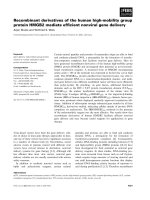
significant (P < 0.05; Table 2). The serum levels of
TNF-α and IL-6 were positively correlated with
HMGB1 levels both at PPHN onset (r = 0.832 and
0.866, respectively, P < 0.05) and after remission (r =
0.873 and 0.843, respectively, P < 0.05; Figure 1).
Animal experiment results
mPAP at each time point
The mPAP of the PPHN group was higher than
that of control group at each time point (respectively,
P < 0.05; Figure 2). Compared to that at the 24 h time
point, mPAP was significantly increased on day 3
time point in the control group (P < 0.05; Figure 2), but
was not increased in the PPHN group (P > 0.05;
Figure 2).
Table 2. Serum HMGB1, TNF-α and IL-6 levels among three
situations
Group
number
Control group
PPHN onset
PPHN
remission
F
P
10
12
12
HMGB1
(ng/ml)
2.37 ± 0.88
33.19 ± 9.45a
13.42 ± 2.14bc
TNF-α (pg/ml)
IL-6 (pg/ml)
3.14 ± 1.30
40.41 ± 14.3a
15.12 ± 2.45bc
3.47 ± 0.90
32.98 ± 13.42a
11.75 ± 2.77bc
81.171
0.000
53.354
0.000
39.089
0.000
a: The PPHN onset group compared with the normal cord blood group: Pa.HMGB1 =
0.000, Pa.TNF-α = 0.000, Pa.IL-6 = 0.000;
b: The after PPHN remission group compared with the normal cord blood group:
Pb.HMGB1 = 0.000, Pb.TNF-α = 0.003, Pb.IL-6 = 0.024;
c: The after PPHN remission group compared with the PPHN onset group:Pc.HMGB1
= 0.000, Pc.TNF-α = 0.000, Pc.IL-6 = 0.000.
Int. J. Med. Sci. 2019, Vol. 16
1127
Figure 3. Trends in serum HMGB1 levels at different time points of persistent
pulmonary hypertension of the newborn (PPHN) based on a rat model. Serum
HMGB1 levels were stable at different time points in the control group; however,
serum HMGB1 levels in the PPHN group first increased and then decreased. **P <
0.01 vs. control; ***P < 0.001 vs. control; n = 10.
Pulmonary vascular histopathology
Figure 1. Correlation between serum HMGB1 levels and serum TNF-α/IL-6 levels in
newborns with persistent pulmonary hypertension of the newborn (PPHN). a.
Correlation between TNF-α/IL-6 levels and HMGB1 levels at PPHN onset. b.
Correlation between TNF-α/IL-6 levels and HMGB1 levels after PPHN remission; n =
12.
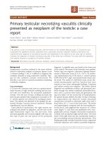
Compared to that in the normal control group,
H&E staining of lung tissue in the hypoxia-induced
PPHN animal model on day 3 showed that the wall of
the pulmonary arteriole accompanied by the terminal
bronchioles was thicker and that the lumen was
narrower; the thickened arteriole wall was mainly
caused by thickened medial vessel walls (Figure 4).
The MT% in the PPHN group was 7.2 ± 4.9%,
compared to 5.9 ± 4.7% in the control group. Although
there was no significant difference between the two
groups (P > 0.05), the MT% of the PPHN group
increased by 20.6% compared to that in the control
group.
Western blot analysis of lung tissue from PPHN rats
Figure 2. Changes in mean pulmonary artery pressure (mPAP) at different time
points after hypoxia-induced persistent pulmonary hypertension of the newborn
(PPHN). *P < 0.05 vs. control; **P < 0.01 versus control; #P < 0.05 vs. 24 h time point;
n = 4.
Serum HMGB1 levels at each time point
Serum levels of HMGB1 were basically stable in
11–14 day old control rats. Within 0–3 days after
PPHN, HMGB1 levels in the PPHN group increased
by 1.5–2 fold compared to those in the control group
at each time point, peaking 24 h after PPHN.
Differences between the PPHN and control groups
were statistically significant (respectively, P < 0.05;
Figure 3), but there were no differences among
different time points in the PPHN group (F = 2.134, P
> 0.05).
HMGB1 expression in the lungs of PPHN
neonatal rats was significantly higher than that in the
control group at 2, 8, and 24 h, as well as day
3(respectively, P < 0.05; Figure 5). In the PPHN group,
HMGB1 levels were significant different among
different time points (F = 14.136, P = 0.000), and the
expression trend showed a significant increase at 2 h,
peaking at 24 h, and then a decrease at day3. HMGB1
levels at day 3 were significantly lower than those at
24 h in the PPHN group (P < 0.001; Figure 5).
Discussion
PPHN comprises three types (primary,
congenital,
and
secondary),
with
different
mechanisms of pathogenesis. PPHN can also be
divided into pulmonary vascular underdevelopment,
pulmonary
vascular
mal-development,
and
pulmonary vascular maladaptation depending on
different pathological changes. Clearly, the etiology
Int. J. Med. Sci. 2019, Vol. 16
1128
and pathogenesis of PPHN are complex [20]. In recent
years, increasing numbers of studies have found that
extensive inflammatory cytokine dysregulation can
trigger and aggravate the abnormal contraction and
proliferation of pulmonary vascular smooth muscle
cells, resulting in pulmonary artery wall remodeling.
This suggests that the abnormal expression of
inflammatory cytokines is related to the occurrence
and development of pulmonary hypertension, and
accordingly, the role of inflammation in the
development of pulmonary hypertension is being
increasingly studied [21-23]. This study found the
following results: (1) serum levels of HMGB1 in
newborns with PPHN were significantly increased
early after PPHN onset, and then decreased after
remission, and were positively correlated with levels
of the classic inflammatory factors TNF-α and IL-6. (2)
In a rat model of PPHN, 7-day continuous hypoxia
can induce an increase in pulmonary artery pressure
lasting for 3 d, and histopathological analysis also
showed thickening of the medial
vessels of pulmonary arterioles
and lumenal stenosis, although
pulmonary arteriole changes did
not reach the level of irreversible
vascular remodeling on the 3rd
day after PPHN. (3) ELISA and
western
blot
analyses
demonstrated similar short-term
increases in HMGB1 levels in the
serum and lung tissue of PPHN
rats after PPHN onset, peaking
after 8–24 h, and slightly
decreasing,
but
remaining
significantly higher than those in
the control group, at day 3. These
results indicate that changes in
HMGB1 levels are related to the
occurrence and development of
PPHN. In hypoxia-induced PPHN
models,
pulmonary
arterial
changes might be dominated by
vasospasms, and during this
mode of remodeling, HMGB1
levels are more sensitive to
changes than tissue histology.
This provides an objective basis
for using HMGB1 as an early
diagnostic marker of PPHN and
to monitor the PPHN process.
HMGB1 is a single-chain
polypeptide consisting of 215
amino acids. In disease states, this
protein can promote local and
systemic inflammatory responses
through its passive secretion from
necrotic cells and active secretion
from immune cells including
macrophages, dendritic cells, and
natural killer cells. It can also
regulate the immune reaction by
promoting the production of
other inflammatory cytokines and
Figure 4. H&E staining of pulmonary arterioles in a rat model of PPHN. Morphology of lung tissue and right middle
activating different immune cells
lobe in experimental neonatal rats(A, B).The wall of pulmonary arterioles accompanied with terminal bronchioles in
normal control rats (C, D) and in PPHN rats (E, F). scale bar: 20 µm; n=4.
[24, 25]. HMGB1 is a central
Int. J. Med. Sci. 2019, Vol. 16
component of the inflammatory network, as its
secretion has an amplifying effect and it can also
regulate the secretion of other inflammatory cytokines
[26]. This clinical study demonstrated that serum
HMGB1, TNF-α, and IL-6 levels are significantly
increased with PPHN onset and significantly
decreased with PPHN alleviation. TNF-α and IL-6
expression was significantly positively correlated
with HMGB1 expression in the serum of newborns
with PPHN. These results suggested that
hypoxia-induced HMGB1 and inflammatory release
are closely related to the occurrence and development
of PPHN. There are two possible mechanisms that
could explain these changes (as follows): (1)
hypoxia-induced HMGB1 release from activated
pulmonary macrophages causes inflammation and
directly leads to vascular endothelial cell injury,
endothelial cell gap widening, increases in endothelial
permeability, and changes in pulmonary vascular
endothelial cell cytoskeletal remodeling, proliferation,
and contraction in a dose-dependent manner [27, 28].
(2) Extracellular HMGB1 stimulates the production of
reactive
oxygen
species
and
downstream
inflammatory cytokines inducing oxidative stress and
TNF-α, among others; meanwhile, with increases in
pulmonary artery pressure, blood flow shearing force
is increased and hypoxia is exacerbated. This would
result in an increase in the generation of reactive
oxygen species by vascular endothelial cells, vascular
smooth muscle cells, and adventitial fibroblasts,
further stimulating the expression of HMGB1 and its
receptors, and forming a positive feedback loop [11].
Animal results showed that both the serum
concentrations of HMGB1 and the expression of
HMGB1 in lung tissues began to increase 2 h after
PPHN onset, peaked after 24 h, and decreased at day
3. This dynamic change over time is consistent with a
previous report showing HMGB1 secretion into the
peripheral blood 24–48 h after PPHN onset [29].
In this study, the average gestational age of
newborns with PPHN was 39 weeks and the average
birth weight was 3.3 kg, a suitable weight for full-term
newborns. Most newborns had intrauterine hypoxia
and were diagnosed with PPHN shortly after birth.
Their peripheral blood HMGB1 and inflammatory
cytokine levels were significantly increased,
indicating a link between PPHN and the
hypoxia-induced inflammatory response. The
progression to pulmonary hypertension was
associated with hypoxic pulmonary vasospasm and
pulmonary vascular remodeling, which both
manifested as pulmonary arteriolar stenosis.
1129
Pulmonary vascular remodeling was markedly
increased with MT% as the main manifestation. When
the MT% increases to a certain extent, PPHN changes
are considered irreversible [30]. Animal studies have
reported that pulmonary arterial pressure in neonatal
rats is significantly increased 3–5 d after hypoxia
exposure, but increases in MT% were not obvious
[31]. We also observed thickening of the pulmonary
arteriole walls, accompanied by pulmonary terminal
bronchioles, and that the MT% was not different, as
compared to that in the control group, 3 days after
PPHN induction despite an increase in MT% of 20.6%.
These results indicate that the main pathological
change in the early stage of PPHN is vasospasm;
however, it is possible that pulmonary vessels
undergo vascular remodeling at this time, although it
is unclear when irreversible remodeling of the
pulmonary
arteries
occurs
during
hypoxia
progression. At present, studies examining
hypoxia-induced PPHN have mainly focused on
mediators that lead to irreversible vascular
remodeling, such as endothelin-1 [18], thyroid
hormone receptor interactor 6, cyclin D1 [15], and
vascular endothelial growth factor [32]. Although the
inhibition of HMGB1 reduces the right ventricular
systolic pressure and pulmonary vascular remodeling
in an adult rat model of hypoxia-induced pulmonary
arterial hypertension (PAH) by blocking the
interaction between HMGB1 and the TLR4 adaptor
MD2, little is known about the effects of
HMGB1-mediated inflammation on pulmonary
vasculature in developing animals [33]. Further
studies are needed to explore the role of HMGB1 in
irreversible hypoxia-mediated pulmonary artery
remodeling and related mechanisms during PPHN. In
addition, it is also important to further address the
clinical value of HMGB1 as a diagnostic and
predictive marker of adverse PPHN prognosis.
In summary, the clinical research presented in
this study indicates that serum levels of HMGB1 in
newborns with PPHN are significantly increased
early after PPHN onset, and then decreased after
remission, and that they are positively correlated with
levels of inflammatory factors. Animal experiments
confirmed that HMGB1 levels in the peripheral blood
and lung tissue change with hypoxia-induced PPHN
progression, although there was no significant
pulmonary vascular remodeling in PPHN rats. As an
inflammatory mediator, HMGB1 plays an important
role in the early stages of hypoxia-induced PPHN,
suggesting that it might be a useful early marker for
PPHN diagnosis.
Int. J. Med. Sci. 2019, Vol. 16
1130
Figure 5. HMGB1 expression in the lungs of a rat model of PPHN as detected by western blotting. ***P < 0.001 vs. control; **P <0.01 vs. control; *P < 0.05 vs. control; #P <
0.001 vs. 24 h time point; n = 6–10.
Acknowledgements
We are grateful for strong support from
ultrasound, obstetrics, and central laboratory staff at
the Third Xiangya Hospital of Central South
University and at Beijing Children’s Hospital, Capital
Medical University.
Ethics approval and consent to participate
The clinical part of this study was approved by
the medical ethics committee of the Third Xiangya
Hospital of Central South University and Beijing
Children’s Hospital, Capital Medical University. The
animal study part was approved by the medical ethics
committee of the Third Xiangya Hospital of Central
South University.
Funding
National Natural Science Foundation of China
(81671505).
Author Contributions
TZ JM OY-ZC was involved in data collection,
analysis, and interpretation; wrote the first draft;
performed revisions of the drafted article. HMY, TZ
WHL, DSX, and JM were involved in the conception
and design of the study; involved in data collection,
analysis, and interpretation; performed critical
revisions of the drafted article. TZ and HMY were
involved in data collection, analysis, and
interpretation; performed critical revisions of the
drafted article. All author approved the final version
of the manuscript for submission.
Competing Interests
The authors have declared that no competing
interest exists.
References
1.
Lapointe A, Barrington KJ. Pulmonary hypertension and the asphyxiated
newborn. J Pediatr. 2011; 158: e19-24. doi:10.1016/j.jpeds.2010.11.008.
2. Jain A, McNamara PJ. Persistent pulmonary hypertension of the newborn:
Advances in diagnosis and treatment. Semin Fetal Neonatal Med. 2015; 20:
262-71. doi:10.1016/j.siny.2015.03.001.
3. Fuloria M, Aschner JL. Persistent pulmonary hypertension of the newborn.
Semin Fetal Neonatal Med. 2017; 22: 220-6. doi:10.1016/j.siny.2017.03.004.
4. Storme L, Aubry E, Rakza T, et al. Pathophysiology of persistent pulmonary
hypertension of the newborn: impact of the perinatal environment. Arch
Cardiovasc Dis. 2013; 106: 169-77. doi: 10.1016/j.acvd.2012.12.005.
5. Nair J, Lakshminrusimha S. Update on PPHN: mechanisms and treatment.
Semin Perinatol. 2014; 38: 78-91. doi: 10.1053/j.semperi.2013.11.004.
6. Taleb M, Khuder S, Tinkel J, et al. The diagnostic accuracy of Doppler
echocardiography in assessment of pulmonary artery systolic pressure: a
meta-analysis. Echocardiography. 2013; 30: 258-65. doi: 10.1111/echo.12061.
7. Rabinovitch M, Guignabert C, Humbert M, et al. Inflammation and immunity
in the pathogenesis of pulmonary arterial hypertension. Circ Res. 2014; 115:
165-75. doi: 10.1161/CIRCRESAHA.113.301141.
8. Harbaum L, Renk E, Yousef S, et al. Acute effects of exercise on the
inflammatory state in patients with idiopathic pulmonary arterial
hypertension. BMC Pulm Med. 2016; 16: 145. doi: 10.1186/s12890-016-0301-6.
9. Bauer EM, Shapiro R, Billiar TR, et al. High mobility group Box 1 inhibits
human pulmonary artery endothelial cell migration via a Toll-like receptor 4and interferon response factor 3-dependent mechanism(s). J Biol Chem. 2013;
288: 1365-73. doi: 10.1074/jbc.M112.434142.
10. Bauer EM, Shapiro R, Zheng H, et al. High mobility group box 1 contributes to
the pathogenesis of experimental pulmonary hypertension via activation of
Toll-like
receptor
4.
Mol
Med.
2013;
18:
1509-18.
doi:
10.2119/molmed.2012.00283.
Int. J. Med. Sci. 2019, Vol. 16
1131
11. Janko C, Filipovic M, Munoz LE, et al. Redox modulation of HMGB1-related
signaling. Antioxid Redox Signal. 2014; 20: 1075-85. doi: 10.1089/ars.2013.
5179.
12. Lakshminrusimha S, Keszler M. Persistent Pulmonary Hypertension of the
Newborn. Neoreviews. 2015; 16: e680-92. doi: 10.1542/neo.16-12-e680.
13. Baquero H, Soliz A, Neira F, et al. Oral sildenafil in infants with persistent
pulmonary hypertension of the newborn: a pilot randomized blinded study.
Pediatrics. 2006; 117: 1077-83. doi: 10.1542/peds.2005-0523.
14. Clark RH, Kueser TJ, Walker MW, et al. Low-dose nitric oxide therapy for
persistent pulmonary hypertension of the newborn. Clinical Inhaled Nitric
Oxide Research Group. N Engl J Med. 2000; 342: 469-74. doi:
10.1056/NEJM200002173420704.
15. Xu YP, Zhu JJ, Cheng F, et al. Ghrelin ameliorates hypoxia-induced
pulmonary hypertension via phospho-GSK3 beta/beta-catenin signaling in
neonatal rats. J Mol Endocrinol. 2011; 47: 33-43. doi: 10.1530/JME-10-0143.
16. Xu YP, He Q, Shen Z, et al. MiR-126a-5p is involved in the hypoxia-induced
endothelial-to-mesenchymal transition of neonatal pulmonary hypertension.
Hypertens Res. 2017; 40: 552-61. doi: 10.1038/hr.2017.2.
17. Deruelle P, Balasubramaniam V, Kunig AM, et al. BAY 41-2272, a direct
activator of soluble guanylate cyclase, reduces right ventricular hypertrophy
and prevents pulmonary vascular remodeling during chronic hypoxia in
neonatal rats. Biol Neonate. 2006; 90: 135-44. doi: 10.1159/000092518.
18. Wang L, Zhou Y, Li M, et al. Expression of hypoxia-inducible factor-1alpha,
endothelin-1 and adrenomedullin in newborn rats with hypoxia-induced
pulmonary hypertension. Exp Ther Med. 2014; 8: 335-9. doi:
10.3892/etm.2014.1728.
19. Lu TS, Yiao SY, Lim K, et al. Interpretation of biological and mechanical
variations between the Lowry versus Bradford method for protein
quantification. N Am J Med Sci. 2010; 2: 325-8. doi: 10.4297/najms.2010.2325.
20. Distefano G, Sciacca P. Molecular physiopathogenetic mechanisms and
development of new potential therapeutic strategies in persistent pulmonary
hypertension of the newborn. Ital J Pediatr. 2015; 41: 6. doi:
10.1186/s13052-015-0111-0.
21. Van Marter LJ, Hernandez-Diaz S, Werler MM, et al. Nonsteroidal
antiinflammatory drugs in late pregnancy and persistent pulmonary
hypertension
of
the
newborn.
Pediatrics.
2013;
131:
79-87.
doi:10.1542/peds.2012-0496.
22. Papamatheakis DG, Blood AB, Kim JH, et al. Antenatal hypoxia and
pulmonary vascular function and remodeling. Curr Vasc Pharmacol. 2013; 11:
616-40.
23. Sharma V, Berkelhamer S, Lakshminrusimha S. Persistent pulmonary
hypertension of the newborn. Matern Health Neonatol Perinatol. 2015; 1: 14.
doi: 10.1186/s40748-015-0015-4.
24. Zhu S, Li W, Ward MF, et al. High mobility group box 1 protein as a potential
drug target for infection- and injury-elicited inflammation. Inflamm Allergy
Drug Targets. 2010; 9: 60-72.
25. Weber DJ, Allette YM, Wilkes DS, et al. The HMGB1-RAGE Inflammatory
Pathway: Implications for Brain Injury-Induced Pulmonary Dysfunction.
Antioxid Redox Signal. 2015; 23: 1316-28. doi: 10.1089/ars.2015.6299.
26. Kang R, Chen R, Zhang Q, et al. HMGB1 in health and disease. Mol Aspects
Med. 2014; 40: 1-116. doi: 10.1016/j.mam.2014.05.001.
27. Chan SY, Loscalzo J. Pathogenic mechanisms of pulmonary arterial
hypertension.
J
Mol
Cell
Cardiol.
2008;
44:
14-30.
doi:
10.1016/j.yjmcc.2007.09.006.
28. Wolfson RK, Chiang ET, Garcia JG. HMGB1 induces human lung endothelial
cell cytoskeletal rearrangement and barrier disruption. Microvasc Res. 2011;
81: 189-97. doi: 10.1016/j.mvr.2010.11.010.
29. Sadamura-Takenaka Y, Ito T, Noma S, et al. HMGB1 promotes the
development of pulmonary arterial hypertension in rats. PLoS One. 2014; 9:
e102482. doi: 10.1371/journal.pone.0102482.
30. Xu MH, Gong YS, Su MS, et al. Absence of the adenosine A2A receptor confers
pulmonary arterial hypertension and increased pulmonary vascular
remodeling in mice. J Vasc Res. 2011; 48: 171-83. doi: 10.1159/000316935.
31. Sang K, Zhou Y, Li MX. [Pulmonary vascular remodeling in neonatal rats with
hypoxic pulmonary hypertension.] Zhongguo Dang Dai Er Ke Za Zhi. 2012;
14: 210-4. Chinese.
32. Voelkel NF, Gomez-Arroyo J. The role of vascular endothelial growth factor in
pulmonary arterial hypertension. The angiogenesis paradox. Am J Respir Cell
Mol Biol. 2014; 51: 474-84. doi: 10.1165/rcmb.2014-0045TR .
33. Goldenberg NM, Hu Y, Hu X, et al. Therapeutic Targeting of High-Mobility
Group Box-1 in Pulmonary Arterial Hypertension. Am J Respir Crit Care Med.
2019; 199(12): 1566-9. doi: 10.1164/rccm.201808-1597LE.