Comparison of adhesion prevention capabilities of the modified starch powder-based medical devices 4DryField® PH and Arista™ AH in the Optimized Peritoneal Adhesion Model
Bạn đang xem bản rút gọn của tài liệu. Xem và tải ngay bản đầy đủ của tài liệu tại đây (856.74 KB, 6 trang )
Int. J. Med. Sci. 2019, Vol. 16
Ivyspring
International Publisher
1350
International Journal of Medical Sciences
2019; 16(10): 1350-1355. doi: 10.7150/ijms.33277
Research Paper
Comparison of adhesion prevention capabilities of the
modified starch powder-based medical devices
4DryField® PH and Arista™ AH in the Optimized
Peritoneal Adhesion Model
Daniel Poehnert1*, Lavinia Neubert2*, Juergen Klempnauer1, Paul Borchert2, Danny Jonigk2, Markus
Winny1
1.
2.
Department of General, Visceral and Transplantation Surgery, Hannover Medical School, Hannover, Germany
Institute of Pathology, Hannover Medical School, Hannover, Germany
* These authors contributed equally
Corresponding author: Dr. Daniel Poehnert, PhD. Carl-Neuberg-Strasse 1, D-30625 Hannover (Germany); Tel. +49 511 5326534; Fax +49 511 5324010; E-Mail
© The author(s). This is an open access article distributed under the terms of the Creative Commons Attribution License ( />See for full terms and conditions.
Received: 2019.01.18; Accepted: 2019.07.22; Published: 2019.09.19
Abstract
Adhesion barriers can be based on numerous substances. In the rat Optimized Peritoneal Adhesion
Model (OPAM) the starch-based hemostats 4DryField and Arista were tested for their capability to
act in a preventive manner against adhesion formation (applied as a powder that was mixed in situ
with saline solution to form a barrier gel). Adhesions were scored using the established scoring
systems by Lauder and Hoffmann, as well as histopathologically using the score by Zühlke. Animals
receiving saline solution were used as controls. As previously published, 4DryField reduced
peritoneal adhesions significantly. However, Arista did not lead to a statistically significant reduction
of adhesion formation. When comparing 4DryField and Arista applied in the same manner, only
4DryField was significantly effective in preventing peritoneal adhesions. Histopathological
evaluations confirmed the results of the macroscopic investigation, leading to the conclusion that
starch-based hemostats do not generally have the capability to function as effective adhesion
prevention devices.
Key words: Adhesion prevention, abdominal surgery, rat model OPAM, 4DryField® PH, AristaTM AH
Introduction
Surgery is the most common cause for formation
of peritoneal adhesions. Predisposing factors include
mechanical injury of the peritoneum and local
ischemia due to manipulation and retraction of
abdominal tissues during surgery [1-4]. The incidence
of postoperative adhesion formation ranges from 67
to 93% [5]. Several adhesion prevention barrier agents
addressing this problem are available on the market.
In the majority of cases these agents function as a
physical barrier to separate wound areas at risk of
developing adhesions. These devices include
adhesion barriers made from oxidized regenerative
cellulose [6], polytetrafluoroethylene [7], icodextrin
[8], hyaluronic acid/carboxymethyl cellulose [9] and
starch [10]. Typically, starch-based products are used
solely as hemostats, such as Arista™ AH (Arista;
Davol Inc., USA) [11]. A unique starch-based medical
device is 4DryField® PH (4DryField; PlantTec Medical
GmbH, Germany) as it is the only product proven to
provide hemostasis and prevent the formation of
adhesions. While 4DryField is applied as a powder for
hemostasis, the powder is transformed into a gel by
mixing with saline solution for adhesion prevention.
This raised the question if modified starch
Int. J. Med. Sci. 2019, Vol. 16
powders other than 4DryField might also be capable
of reducing adhesion formation when applied in the
same way as 4DryField. Previously, Hoffmann et al.
[12] found Arista to be moderately effective in
preventing adhesions, whereas no effect was
observed in a study by Singh et al. [13]. Therefore, the
aim of the present study was to test 4DryField and
Arista for their capability in preventing postoperative
peritoneal adhesion formation in a challenging and
well-reproducible rat model, the recently described
Optimized Peritoneal Adhesion Model (OPAM) [14].
This model has been shown to induce severest
adhesions with high reliability and it has already been
utilized successfully to examine the effectiveness of
4DryField compared to a control group [15], as well as
in a comparative study with 4DryField and other
adhesion prevention devices based on different
materials [16]. The model includes abrasion of the
cecum and incision of the abdominal wall, as well as
meso-stitch approximation of these lesions.
Materials and Methods
Animals
Thirty-six male Lewis rats were included in the
study. They were housed under standard conditions,
had access to fresh water at any time and were fed a
standard diet ad libitum. Prior to and after surgery,
daily monitoring of body weight and behavioral
changes assessed animal welfare. Animal experiments
were performed at the central animal laboratory of the
Hanover Medical School, Germany, as well as the
therapeutic experimental unit, Faculty of Medicine,
Nantes, France. All protocols regarding animal life
quality were conducted in accordance with national
and European regulations. The present study was
approved by The Lower Saxony State Office for
Consumer Protection and Food Safety (LAVES
Hannover, Germany; approval code 12/0751) and the
Ethical Committee For Animal Experiments (CEEA)
in Pays de la Loire, France (approved under the
reference APAFIS9771).
Surgical procedures and application of
anti-adhesive agents
General anesthesia was achieved by ketamine
(80 mg/kg body weight) and xylazine (5 mg/kg body
weight) or inhalation of isoflurane 3%. The required
level of narcosis was reached when the flexor reflexes
were suppressed. A 3 cm long median laparotomy
was performed after shaving and sanitizing the
abdomen. Adhesion induction was carried out
according to the OPAM [14]: 1) the cecum was
delivered and kept moist with a watery gauze swab,
the cecal peritoneum was gently abraded repeatedly
1351
over a 1x2 cm area in a standard manner using a dry
gauze until removal of visceral peritoneum resulted in
sub-serosal bleeding and the creation of a
homogenous surface of petechial hemorrhages; 2) the
parietal peritoneum and inner muscle layer were
sharply dissected in order to create a 1x2 cm
abdominal wall defect; 3) both injured areas were
approximated using a non-absorbable suture. Prior to
surgery, animals were randomly assigned to one of
the following three groups: control (n=10),
4DryField-treated (n=16) or Arista-treated (n=10,
carried out in France). Control animals received 1.2 ml
0.9% sterile saline solution intraperitoneally. The two
anti-adhesive agents 4DryField and Arista were each
administered in a total amount of 300 mg
powder/animal. The powder was evenly distributed
on the two defects and then transformed into a gel by
dripping with 1.2 ml sterile 0.9% saline solution
before the approximating suture was placed. The
abdomen was closed using a two-layer closure
technique by consecutive sutures. Following surgery,
the animals were monitored until they were
completely awakened and kept warm using an
infrared lamp. Animals received novaminsulfone or
buprenorphine in a body-weight adapted dose to
minimize postoperative pain. On postoperative day 7,
the animals were sacrificed using CO2 narcosis
followed by cervical disclosure. The peritoneal cavity
was opened by an incision at a left-sided position
remote to the original laparotomy scar to prevent
damaging any potentially formed adhesions.
Specimens of cecum, abdominal wall and adhesions
were harvested for histopathological assessment.
A detailed protocol was generated and provided
to the surgeons in France to ensure uniformity of
execution and, thereby, comparability of the results.
Apart from step-by-step descriptions of the
procedures, photographs illustrated all steps in detail,
particularly the abrasion of the cecum, the dissection
of peritoneum and inner muscle layer, as well as the
application of the adhesion barrier.
Adhesion assessment
The adhesion formation between the defective
abdominal wall and cecum was evaluated
macroscopically by two independent observers
according to the scoring systems by Lauder et al. [17]
and Hoffmann et al. [12]. The Lauder scoring system
(Table 1) takes into account number, strength and
distribution of adhesions in a single score, while the
Hoffmann scoring system (Table 2) consists of three
individual scores for area, extent and strength of
adhesions that are summed up to yield a total score.
Int. J. Med. Sci. 2019, Vol. 16
1352
Table 1: Adhesion scoring system according to Lauder et al. [17]
for Mac OS, GraphPad Software, Inc., La Jolly, USA).
Score
0
1
2
3
4
5
Results
Description
No adhesions
Thin filmy adhesions
More than one thin adhesion
Thick adhesion with focal point
Thick adhesion with planar attachment
Very thick vascularized adhesions or more than one planar adhesion
Table 2: Adhesion scoring system according to Hoffmann et al.
[12]
Score Description
Area score
0
No adhesion
1
Cecum to bowel adhesion
2
Cecum to sidewall adhesion over less than 25% of the abraded surface
area
3
Cecum to sidewall adhesion between 25 and 50% of the abraded
surface area
4
Cecum to sidewall adhesion over more than 50% of the surface area
Strength score
0
No adhesion
1
Gentle traction required to break adhesion
2
Blunt dissection required to break adhesion
3
Sharp dissection required to break adhesion
Extent score
0
No adhesion
1
Filmy adhesion
2
Vascularized adhesion
3
Opaque or cohesive adhesion
Histology
Surgical specimens were fixed in buffered 4%
formaldehyde solution. After dehydration and
paraffin embedding, serial thin sections of 1–2 μm
were mounted on glass slides, stained with standard
Hematoxylin and Eosin (HE), Elastika-van-Gieson
(EvG) and periodic acid-Schiff (PAS) staining (Sigma
Aldrich Co Ltd, USA) and light microscope
examinations were performed by experienced
pathologists.
The quantitative analysis of the histologic
stainings was performed using Zühlke’s microscopic
adhesion classification. This system has already been
established for grading of peritoneal adhesions
induced with models very similar to OPAM [19, 20].
Statistical analyses
Adhesion scores are presented as arithmetic
means with standard deviations (SD). Since most of
the data sets did not follow a Gaussian distribution (as
determined using the D’Agostino-Pearson normality
test) the multiple comparisons of adhesion scores of
the
three
groups
were
performed
using
Kruskal-Wallis test followed by Dunn’s multiple
comparisons test for non-parametric data (which
utilizes correction for multiple comparison by
statistical hypothesis testing). Groups were defined to
be significantly different if p<0.05. Statistical analyses
were performed using GraphPad Prism (Version 7.0b
All animals showed comparable viability and
body weight development. None of the animals had
to be sacrificed prematurely due to complications; all
36 animals completed the study.
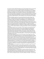
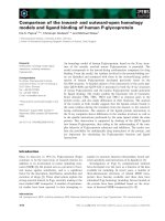
Adhesion development
In the control group, 9 of 10 animals showed
peritoneal adhesions, which were rated with the
maximum Lauder score, as well as the maximum
scores regarding all of the Hoffmann categories
(Figure 1A,B). None of the sixteen 4DryField-treated
animals developed any adhesions (Figure 1C,D). In
contrast, all 10 Arista-treated animals developed
peritoneal adhesions (Figure 1E,F). Two developed
filmy adhesions, with a Lauder score of 1 each. The
total Hoffmann scores of these two animals differed
and were 3 and 7, respectively. The other eight
Arista-treated animals developed severe adhesions
with Lauder scores of 4 (n=6) or 5 (n=2) and total
Hoffmann scores of 8 (n=4), 9 (n=3) or 10 (n=1). The
mean score value of each group was calculated and
tested for significant differences (Table 3). Herein,
4DryField PH reduced the incidence and severity of
peritoneal adhesion formation significantly compared
to the control, as well as to the Arista-treatment group
and concerning every evaluated scoring system. In
contrast, Arista-treatment did not lead to a
statistically significant reduction of adhesion
formation in comparison to control animals.
Table 3: Microscopic adhesion classification according to Zühlke
et al. [18]
Score Description
0
No adhesions
1
Weak connective tissue, rich cell, new and old fibrin, thin reticulin
fibrils
2
Connective tissue which has cells and capillaries. few collagen fibers
3
Thicker connective tissue. Few cells and elastic and smooth muscle
fibers, more vessels
4
Old and thick granulation tissue, poor cells, difficult separation of
serosal surfaces
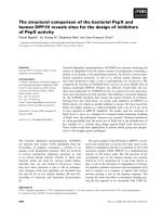
Histological Evaluation
Figure 2 shows representative PAS-stained
tissue slides from all three groups. Figure 2A shows a
control animal where the smooth muscle layers of the
cecum (top) are fused to skeletal muscles of the
abdominal wall (bottom) via dense granulating tissue.
The histological findings support the macroscopic
observation that both, cecum and abdominal wall,
could not readily be separated by mechanical force.
Figure 2B shows cecal and Figure 2C abdominal wall
tissue of an animal from the 4DryField group.
Int. J. Med. Sci. 2019, Vol. 16
1353
animal was scored 1, one was scored 2, four were
scored 3, and four were scored 4.
Like for the macroscopic adhesion assessment
the mean scores were calculated and tested for
significant differences (Table 5). The results were
conform with the macroscopic assessment, the
4DryField treated animals scored significantly better
results than the Arista treated ones as well as the
control animals, while the Arista group did not show
statistically significant differences to the control.
Discussion
As shown in previous studies [14-16], the OPAM
consistently induced severe peritoneal adhesions after
cecal abrasion and creation of abdominal wall defects
in rats.
4DryField
revealed
excellent
adhesion
prevention capabilities, completely preventing the
formation of any adhesions. Furthermore, a
newly-formed mesothelial layer was found by
histopathological assessments of the previously
injured sides. 4DryField could be shown to be highly
effective in preventing peritoneal adhesions in
previous studies, being prophylactically applied
either as a preformed gel or as powder that was
transformed in situ into a gel by adding saline solution
[15, 16].
Figure 1: Representative photographs of the pathological evaluation of control
(A,B), 4DryField- (C,D) and Arista-treated (E,F) rats on day 7.
In contrast to 9 of the 10 control animals, no
agglutinations occurred in the 4DryField group.
Furthermore, in all animals of the 4DryField group
the lesions of the cecum and the abdominal wall
defect had healed, and both featured neomesothelial
cell coverage. The former abdominal wall defect was
filled with fibrous tissue, which still contained slight
remnants of 4DryField particles. Figure 2D shows an
animal from the Arista group. As in Figure 2A the
smooth muscles of the cecum (top) were fused to the
skeletal muscles of the abdominal wall (bottom) via
dense granulation tissue, preventing separation of
cecum and abdominal wall by mechanical force.
The microscopic classification of the adhesions
according to Zühlke et al. [18] was performed all
animals. In the control group one animal was scored
0, two were scored 3 and seven were scored 4, in the
4DryField group the microscopic assessment was
equivalent to the macroscopic investigation with all
16 animals being scored 0. In the Arista group one
Table 4: Arithmetic mean values (AM), standard deviations (SD)
and p-values in comparison to the control (p (ctrl)) or 4DryField (p
(4DF)) groups (statistically significant difference if p<0.05, *)
Score
Lauder
Hoffmann Area
Hoffmann Strength
Hoffmann Extent
Hoffmann Total
Group
control
4DryField
Arista
control
4DryField
Arista
control
4DryField
Arista
control
4DryField
Arista
control
4DryField
Arista
AM
4.5
0.0
3.6
3.6
0.0
2.4
2.7
0.0
2.7
2.7
0.0
2.8
9.0
0.0
7.9
SD
1.6
0.0
1.4
1.3
0.0
0.8
0.9
0.0
0.7
0.9
0.0
0.6
3.2
0.0
1.9
p (ctrl)
p (4DF)
<0.0001*
0.6512
0.0008*
<0.0001*
0.4556
0.0013*
<0.0001*
>0.9999
<0.0001*
<0.0001*
>0.9999
<0.0001*
<0.0001*
0.4565
0.0013*
Table 5: Arithmetic mean values (AM), standard deviations (SD)
and p-values in comparison to the control (p (ctrl)) or 4DryField (p
(4DF)) groups (statistically significant difference with p<0.05, *)
Score
Zühlke
Group
control
4DryField
Arista
AM
3.4
0.0
3.1
SD
1.3
0.0
1.0
p (ctrl)
p (4DF)
<0.0001*
>0.9999
0.0001*
Int. J. Med. Sci. 2019, Vol. 16
1354
Figure 2: Representative histological slides (PAS-stained) of animals from the control (A), 4DryField (B, C) and Arista (D) groups. Black arrows indicate neomesothelial
coverage.
The adhesion prevention capabilities of Arista
were examined for the first time in 2009 by Hoffmann
et al. [12]. Although the authors found the adhesion
development to be significantly reduced in
comparison to a control group, the adhesion
reduction was still limited with an adhesion score of
3.9 (Arista) vs. 6.0 (control). In 2013, Singh et al.
challenged these results in a randomized-controlled
trial using Arista in a rat model with adhesion
induction at the cecum and the uterine horn.
Adhesion prevention capabilities of Arista were
found to be not different from those of the control
group, which received Ringer’s lactate solution [13].
In our present study, Arista did not lead to a
statistically significant reduction of adhesion
formation compared to control animals using Lauder
and Hoffmann scoring systems, as well as systematic
histopathological examinations using the Zühlke
microscopic classification system and confirming the
macroscopic results. The microscopic analysis showed
tight agglutinations of cecum and abdominal wall via
granulating tissue, comparable to those of the control
animals. When comparing 4DryField and Arista
applied in the same manner, 4DryField resulted in a
significantly more effective reduction of adhesion
scores.
Limited comparability of the results arising from
differing surgical performance at the two study
centers can be excluded due to strict monitoring of the
comparability as described above. Additionally, the
OPAM has been used at the Hanover Medical School
extensively [14-16] and different surgeons have
performed surgeries following this protocol in the
past, but a correlation of results with the respective
surgeon has never been observed. Correspondingly,
the model has been shown to be highly reliable and
very robust.
In summary, in this experimental animal model
of severe peritoneal adhesion induction only
4DryField but not Arista was effective in reducing
postoperative adhesion formation when both devices
were applied in the same manner. Our results show
that modified starch-based powder hemostats are not
naturally capable to reduce the formation of
peritoneal adhesions. Instead, the effectiveness
depends on the specific properties of the individual
product, which are often not reported in detail and
might be of interest for further investigations.
Acknowledgments
The authors would like to thank Dres. Valérie
Dumay and Pierre Layrolle for generous sharing of
data obtained in the Therapeutic Experimental Unit,
Faculty of Medicine, Nantes, France. They performed
the Arista experiments, funded by PlantTec Medical
GmbH, Germany. The authors are also grateful to
Valentina Osmani for editing the manuscript.
Competing Interests
The authors have declared that no competing
interest exists.
References
1.
2.
3.
4.
5.
Ellis H. The clinical significance of adhesions: focus on intestinal obstruction.
Eur J Surg Suppl 1997:5-9.
Lehmann-Willenbrock EL, Mecke H, Riedel HH. Sequelae of Appendectomy,
with Special Reference to Intra-Abdominal Adhesions, Chronic Abdominal
Pain, and Infertility. Gynecologic and Obstetric Investigation 1990;29:241-5.
Luijendijk RW, de Lange DC, Wauters CC, et al. Foreign material in
postoperative adhesions. Ann Surg 1996;223:242-8.
Menzies D. Postoperative Adhesions - their Treatment and Relevance in
Clinical Practice. Ann R Coll Surg Engl 1993;75:147-53.
Beyene RT, Kavalukas SL, Barbul A. Intra-abdominal adhesions: Anatomy,
physiology, pathophysiology, and treatment. Curr Probl Surg 2015;52:271-319.
Int. J. Med. Sci. 2019, Vol. 16
6.
7.
8.
9.
10.
11.
12.
13.
14.
15.
16.
17.
18.
19.
20.
1355
Sawada T, Nishizawa H, Nishio E, Kadowaki M. Postoperative adhesion
prevention with an oxidized regenerated cellulose adhesion barrier in infertile
women. J Reprod Med 2000;45:387-9.
Group TMAMS. An expanded polytetrafluoroethylene barrier (Gore-Tex
Surgical Membrane) reduces post-myomectomy adhesion formation. The
Myomectomy Adhesion Multicenter Study Group. Fertil Steril 1995;63:491-3.
Sakari T, Sjodahl R, Pahlman L, Karlbom U. Role of icodextrin in the
prevention of small bowel obstruction. Safety randomized patients control of
the first 300 in the ADEPT trial. Colorectal Dis 2016;18:295-300.
Joung JY, Ha YS, Singer EA, et al. Use of a hyaluronic
acid-carboxymethylcellulose adhesion barrier on the neurovascular bundle
and prostatic bed to facilitate earlier recovery of erectile function after
robot-assisted prostatectomy: an initial experience. J Endourol 2013;27:1230-5.
Korell M, Ziegler N, De Wilde RL. Use of Modified Polysaccharide
4DryField® PH for Adhesion Prevention and Hemostasis in Gynecological
Surgery: A Two-Center Observational Study by Second-Look Laparoscopy.
Biomed Res Int 2016;2016:1-9.
Bruckner BA, Blau LN, Rodriguez L, et al. Microporous polysaccharide
hemosphere absorbable hemostat use in cardiothoracic surgical procedures.
Journal of Cardiothoracic Surgery 2014;9.
Hoffmann NE, Siddiqui SA, Agarwal S, et al. Choice of hemostatic agent
influences adhesion formation in a rat cecal adhesion model. J Surg Res
2009;155:77-81.
Singh P, Vasques D, Deleon F. Microporous Polysaccharide Hemospheres for
Adhesion Prevention: A Randomized Controlled Trial. Journal of Gynecologic
Surgery 2013;29:196-202.
Poehnert D, Abbas M, Kreipe H-H, Klempnauer J, Winny M. High
reproducibility of adhesion formation in rat with meso-stitch approximation
of injured cecum and abdominal wall. Int J Med Sci 2015;12:1-6.
Poehnert D, Abbas M, Kreipe H-H, Klempnauer J, Winny M. Evaluation of
4DryField® PH as Adhesion Prevention Barrier Tested in an Optimized
Adhesion Model (OPAM) in Rats. Eur Surg Res 2015;55:341-51.
Poehnert D, Grethe L, Maegel L, et al. Evaluation of the Effectiveness of
Peritoneal Adhesion Prevention Devices in a Rat Model. Int J Med Sci
2016;13:524-32.
Lauder CI, Garcea G, Strickland A, Maddern GJ. Use of a modified
chitosan-dextran gel to prevent peritoneal adhesions in a rat model. J Surg Res
2011;171:877-82.
Zühlke HV, Lorenz EMP, Straub EM, Savvas V. Pathophysiologie und
Klassifikation von Adhäsionen. 1990;:1009-16.
Karaca G, Aydin O, Pehlivanli F, et al. Effect of ankaferd blood stopper in
experimental peritoneal adhesion model. Ann Surg Treat Res 2016;90:213-7.
Gokcelli U, Ercan UK, Ilhan E, Argon A, Cukur E, Ureyen O. Prevention of
Peritoneal Adhesions by Non-Thermal Dielectric Barrier Discharge Plasma
Treatment on Mouse Model: A Proof of Concept Study. J Invest Surg
2019;:1-10.