Uric acid in the pathogenesis of metabolic, renal, and cardiovascular diseases: A review
Bạn đang xem bản rút gọn của tài liệu. Xem và tải ngay bản đầy đủ của tài liệu tại đây (1.26 MB, 12 trang )
Journal of Advanced Research 8 (2017) 537–548
Contents lists available at ScienceDirect
Journal of Advanced Research
journal homepage: www.elsevier.com/locate/jare
Review
Uric acid in the pathogenesis of metabolic, renal, and cardiovascular
diseases: A review
Usama A.A. Sharaf El Din a,⇑, Mona M. Salem b, Dina O. Abdulazim c
a
Nephrology Unit, Internal Medicine Department, School of Medicine, Cairo University, Egypt
Endocrinology Unit, Internal Medicine Department, School of Medicine, Cairo University, Egypt
c
Rheumatology and Rehabilitation Department, School of Medicine, Cairo University, Egypt
b
g r a p h i c a l a b s t r a c t
a r t i c l e
i n f o
Article history:
Received 11 September 2016
Revised 26 November 2016
Accepted 27 November 2016
Available online 3 December 2016
Keywords:
Uric acid
Insulin resistance
Non-alcoholic fatty liver disease
Acute kidney injury
Chronic kidney disease
Cardiovascular disease
a b s t r a c t
The association between uric acid (UA) on one side and systemic hypertension (Htn), dyslipidemia, glucose intolerance, overweight, fatty liver, renal disease and cardiovascular disease (CVD) on the other side
is well recognized. However, the causal relationship between UA and these different clinical problems is
still debatable. The recent years have witnessed hundreds of experimental and clinical trials that favored
the opinion that UA is a probable player in the pathogenesis of these disease entities. These studies disclosed the strong association between hyperuricemia and metabolic syndrome (MS), obesity, Htn, type 2
diabetes mellitus (DM), non-alcoholic fatty liver disease, hypertriglyceridemia, acute kidney injury,
chronic kidney disease (CKD), coronary heart disease (CHD), heart failure and increased mortality among
cardiac and CKD patients. The association between UA and nephrolithiasis or preeclampsia is a nondebatable association. Recent experimental trials have disclosed different changes in enzyme activities
induced by UA. Nitric oxide (NO) synthase, adenosine monophosphate kinase (AMPK), adenosine
monophosphate dehydrogenase (AMPD), and nicotinamide adenine dinucleotide phosphate (NADPH)oxidase are affected by UA. These changes in enzymatic activities can lead to the observed biochemical
and pathological changes associated with UA. The recent experimental, clinical, interventional, and epidemiologic trials favor the concept of a causative role of UA in the pathogenesis of MS, renal, and CVDs.
Ó 2016 Production and hosting by Elsevier B.V. on behalf of Cairo University. This is an open access article
under the CC BY-NC-ND license ( />
Peer review under responsibility of Cairo University.
⇑ Corresponding author. Fax: +20 222753890.
E-mail address: (U.A.A. Sharaf El Din).
/>2090-1232/Ó 2016 Production and hosting by Elsevier B.V. on behalf of Cairo University.
This is an open access article under the CC BY-NC-ND license ( />
538
U.A.A. Sharaf El Din et al. / Journal of Advanced Research 8 (2017) 537–548
Introduction
UA is a weak acid (M.W. = 168) produced in the liver, muscles,
and intestines [1]. Purines are the precursors of UA. Xanthine
oxidoreductase (XO) is the enzyme responsible for UA production.
Exogenous sources that can increase serum UA include fatty meat,
organ meat, and seafood [2]. Fructose is another source of exogenous UA. Fructose is present in fruits and added sugar. Fructokinase enzyme catalyzes the phosphorylation of fructose by
consuming adenosine triphosphate (ATP). Adenosine monophosphate (AMP) thus generated finally converts to UA [3]. UA was
incriminated in the pathogenesis of gout and kidney stones. However, for more than 140 years ago, high serum UA (SUA) was proposed in association with other diseases including Htn [4], CKD
and DM [5]. The association between hyperuricemia and CHD
was first reported in 1951 [6]. SUA bears a highly significant positive correlation with insulin resistance (IR) and insulin response to
oral glucose load. Hyperuricemia encountered in case of increased
IR is the sequence of decreased renal urate clearance [7]. Accumulating data point toward a possible etiologic role of increased UA in
the pathogenesis of MS, CVD and renal disease [8]. Experimental
and clinical trials have demonstrated the reversal or amelioration
of different diseases associated with hyperuricemia after administration of hypouricemic agents. These agents are either inhibitors
of the XO enzyme or stimulants of renal UA excretion. This later
group supports that the therapeutic effect is a consequence of UA
lowering rather than inhibition of release of free oxygen radicals
on inhibition of XO enzyme. In this review, we are going to discuss
the possible impact of hyperuricemia on metabolic, renal, and
CVDs.
Uric acid and metabolic syndrome
MS is a group of clinical and laboratory abnormalities. Out of
the five established manifestations, three or more are needed to
diagnose MS. These manifestations are (1) waist circumference P 90 and 80 cm in men and women respectively; (2) serum
triglyceride P 150 mg/dL; (3) high-density lipoprotein cholesterol
(HDLc) < 40 and 50 mg/dL in men and women respectively; (4)
blood pressure (BP) P 130/85 mmHg; and (5) fasting blood
sugar P 100 mg/dL [9]. The different manifestations of MS are considered as consequences of excess fat deposition in the adipose tissue [10]. Excess intake of sugars beside purine rich foods can lead
to increased incidence of hyperuricemia, obesity and DM [11]. In
adults with normal body mass index, MS is 10 times higher in
those having SUA P 10 mg/dL compared to those with
SUA < 6 mg/dL [12]. The hazard ratio of incident MS shows a steady
increase when normal adults were allocated into four quartiles
according to SUA. These results were still observed after considering the body composition [13]. When children (10–15 years at
baseline) were followed for 10 years, high SUA was a significant
predictor of incident MS in male subjects [14]. On the other hand,
when elderly hyperuricemic subjects above sixty-five years were
followed for more than 4 years, only female subjects showed
increased incidence of MS [15]. Another prospective study assessed
1511 men and women 55–80 years old, who were not affected initially by any of the components of MS. Follow-up has demonstrated a significantly higher incidence of many components of
MS, namely, hypertriglyceridemia, low HDL, and Htn in subjects
with highest sex-adjusted quartile of UA [16]. A meta-analysis of
eleven studies of more than fifty-four thousand participants
showed that elevated SUA is associated with increased risk of MS
and non-alcoholic fatty liver disease (NAFLD) [17]. By inhibiting
endothelial NO synthase, decreased NO might underlie insulin
resistance [18]. Hyperuricemia is significantly associated with
insulin resistance in normal subjects and to lesser extent in type
1 diabetic subjects [19]. Lowering SUA by a uricosuric agent [20]
or allopurinol [21] is associated with improved insulin sensitivity
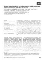
in human subjects (Fig. 1).
Glucose intolerance and diabetes mellitus
The link of UA to hyperglycemia was first described in the nineteenth century [22]. Elevated SUA predicted DM and insulin resistance in a fifteen-year follow-up study. Baseline SUA in this cohort
of 5012 young adults was not associated with a change in serum
insulin, indicating that hyperuricemia is an independent risk factor
for insulin resistance and type 2 DM [23]. High normal SUA was
also associated with future development of type 2 DM among lean
healthy and normoglycemic women [24]. Increased hepatic glucose production is a distinguished feature of insulin resistance
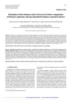
and type 2 DM. Intracellular UA stimulates AMPD and inhibits
AMPK enzyme activity (Fig. 2). Intracellular AMPK inhibits hepatic
gluconeogenesis. AMPD stimulates hepatic gluconeogenesis [25].
Decreased endothelial NO synthase (eNOS) activity in hyperuricemic patients causes increased insulin resistance [18,19].
Treatment of asymptomatic hyperuricemic personnel with allopurinol for 3 months results in significant decrease in insulin resistance and inflammation parameters [21].
Fig. 1. Effect of intra-cellular uric acid on nitric oxide synthesis within vascular endothelium UA = uric acid; NO = nitric oxide; FMD = flow mediated dilation; Htn = systemic
hypertension.
U.A.A. Sharaf El Din et al. / Journal of Advanced Research 8 (2017) 537–548
Fig. 2. Intra-cellular uric acid stimulates gluconeogenesis. UA = uric acid; AMPD = adenosine monophosphate dehydrogenase; AMPK = adenosine monophosphate
protein kinase.
However, genetic epidemiologic studies also called Mendelian
randomization studies failed to prove an association between UA
and type 2 DM [26,27]. Genetic polymorphisms of a wellcharacterized serologic variant can be utilized to study the effect
of this variant on disease risk. A total of 28 genetic loci were recognized to significantly associate with SUA concentration [28]. The
knowledge of genetic regulation of SUA allows the use of Mendelian randomization to examine the possible causal relation
between SUA and type 2 DM risk. The genetic score in these 2 articles is mainly based on genes that control UA transport between
extracellular and intracellular compartments and, hence, may dissociate the physiological serum-intracellular relationship. Intracellular UA is postulated as the cause of insulin resistance and
enhanced gluconeogenesis [29]. It is not known whether the score
alters the extracellular-intracellular equilibrium. The genetic score
may dissociate this equilibrium and then can lead to the incorrect
conception that SUA is not a risk factor for diabetes [29]. Unfortunately, we did not encounter large randomized controlled clinical
studies looking for impact of SUA lowering on the development
of MS and type 2 DM.
Systemic hypertension
The chance to develop Htn is greater in hyperuricemic male and
female subjects; this chance augments in older age [14,30,31].
Increased SUA increases the chance of non-dipping [32]. A similar
finding is encountered among CKD patients [33]. Increased SUA is
significantly associated with the development of new-onset primary Htn in children [34]. In a recent meta-analysis of 25 moderate to high-quality studies selected from all the clinical trials with
SUA as exposure and incident systemic Htn as outcome variables
through September 2013, these 25 studies of 97,824 participants
have shown that high SUA significantly predicts systemic Htn
[35]. Among 118 thousand healthy subjects 40–70 years old that
were screened for SUA during 2002, a quarter of them developed
systemic Htn over the following 10 years. Those with SUA higher
than 3 mg/dL had a greater chance to develop Htn. The higher
the SUA within the normal range, the greater was the risk to
develop Htn [36]. The association between high SUA and Htn is
stronger in younger ages and in females [37,38]. High SUA is one
of the major predictors of worse BP control [39–42]. SUA significantly correlates with sympathetic domain parameters among
pre-hypertensive and hypertensive personnel [43]. The in vivo rise
539
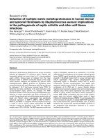
Fig. 3. Uric acid as mediator of systemic hypertension. ENaC = epithelial sodium
channels; Na = sodium; HTn = systemic hypertension.
of SUA in rats induces the epithelial sodium channel (ENaC) in the
distal nephron with consequent decrease in renal sodium excretion
[44] (Fig. 3). One of the important determinants of SUA is the glucose transporter ‘‘GLUT9” gene [45]. GLUT9 transports UA. GLUT9
gene polymorphism confirmed a causal relationship of hyperuricemia for systemic Htn in a family study [46]. Genotype variants
of GLUT9 associated with decreased SUA are associated with a significant decline of BP in different salt intake situations [47]. In adolescents with obesity and prehypertension, allopurinol or
probenecid achieved marked control of ambulatory BP [48]. Allopurinol was also associated with significant decrease in office BP
and body weight and increase in the percentage of dippers among
overweight prehypertensive subjects [49]. Six months of febuxostat treatment resulted in a significant decrease in plasma renin
activity and plasma concentration of aldosterone and a significant
increase in estimated glomerular filtration rate (eGFR) in hyperuricemic hypertensive patients [50].
Adiposity
Excess fat accumulation in MS involves adipose tissue, hepatocytes and increased level of serum triglycerides [51]. NAFLD is
characterized by triglyceride accumulation by variable degree
within hepatocytes [52]. NAFLD is the hepatic manifestation of
MS. Recent studies point toward UA as an important factor underlying excess fat storage [25,53–55]. UA up-regulates the fructokinase enzyme within human hepatocytes. This up-regulation is
UA concentration dependent with stepladder increase in fructokinase activity with increasing the intracellular UA concentration
from 4 to 12 mg/dL. This up-regulation is blocked on adding either
probenecid or allopurinol. Stimulation of fructokinase mediates
fructose-induced hepatic steatosis [56]. AMPK and AMPD within
hepatocytes are incriminated in the developments of hepatic
steatosis. When AMPK activity is reduced excess fat infiltration
occurs, while its stimulation can prevent steatosis through
increased fat oxidation and inhibition of lipogenesis. AMPD has
opposing effect on fat deposition within the hepatocytes. AMPD
activation increases intracellular UA synthesis [57]. Intracellular
UA inhibits AMPK activity [58]. Two years ago, a novel mechanism
of UA induced fatty liver was demonstrated. UA induced hepatocyte endoplasmic reticulum stress within hepatocytes. Associated
with this increased stress, the sterol regulatory element-binding
protein (SREBP) undergoes cleavage and nuclear translocation
540
U.A.A. Sharaf El Din et al. / Journal of Advanced Research 8 (2017) 537–548
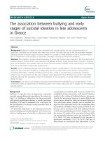
Fig. 4. Pathways of lipogenesis activated by intra-cellular uric acid. UA = uric acid; AMPD = adenosine monophosphate dehydrogenase; AMPK = adenosine monophosphate
protein kinase; ROS = reactive oxygen species; ER = endoplasmic reticulum.
and stimulates triglyceride accumulation within hepatocytes [54]
(Fig. 4).
Among the different components of MS, hypertriglyceridemia
carries the strongest association with hyperuricemia [59,60]. The
mechanism of this strong association is not yet known.
Excess fructose or sucrose intake can induce obesity beside
other features of MS [11]. In contrast, if animals are fed either glucose or starch of equivalent caloric value fewer features of MS are
observed [61]. These findings point to the ability of fructose to
induce visceral fat accumulation compared to isocaloric glucose
intake. Increased fructose intake is associated with intracellular
depletion of ATP, increased AMP and increased intra-cellular production of UA. This is followed by increased SUA [62]. Increased
SUA is an independent predictor of obesity [63]. URAT1 is one of
the transporters of UA. URAT1 mediates intracellular shift of UA.
This transporter is encountered within the adipocyte membrane
[64]. Adipose tissue can also generate UA. Adipocytes have XO that
can produce intracellular UA [65]. While extracellular UA acts as
strong antioxidant, it acts as a pro-oxidant inside the cell where
it stimulates NADPH oxidase enzyme causing increased intracellular oxidative stress, mitochondrial injury, and ATP depletion
[64,66,67] (Fig. 5). XO increases fat deposition within adipocytes.
XO knock-out mice get 50% reduction of their fat compared to wild
mice [65]. Genetic polymorphism of URAT1 gene was associated
with body mass index (BMI), waist circumference, and MS. Intracellular concentration of UA looks as an important determinant
of obesity [68].
Fig. 5. Intracellular uric acid as a pro-oxidant agent. UA = uric acid; ROS = reactive
oxygen species.
falls in cases of CKD, compensatory increase in intestinal secretion
of UA ensues [76,77]. Whether UA is a cause or an association to
renal diseases is a question that still waits for a definitive answer.
We hope we can settle this controversy in the present review.
Uric acid and the kidney
Nephrolithiasis
The kidney is responsible for elimination of 70% of the daily UA
production [69]. Renal handling of UA includes glomerular filtration, proximal tubular reabsorption, secretion and post-secretory
reabsorption [70]. ABCG2 that secretes UA is restricted to the proximal straight tubule (S3 segment) [71]. URAT1 is a voltage-driven
urate transporter located in the brush border of proximal convoluted tubules (PCT) and efficiently reabsorbs glomerular-filtrated
UA [1,72,73]. The reabsorbed UA is then driven out of PCT cells
through the basolateral membrane. The glucose transporter 9
(GLUT9) is involved in this extracellular efflux of UA [74]. ABCG2
is also expressed in the liver and intestine [75]. As UA excretion
Increased SUA and high animal protein diet can cause hyperuricosuria. Uricosuric agents used to treat hyperuricemia can aggravate hyperuricosuria. UA within the urine (UUA) tends to
crystalize when urine pH is low. Insulin resistance, obesity, high
animal protein intake and gout can decrease urine pH. Hyperuricosuria in the presence of acidic urine especially in case of low urine
volume can result in formation of urate stones [78]. In type 1 DM
adolescents, UUA is significantly higher and urine pH is lower compared to non-diabetic controls [79]. DM patients are more prone to
develop urate stones [80].
U.A.A. Sharaf El Din et al. / Journal of Advanced Research 8 (2017) 537–548
Chronic kidney disease
For the last one and half centuries, the association of gout with
CKD is well recognized [81]. However, it was not known which
came first. The decrease in GFR is associated with UA retention
[76]. The evidence of the offending action of UA was clearly
demonstrated in experimental studies. Most of the animals have
low SUA thanks to the existence of the uricase enzyme that breaks
down UA. To raise SUA in these animals, oxonic acid is used to inhibit the uricase enzyme. By increasing SUA, animals develop systemic Htn, glomerular Htn, glomerulosclerosis, and interstitial
fibrosis [81–85]. These changes were attributed to activation of
NADPH oxidase enzyme causing increased intracellular oxidative
stress, mitochondrial injury, ATP depletion [66,67], endothelial
injury, renin – angiotensin system (RAS) activation and increased
epithelial-mesenchyme transition (EMT). Increased EMT was
proved by decreased E-cadherin expression and an increased asmooth muscle actin and vimentin. Excess interstitial infiltration
by fibroblasts and progressive interstitial fibrosis eventually
ensues [86] (Fig. 6). On the other hand, some early clinical studies
denied UA as a risk of incident CKD [87–91]. Definition of CKD in
these articles was not precise. In one of these articles, serum creatinine of 2 mg/dL was considered the cutoff point [87]. In another
study the follow-up period was relatively short to detect change
in serum creatinine in healthy cohort at basal assessment [88].
Before the introduction of UA lowering agents, up to a quarter of
gouty patients developed proteinuria. Histologic examination of
the kidneys in these patients revealed nonspecific changes, namely
arteriosclerosis, glomerulosclerosis, and interstitial fibrosis. In
addition, collecting ducts and the medullary interstitium in some
of these patients showed focal deposition of monosodium urate
crystals with secondary inflammatory response. This inflammatory
response is in the form of focal granulomatous reaction with dense
accumulation of macrophages and T-lymphocytes. Tubular cells
within the inflammatory exudate showed a sixfold increase in
macrophage migration inhibitory factor (MIF) mRNA, compared
with uninvolved areas [92]. These changes were described as
‘‘gouty nephropathy” or ‘‘chronic urate or UA nephropathy” [93].
However, the focal nature of urate deposits and of the inflammatory response can’t explain the diffuse pathology of CKD encountered in these cases [94]. It is worth mentioning that urate
deposits could be detected in autopsies that lack evidence of CKD
[95]. Irrespective of the baseline eGFR, SUA significantly predicted
CKD progression over 5 years of follow-up of a cohort of IgA
nephropathy patients [96]. SUA proved as a strong predictor for
the development of increased urine albumin excretion rate (UAER)
on follow-up of normoalbuminuric type 1 diabetic patients for
6 years. For every 1 mg/dL increase in SUA, the risk of development
of albuminuria increased by 80% [97]. In a recent cohort study of
541
3605 normal subjects having normal kidney functions, the subjects
were categorized according to the longitudinal follow-up of SUA
into persistently low, fluctuating with declining or rising SUA,
and persistently high SUA. Incident CKD was significantly higher
in categories with rising or persistently high SUA [98]. SUA is associated with resistive indices within renal arteries estimated by
Doppler study [99]. In another study in Type 1 DM there was a
2.4-fold increase in the unadjusted risk of eGFR loss in patients
having SUA > 6.6 mg/dL compared to those with lower level
[100]. In a study of 263 type 1 DM newly diagnosed, SUA was a significant independent predictor of macroalbuminuria after 18 years
[101]. In a recent study, insulin sensitivity was significantly higher
in type 1 DM who had regression of albuminuria compared to
those who did not [102]. In a longitudinal study of a cohort of
20,142 type 2 DM patients having eGFR > 60 mL/min and normal
UAER, De Cosmo et al., looked at the incidence of eGFR < 60 mL/
min., increased UAER or both over 4 years of follow-up. They
assessed the association of SUA quintiles with the onset of these
CKD components using regression analysis to adjust for different
confounders. 7.9% of patients developed eGFR < 60 mL/min, 14.1%
developed increased UAER and 2% of patients developed both components. The higher the SUA quintile the higher is the relative risk
ratio of eGFR decline. In patients who developed eGFR decline,
there was a significant association of SUA with albuminuria
[103]. These findings are supported by more recent results
reported in Japan [104]. A cross-sectional study of more than three
thousand type 2 DM patients looked for UA effect on the prevalence of diabetic kidney disease (DKD). 68% of the hyperuricemic
had DKD versus 41.5% with normal UA [105]. When the data of
seventy liver transplantation children were revised, a cumulative
incidence of hyperuricemia of 32% over ten-year postoperative
was observed. All these children underwent annual estimation of
SUA, inulin and urate clearance. Decreased urate clearance was
the main cause of hyperuricemia. SUA tended to predict the development of CKD [106]. A more interesting prospective observation
study of a cohort of 900 healthy adult blood donors that were followed for 5 years showed that the basal SUA was a significant predictor of eGFR decline even after multivariate regression analysis
[107]. The drawback of this trial is the lack of serial estimation of
SUA and the limited number of females. However, this study is distinguished because the subjects were healthy normotensive subjects lacking signs of CKD on entry to the study. Another
prospective study of 21,475 healthy volunteers followed for seven
years looked for the association of UA level with incident CKD
defined as eGFR < 60 mL/min/1.73 m2. UA between 7 and 8.9 mg/
dL was associated with almost doubling and level above 9 mg/dL
was associated with tripling of incident CKD [108]. A Japanese 5year follow-up study of more than two thousand healthy adults
above the age of 40 years without CKD showed that
Fig. 6. Different pathogenic mechanisms of kidney injury possibly induced by uric acid. UA = uric acid; ROS = reactive oxygen species; MCPl = Macrophage chemo-attractant
protein-1; RAS = renin angiotensin system; EMT = epithelium mesenchyme transition; VSMC = vascular smooth muscle cells.
542
U.A.A. Sharaf El Din et al. / Journal of Advanced Research 8 (2017) 537–548
SUA > 5.9 mg/dL is a significant risk factor for CKD and proteinuria
[104]. A recent meta-analysis and review of 13 studies containing
more than one hundred and ninety thousand participants tried to
find out whether UA is an independent risk factor of incident
CKD. This study confirmed that UA is an independent risk factor
for the development of CKD in non-CKD healthy persons with no
discrimination between male and female sex. The longer the
follow-up the stronger is this association [109]. Glucose concentration in the glomerular ultrafiltrate is similar to serum concentration. This glucose is reabsorbed by the PCT. Sodium-glucose cotransporter 2 (SGLT2) present in the apical membrane is responsible for absorption of 90% of this glucose [110]. In case of hyperglycemia, SGLT2 is over expressed to increase glucose absorption
[111]. Intracellular glucose increases leading to increased activity
of polyol pathway leading to increased fructose synthesis. Intracellular fructose metabolism leads to increased UA synthesis
[112,113]. Fructokinase knockout mice are protected against the
renal degenerative changes associated with aging and increased
salt intake [114]. In a recent study of 422 type 2 DM for more than
fifteen years that were followed for up to 77 months, patients with
SUA > 7 mg/dL in males and >6 mg/dL in females had a significantly higher rate of DKD progression, and overall mortality
[115]. In a meta-analysis of 24 studies with twenty-five plus
patients with CKD, elevated SUA is significantly associated with
risk of mortality in these patients [116]. GLUT9 polymorphism is
strongly associated with SUA in healthy subjects in the general
population that have normal kidney function. In a cohort of 755
CKD patients, GLUT9 polymorphism predicted progression [117].
A causal relation of UA to CKD progression could be realized based
on this study. In a retrospective cohort study of 803 CKD patients,
propensity score analysis using three different methods showed a
consistent impact of high UA on progression to end-stage renal disease (ESRD) [118]. XO inhibitors possibly delay the progression of
CKD in adult hyperuricemic and hypertensive patients [119]. The
target SUA should be <6.5 mg/dL to delay progression [77,118].
Acute kidney injury (AKI)
In 37 patients who underwent cardiac surgery, SUA was
assessed 1 hour postoperative. A significant positive correlation
between SUA, on one hand, and urine neutrophil gelatinaseassociated lipocalin (NGAL) estimated 1 h, 6 h and 24 h postoperative, and serum creatinine measured 1 day, 2 days and 3 days postoperative respectively on the other hand. There was also a
significant negative correlation between SUA and the kinetic eGFR
measured 1, 2, 3 and 4 days postoperative respectively. These findings illustrated that the rise of UA one-hour postoperative precedes
and significantly predicts subsequent development of AKI [120]. In
another trial in patients undergoing open-heart surgery, SUA in
blood samples collected 2 h postoperative had a stronger predictive value for AKI and the need for renal replacement therapy
(RRT) in comparison with serum and urine NGAL [121]. Preoperative UA level was also a strong predictor of postoperative AKI. In
patients undergoing radical cystectomy, preoperative SUA was an
independent predictor of postoperative AKI [122]. In a retrospective study of more than two thousand patients who underwent
coronary bypass surgery, preoperative SUA was a strong predictor
for the development of postoperative AKI [123]. UA was not only a
predictor of postoperative AKI but also predicted AKI in patients
having burns [124] or those with sepsis [125]. In a retrospective
analysis of all patients admitted to a tertiary hospital over 2 years,
and after consideration of logistic regression analysis, patients having SUA > 9.4 mg/dL on hospital admission had significantly the
highest risk to develop AKI during their hospital stay. On the other
hand, those having UA < 4.5 mg/dL were at lowest risk [126]. The
strength of this study is based on many points: 1st is wide spec-
trum of the patients’ primary disease, including infectious, cardiovascular, gastrointestinal, hematology/oncology, and respiratory
disorders. The 2nd point is the graded association of UA with the
development of AKI. A similar retrospective study in another hospital has shown similar results [127]. When more than eleven
thousands of participants were followed for about twelve years,
823 of them were admitted to the hospital because of AKI.
SUA > 5 mg/dL was independently associated with these admissions. The risk of AKI was 16% higher with each 1 mg increase in
SUA [128]. SUA level is a significant predictor of contrast-induced
nephropathy (CIN) [129,130]. UA lowering with allopurinol in
addition to saline hydration was associated with significantly
lower incidence of CIN compared to saline hydration alone or saline hydration plus N-acetyl cysteine [130]. UA potentially mediates
AKI through vascular, pro-oxidative and inflammatory mechanisms [131]. UA inhibits endothelial NO synthesis, and thus promotes vasospasm in afferent and, to less extent, in the efferent
arterioles [82,132]. UA inhibits capillary endothelial cells’ proliferation and migration [133]. It can also induce endothelial apoptosis
[132]. UA also correlates with pre-glomerular arteriolopathy in
human beings, an obstacle to renal autoregulation in condition of
renal hypoperfusion [134]. As mentioned above, UA stimulates
NADPH oxidase with consequent increase in oxidant stress. The
increased oxidant stress stimulates production of macrophage
chemo-attractant factor (MCP1) within vascular smooth muscle
cells (VSMCs) [135]. Hyperuricemic rats show increased macrophage infiltration of their kidneys [83]. Administration of an
NADPH oxidase inhibitor inhibited MCP1 production within
VSMCs [135] (Fig. 6).
Preeclampsia (PE)
PE complicates 5–10% of pregnancies worldwide [136]. Affected
women usually have profound long-term consequences [137]. PE is
characterized by Htn, proteinuria, and edema that develop after
20 weeks of pregnancy [138]. Decreased placental perfusion due
to impaired remodeling of spiral arteries might result in hypoxia
[139]. UA level showed high correlation with BP in cases of PE
[140]. In pregnant ladies suffering PE, serum tumor necrosis factor
a (TNFa) and ICAM1 were significantly higher than control or
hypertensive pregnant ladies. Subcutaneous blood vessels showed
intense staining with these 2 agents. SUA showed positive correlation with TNF a and ICAM1 in PE patients [141].
Uric acid and cardiovascular system (CVS)
Whether SUA is merely a risk marker or a risk factor for CV disease, or whether hypouricemic agents affect outcomes is still a
matter of debate [142]. The association between SUA and different
CVD might be confounded by different factors frequently encountered in cardiac patients. These factors include Htn, dyslipidemia,
DM, alcohol consumption, hypothyroidism and diuretic use
[143]. Independent of any CV risk factor, increased SUA level, even
within the normal range, is a risk factor for impaired flowmediated dilation (FMD) of brachial artery (Fig. 1), increased carotid intima-media thickness (IMTc), and increased stiffness of the
aorta in healthy subjects [144–149]. In non-diabetic CKD patients
(stage 3–5) who lack evidence of CVD and were not treated with
either RAS blockers or statins, FMD inversely correlated with SUA
[150]. Treatment of hyperuricemic type 2 DM patients with allopurinol for 3 years succeeded to reduce carotid IMT [151]. UA stimulates platelet-derived growth factor receptor b (PDGFRb)
phosphorylation in the rat aorta [152]. This discovery would
explain the VSMC proliferation and CVD in hyperuricemic patients.
When isolated human umbilical vein endothelial cells (HUVECs)
U.A.A. Sharaf El Din et al. / Journal of Advanced Research 8 (2017) 537–548
were exposed to 6 mg and 9 mg/dL UA, significant increase in
intracellular free oxygen species was followed by senescence and
apoptosis of these cells. Senescence and apoptosis of HUVECs were
ameliorated on addition of either probenecid or an antioxidant like
N-acetyl cysteine or tempol. In addition, UA increased expression
of the different elements of RAS within HUVECs [153]. When
human aortic endothelial cells (HAECs) are exposed to high UA
concentration for 48 h, a significant decline in eNOS activity was
observed. There was also 50% reduction in mitochondrial DNA
level, a decrease in mitochondrial mass and a significant reduction
in basal concentration of ATP. The higher the concentration of UA
within the culture medium the greater was the reduction in intracellular ATP concentration [66] (Fig. 7).
On the other hand, some studies failed to demonstrate UA as
independent CVD risk factor [154]. Analysis of data obtained from
6763 participants in the Framingham heart study failed to demonstrate a significant association between SUA and CHD and CV mortality [155]. However, many of the epidemiologic data collected in
recent years favor the association between SUA and the risk of
CVD. A recent study showed SUA as independent predictor of
CHD [156]. In a prospective study of more than fifty thousand male
subjects with history of gout in the Health Professionals Follow-Up
Study, the relation between history of gout and the development of
CVD was examined. After follow-up for twelve years, patients with
history of gout were found at greater risk of CV mortality, mainly
due to CHD [157]. Increased SUA is associated with the unstable
coronary lipid-rich plaques [158]. SUA predicts HF in patients with
stable CHD. This predictability is muffled, but not abolished, by different confounders [159]. SUA was measured in 705 cases of both
sexes that underwent coronary angiography. 41% of cases had normal angiography and were considered the control group. A significant positive correlation between SUA and the severity of CHD
score was encountered [160]. After measurement of SUA of over
400.000 in checkup centers in Stockholm, these candidates were
followed for 7–17 years. The higher the basal SUA in this middleaged population the higher is the chance to develop acute myocardial infarction (AMI), heart failure (HF), and stroke [161]. SUA is a
significant predictor of poor outcomes in AMI patients complicated
with reduced LV function, HF, or both [162]. The Pooled data from
eleven studies that evaluated the prognostic importance of SUA
demonstrated that hyperuricemia can significantly predict allcause mortality in HF patients [163]. These data are also observed
in HF patients with preserved ejection fraction [164] and in
patients hospitalized with severely decompensated acute HF
[165,166]. The relation of SUA with acute HF outcome is weakened
with deterioration of kidney function [167]. The association
543
between SUA and ischemic stroke is debatable. While some accuse
SUA as predictor of magnitude of infarct [168], most found SUA to
play a favorable role [169–171]. Allopurinol succeeded to improve
mortality rate in HF patients with history of gout [172]. However,
in a more recent trial, allopurinol failed to improve left ventricular
ejection fraction, or exercise capacity after 6 months in patients
with HF and hyperuricemia [173]. In a cohort of 557 healthy subjects, 415 of whom were women, aged 60 years and older, men
with higher SUA (>5.5 mg/dL) had significantly higher left ventricular mass compared to men with lower level [174]. The association
between SUA and left ventricular hypertrophy (LVH) is more likely
in women than in men when they have CKD [175]. In patients with
LVH and preserved ejection fraction, SUA is associated with diastolic dysfunction in women only [176]. 37% of kidney transplant
recipients that had normal graft function developed persistent
hyperuricemia within the 1st post-transplant year. Hyperuricemia
in these patients was significantly associated with Htn, increased
pulse wave velocity, and LVH [177]. Treatment with allopurinol
improved left ventricular function and coronary flow reserve in
patients with dilated cardiomyopathy and concomitantly elevated
SUA [178]. The association between SUA and the major cardiovascular adverse events following acute coronary syndrome is stronger in women compared to men [179]. It seems that this
association in patients with normal kidney function is observed
in older aged women. SUA was found as independent predictor
of LVH in postmenopausal but not in premenopausal women
[180]. In type 2 DM hyperuricemia was significantly associated
with atrial fibrillation independent of other risk factors and all
potential confounders [181]. In 200 hypertensive patients that
have normal treadmill exercise test, patients with erectile dysfunction have significantly higher SUA [182]. In persons with elevated
level of HDLc, SUA is associated with an increased risk of idiopathic
venous thromboembolism [183]. In patients with hypertrophic
cardiomyopathy, SUA is a significant predictor of adverse outcome
[184]. Increased SUA was appointed as independent risk factor for
overall mortality and CV mortality [185,186]. The relationship
between SUA and CV mortality is higher in the lowest and highest
quintiles in both men and women [187]. A prospective analysis of
329 patients with ST-elevation myocardial infarction (STEMI) and
eGFR < 60 mL/min/1.73 m2 treated with percutaneous coronary
intervention (PCI) disclosed a strong correlation of SUA with 1year mortality [188]. A recent meta-analysis of six studies, including more than 200.000 patients showed that hyperuricemia independently increases the risk of mortality from CVD and CHD
[189]. The knowledge of genetic regulation of SUA allows the use
of Mendelian randomization to examine the possible causal rela-
Fig. 7. Different vascular injury mechanisms possibly mediated by uric acid. UA – uric acid; ROS = reactive oxygen species l; RAS = renin angiotensin system; PDGFR – platelet
derived growth factor receptor.
544
U.A.A. Sharaf El Din et al. / Journal of Advanced Research 8 (2017) 537–548
tion between SUA and cardiovascular risk. Genotype precedes life
events and is not affected by lifestyle [190]. This analysis disclosed
a causal relation between SUA on one hand and CHD, cardiovascular mortality and sudden cardiac death on the other hand [191].
These results criticize the hypothesis that the effect observed with
high SUA is not due to the molecule itself but due to the induction
of the XO and the effect of XO inhibitors is secondary to inhibition
of the enzyme rather than the consequent control of SUA. XO activation results in increased production of free oxygen radicals with
consequent increased oxidative stress and triggered inflammation.
XO inhibitors can abolish this oxidative stress and burns out the
consequent inflammation [192].
Conclusions
According to the recent experimental and clinical trials and to
the therapeutic interventions and the Mendelian randomization
studies it seems that UA is a real risk factor for the development
of metabolic, renal and CVDs. The intracellular UA seems to be
more pathogenic. The cell membrane urate transporters are
responsible for the intra-extracellular UA shift, and hence, they
are important determinants of the offending role of UA. These
studies have also demonstrated that low SUA levels might carry
high risk similar to the high levels. Based on these facts, more
interventional studies are needed to optimize the therapeutic management of this evolving risk factor. These studies should highlight
when to treat, the target SUA level and the long-term safety of the
different hypouricemic agents.
Conflict of interest
The authors have declared no conflict of interest.
Compliance with ethics requirements
This article does not contain any studies with human or animal
subjects.
References
[1] Hediger MA, Johnson RJ, Miyazaki H, Endou H. Molecular physiology of urate
transport. Physiology 2005;20:125–33.
[2] Kang DH, Chen W. Uric acid and chronic kidney disease: new understanding
of an old problem. Semin Nephrol 2011;31(5):447–52.
[3] Perheentupa J, Raivio K. Fructose-induced hyperuricaemia. Lancet 1967;2
(7515):528–31.
[4] Mahomed FA. On chronic Bright’s disease, and its essential symptoms. Lancet
1879;1:399–401.
[5] Haig A. Uric acid as a factor in the causation of disease. London: J&A Churchill;
1897.
[6] Gertler MM, Garn SM, Levine SA. Serum uric acid in relation to age and
physique in health and in coronary heart disease. Ann Intern Med 1951;34
(6):1421–31.
[7] Facchini F1, Chen YD, Hollenbeck CB, Reaven GM. Relationship between
resistance to insulin-mediated glucose uptake, urinary uric acid clearance,
and plasma uric acid concentration.. JAMA 1992;266(21):3008–11.
[8] Nakagawa T, Kang DH, Feig D, Sanchez-Lozada LG, Srinivas TR, Sautin Y, et al.
Unearthing uric acid: an ancient factor with recently found significance in
renal and cardiovascular disease. Kidney Int 2006;69(10):1722–5.
[9] Alberti KG, Eckel RH, Grundy SM, Zimmet PZ, Cleeman JI, Donato KA, et al.
Harmonizing the metabolic syndrome: a joint interim statement of the
international diabetes federation task force on epidemiology and prevention;
national heart, lung, and blood institute; american heart association; world
heart federation; international atherosclerosis society; and international
association for the study of obesity. Circulation 2009;120(16):1640–5.
[10] Johnson RJ, Stenvinkel P, Martin SL, Jani A, Sánchez-Lozada LG, Hill JO, et al.
Redefining metabolic syndrome as a fat storage condition based on studies of
comparative physiology. Obesity (Silver Spring) 2013;21(4):659–64.
[11] Bocarsly ME, Powell ES, Avena NM, Hoebel BG. High-fructose corn syrup
causes characteristics of obesity in rats: increased body weight, body fat and
triglyceride levels. Pharmacol Biochem Behav 2010;97:101–6.
[12] Choi HK, Ford ES. Prevalence of the metabolic syndrome in individuals with
hyperuricemia. Am J Med 2007;120(5):442–7.
[13] Yu TY, Jee JH, Bae JC, Jin SM, Baek JH, Lee MK, et al. Serum uric acid: a strong
and independent predictor of metabolic syndrome after adjusting for body
composition. Metabolism 2016;65(4):432–40.
[14] Sun HL, Pei D, Lue KH, Chen YL. Uric acid levels can predict metabolic
syndrome and hypertension in adolescents: a 10-year longitudinal study.
PLoS ONE 2015;10(11):e0143786.
[15] Zurlo A, Veronese N, Giantin V, Maselli M, Zambon S, Maggi S, et al.
High serum uric acid levels increase the risk of metabolic syndrome in
elderly women: the PRO.V.A study. Nutr Metab Cardiovasc Dis 2016;26
(1):27–35.
[16] Babio N, Martínez-González MA, Estruch R, Wärnberg J, Recondo J, OrtegaCalvo M, et al. Associations between serum uric acid concentrations and
metabolic syndrome and its components in the PREDIMED study. Nutr Metab
Cardiovasc Dis 2015 Feb;25(2):173–80.
[17] Yuan H, Yu C, Li X, Sun L, Zhu X, Zhao C, et al. Serum uric acid levels and risk of
metabolic syndrome: a dose-response meta-analysis of prospective studies. J
Clin Endocrinol Metab 2015;100(11):4198–207.
[18] Roy D, Perreault M, Marette A. Insulin stimulation of glucose uptake in
skeletal muscles and adipose tissues in vivo is NO dependent. Am J Physiol
1998;274(4 Pt 1). E692-9.
[19] Bjornstad P, Snell-Bergeon JK, McFann K, Wadwa RP, Rewers M, Rivard CJ,
et al. Serum uric acid and insulin sensitivity in adolescents and adults with
and without type 1 diabetes. J Diabetes Complications 2014;28(3):298–304.
[20] Ogino K, Kato M, Furuse Y, Kinugasa Y, Ishida K, Osaki S, et al. Uric acidlowering treatment with benzbromarone in patients with heart failure: a
double-blind placebo-controlled crossover preliminary study. Circ Heart Fail
2010;3:73–81.
[21] Takir M, Kostek O, Ozkok A, Elcioglu OC, Bakan A, Erek A, et al. Lowering uric
acid with allopurinol improves insulin resistance and systemic inflammation
in asymptomatic hyperuricemia. J Investig Med 2015;63(8):924–9.
[22] Duckworth D. A treatise on gout. London: C Griffin & Co; 1889. p. 476.
[23] Krishnan E, Pandya BJ, Chung L, Hariri A, Dabbous O. Hyperuricemia in young
adults and risk of insulin resistance, prediabetes, and diabetes: a 15-year
follow-up study. Am J Epidemiol 2012;176(2):108–16.
[24] Shani M, Vinker S, Dinour D, Leiba M, Twig G, Holtzman EJ, et al. High normal
uric acid levels are associated with an increased risk of diabetes in lean,
normoglycemic healthy women. J Clin Endocrinol Metab 2016;101
(10):3772–8.
[25] Cicerchi C, Li N, Kratzer J, Garcia G, Roncal-Jimenez CA, Tanabe K, et al. Uric
acid-dependent inhibition of AMP kinase induces hepatic glucose production
in diabetes and starvation: evolutionary implications of the uricase loss in
hominids.. FASEB J 2014(8):3339–50.
[26] Pfister R, Barnes D, Luben R, Forouhi NG, Bochud M, Khaw KT, et al. No
evidence for a causal link between uric acid and type 2 diabetes: a Mendelian
randomisation approach. Diabetologia 2011;54(10):2561–9.
[27] Sluijs I1, Holmes MV2, van der Schouw YT3, Beulens JW3, Asselbergs FW4,
Huerta JM5, et al., A Mendelian Randomization Study of Circulating Uric Acid
and Type 2 Diabetes. Diabetes. 2015 Aug; 64(8):3028–36.
[28] Köttgen A, Albrecht E, Teumer A, Vitart V, Krumsiek J, Hundertmark C, et al.
Genome-wide association analyses identify 18 new loci associated with
serum urate concentrations. Nat Genet 2013;45:145–54.
[29] Johnson RJ, Merriman T, Lanaspa MA. Causal or noncausal relationship of uric
acid with diabetes. Diabetes 2015;64(8):2720–2.
[30] Wei F, Sun N, Cai C, Feng S, Tian J, Shi W, et al. Associations between serum
uric acid and the incidence of hypertension: a Chinese senior dynamic cohort
study. J Transl Med 2016;14(1):110.
[31] Yokoi Y, Kondo T, Okumura N, Shimokata K, Osugi S, Maeda K, et al. Serum
uric acid as a predictor of future hypertension: stratified analysis based on
body mass index and age. Prev Med 2016;9(90):201–6.
[32] Giallauria F, Predotti P, Casciello A, Grieco A, Russo A, Viggiano A, et al. Serum
uric acid is associated with non-dipping circadian pattern in young patients
(30–40 years old) with newly diagnosed essential hypertension. Clin Exp
Hypertens 2016;38(2):233–7.
[33] Ahbap E, Sakaci T, Kara E, Sahutoglu T, Koc Y, Basturk T, et al. Serum uric acid
levels and inflammatory markers with respect to dipping status: a
retrospective analysis of hypertensive patients with or without chronic
kidney disease. Clin Exp Hypertens 2016;38(6):555–63.
[34] Feig DI, Johnson RJ. Hyperuricemia in childhood primary hypertension.
Hypertension 2003;42:247–52.
[35] Wang J, Qin T, Chen J, Li Y, Wang L, Huang H, et al. Hyperuricemia and risk of
incident hypertension: a systematic review and meta-analysis of
observational studies. PLoS ONE 2014;9(12):e114259.
[36] Leiba A, Vinker S, Dinour D, Holtzman EJ, Shani M. Uric acid levels within the
normal range predict increased risk of hypertension: a cohort study. J Am Soc
Hypertens. 2015;9(8):600–9.
[37] Yokokawa H, Fukuda H, Suzuki A, Fujibayashi K, Naito T, Uehara Y, et al.
Association between serum uric acid levels/hyperuricemia and hypertension
among 85,286 japanese workers. J Clin Hypertens (Greenwich) 2016 Jan;18
(1):53–9.
[38] Lee JJ, Ahn J, Hwang J, Han SW, Lee KN, Kim JB, et al. Relationship between
uric acid and blood pressure in different age groups. Clin Hypertens 2015
Jul;15(21):14.
[39] Cicero A, Rosticci M, Tartagni E, Parini A, Grandi E, D’Addato S, et al. Serum
uric acid level, but not renal function or arterial stiffness, is associated to
worse blood pressure control in general practice: data from the brisighella
heart study. J Hypertens 2015;33(Suppl 1):e22.
U.A.A. Sharaf El Din et al. / Journal of Advanced Research 8 (2017) 537–548
[40] Viazzi F, Rebora P, Giussani M, Orlando A, Stella A, Antolini L, et al. Increased
serum uric acid levels blunt the antihypertensive efficacy of lifestyle
modifications in children at cardiovascular risk. Hypertension 2016;67
(5):934–40.
[41] Cicero AF, Rosticci M, Fogacci F, Grandi E, D’Addato S, Borghi C, et al.
High serum uric acid is associated to poorly controlled blood pressure and
higher arterial stiffness in hypertensive subjects. Eur J Intern Med
2017;37:38–42.
[42] Cho J, Kim C, Kang DR, Park J. Hyperuricemia and uncontrolled hypertension
in treated hypertensive patients: K-MetS study. Medicine (Baltimore)
2016;95(28):e4177.
[43] Kunikullaya KU, Purushottam N, Prakash V, Mohan S, Chinnaswamy R.
Correlation of serum uric acid with heart rate variability in hypertension.
Hipertens Riesgo Vasc 2015;32(4):133–41.
[44] Xu W, Huang Y, Li L, Sun Z, Shen Y, Xing J, et al. Hyperuricemia induces
hypertension through activation of renal epithelial sodium channel (ENaC).
Metabolism 2016;65(3):73–83.
[45] Clémençon B, Lüscher BP, Fine M, Baumann MU, Surbek DV, Bonny O, et al.
Expression, purification, and structural insights for the human uric acid
transporter, GLUT9, using the Xenopus laevis oocytes system. PLoS ONE
2014;9(10):e108852.
[46] Mallamaci F, Testa A, Leonardis D, Tripepi R, Pisano A, Spoto B, et al. A
polymorphism in the major gene regulating serum uric acid associates with
clinic SBP and the white-coat effect in a family-based study. J Hypertens
2014;32(8):1621–8.
[47] Parsa A, Brown E, Weir MR, Fink JC, Shuldiner AR, Mitchell BD, et al.
Genotype-based changes in serum uric acid affect blood pressure. Kidney Int
2012;81(5):502–7.
[48] Soletsky B, Feig DI. Uric acid reduction rectifies prehypertension in obese
adolescents. Hypertension 2012;60(5):1148–56.
[49] Madero M, Rodríguez Castellanos FE, Jalal D, Villalobos-Martín M, Salazar J,
Vazquez-Rangel A, et al. A pilot study on the impact of a low fructose diet and
allopurinol on clinic blood pressure among overweight and prehypertensive
subjects: a randomized placebo controlled trial. J Am Soc Hypertens 2015;9
(11):837–44.
[50] Tani S, Nagao K, Hirayama A. Effect of febuxostat, a xanthine oxidase
inhibitor, on cardiovascular risk in hyperuricemic patients with
hypertension: a prospective, open-label, Pilot Study. Clin Drug Investig
2015;35(12):823–31.
[51] Kanbay M, Jensen T, Solak Y, Le M, Roncal-Jimenez C, Rivard C, et al. Uric acid
in metabolic syndrome: from an innocent bystander to a central player. Eur J
Int Med 2016;29:3–8.
[52] Marchesini G, Bugianesi E, Forlani G, Cerrelli F, Lenzi M, Manini R, et al.
Nonalcoholic fatty liver, steatohepatitis, and the metabolic syndrome.
Hepatology 2003;37(4):917–23.
[53] Lanaspa MA, Sanchez-Lozada LG, Choi YJ, Cicerchi C, Kanbay M, RoncalJimenez CA, et al. Uric acid induces hepatic steatosis by generation of
mitochondrial oxidative stress: potential role in fructose-dependent and independent fatty liver. J Biol Chem 2012;287(48):40732–44.
[54] Choi YJ, Shin HS, Choi HS, Park JW, Jo I, Oh ES, et al. Uric acid induces fat
accumulation via generation of endoplasmic reticulum stress and SREBP-1c
activation in hepatocytes. Lab Invest 2014 Oct;94(10):1114–25.
[55] Liang J, Pei Y, Gong Y, Liu XK, Dou LJ, Zou CY, et al. Serum uric acid and nonalcoholic fatty liver disease in non-hypertensive Chinese adults: the
Cardiometabolic Risk in Chinese (CRC) study. Eur Rev Med Pharmacol Sci
2015;19(2):305–11.
[56] Lanaspa MA, Sanchez-Lozada LG, Cicerchi C, Li N, Roncal-Jimenez CA,
Ishimoto T, et al. Uric acid stimulates fructokinase and accelerates fructose
metabolism in the development of fatty liver. PLoS ONE 2012;7(10):e47948.
doi: />[57] Lanaspa MA, Epperson LE, Li N, Cicerchi C, Garcia GE, Roncal-Jimenez CA, et al.
Opposing activity changes in AMP deaminase and AMP-activated protein
kinase in the hibernating ground squirrel. PLoS ONE 2015;10(4):e0123509.
doi: />[58] Lanaspa MA, Cicerchi C, Garcia G, Li N, Roncal-Jimenez CA, Rivard CJ, et al.
Counteracting roles of AMP deaminase and AMP kinase in the development of
fatty liver. PLoS ONE 2012;7(11):e48801. doi: />journal.pone.0048801.
[59] Conen D, Wietlisbach V, Bovet P, Shamlaye C, Riesen W, Paccaud F, et al.
Prevalence of hyperuricemia and relation of serum uric acid with
cardiovascular risk factors in a developing country. BMC public health
2004;4:9.
[60] Shih MH, Lazo M, Liu SH, Bonekamp S, Hernaez R, Clark JM. Association
between serum uric acid and nonalcoholic fatty liver disease in the US
population. J Formos Med Assoc 2015;114(4):314–20.
[61] Roncal-Jimenez CA, Lanaspa MA, Rivard CJ, Nakagawa T, Sanchez-Lozada LG,
Jalal D, et al. Sucrose induces fatty liver and pancreatic inflammation in male
breeder rats independent of excess energy intake. Metabolism
2011;60:1259–70.
[62] Johnson RJ, Nakagawa T, Sanchez-Lozada LG, Shafiu M, Sundaram S, Le M,
et al. Sugar, uric acid, and the etiology of diabetes and obesity. Diabetes
2013;62(10):3307–15.
[63] Masuo K, Kawaguchi H, Mikami H, Ogihara T, Tuck ML. Serum uric acid and
plasma norepinephrine concentrations predict subsequent weight gain and
blood pressure elevation. Hypertension 2003;42:474–80.
545
[64] Sautin YY, Nakagawa T, Zharikov S, Johnson RJ. Adverse effects of the classic
antioxidant uric acid in adipocytes: NADPH oxidase-mediated oxidative/
nitrosative stress. Am J Physiol Cell Physiol 2007;293:C584–96.
[65] Cheung KJ, Tzameli I, Pissios P, Rovira I, Gavrilova O, Ohtsubo T, et al.
Xanthine oxidoreductase is a regulator of adipogenesis and PPARc activity.
Cell Metab 2007;5:115–28.
[66] Sánchez-Lozada LG, Lanaspa MA, Cristóbal-García M, García-Arroyo F, Soto V,
Cruz-Robles D, et al. Uric acid-induced endothelial dysfunction is associated
with mitochondrial alterations and decreased intracellular ATP
concentrations. Nephron Exp Nephrol 2012;121(3–4). e71-8.
[67] Cristóbal-García M, García-Arroyo FE, Tapia E, Osorio H, Arellano-Buendía AS,
Madero M, et al. Renal oxidative stress induced by long-term hyperuricemia
alters mitochondrial function and maintains systemic hypertension. Oxid
Med Cell Longev 2015;2015:535686.
[68] Shafiu M, Johnson RJ, Turner ST, Langaee T, Gong Y, Chapman AB, et al. Urate
transporter gene SLC22A12 polymorphisms associated with obesity and
metabolic syndrome in Caucasians with hypertension. Kidney Blood Press Res
2012;35(6):477–82.
[69] Maesaka JK, Fishbane S. Regulation of renal urate excretion: a critical review.
Am J Kidney Dis 1998;32:917–33.
[70] Roch-Ramel F, Werner D, Guisan B. Urate transport in brushborder
membrane of human kidney. Am J Physiol Renal Physiol 1994;266:F797–805.
[71] Taniguchi K, Tamura Y, Kumagai T, Shibata S, Uchida S. Stimulation of V1a
receptor increases renal uric acid clearance via urate transporters: insight
into pathogenesis of hypouricemia in SIADH. Clin Exp Nephrol 2016;20
(6):845–52.
[72] Anzai N, Ichida K, Jutabha P, Kimura T, Babu E, Jin CJ, et al. Plasma urate level
is directly regulated by a voltage-driven urate efflux transporter URATv1
(SLC2A9) in humans. J Biol Chem 2008;283(40):26834–8.
[73] Enomoto A, Kimura H, Chairoungdua A, Shigeta Y, Jutabha P, Cha SH, et al.
Molecular identification of a renal urate anion exchanger that regulates blood
urate levels. Nature 2002;417(6887):447–52.
[74] Bobulescu IA, Moe OW. Renal transport of uric acid: evolving concepts and
uncertainties. Adv Chronic Kidney Dis 2012;19(6):358–71.
[75] Huls M, Brown CD, Windass AS, Sayer R, van den Heuvel JJ, Heemskerk S, et al.
The breast cancer resistance protein transporter ABCG2 is expressed in the
human kidney proximal tubule apical membrane. Kidney Int 2008;73
(2):220–5.
[76] Yano H, Tamura Y, Kobayashi K, Tanemoto M, Uchida S. Uric acid transporter
ABCG2 is increased in the intestine of the 5/6 nephrectomy rat model of
chronic kidney disease. Clin Exp Nephrol 2014;18(1):50–5.
[77] Kumagai T, Ota T, Tamura Y, Chang WX, Shibata S, Uchida S. Time to target
uric acid to retard CKD progression. Clin Exp Nephrol 2017;21(2):182–92.
[78] DiBianco JM, Jarrett TW, Mufarrij P. Metabolic syndrome and nephrolithiasis
risk: should the medical management of nephrolithiasis include the
treatment of metabolic syndrome? Rev Urol 2015;17(3):117–28.
[79] Bjornstad P, Roncal C, Milagres T, Pyle L, Lanaspa MA, Bishop FK, et al.
Hyperfiltration and uricosuria in adolescents with type 1 diabetes. Pediatr
Nephrol 2016(5):787–93.
[80] Lieske JC, de la Vega LS, Gettman MT, Slezak JM, Bergstralh EJ, Melton 3rd LJ,
et al. Diabetes mellitus and the risk of urinary tract stones: a populationbased case-control study. Am J Kidney Dis 2006;48(6):897–904.
[81] Johnson RJ, Nakagawa T, Jalal D, Sánchez-Lozada LG, Kang DH, Ritz E. Uric acid
and chronic kidney disease: which is chasing which? Nephrol Dial Transplant
2013;28(9):2221–8.
[82] Sanchez-Lozada LG, Tapia E, Santamaria J, Avila-Casado C, Soto V,
Nepomuceno T, et al. Mild hyperuricemia induces vasoconstriction and
maintains glomerular hypertension in normal and remnant kidney rats.
Kidney Int 2005;67:237–47.
[83] Mazzali M, Hughes J, Kim YG, Jefferson JA, Kang DH, Gordon KL, et al. Elevated
uric acid increases blood pressure in the rat by a novel crystal-independent
mechanism. Hypertension 2001;38:1101–6.
[84] Nakagawa T, Mazzali M, Kang DH, Kanellis J, Watanabe S, Sanchez-Lozada LG,
et al. Hyperuricemia causes glomerular hypertrophy in the rat. Am J Nephrol
2003;23:2–7.
[85] Nakagawa T, Mazzali M, Kang DH, Sánchez-Lozada LG, Herrera-Acosta J,
Johnson RJ. Uric acid – a uremic toxin? Blood Purif 2006;24(1):67–70.
[86] Ryu E-S, Kim MJ, Shin H-S, Jang YH, Choi HS, Jo I, et al. Uric acid-induced
phenotypic transition of renal tubular cells as a novel mechanism of chronic
kidney disease. Am J Physiol Renal Physiol 2013;304. F471-80.
[87] Campion EW, Glynn RJ, DeLabry LO. Asymptomatic hyperuricemia. Risks and
consequences in the Normative Aging Study. Am J Med 1987;82:421.
[88] Langford HG, Blaufox MD, Borhani NO, Curb JD, Molteni A, Schneider KA, et al.
Is thiazide-produced uric acid elevation harmful? Analysis of data from the
hypertension detection and follow-up program. Arch Intern Med
1987;147:645.
[89] Hall AP, Barry PE, Dawber TR, McNamara PM. Epidemiology of gout and
hyperuricemia. A long-term population study. Am J Med 1967;42:27.
[90] Fessel WJ. Renal outcomes of gout and hyperuricemia. Am J Med 1979;67:74.
[91] Liang MH, Fries JF. Asymptomatic hyperuricemia: the case for conservative
management. Ann Intern Med 1978;88:666.
[92] Kim YG, Huang XR, Suga S, Mazzali M, Tang D, Metz C, et al. Involvement of
macrophage migration inhibitory factor (MIF) in experimental uric acid
nephropathy. Mol Med 2000;6(10):837–48.
[93] Talbott JH, Terplan KL. The kidney in gout. Medicine 1960;39:405–67.
546
U.A.A. Sharaf El Din et al. / Journal of Advanced Research 8 (2017) 537–548
[94] Linnane JW, Burry AF, Emmerson BT. Urate deposits in the renal medulla.
Prevalence and associations. Nephron 1981;29(5–6):216–22.
[95] Beck LH. Requiem for gouty nephropathy. Kidney Int 1986;30(2):280–7.
[96] Shi Y, Chen W, Jalal D, Li Z, Chen W, Mao H, et al. Clinical outcome of
hyperuricemia in IgA nephropathy: a retrospective cohort study and
randomized controlled trial. Kidney Blood Press Res 2012;35(3):153–60.
[97] Jalal DI, Rivard CJ, Johnson RJ, Maahs DM, McFann K, Rewers M, et al.
Serum uric acid levels predict the development of albuminuria over
6 years in patients with type 1 diabetes: findings from the Coronary Artery
Calcification in Type 1 Diabetes study. Nephrol Dial Transplant 2010;25
(6):1865–9.
[98] Chou YC, Kuan JC, Yang T, Chou WY, Hsieh PC, Bai CH, et al. Elevated uric acid
level as a significant predictor of chronic kidney disease: a cohort study with
repeated measurements. J Nephrol 2015;28(4):457–62.
[99] Geraci G, Mulè G, Mogavero M, Geraci C, Nardi E, Cottone S. Association
between uric acid and renal hemodynamics: pathophysiological implications
for renal damage in hypertensive patients. J Clin Hypertens (Greenwich)
2016;18(10):1007–14.
[100] Ficociello LH, Rosolowsky ET, Niewczas MA, Maselli NJ, Weinberg JM,
Aschengrau A, et al. High-normal serum uric acid increases risk of early
progressive renal function loss in type 1 diabetes: results of a 6-year followup. Diabetes Care 2010;33(6):1337–43.
[101] Hovind P, Rossing P, Tarnow L, Johnson RJ, Parving HH. Serum uric acid as a
predictor for development of diabetic nephropathy in type 1 diabetes: an
inception cohort study. Diabetes 2009;58(7):1668–71.
[102] Bjornstad P, Maahs DM, Johnson RJ, Rewers M, Snell-Bergeon JK. Estimated
insulin sensitivity predicts regression of albuminuria in Type 1 diabetes.
Diabet Med 2015;32(2):257–61.
[103] De Cosmo S, Viazzi F, Pacilli A, Giorda C, Ceriello A, Gentile S, et al. Serum uric
acid and risk of CKD in Type 2 diabetes. Clin J Am Soc Nephrol 2015;10
(11):1921–9.
[104] Takae K, Nagata M, Hata J, Mukai N, Hirakawa Y, Yoshida D, et al. Serum uric
acid as a risk factor for chronic kidney disease in a Japanese community – the
Hisayama study. Circ J 2016;80(8):1857–62.
[105] Yan D, Tu Y, Jiang F, Wang J, Zhang R, Sun X, et al. Uric acid is independently
associated with diabetic kidney disease: a cross-sectional study in a Chinese
population. PLoS ONE 2015;10(6):e0129797. doi: />journal.pone.0129797.
[106] Harambat J, Dubourg L, Ranchin B, Hadj-Aïssa A, Fargue S, Rivet C, et al.
Hyperuricemia after liver transplantation in children. Pediatr Transplant
2008;12(8):847–53.
[107] Bellomo G, Venanzi S, Verdura C, Saronio P, Esposito A, Timio M. Association
of uric acid with change in kidney function in healthy normotensive
individuals. Am J Kidney Dis 2010;56(2):264–72.
[108] Obermayr RP, Temml C, Gutjahr G, Knechtelsdorfer M, Oberbauer R, KlauserBraun R. Elevated uric acid increases the risk for kidney disease. J Am Soc
Nephrol 2008;19(12):2407–13.
[109] Li L, Yang C, Zhao Y, Zeng X, Liu F, Fu P. Is hyperuricemia an independent risk
factor for new-onset chronic kidney disease? A systematic review and metaanalysis based on observational cohort studies. BMC Nephrol 2014;27
(15):122.
[110] List JF, Whaley JM. Glucose dynamics and mechanistic implications of SGLT2
inhibitors in animals and humans. Kidney Int Suppl 2011;120. S20-7.
[111] Freitas HS, Anhê GF, Melo KF, Okamoto MM, Oliveira-Souza M, Bordin S, et al.
Na(+)-glucose transporter-2 messenger ribonucleic acid expression in kidney
of diabetic rats correlates with glycemic levels: involvement of hepatocyte
nuclear factor-1alpha expression and activity. Endocrinology 2008;149
(2):717–24.
[112] Nakagawa T, Hu H, Zharikov S, Tuttle KR, Short RA, Glushakova O, et al. A
causal role for uric acid in fructose-induced metabolic syndrome. Am J
Physiol Renal Physiol 2006;290(3). F625-31.
[113] Bjornstad P, Lanaspa MA, Ishimoto T, Kosugi T, Kume S, Jalal D, et al.
Fructose and uric acid in diabetic nephropathy. Diabetologia 2015;58
(9):1993–2002.
[114] Roncal-Jimenez CA, Ishimoto T, Lanaspa MA, Milagres T, Andres-Hernando A,
Jensen T, et al. Aging-associated renal disease in mice is fructokinase
dependent. Am J Physiol Renal Physiol 2016;311(4):F722–30. doi: http://
dx.doi.org/10.1152/ajprenal.00306.2016.
[115] Bartáková V, Kuricová K, Pácal L, Nová Z, Dvorˇáková V, Švrcˇková M, et al.
Hyperuricemia contributes to the faster progression of diabetic kidney
disease in type 2 diabetes mellitus. J Diabetes Complications 2016;30
(7):1300–7.
[116] Xia X, Luo Q, Li B, Lin Z, Yu X, Huang F. Serum uric acid and mortality in
chronic kidney disease: a systematic review and meta-analysis. Metabolism
2016;65(9):1326–41.
[117] Testa A, Mallamaci F, Spoto B, Pisano A, Sanguedolce MC, Tripepi G, et al.
Association of a polymorphism in a gene encoding a urate transporter with
CKD progression. Clin J Am Soc Nephrol 2014;9(6):1059–65.
[118] Uchida S, Chang WX, Ota T, Tamura Y, Shiraishi T, Kumagai T, et al. Targeting
uric acid and the inhibition of progression to end-stage renal disease – a
propensity score analysis. PLoS ONE 2015;10(12):e0145506. doi: http://dx.
doi.org/10.1371/journal.pone.0145506.
[119] Kohagura K, Tana T, Higa A, Yamazato M, Ishida A, Nagahama K, et al. Effects
of xanthine oxidase inhibitors on renal function and blood pressure in
hypertensive patients with hyperuricemia. Hypertens Res 2016 Aug;39
(8):593–7.
[120] Ejaz AA, Alquadan KF, Dass B, Shimada M, Kanbay M, Johnson RJ. Effects of
serum uric acid on estimated GFR in cardiac surgery patients: a pilot study.
Am J Nephrol 2015;42(6):402–9.
[121] Gaipov A, Solak Y, Turkmen K, Toker A, Baysal AN, Cicekler H, et al. Serum uric
acid may predict development of progressive acute kidney injury after open
heart surgery. Ren Fail 2015;37(1):96–102.
[122] Joung KW, Choi SS, Kong YG, Yu J, Lim J, Hwang JH, et al. Incidence and risk
factors of acute kidney injury after radical cystectomy: importance of
preoperative serum uric acid level. Int J Med Sci 2015 Jul 16;12(7):599–604.
[123] Lee EH, Choi JH, Joung KW, Kim JY, Baek SH, Ji SM, et al. Relationship between
serum uric acid concentration and acute kidney injury after coronary artery
bypass surgery. J Korean Med Sci 2015;30(10):1509–16.
[124] Liang J, Zhang P, Hu X, Zhi L. Elevated serum uric acid after injury correlates
with the early acute kidney in severe burns. Burns 2015 Dec;41(8):1724–31.
[125] Akbar SR, Long DM, Hussain K, Alhajhusain A, Ahmed US, Iqbal HI, et al.
Hyperuricemia: an early marker for severity of illness in sepsis. Int J Nephrol
2015;2015:301021.
[126] Cheungpasitporn W, Thongprayoon C, Harrison AM, Erickson SB. Admission
hyperuricemia increases the risk of acute kidney injury in hospitalized
patients. Clin Kidney J 2016;9(1):51–6.
[127] Otomo K, Horino T, Miki T, Kataoka H, Hatakeyama Y, Matsumoto T, et al.
Serum uric acid level as a risk factor for acute kidney injury in hospitalized
patients: a retrospective database analysis using the integrated medical
information system at Kochi Medical School hospital. Clin Exp Nephrol
2016;20(2):235–43.
[128] Greenberg K, McAdams-DeMarco MA, Köttgen A, Appel LJ, Coresh J, Grams
ME. Plasma urate and risk of a hospital stay with AKI: the atherosclerosis risk
in communities study. Clin J Am Soc Nephrol 2015;10(5):776–83.
[129] Mendi MA, Afsar B, Oksuz F, Turak O, Yayla C, Ozcan F, et al. Uric acid is a
useful tool to predict contrast-induced nephropathy. Angiology 2017;68
(7):627–32.
[130] Kanbay M, Solak Y, Afsar B, Nistor I, Aslan G, Çag˘layan OH, et al. Serum uric
acid and risk for acute kidney injury following contrast: an evaluation of
epidemiology, clinical trials, and potential mechanisms. Angiology 2017;68
(2):132–44.
[131] Ejaz AA, Mu W, Kang DH, Roncal C, Sautin YY, Henderson G, et al. Could uric
acid have a role in acute renal failure? Clin J Am Soc Nephrol 2007;2
(1):16–21.
[132] Li P, Zhang L, Zhang M, Zhou C, Lin N. Uric acid enhances PKC-dependent
eNOS phosphorylation and mediates cellular ER stress: a mechanism for uric
acid-induced endothelial dysfunction. Int J Mol Med 2016;37(4):989–97.
[133] Kang DH, Park SK, Lee IK, Johnson RJ. Uric acid-induced C-reactive protein
expression: implication on cell proliferation and nitric oxide production of
human vascular cells. J Am Soc Nephrol 2005;16(12):3553–62.
[134] Johnson RJ, Segal MS, Srinivas T, Ejaz A, Mu W, Roncal C, et al. Essential
hypertension, progressive renal disease, and uric acid: a pathogenetic link? J
Am Soc Nephrol 2005;16(7):1909–19.
[135] Kanellis J, Watanabe S, Li JH, Kang DH, Li P, Nakagawa T, et al. Uric acid
stimulates monocyte chemoattractant protein-1 production in vascular
smooth muscle cells via mitogen-activated protein kinase and
cyclooxygenase-2. Hypertension 2003;41(6):1287–93.
[136] Yakoob MY. Vitamin D deficiency during pregnancy and the risk of
preeclampsia. J Pak Med Assoc 2011;61(8):827–8.
[137] Amaral LM, Cunningham Jr MW, Cornelius DC, LaMarca B. Preeclampsia:
long-term consequences for vascular health. Vasc Health Risk Manage
2015;11:403–15.
[138] Smárason AK, Allman KG, Young D, Redman CW. Elevated levels of serum
nitrate, a stable end product of nitric oxide, in women with pre-eclampsia. Br
J Obstet Gynaecol 1997;104(5):538–43.
[139] Raijmakers MT, Dechend R, Poston L. Oxidative stress and pre-eclampsia:
rationale for antioxidant clinical trials. Hypertension 2004;44(4):374–80.
[140] Elmas O, Elmas O, Aliciguzel Y, Simsek T. The relationship between
hypertension and plasma allantoin, uric acid, xanthine oxidase activity and
nitrite, and their predictive capacity in severe preeclampsia. J Obstet
Gynaecol 2016;36(1):34–8.
[141] Zhao J, Zheng DY, Yang JM, Wang M, Zhang XT, Sun L, et al. Maternal serum
uric acid concentration is associated with the expression of tumour necrosis
factor-a and intercellular adhesion molecule-1 in patients with
preeclampsia. J Hum Hypertens 2016;30(7):456–62.
[142] Wu AH, Gladden JD, Ahmed M, Ahmed A, Filippatos G. Relation of serum uric
acid to cardiovascular disease. Int J Cardiol 2016;15(213):4–7.
[143] Dogan M, Uz O, Aparci M, Atalay M. Confounders of uric acid level for
assessing cardiovascular outcomes. J Geriatr Cardiol 2016;13(2):197–8.
[144] Erdogan D, Gullu H, Caliskan M, Yildirim E, Bilgi M, Ulus T, et al. Relationship
of serum uric acid to measures of endothelial function and atherosclerosis in
healthy adults. Int J Clin Pract 2005;59(11):1276–82.
[145] Kato M, Hisatome I, Tomikura Y, Kotani K, Kinugawa T, Ogino K, et al. Status
of endothelial dependent vasodilation in patients with hyperuricemia. Am J
Cardiol 2005;96(11):1576–8.
[146] Zoccali C, Maio R, Mallamaci F, Sesti G, Perticone F. Uric acid and endothelial
dysfunction in essential hypertension. J Am Soc Nephrol 2006;17:1466–71.
[147] Mehta T, Nuccio E, McFann K, Madero M, Sarnak MJ, Jalal D. Association of
uric acid with vascular stiffness in the Framingham heart study. Am J
Hypertens 2015;28(7):877–83.
[148] Nagayama D, Yamaguchi T, Saiki A, Imamura H, Sato Y, Ban N, et al. High
serum uric acid is associated with increased cardio-ankle vascular index
U.A.A. Sharaf El Din et al. / Journal of Advanced Research 8 (2017) 537–548
[149]
[150]
[151]
[152]
[153]
[154]
[155]
[156]
[157]
[158]
[159]
[160]
[161]
[162]
[163]
[164]
[165]
[166]
[167]
[168]
[169]
[170]
[171]
[172]
(CAVI) in healthy Japanese subjects: a cross-sectional study. Atherosclerosis
2015;239(1):163–8.
Chen Y, Xu B, Sun W, Sun J, Wang T, Xu Y, et al. Impact of the serum uric acid
level on subclinical atherosclerosis in middle-aged and elderly chinese. J
Atheroscler Thromb 2015;22(8):823–32.
Kanbay M, Yilmaz MI, Sonmez A, Turgut F, Saglam M, Cakir E, et al. Serum
uric acid level and endothelial dysfunction in patients with nondiabetic
chronic kidney disease. Am J Nephrol 2011;33:298–304.
Liu P, Wang H, Zhang F, Chen Y, Wang D, Wang Y. The effects of allopurinol on
the carotid intima-media thickness in patients with Type 2 diabetes and
asymptomatic hyperuricemia: a three-year randomized parallel-controlled
study. Intern Med 2015;54(17):2129–37.
Kırça M, Og˘uz N, Çetin A, Uzuner F, Yesßilkaya A. Uric acid stimulates
proliferative pathways in vascular smooth muscle cells through the
activation of p38 MAPK, p44/42 MAPK and PDGFRb. J Recept Signal
Transduct Res 2016;12:1–7.
Yu MA, Sánchez-Lozada LG, Johnson RJ, Kang DH. Oxidative stress with an
activation of the renin-angiotensin system in human vascular endothelial
cells as a novel mechanism of uric acid-induced endothelial dysfunction. J
Hypertens 2010;28(6):1234–42.
Reschke LD, Miller 3rd ER, Fadrowski JJ, Loeffler LF, Holmes KW, Appel LJ,
et al. Elevated uric acid and obesity-related cardiovascular disease
risk factors among hypertensive youth. Pediatr Nephrol 2015;30
(12):2169–76.
Culleton BF, Larson MG, Kannel WB, Levy D. Serum uric acid and risk for
cardiovascular disease and death: the Framingham Heart Study. Ann Intern
Med 1999;131(1):7–13.
Jayashankar CA, Andrews HP, Vijayasarathi Pinnelli VB, Shashidharan B,
Nithin Kumar HN, Vemulapalli S. Serum uric acid and low-density lipoprotein
cholesterol levels are independent predictors of coronary artery disease in
Asian Indian patients with type 2 diabetes mellitus. J Nat Sci Biol Med 2016;7
(2):161–5.
Choi HK, Curhan G. Independent impact of gout on mortality and risk for
coronary heart disease. Circulation 2007;116(8):894–900.
Ando K, Takahashi H, Watanabe T, Daidoji H, Otaki Y, Nishiyama S, et al.
Impact of serum uric acid levels on coronary plaque stability evaluated using
integrated backscatter intravascular ultrasound in patients with coronary
artery disease. J Atheroscler Thromb 2016;23(8):932–9.
Eisen A, Benderly M, Goldbourt U, Haim M. Is serum uric acid level an
independent predictor of heart failure among patients with coronary artery
disease? Clin Cardiol 2013;36(2):110–6.
Ekici B, Kütük U, Alhan A, Töre HF. The relationship between serum uric acid
levels and angiographic severity of coronary heart disease. Kardiol Pol
2015;73(7):533–8.
Holme I, Aastveit AH, Hammar N, Jungner I, Walldius G. Uric acid and risk of
myocardial infarction, stroke and congestive heart failure in 417,734 men
and women in the Apolipoprotein MOrtality RISk study (AMORIS). J Intern
Med 2009;266(6):558–70.
von Lueder TG, Girerd N, Atar D, Agewall S, Lamiral Z, Kanbay M, et al. Serum
uric acid is associated with mortality and heart failure hospitalizations in
patients with complicated myocardial infarction: findings from the High-Risk
Myocardial Infarction Database Initiative. Eur J Heart Fail 2015;17
(11):1144–51.
Huang H, Huang B, Li Y, Huang Y, Li J, Yao H, et al. Uric acid and risk of heart
failure: a systematic review and meta-analysis. Eur J Heart Fail 2014;16
(1):15–24.
Shimizu T, Yoshihisa A, Kanno Y, Takiguchi M, Sato A, Miura S, et al.
Relationship of hyperuricemia with mortality in heart failure patients with
preserved ejection fraction. Am J Physiol Heart Circ Physiol 2015;309:
H1123–9.
Okazaki H, Shirakabe A, Kobayashi N, Hata N, Shinada T, Matsushita M, et al.
The prognostic impact of uric acid in patients with severely decompensated
acute heart failure. J Cardiol 2016;68(5):384–91.
Palazzuoli A, Ruocco G, Pellegrini M, Beltrami M, Giordano N, Nuti R, et al.
Prognostic significance of hyperuricemia in patients with acute heart failure.
Am J Cardiol 2016;117(10):1616–21.
Huang WM, Hsu PF, Cheng HM, Lu DY, Cheng YL, Guo CY, et al. Determinants
and prognostic impact of hyperuricemia in hospitalization for acute heart
failure. Circ J 2016;80(2):404–10.
Chiquete E, Ruiz-Sandoval JL, Murillo-Bonilla LM, Arauz A, Orozco-Valera DR,
Ochoa-Guzmán A, et al. Serum uric acid and outcome after acute ischemic
stroke: PREMIER study. Cerebrovasc Dis 2013;35(2):168–74.
Wang Z, Lin Y, Liu Y, Chen Y, Wang B, Li C, et al. Serum uric acid levels and
outcomes after acute ischemic stroke. Mol Neurobiol 2016;53(3):1753–9.
Yu X, Shi J, Jiang C, Xu J, You S, Cao Y, et al. Association study of serum uric
acid levels with clinical outcome and hemorrhagic transformation in stroke
patients with rt-PA intravenous thrombolysis. Zhonghua Yi Xue Za Zhi
2015;95(29):2351–4.
Wu H, Jia Q, Liu G, Liu L, Pu Y, Zhao X, et al. Decreased uric acid
levels correlate with poor outcomes in acute ischemic stroke patients, but
not in cerebral hemorrhage patients. J Stroke Cerebrovasc Dis 2014;23
(3):469–75.
Kelkar A, Kuo A, Frishman WH. Allopurinol as a cardiovascular drug. Cardiol
Rev 2011;19(6):265–71.
547
[173] Givertz MM, Anstrom KJ, Redfield MM, Deswal A, Haddad H, Butler J, et al.
Effects of xanthine oxidase inhibition in hyperuricemic heart failure patients:
the xanthine oxidase inhibition for hyperuricemic heart failure patients
(EXACT-HF) study. Circulation 2015;131(20):1763–71.
[174] Marotta T, Liccardo M, Schettini F, Verde F, Ferrara AL. Association of
hyperuricemia with conventional cardiovascular risk factors in elderly
patients. J Clin Hypertens (Greenwich) 2015;17(1):27–32.
[175] Yoshitomi R, Fukui A, Nakayama M, Ura Y, Ikeda H, Oniki H, et al. Sex
differences in the association between serum uric acid levels and cardiac
hypertrophy in patients with chronic kidney disease. Hypertens Res 2014;37
(3):246–52.
[176] Nogi S, Fujita S, Okamoto Y, Kizawa S, Morita H, Ito T, et al. Serum uric
acid is associated with cardiac diastolic dysfunction among women with
preserved ejection fraction. Am J Physiol Heart Circ Physiol 2015;309(5).
H986-94.
[177] Erkmen Uyar M, Sezer S, Bal Z, Guliyev O, Tutal E, Kulah E, et al. Posttransplant Hyperuricemıa as a Cardıovascular Risk Factor. Transplant Proc
2015;47(4):1146–51.
[178] Erdogan D, Tayyar S, Ali Uysal BA, Icli A, Karabacak M, Ozaydin M, et al.
Effects of allopurinol on coronary microvascular and left ventricular function
in patients with idiopathic dilated cardiomyopathy. Can J Cardiol
2012;28:721–7.
[179] Kawabe M, Sato A, Hoshi T, Sakai S, Hiraya D, Watabe H, et al.
Gender differences in the association between serum uric acid and
prognosis in patients with acute coronary syndrome. J Cardiol 2016;67
(2):170–6.
[180] Yu S, Yang H, Guo X, Zheng L, Sun Y. Hyperuricemia is independently
associated with left ventricular hypertrophy in post-menopausal women but
not in pre-menopausal women in rural Northeast China. Gynecol Endocrinol
2015;31(9):736–41.
[181] Mantovani A, Rigolon R, Pichiri I, Pernigo M, Bergamini C, Zoppini G, et al.
Hyperuricemia is associated with an increased prevalence of atrial fibrillation
in hospitalized patients with type 2 diabetes. J Endocrinol Invest 2016;39
(2):159–67.
[182] Aribas A, Kayrak M, Ulucan S, Keser A, Demir K, Alibasic H, et al. The
relationship between uric acid and erectile dysfunction in hypertensive
subjects. Blood Press 2014;23:370–6.
[183] Yu M, Ling K, Teng Y, Li Q, Mei F, Li Y, et al. Serum uric acid is associated with
increased risk of idiopathic venous thromboembolism in high HDL-C
population: a case-control study. Exp Ther Med 2016;11(6):2314–20.
[184] Zhu L, Wang J, Wang Y, Jia L, Sun K, Wang H, et al. Plasma uric acid as a
prognostic marker in patients with hypertrophic cardiomyopathy. Can J
Cardiol 2015;31(10):1252–8.
[185] Stack AG, Hanley A, Casserly LF, Cronin CJ, Abdalla AA, Kiernan TJ, et al.
Independent and conjoint associations of gout and hyperuricaemia with total
and cardiovascular mortality. QJM 2013;106(7):647–58.
[186] Wu CY, Hu HY, Chou YJ, Huang N, Chou YC, Lee MS, et al. High serum uric acid
levels are associated with all-cause and cardiovascular, but not cancer,
mortality in elderly adults. J Am Geriatr Soc 2015;63(9):1829–36.
[187] Zhang W, Iso H, Murakami Y, Miura K, Nagai M, Sugiyama D, et al. Serum uric
acid and mortality form cardiovascular disease: EPOCH-JAPAN study. J
Atheroscler Thromb 2016;23(6):692–703.
[188] Lazzeri C, Valente S, Chiostri M, Gensini GF. Long-term prognostic role of uric
acid in patients with ST-elevation myocardial infarction and renal
dysfunction. J Cardiovasc Med (Hagerstown) 2015;16(11):790–4.
[189] Clarson LE, Chandratre P, Hider SL, Belcher J, Heneghan C, Roddy E, et al.
Increased cardiovascular mortality associated with gout: a systematic review
and meta-analysis. Eur J Prev Cardiol 2015;22(3):335–43.
[190] Keenan T, Zhao W, Rasheed A, Ho WK, Malik R, Felix JF, et al.
Causal assessment of serum urate levels in cardiometabolic diseases
through a mendelian randomization study. J Am Coll Cardiol 2016;67
(4):407–16.
[191] Kleber ME, Delgado G, Grammer TB, Silbernagel G, Huang J, Krämer BK, et al.
Uric acid and cardiovascular events: a mendelian randomization study. J Am
Soc Nephrol 2015;26(11):2831–8.
[192] Doehner W, Jankowska EA, Springer J, Lainscak M, Anker SD. Uric acid and
xanthine oxidase in heart failure – emerging data and therapeutic
implications. Int J Cardiol 2016;15(213):15–9.
Usama A.A. Sharaf El Din Emeritus Professor of
Internal Medicine and Nephrology at Cairo University.
Authorized more than 70 articles in the field of Internal
Medicine, Nephrology and diabetes, many of them are
peer reviewed with more than 200 citations. Chairman
of the Vascular Calcification group. Peer reviewer in 10
international indexed medical journals.
548
U.A.A. Sharaf El Din et al. / Journal of Advanced Research 8 (2017) 537–548
Mona M. Salem: Professor of Internal Medicine and
Endocrinology, Cairo University. Member of the vascular
calcification group.
Dina O. Abdulazim, Resident of Rheumatology and
Rehabilitation sarting in May 2013 Till May 2016. M.Sc.
Rheumatology and Rehabilitation May, 2016. Specialist
o Rheumatology.