Overexpression of lncRNA H19/miR-675 promotes tumorigenesis in head and neck squamous cell carcinoma
Bạn đang xem bản rút gọn của tài liệu. Xem và tải ngay bản đầy đủ của tài liệu tại đây (1.36 MB, 9 trang )
Int. J. Med. Sci. 2016, Vol. 13
Ivyspring
International Publisher
914
International Journal of Medical Sciences
2016; 13(12): 914-922. doi: 10.7150/ijms.16571
Research Paper
Overexpression of lncRNA H19/miR-675 promotes
tumorigenesis in head and neck squamous cell
carcinoma
Guo-fang Guan1, De-jun Zhang1, Lian-ji Wen1, Ding Xin1, Yan Liu1, Duo-jiao Yu1, Kai Su1, Lin Zhu1,
Ying-yuan Guo1, Ke Wang2
1.
2.
Department of Otolaryngology, Head and Neck Surgery, The Second Hospital of Jilin University, Changchun 130041, P. R. China
Department of Respiratory Medicine, The Second Hospital of Jilin University, Changchun 130041, P. R. China.
Corresponding author: Ke Wang. Department of Respiratory Medicine, The Second Hospital of Jilin University, Changchun 130041, P. R. China. Tel:
13844097779. E-mail:
© Ivyspring International Publisher. Reproduction is permitted for personal, noncommercial use, provided that the article is in whole, unmodified, and properly cited. See
for terms and conditions.
Received: 2016.06.22; Accepted: 2016.10.07; Published: 2016.11.09
Abstract
There is accumulating evidence indicating that long non-coding RNA H19 and its mature product
miR-675 play essential roles for tumor growth and progression. However, their prognostic value in
human head and neck squamous cell carcinoma (HNSCC), particular in laryngeal carcinoma,
remains to be elucidated. In this study, we observed that both H19 and miR-675 were significantly
overexpressed in a cohort of 65 primary tumor samples and two HNSCC cell lines. Importantly,
when paired with patient follow-up data, higher expression of either H19 or miR-675 was
significantly correlated with higher risk of patient relapse, and associated with worse overall
survival and poor disease-free survival. Knockdown miR-675 caused significant reduction of cell
viability, migratory and invasive capabilities. Taken together, these results suggest that the strong
correlation of H19 overexpression together with higher miR-675 and lymph node metastases
could be useful predictive markers, indicating a potentially therapeutic strategy for HNSCC
patients.
Key words: head and neck squamous cell carcinoma, H19, miR-675, prognostic predictor, proliferation.
1. Introduction
As genome-wide expression analysis has been
continue to improve, led to increase insights into the
molecular events underlying human biology and
disease. The recent Encyclopedia of DNA Elements
(ENCODE) project has identified and characterized
annotated and novel RNAs transcripts that are
enriched in either of nucleus and cytosol by RNA-seq
[1]. Their results reveal that more than 70% of the
human genome to be covered by either processed or
primary transcripts, including thousands of
non-coding RNAs [1-3].
Long non-coding RNAs (lncRNAs) are a novel
class of non-protein coding molecules with more than
200 bases in length. Although lncRNAs share many
features of miRNAs, increasing evidence suggests that
lncRNAs have essential roles in diverse physiological
cellular processes, such as decoys, scaffolds for
interacting
proteins,
chromatin
remodeling,
post-transcriptional modifications [4]. lncRNAs
deregulated expression has recently been implicated
in the pathogenesis of many types of cancer. MALAT1
has been reported to be up-regulated in lung [5] and
colorectal cancer [6]. Overexpression of MALAT1
increases cell proliferation, induces cell migration and
correlates with tumor metastasis [6]. Other most well
established lncRNA, HOTAIR has been observed
overexpress in different type of cancers and is
positively associated with patients’ survival in
hepatocellular carcinoma [7] and colorectal cancers
[8]. H19 was first reported up-regulated in bladder
carcinoma and has been recognized as a predict
marker for early recurrence [9]. Subsequently, in a
Int. J. Med. Sci. 2016, Vol. 13
cohort of 80 archival paraffin embedded hepatic
metastases samples, H19 RNA was found
overexpression in 64 of 80 (80%) patients [10].
Furthermore, emerging evidence from recent studies
has shown that H19 promotes human pancreatic
tumors progression [11] and affects cell proliferation
through c-Myc. Overexpression of H19 predicts poor
prognosis in gastric cancer and non-small cell lung
cancer patients [12, 13]. In contrast to miRNAs,
lncRNAs also function as precursors or spongers for
miRNAs. As a precursor of miR-675, the
tumoriogenesis role of H19 may go through
miR-675[14]. In human colon cancer cell lines and
primary human colorectal cancer, both H19 and
miR-675 have been found to up-regulate and affect
colorectal cancer development through suppressing
RB [15].
Head and neck squamous cell carcinoma
(HNSCC) is the sixth most common malignant tumor
worldwide [16], and laryngeal carcinoma accounts for
30% to 40% of all malignant head and neck tumors.
Despite recent advances in combination with
chemotherapy for more advanced diseases, the 5-year
915
overall survival remains poor. As far as several
predisposing factors are concerned, including
predominantly smoking and alcohol consumption
and other possible risk factors such as HPV and EBV
infections, work environment and nutrition, the
mechanism of carcinogenesis in the HNSCC remains
elusive, underlining the importance to better
understand the molecular basis and developing novel
therapeutic strategies to target this disease. The
oncogenic role of H19 in different cancers has been
investigated. However, there is no evidence whether
H19 and miR-675 are involved in human HNSCC. We
therefore evaluate the expression of H19 and miR-675
and its correlation with clinicopathologic features in
HNSCC patients.
2. Results
2.1. H19 and miR-675 were significantly
up-regulated in HNSCC patients and
associated with tumor recurrence
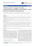
Quantitative real-time PCR was performed on 62
HNSCC (46 larynx, 14 hypopharynx and 2
oropharynx) and 19 adjacent normal tissues to assess
H19
and
miR-675
expression. The expression
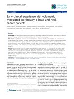
of H19 was significantly
higher in the tumor cohorts
compared
with
the
adjacent normal tissues (p
=0.0063,
Fig
1A).
Furthermore,
when
comparing H19 expression
in
recurrence
with
non-recurrence
patient’s
group,
H19
was
significantly higher in the
patients that eventually
relapsed (p=0.052; Fig 1B).
Figure 1. Prognostic significance of
H19 in patients with head neck
squamous cell carcinoma. (A). Relative
expression of H19 in HNSCC compared
with normal tissues. (B). non-recurrence
compared with recurrence samples.
Kaplan-Meier analysis was performed
for overall survival and disease free
survival. Patients with high expression of
H19 exhibited significantly worse overall
survival (C) and shortened disease-free
survival (D) than that with low
expression of H19 as defined by log-rank
test.
Int. J. Med. Sci. 2016, Vol. 13
916
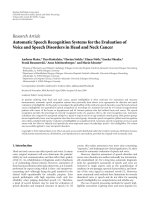
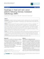
Figure 2. Prognostic values of miR-675 in patients with head neck squamous cell carcinoma. (A). Relative expression of miR-675 in HNSCC compared with normal
tissues. (B). non-recurrence compared with recurrence samples. Kaplan-Meier analysis was performed for overall survival and disease free survival. Patients with high
expression of miR-675 exhibited significantly worse overall survival (C) and shortened disease-free survival (D) than that with low expression of miR-675 as defined
by log-rank test.
H19 is a precursor of miR-675 and its function
could be at least through derived miR-675.
Unsurprisingly, qRT-PCR revealed that miR-675 was
positively concordant with H19 expression and
significantly overexpressed in the tumors compared
with the adjacent normal tissues (p=0.001, Fig 2A).
2.2. High expression of H19 and miR-675 were
correlated with poor prognosis in HNSCC
patients
In order to investigate the association between
H19 expression and patient survival, the patients
were dichotomized into H19 high (n=37) and low
(n=25) groups by using the median expression value
of H19. The Kaplan-Meier survival curves showed
that patients with higher H19 expression had worse
overall survival (OS) and disease-free survival (DFS)
compared with those with lower H19 expression by
log-rank test (p=0.0048 and p=0.0052, respectively. Fig
1 C and D). Similarly, miR-675 expression level was
observed to be correlated significant for overall
survival (OS) and disease-free survival (DFS). Higher
expression of miR-675 was associated with patient
recurrences (p =0.003, Fig 2B) and had worse overall
survival (p=0.024) and DFS (p=0.045) than those with
low miR-675 expression group (Fig 2C and 2D).
When comparing H19 and miR-675 expression
with gender, age, tumor stage, primary tumor
location and lymph node metastases, univariate
analysis showed that overexpression of miR-675
significantly correlated with patients have higher
expression of H19 and lymph node metastases, but
not with sex, age and primary tumor location (Table 2
(A)) for both OS and DFS. By using Cox proportional
hazard regression model, multivariate analysis
Int. J. Med. Sci. 2016, Vol. 13
917
revealed that H19 overexpression together with
higher miR-675 and lymph node metastases were
independent prognostic factors for poor disease-free
survival (Table 2 (B); H19; p=0.017; Hazard ratio: 4.11;
95% CI between 1.27 to 13.21). Likewise, H19 was also
significantly correlated with overall survival when
controlled for the clinical prognostic factors (Table 2
(B); p= 0.0025). The hazard ratio for the effect of H19
on survival when controlled for clinical factors was
14.29, 95% CI was between 3.43 and 59.41.
Table 2. Analysis of prognostic parameters.
Variable
(A). Univariate
analysis
Age at surgery (>64
v.s. <=64)
Gender
(Male/Female)
Primary location
(Larynx/others)
Tumor stage
(T1-2/T3-4)
Relapse v.s.
Non-relapse
High H19 (Y/N)
High miR-675
(Y/N)
OS
(p value)
DFS
(p value)
0.78
0.79
0.94
0.76
0.59
0.37
0.057
0.059
0.0012
0.0055
0.0017
0.021
0.021
0.0067
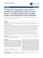
To evaluate the effects of miR-675 depletion, cells
were transfected with either scramble controls (SC) or
anti-miR-675. Expression of miR-675 was significantly
reduced by 65% and 70% respectively, at 48 and 72
hours post-transfection (Fig 3C left) in Hep-2 cells.
Similarly, in Fadu cells, miR-675 was significantly
suppressed by 65% at 48 hours post-transfection and
continuously declined at 72 hours (Fig 3C right).
Furthermore, transfection with anti-miR-675 led to
significantly decreased cell viability in both Hep-2
and Fadu cells, by 30% and 55% respectively, at 48
hours post-transfection compared with controls (Fig
3D). Taken together, these data indicate that
overexpressions of H19 and miR-675 are common
events in HNSCC, effective suppression of miR-675
warrant further investigation for potential therapeutic
applications.
2.4. miR-675 enhanced cell migration and
invasion.
(B). Multivariate
analysis
OS (HR, 95%CI) p value
DFS (HR, 95%CI) p
value
Tstage
Lymph node
metastases
High H19
High miR-675
1.086(1.018-1.15) 0.012
0.049(0.0060-0.41) 0.0053
1.098(1.026-1.17) 0.0064
0.029(0.0033-0.25) 0.0014
14.29(3.43-59.41) 0.0025
2.52(1.75-8.45)
0.013
4.11(1.27- 13.21)
3.26(0.94-10.71)
0.017
0.051
OS-overall survival; DFS-disease-free survival; HR-Hazard ratio; CI-confidence
interval.
2.3. miR-675 promoted cell proliferation in
HNSCC cells
Given the evidence that overexpression of H19
and miR-675 were associated with poor clinical
outcomes in HNSCC, particularly in laryngeal
carcinoma, we further assessed the biological
significance of depletion of miR-675 in 2 HNSCC cell
lines; Hep-2, a laryngeal carcinoma line and Fadu, a
hypopharyngeal
carcinoma
line.
H19
was
significantly overexpressed in two examined cancer
lines, when compared with immortalized normal
human oral epithelial cells (iNOE; Fig 3A). As
expected, both cancer cell lines demonstrated higher
level of miR-675 expression compared with that of
normal (Fig 3B).
As miR-675 up-regulation was observed to be
associated with recurrence in our patients, we
performed migration and invasion assays on Hep-2
cells. Compared with their corresponding negative
controls, transfection with antimiR-675 significantly
reduced migrate capacities of Hep-2 cells by 40% and
resulted in reduction of invasion of Hep-2 cells by
30%, as shown in Figure 4.
3. Discussion
It has become increasing evidence that
lncRNAs play essential roles for human tumour
growth and have been found to be key regulators of
the complex protein signaling pathways underling
carcinogenesis. In the present study, we found that
clinically, high expression levels of H19 correlated
with patient clinical outcome in our cohort of HNSCC
patients. miR-675 expression was positively
correlated with H19. Moreover, both high expression
of H19 and miR-675 significantly correlated with poor
overall
survival
and
disease-free
survival.
Furthermore, H19 overexpression together with
higher miR-675 and lymph node metastases were
identified as independent prognostic factors for
HNSCC patients. These finding further supported by
the evidence of that depletion of miR-675 inhibiting
laryngeal and hypopharyngeal cancer cells
proliferation, migration and invasion through
involvement in cellular processes.
H19 is an imprinted long non-coding RNA gene.
Emerging evidence indicates that H19 is frequently
overexpressed in the majority of human malignancies.
Although it acts as an oncogene in breast [17], bladder
[18], glioma [19] and prostate [20] cancers, some
studies also show its controversial role as a tumor
Int. J. Med. Sci. 2016, Vol. 13
suppressor in different tumor types [21] [22]. In
HNSCC, a study by Esteves LI [23] has shown a
significant correlation between H19 expression and
tumor recurrence. Consistence with their observation,
H19 expression in our cohort demonstrated
significant up-regulation in cancer than that in
918
normal. Importantly, the patients with higher H19
expression had worse overall survival and
disease-free survival compared with those with lower
H19 expression. These results suggest that H19
contributes to oncogenesis of HNSCC.
Figure 3. Down-regulation of miR-675 reduced cell proliferation in squamous cell carcinoma cells. (A & B). Quantitative real-time PCR (qRT-PCR) for H19 and
miR-675 expressions in head neck squamous cell carcinoma cells (Hep2 and Fadu) compared with normal epithelial cells (iNOE). (C). Equal amount of Anti-miR-675
(40 nM) or scrambled control (SC) was transfected into either Hep-2 cells or Fadu cells and significantly reduced miR-675 expression in both cells, compared with
scrambled control (SC) or transfection reagent alone (Lipo). (D). Cell viability was assessed in both Hep2 and Fadu cells by the MTT assay at 24–72 hours
post-transfection. Each datum represents the mean fold change ± SE in triplicates. *p<0.05. **p<0.01.
Int. J. Med. Sci. 2016, Vol. 13
919
Figure 4. Depletion of miR-675 reduced cell migration and invasion. Top panel--- Representative images of migration assay depict migratory ability after transfection
with antimiR-675 compared to scramble control. Bottom panel--- Representative images of invasive assay depict invasiveness after transfection with antimiR-675
compared to scramble control.
miR-675 is excised from first exon of H19. Both
H19 and miR-675 are believed to be involved in
tumoriogenesis. It was first described by Tsang WP
[15], in which H19-derived miR-675 regulated
primary human colorectal cancer development
through down-regulation of tumor suppressor RB.
Subsequently, a strong relationship between H19 and
miR-675 was confirmed in various studies.
H19/miR-675 targeted tumor suppressor Runt
Domain Transcription Factor1 and regulated
gastric cancer proliferation and development [24].
H19 derived miR-675 promoted glioma cell invasion
[19] and enhanced tumorigenesis and metastasis
through downregulating c-CbI/CbI-b in breast cancer
cells [25].
The precise underlying mechanism of
H19/miR-675 in tumorigenesis remains unclear. H19
involves in the complex biological process of
oncogenesis [2, 17, 26]. Genomic imprinting is an
inherited epigenetic phenomenon, loss of imprinting
at the IGF2 and H19 loci play a role in the oncogenesis
of head and neck carcinoma [27]. H19 expression in
many cancers is usually associated with the function
of methylation. H19 CBS6 methylation potentially
represents a novel clinically relevant epigenetic
marker for the recurrence and progression of
esophageal squamous cell carcinoma [28]. There are
clear evidences show that TGF-β can induce the
expression of H19 along with EMT markers, such as
Slug, which also contribute to tumor metastasis and
dependent on H19[17]. Recent study has
demonstrated that H19 functions as a miRNA
sponges to promoting EMT in colorectal cancer
through activating Wnt signaling pathway [29]. On
one hand, H19 overexpression increases the cellular
abundance of miR-675 [30] and regulates intestinal
epithelial barrier function via miR-675 by interacting
with RNA-binding protein HuR [31]. On the other
hand, miR-675 up-regulates long noncoding RNA
H19 through activating EGR1 in human liver cancer
[32]. In our study, miR-675 expression was positively
correlated with H19 over-expression in HNSCC.
Overexpression of miR-675 together with higher H19
and lymph node metastases were independent
prognostic factors for poor disease-free survival.
Furthermore, our results were in line with their
observation, in that knock- down miR-675 caused
significant reduction of cell viability, migratory and
invasive capabilities in carcinoma lines, indicating
miR-675 involving in HNSCC tumor progression.
Nonetheless, further studies in the function of
H19/miR-675 axis in the molecular mechanism of
HNSCC tumor progression in a large cohort of
samples may validate the exploratory findings.
Int. J. Med. Sci. 2016, Vol. 13
4. Material and Methods
4.1. Patient Characteristics
Sixty-five head neck squamous cell
carcinoma formalin-fixed and paraffin-embedded
samples were collected during the period of 2001-2011,
with approval from the Second Hospital of Jilin
University. Among of them, sixty-two tissue samples
had sufficient tumor cell contains and have been
involved in the study. The clinical, demographic and
tumor-related characteristics of patients have been
descripted in Table 1. The predominant primary
sub-site was larynx (46 of 62, or 74.2%). The majority
of patients were male (48 or 77.4 %) and median age
was 63.8 years. More than half of patients (59.6%) had
lymph node metastasis at the diagnosis and 71%
patients had locally advanced disease with stage III
and IV. The average follow-up time was 3.09 years.
During the follow-up period, 25 patients were dead
(40.3%) and all of the patients were treated neither
with radiotherapy nor with chemotherapy prior to the
surgery. Histologically confirmed, adjacent normal
tissues from 19 samples were able to obtain either
from separate block or from macro-dissected blocks of
patients.
920
using the LipofectAMINE 2000 (Invitrogen) reverse
transfection protocol, according to the manufacturer’s
instructions. AntagomiR-675 and anti-miR scramble
negative control 1 (SC; Ambion) were transfected at a
final concentration of 40 nmol/L to a 96-well plate.
Table 1. Clinical Characteristics of Patients.
Parameter
Gender
Male
Female
Age (years)
>64
<=64
Primary location
Larynx
Hypo-&others
Tumor stage
T1-2
T3-4
Relapse
No
Yes
N=62 (%)
H19
p value mir-675
p value
expression*
expressio
n*
48
14
77
23
20.11
13.08
0.36
18.88
8.15
0.36
33
29
53
47
25.01
11.15
0.27
20.79
11.51
0.3
46
16
74
26
22.04
8.41
0.77
17.66
12.98
0.92
18
44
29
71
15.22
19.87
0.16
15.59
16.81
0.77
25
37
40
60
7.48
25.98
0.038
7.61
22.44
0.017
* represent the median of H19 or miR-675 expression.
4.2. Sample preparation
4.4. RNA and miRNAs extraction
All studied tumour (n=65) and adjacent normal
(n=19)
specimens
were
assessed
on
hematoxylin-and-eosin-stained (H&E) slides using
standard diagnostic criteria. 62 tumor specimens were
confirmed with more than 60% of malignant cells and
were selected for the analysis. Based on the H&E
review, the samples were macro-dissected. This study
was approval from the Second Hospital of Jilin
University and all patients have consented for the
study.
Total RNA containing miRNAs were extracted
from either primary tumor tissues or cell lines using
RecoverAll Total Nucleic Acid Isolation kit (Ambion)
according to the manufacturer’s instructions. Briefly,
the
macrodissected
FFPE
samples
were
de-paraffinized with xylene, washed with 100%
ethanol. The pellet then was digested by proteinase K.
After incubation the pellet in different heating blocks,
total RNA and miRNAs were isolated through a filter
cartridge and treated with DNase. The samples were
then stored in -80ºC until use.
4.3. Cell lines and transfection
The human laryngeal cancer cell line, Hep-2, was
purchased from the China Center for Type Culture
Collection and maintained at 37°C, 5% CO2 in RPMI
1640 complete medium (Gibco, USA) with 10% fetal
bovine serum (FBS, Gibco, USA). The human
hypopharyngeal squamous carcinoma cell line, Fadu,
was purchased from American Type Culture
Collection and were maintained in Minimum
Essential Medium, supplemented with 10% Fetal
Bovine Serum (FBS), 1.5 g/L bicarbonate, and 1 mM
pyruvate. The immortalized normal human oral
epithelial cells (iNOE) were purchased commercially
and cultured in the recommended medium. All cells
were maintained in a 37°C incubator with humidified
5% CO2. To knockdown endogenous miR-675
expression, Hep-2 and Fadu cells were transfected
4.5. Real-Time Quantification of H19 and
microRNA-675 expression
For H19 mRNA expression, the reverse
transcription was performed using SuperScript III
Reverse Transcriptase (Invitrogen Corp.) according to
the manufacturer’s recommendations. qRT-PCR
analyses were performed using SYBR Green Master
Mix (Applied Biosystems) and the ABI PRISM 7900
Sequence Detection System (Applied Biosystems Inc.,
Foster City, CA, USA). As described previously [15],
the primers used for H19 were: forward:
5′-TACAACCACTGCACTACCTG-3′;
reverse:
5′-TGGAATGCTTGAAGGCTGCT-3′. Compared with
control specimens, the relative fold change in mRNA
expression was calculated using the 2−ΔΔCt method
and was normalized to that of an endogenous gene
Int. J. Med. Sci. 2016, Vol. 13
(GAPDH), which was amplified in parallel as the
internal control. For miR-675 expression, the RNA
was reverse-transcribed with a MultiScribe reverse
transcriptase (Applied Biosystems) by using a
stem-loop RT primer specifically hybridises with a
miR molecule. The RT products were subsequently
amplified with sequence-specific primers using
Taqman MicroRNA Assay kit (Applied Biosystems)
by the Applied Biosystems 7900 HT Real-Time PCR
system. RNU48 was amplified in parallel and used as
an endogenous control.
4.6. MTT cell proliferation assay
The cell proliferation and cytotoxicity of
introducing anti-miR-675 were assessed by using a
3-[4,5-dimethylthiazol-2-yl]-2,5-diphenyltetrazolium
bromide (MTT) staining kit (Sigma, USA). Cells were
reverse transfected with either SC control,
anti-miR-675 or Lipofectamine 2000 and seeded onto
96-well plates (5 x 10³ cells/per well). Cell viability
was measured at 24, 48 and 72 hours’
post-transfection by a microplate reader.
4.7. Cell migration and invasion assays
Cell migration and invasion were assessed by
using BD BioCoat Matrigel Invasion Chambers and
Control Inserts (BD Biosciences). The cells were
transfected with either anti-miR-675 or SC (40nM),
then seeded on either control inserts (polyethylene
terephthalate membrane) or trans-well chambers with
Matrigel. Two ml RPMI supplemented with 15% FBS
was added to the lower chamber, served as the
chemo-attractant. 0.5x10⁴ transfected Fadu cells were
re-suspended in RPMI plus 1% FBS, added to the
upper chamber (0.5 mls). Twenty hours later,
migrating or invading cells attached to the lower
surface of the membrane insert were fixed and
stained, then counted under a microscope. Relative
migration was calculated by comparison with cells
transfected with the negative control. The percentage
invasion was calculated based on the number of cells
which have invaded through the Matrigel insert,
divided by the number of cells which have migrated
through the control insert.
921
significance, they were then further analysed in a
multivariate Cox’s proportional hazards regression
model. A p-value of less than 0.05 was considered to
be statistically significant.
5. Conclusions
In summary, our results indicate that H19 and
miR-675 are significantly overexpressed in HNSCC
cancer cells and patients. Higher expression of H19
and miR-675 are correlated with poor prognosis. The
strong correlation of H19 overexpression together
with higher miR-675 and lymph node metastases
could be useful predictive markers, indicating a
potentially therapeutic strategy for HNSCC patients.
Acknowledgments
The authors acknowledge the financial support
of the Science and Technology R&D program of Jilin
Province, China (20150101140JC).
Conflict of interest
The authors report no conflict of interest in this
work.
References
1.
2.
3.
4.
5.
6.
7.
8.
9.
10.
4.8. Statistical Analysis
The differences expression of H19 and miR-675
between each group were evaluated by unpaired,
two-tailed Student’s t-test. Overall survival (OS) was
defined from the time of diagnosis to date of death.
Disease-free survival (DFS) was defined from the time
of diagnosis to the date of first failure. Survival curves
were generated using the Kaplan-Meier method and
were compared by means of the log-rank test. In the
univariate model, if factors with prognostic
11.
12.
13.
14.
Djebali S, Davis CA, Merkel A, Dobin A, Lassmann T, Mortazavi A, et al.
Landscape of transcription in human cells. Nature. 2012; 489: 101-8.
Mattick JS. Long noncoding RNAs in cell and developmental biology.
Seminars in cell & developmental biology. 2011; 22: 327.
Mele M, Ferreira PG, Reverter F, DeLuca DS, Monlong J, Sammeth M, et al.
Human genomics. The human transcriptome across tissues and individuals.
Science. 2015; 348: 660-5.
Cheetham SW, Gruhl F, Mattick JS, Dinger ME. Long noncoding RNAs and
the genetics of cancer. British journal of cancer. 2013; 108: 2419-25.
Schmidt LH, Spieker T, Koschmieder S, Schaffers S, Humberg J, Jungen D, et
al. The long noncoding MALAT-1 RNA indicates a poor prognosis in
non-small cell lung cancer and induces migration and tumor growth. Journal
of thoracic oncology : official publication of the International Association for
the Study of Lung Cancer. 2011; 6: 1984-92.
Xu C, Yang M, Tian J, Wang X, Li Z. MALAT-1: a long non-coding RNA and
its important 3' end functional motif in colorectal cancer metastasis.
International journal of oncology. 2011; 39: 169-75.
Yang Z, Zhou L, Wu LM, Lai MC, Xie HY, Zhang F, et al. Overexpression of
long non-coding RNA HOTAIR predicts tumor recurrence in hepatocellular
carcinoma patients following liver transplantation. Annals of surgical
oncology. 2011; 18: 1243-50.
Kogo R, Shimamura T, Mimori K, Kawahara K, Imoto S, Sudo T, et al. Long
noncoding RNA HOTAIR regulates polycomb-dependent chromatin
modification and is associated with poor prognosis in colorectal cancers.
Cancer research. 2011; 71: 6320-6.
Ariel I, de Groot N, Hochberg A. Imprinted H19 gene expression in
embryogenesis and human cancer: the oncofetal connection. American journal
of medical genetics. 2000; 91: 46-50.
Fellig Y, Ariel I, Ohana P, Schachter P, Sinelnikov I, Birman T, et al. H19
expression in hepatic metastases from a range of human carcinomas. Journal
of clinical pathology. 2005; 58: 1064-8.
Sorin V, Ohana P, Gallula J, Birman T, Matouk I, Hubert A, et al.
H19-promoter-targeted therapy combined with gemcitabine in the treatment
of pancreatic cancer. ISRN oncology. 2012; 2012: 351750.
Zhang EB, Han L, Yin DD, Kong R, De W, Chen J. c-Myc-induced, long,
noncoding H19 affects cell proliferation and predicts a poor prognosis in
patients with gastric cancer. Medical oncology. 2014; 31: 914.
Zhang E, Li W, Yin D, De W, Zhu L, Sun S, et al. c-Myc-regulated long
non-coding RNA H19 indicates a poor prognosis and affects cell proliferation
in non-small-cell lung cancer. Tumour biology : the journal of the International
Society for Oncodevelopmental Biology and Medicine. 2016; 37: 4007-15.
Matouk IJ, Halle D, Gilon M, Hochberg A. The non-coding RNAs of the
H19-IGF2 imprinted loci: a focus on biological roles and therapeutic potential
in Lung Cancer. Journal of translational medicine. 2015; 13: 113.
Int. J. Med. Sci. 2016, Vol. 13
922
15. Tsang WP, Ng EK, Ng SS, Jin H, Yu J, Sung JJ, et al. Oncofetal H19-derived
miR-675 regulates tumor suppressor RB in human colorectal cancer.
Carcinogenesis. 2010; 31: 350-8.
16. Jemal A, Bray F, Center MM, Ferlay J, Ward E, Forman D. Global cancer
statistics. CA: a cancer journal for clinicians. 2011; 61: 69-90.
17. Matouk IJ, Raveh E, Abu-lail R, Mezan S, Gilon M, Gershtain E, et al. Oncofetal
H19 RNA promotes tumor metastasis. Biochimica et biophysica acta. 2014;
1843: 1414-26.
18. Byun HM, Wong HL, Birnstein EA, Wolff EM, Liang G, Yang AS. Examination
of IGF2 and H19 loss of imprinting in bladder cancer. Cancer research. 2007;
67: 10753-8.
19. Shi Y, Wang Y, Luan W, Wang P, Tao T, Zhang J, et al. Long non-coding RNA
H19 promotes glioma cell invasion by deriving miR-675. PloS one. 2014; 9:
e86295.
20. Ribarska T, Goering W, Droop J, Bastian KM, Ingenwerth M, Schulz WA.
Deregulation of an imprinted gene network in prostate cancer. Epigenetics.
2014; 9: 704-17.
21. Cui H, Hedborg F, He L, Nordenskjold A, Sandstedt B, Pfeifer-Ohlsson S, et al.
Inactivation of H19, an imprinted and putative tumor repressor gene, is a
preneoplastic event during Wilms' tumorigenesis. Cancer research. 1997; 57:
4469-73.
22. Fukuzawa R, Umezawa A, Ochi K, Urano F, Ikeda H, Hata J. High frequency
of inactivation of the imprinted H19 gene in "sporadic" hepatoblastoma.
International journal of cancer. 1999; 82: 490-7.
23. Esteves LI, Javaroni AC, Nishimoto IN, Magrin J, Squire JA, Kowalski LP, et al.
DNA methylation in the CTCF-binding site I and the expression pattern of the
H19 gene: does positive expression predict poor prognosis in early stage head
and neck carcinomas? Molecular carcinogenesis. 2005; 44: 102-10.
24. Zhuang M, Gao W, Xu J, Wang P, Shu Y. The long non-coding RNA
H19-derived miR-675 modulates human gastric cancer cell proliferation by
targeting tumor suppressor RUNX1. Biochemical and biophysical research
communications. 2014; 448: 315-22.
25. Vennin C, Spruyt N, Dahmani F, Julien S, Bertucci F, Finetti P, et al. H19 non
coding RNA-derived miR-675 enhances tumorigenesis and metastasis of
breast cancer cells by downregulating c-Cbl and Cbl-b. Oncotarget. 2015; 6:
29209-23.
26. Nakao M, Sasaki H. Genomic imprinting: significance in development and
diseases and the molecular mechanisms. Journal of biochemistry. 1996; 120:
467-73.
27. Rainho CA, Kowalski LP, Rogatto SR. Loss of imprinting and loss of
heterozygosity on 11p15.5 in head and neck squamous cell carcinomas. Head
& neck. 2001; 23: 851-9.
28. Gao T, He B, Pan Y, Gu L, Chen L, Nie Z, et al. H19 DMR methylation
correlates to the progression of esophageal squamous cell carcinoma through
IGF2 imprinting pathway. Clinical & translational oncology : official
publication of the Federation of Spanish Oncology Societies and of the
National Cancer Institute of Mexico. 2014; 16: 410-7.
29. Liang WC, Fu WM, Wong CW, Wang Y, Wang WM, Hu GX, et al. The lncRNA
H19 promotes epithelial to mesenchymal transition by functioning as miRNA
sponges in colorectal cancer. Oncotarget. 2015; 6: 22513-25.
30. Keniry A, Oxley D, Monnier P, Kyba M, Dandolo L, Smits G, et al. The H19
lincRNA is a developmental reservoir of miR-675 that suppresses growth and
Igf1r. Nature cell biology. 2012; 14: 659-65.
31. Zou T, Jaladanki SK, Liu L, Xiao L, Chung HK, Wang JY, et al. H19 Long
Noncoding RNA Regulates Intestinal Epithelial Barrier Function via
MicroRNA 675 by Interacting with RNA-Binding Protein HuR. Molecular and
cellular biology. 2016; 36: 1332-41.
32. Li H, Li J, Jia S, Wu M, An J, Zheng Q, et al. miR675 upregulates long
noncoding RNA H19 through activating EGR1 in human liver cancer.
Oncotarget. 2015; 6: 31958-84.