Fractional flow reserve in non-culprit coronary arteries of patients with acute ST elevation myocardial infarction
Bạn đang xem bản rút gọn của tài liệu. Xem và tải ngay bản đầy đủ của tài liệu tại đây (556.5 KB, 8 trang )
JOURNAL OF MEDICAL RESEARCH
FRACTIONAL FLOW RESERVE IN NON-CULPRIT CORONARY
ARTERIES OF PATIENTS WITH ACUTE ST ELEVATION
MYOCARDIAL INFARCTION
Vu Quang Ngoc1, Ken Kozuma2, Nguyen Quoc Thai1, Pham Manh Hung3
Vietnam National Heart Institude, Bach Mai Hospital, Hanoi, Vietnam
2
Department of Cardiology, Teikyo University, Tokyo, Japan
3
Department of Cardiology, Hanoi Medical University, Hanoi, Vietnam
1
Multi-vessel disease (MVD) with stenotic lesions other than the culprit artery (the so-called nonculprit artery) - is present in 40-60% of acute ST elevation myocardial infarction (STEMI) patients, which
is a determintant of higher risk of death and re-intervention compared to single vessel dissease [1]. In the
presence of non-culprit coronary stensosis, the optimal therapy for that is still a matter of debate. While
guidelines discourage a concomitant treatment of infarct- and non-infarct-related arteries, recent studies
document advantages of a complete (preventive) revascularization during primary PCI, which may result in
overtreatment, as angiography alone does not provide robust information about the functional severity of
MVD. Fractional flow reserve (FFR) measurements have been established in this acute setting as a possibly
valuable guide for non-culprit lesions after uncomplicated primary PCI accordingly. FFR value ≤ 0.80
has been determined to be predictive of functional significance and, in addition, is the threshold at which
revascularisation should be considered. The clinical implications of an FFR-guided treatment strategy in
STEMI patients with MVD have been proved in a variety of randomized clinical trials CVLPRIT [2], DANAMI3-PRIMULTI [3]. In Vietnam, FFR has been validated in a large number of studies but limited to data of
patients with stable ischemic heart disease [4]. We undertook the study to assess the FFR of non-culprit
arteries in patients with acute STEMI and MVD after uncomplicated primary PCI. 81 acute STEMI patients
at 2 institutions (Vietnam National Heart Institute - Hanoi - Vietnam and Teikyo University Hospital - Tokyo
- Japan), who met the inclusion criteria, were enrolled in the prospective, non-randomized study from Nov
2017 to Sept 2018. The mean age was 60.9 ± 12.2 (yrs). 63% of patients were male. The most common
culprit artery was LAD (57.6%). 60.5% involved 2 vessels, and 39.5% involved 3 vessels. Mean percentage
diameter stenosis (PDS) was 55.17 ± 9.85%. FFR of 135 non-culprit lesions contained of 23.8% lesions
with FFR ≤ 0.80, 76.2% lesions with FFR > 0.80. The mean FFR value was 0,82 ± 0,16. The study showed
100% of technical success rate, and 99.3% procedural success rate. FFR revealed correlation with minimum
lumen diameter (MLD), inverse correlation with lesion length (LL), but no correlation with PDS. Measuring
FFR of non-culprit artery after uncomplicated primary PCI setting is safe and provides helpful information
on functionally ischemic impact, and further, on revascularization strategy in STEMI patients with MVD.
Keywords: acute myocardial infarction, STEMI, primary coronary intervention, fractional flow reserve.
I. INTRODUCTION
Acute ST segment elevation myocardial
infarction (STEMI) most commonly occurs
Corresponding author: Vu Quang Ngoc, Vietnam
National Heart Institude, Bachmai Hospital
Email:
Received: 27/11/2018
Accepted: 12/03/2019
JMR 118 E4 (2) - 2019
when thrombus formation results in complete
occlusion of a major epicardial coronary vessel.
The most serious form of acute coronary
syndromes, STEMI is a life-threatening, timesensitive emergency that must be diagnosed
and treated promptly via primary percutaneous
coronary intervention (PCI) to restore blood
105
JOURNAL OF MEDICAL RESEARCH
flow as soon as possible in the occluded vessel.
Multi-vessel disease (MVD) is present in about
40-60% of patients with STEMI referred for PCI,
which is a determinant of higher risk of death and
revascularization compared to single vessel
disease [1]. Although the presence of MVD
has been associated with a worse prognosis,
not all studies have shown improved outcomes
when these so called "non-culprit lesions" are
treated with PCI. In theory, one might argue
that this is because the lesion in the non-infarct
artery is an "innocent bystander" and therefore
should be approached in much the same
way one approaches stable ischemic heart
disease. Opponents to this argument might
propose that these non-culprit lesions may
also be biologically active as there are often
multiple complex plaques in patients with acute
myocardial infarction shown in various studies,
and therefore these arteries warrant treatment
in much the same way one would approach any
unstable lesion. There have been a number
of studies including CVLPRIT [2], DANAMI3-PRIMULTI [3] which showed benefits of
total revascularization (culprit + nonculprit
artery PCI), but PRAGUE-13 study brought
reverse outcomes. Measuring Fractional flow
reserve - FFR in non-culprit coronary lesions
would provide interventional cardiologists
with appropriate information of hemodynamic
significance of the lesions. FFR value > 0.80
has been determined to be predictive of
functional significance and, in addition, is the
threshold at which revascularization should be
considered, while FFR value > 0.80 is safe for
conservatively medical treatment. In Vietnam,
FFR has been validated in a variety of studies
but limited to data of patients with stable
ischemic heart disease [4]. We undertook the
study to assess the Fractional Flow Reserve
in non-culprit coronary arteries of patients with
106
acute ST elevation myocardial infarction.
II. STUDY POPULATION AND
STUDY METHOD
1. Study population
The study was conducted from Nov - 2017
to Sep - 2018 with 81 consecutive acute
STEMI patients who received primary PCI in
Vietnam National Heart Institute (n = 31) and
Cardiovascular Division - Teikyo University Tokyo - Japan (n = 50).
Inclusion criteria
• Patient ≥ 18 years old
• Acute STEMI indicated for primary PCI
within 12h (from symptoms onset) or > 12h if
persistent ischemic demonstration.
• At least one non-culprit coronary artery
lesion with diameter stenosis of 40% to < 70%
on QCA [5], [6].
• Coronary vessel diameter ≥ 2.0 mm
Exclusion criteria
• Severe heart failure, cardiac shock, Killip
III/IV on admission or after culprit coronary
revascularization.
• STEMI related to in-stent thrombosis
• Unsuccessful
primary
PCI
or
complicated primary PCI (inability of stent
deployment to culprit vessel, or TIMI flow 0 1 post PCI, residual stenosis > 20%, coronary
dissection or rupture).
• Non-culprit lesions of < 40% or > 70% of
diameter stenosis (on QCA).
• Stenosis of left main stem > 50%
• Non-culprit artery with TIMI flow II
• Chronic total occlusion of non-culprit.
• Bypass graft lesions.
• Syntax score > 22
• Inappropriate anatomical features for
pressure wire passage.
• Medical history of allergy to any of the
following medication: Aspirin, Clopidogrel,
JMR 118 E4 (2) - 2019
JOURNAL OF MEDICAL RESEARCH
Ticagrelor, Heparin, contrast agents, or
papaverine.
• Prolonged bradycardia, AV block, long QT
syndrome.
• Anticoagulation disorders or recent history
of bleeding (cerebral, gastrointestinal or
genitourinary) within 3 months.
• End-stage renal disease, severe sepsis,
end-stage cancer, or other medical conditions
with estimated life expectancy of less than 1
year.
• Pregnancy.
• Refuse to enroll in the study.
2. Study method
Study design: prospective observational
cohort.
Sampling
method:
non-randomized,
consecutive sampling. All acute STEMI
patients with MVD admitted to the National
Heart Institute – Bach Mai Hospital and
Cardiovascular Division - Teikyo University
- Tokyo - Japan, for primary PCI, who are
eligible for study inclusion criteria. Non-culprit
coronary lesions were assessed anatomically
by Quantitative Coronary Angiography (QCA)
and functionally by Fractional flow reserve per
protocol [5].
FFR measuring requires the use of a specific
PressureWire (solid-state sensor mounted
on a floppy-tipped 0.014- inch guidewire) (St.
Jude Medical Inc., Minneapolis, Minnesota
and Uppsala, Sweden). Before introducing
the sensor into the vessel to be studied, the
pressures recorded by the sensor and by the
guiding catheter should be equalized.
A 200 mcg bolus of intracoronary nitrate,
followed by papaverine (10mg in the right
coronary artery, 20 mg in the left coronary
artery LCA), allows the abolition of any form
of epicardial vasoconstriction and hyperemia.
All procedures were performed during index
hospitalization.
Statistical Analysis: Continuous variables
are presented as mean ± SD or median and
inter-quartile range from the 25th to the 75th
percentile; categorical data are presented as
numbers and percentages, as appropriate.
P values smaller than 0.05 were considered
as statistically significant. Analyses were
performed with SPSS 20.0 (IBM, Inc, New
York).
Ethical approval provided by Bach Mai
University hospital and Teikyo University
hospital.
III. RESULTS
1. Baseline parameters
From Nov 2017 to Sep 2018 at Vietnam National Heart Institute and Cardiovascular Division
- Teikyo University - Tokyo - Japan, FFR measurements were done on 81 STEMI patients, who
received primary PCI, with 135 non-culprit coronary arteries of moderate stenosis (40 - 70% by
QCA). Male/female ratio was 1.7/1. The major risk factors included hypertension (67.9%) and
smoking (55.6%).
Table 1. Baseline parameters
Parameter
Male (%)
Mean age (yrs)
JMR 118 E4 (2) - 2019
N
51 (63%)
65.7 ± 12.4
107
JOURNAL OF MEDICAL RESEARCH
Parameter
N
Hypertension (%)
55 (67.9%)
Smoking (%)
45 (55.6%)
Dislipidemia (%)
41(50.6%)
Diabetes mellitus (%)
27 (33.3%)
Duration of symptom onset (hrs)
9.24 ± 2.9
Left ventricular ejection fraction (%)
42.2 ± 6.7
Creatinin (μmol/l)
92.9 ± 18.7
Table 2. Non-culprit coronary artery characteristics
Parameter
N
Number of diseased vessel n (%)
2-vessel disease
49 (60.5%)
3-vessel disease
32 (39.5%)
Syntax score (points)
17.45 ± 2.69
Lesion type n (%)
Type A
11 (8.1%)
Type B1
39 (28.9%)
Type B2
66 (48.9%)
Type C
19 (14.1%)
QCA parameters of non-culprit coronary lesions
Reference vessel diameter - RVD (mm)
2.88 ± 0.51
Minimal lumen diameter - MLD (mm)
1.43 ± 0.27
Percentage of diameter stenosis - PDS (%)
55.17 ± 9.85
Lesion length - LL (mm)
22.45 ± 7.62
2. Fractional flow reserve of non-culprit coronary arteries
2.1. FFR measurement
FFR evaluation were performed via radial access with 6F guiding catheter in a large proportion of
patients (96.3%). Femoral access was chosen among 3 cases (3.7%). The time from primary PCI
108
JMR 118 E4 (2) - 2019
JOURNAL OF MEDICAL RESEARCH
to FFR measurement ranged on an average of 2.65 ± 1.09 days. Among 135 non-culprit lesions,
the percentage of significantly functional stenosis that required revascularization was 23.8%, while
the rest 76.2% of lesions not contributed to physiological impact.
The mean FFR value was 0.82 ± 0.09. Lesion distribution was as followed: 22.9% in proximal
left descending artery (LAD), 31.3% in the mid LAD, 4.2% in distal LAD, 4.2% in proximal circumflex
(LCx), 14.6% in mid LCx, 8.4% in proximal right coronary artery (RCA), 12.5% in mid RCA, and 1.9%
in distal RCA; There was 1 coronary dissection complication related to pressure wire manipulation.
2.2. Correlation between FFR and QCA parameters of non-culprit coronary lesions.
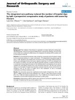
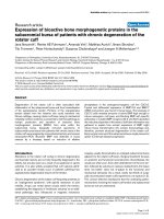
Figure 1. Correlation between FFR and PDS.
No significant correlation was found between FFR and PDS.
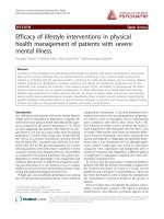
Figure 2. Correlation between FFR and MLD.
JMR 118 E4 (2) - 2019
109
JOURNAL OF MEDICAL RESEARCH
MLD was weakly correlated with FFR (r = 0,81, p = 0,04). In other words, the less the MLD, the
lower the FFR.
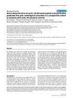
Figure 3. Correlation between FFR and lesion length.
Lesion length was moderately inversely correlated with FFR (r = - 0.38, p = 0.045).
III. DISCUSSION
Baseline parameters: patients' mean age
was 65.7 ± 12.4 (years). A larger proportion
of patients were male (63%). The major risk
factors include hypertension 67.9%, smoking
55.6% and diabetes mellitus 50.6%. Similar
findings were also reported in various studies
[1-3].
The percentage of 2 - vessel disease was
60.5%. The complexity of coronary disease was
graded by Syntax score (based on segment
involved, chronic total ccclusion, tortuosity,
angulation, calcification severity, lesion length,
bifurcation...). Syntax score > 22 is known to be
not only an independent predictor of MACEs in
ACS patients with multivessel disease, but also
an indication for early referral to coronary artery
bypass grafting. The mean syntax score in our
study was 17.45 ± 2.69 points as only patients
whose Syntax score ≤ 22 were enrolled. The
110
mean RVD was 2.7 ± 0.5 mm and the mean LL
was 22.1 ± 7.0 mm.
Angiographical characteristics of nonculprit coronary lesions: The complexity of
non-culprit coronary lesions was evaluated
angiographically based on AHA/ACC 1988
classification, which ranges from type A, type
B1, type B2, to type C (the most complex).
A 100% success rate was achieved with
pressurewire passage distal to stenotic nonculprit lesions, as most of them were classified
of type A, B1, and B2. The pressurewire was
also passed through all 19 complex lesions
(type C) thank to unique handling characteristic
and flexible tip that in not so much different from
regular workhorse wires. We reported 1 case
of left main coronary dissection complication
related to pressure wire manipulation, which
required immediate stent implantation and
JMR 118 E4 (2) - 2019
JOURNAL OF MEDICAL RESEARCH
lead to uncomplicated discharge 2 days after
the procedure. In brief, our technical success
rate approached 99.3%. The time from primary
PCI to FFR assessment was on the average of
3.14 ± 1.05 days.
Correlation between QCA parameters
and FFR of non-culprit artery: In acute
STEMI patients required emergent coronary
angiography, after the culprit artery is
determined with certainty (often based on
the presence of thrombus, no flow or slow
flow at stenotic site...), revascularization and
stent implantation is immediately performed to
restore blood flow to the infarcted myocardial
territory. The severity of non-culprit artery
stenosis is then estimated based on visual
assessment, which is commonly applied in
clinical practice. This method enables quick
evaluation but subjective and individual-based
decision making. While QCA was chosen as
the method of evaluation in our study, PDS
was not shown to be correlated with FFR (r =
- 0.057, p = 0.46). A number of studies stated
weak correlation between the two, but Park et
al [1] found a significantly inversely correlation.
Our findings demonstrated a non-significant
difference of PDS between FFR > 0,80 group
and FFR ≤ 0.80 group (p = 0.65). The weak
correlation of PDS and FFR was also mentioned
by Belle et al [6]. Data from meta-analysis [7]
suggested that QCA does not help determine
the functional significance in coronary lesions.
Although MLD is not a parameter of
choice for interventional cardiologists in their
clinical practice to decide whether or not to
revascularize, our study found the mean MLD
in FFR ≤ 0.80 group was significantly lower
than that of FFR > 0.80 group (p = 0.041).
There was a mild correlation between FFR
and MLD (r = 0.181, p = 0.04), which was also
reported in DANAMI-3-PRIMULTI trial [3]. The
JMR 118 E4 (2) - 2019
smaller the MLD, the lower the FFR value.
In our study, lesion length in FFR ≤ 0.80
group was significantly greater than that of
FFR > 0.80 group, 24.5 ± 12.5 mm and 17.5
± 8.5 mm respectively (p = 0.016). There was
a moderately reverse correlation between FFR
and lesion length with r = - 0.38 (p = 0.045),
which showed similar findings in CVLPRIT
study [2].
Ntalianis et al [5] investigated the reliability
of FFR of nonculprit coronary stenoses in
101 patients undergoing PCI for an acute
myocardial infarction. FFR measurements
were obtained immediately after PCI of the
culprit stenosis and were repeated 35 ± 4 days
later. The FFR value of the nonculprit stenoses
did not change between the acute and followup (0.77 ± 0.13 vs. 0.77 ± 0.13, respectively,
p > 0.05). During the acute phase of acute
coronary syndromes, the severity of nonculprit
coronary artery stenoses can reliably be
assessed by FFR. This allows a decision about
the need for additional revascularization and
might contribute to a better risk stratification.
V. CONCLUSION
In patients with acute STEMI and MVD,
FFR measurement after primary PCI appeared
to be feasible and revealed hemodynamic
significance of non-culprit artery lesions,
which resulted in appropriate multi-vessel
revascularization strategy in acute setting.
REFERENCES
1. Park DW, Clare RM, et al (2014).
Extent, Location, and Clinical Significance of
Non–Infarct-Related Coronary Artery Disease
Among Patients With ST-Elevation Myocardial
Infarction. JAMA. 312(19), 2019 - 2027.
2. Gershlick AH, Khan JN, Kelly DJ,
et al (2015). Randomized trial of complete
111
JOURNAL OF MEDICAL RESEARCH
versus
lesion-only
revascularization
in
patients undergoing primary percutaneous
coronary intervention for stemi and multivessel
disease: The CvLPRIT trial. J Am Coll Cardiol.
65(10):,963 - 972.
3. Høfsten DE, Kelbæk H, Helqvist S
(2015). The Third DANish Study of Optimal
Acute Treatment of Patients with ST-segment
Elevation Myocardial Infarction: Ischemic
postconditioning or deferred stent implantation
versus conventional primary angioplasty and
complete revascularization versus treatment.
Am Heart J. 169(5), 613 - 621.
4. Đinh Huỳnh Linh, Nguyễn Ngọc
Quang, Phạm Mạnh Hùng (2010). Đánh giá
phân số dự trữ lưu lượng vành của các tổn
thương hẹp vừa động mạch vành. Tạp chí y
học lâm sàng. 59, 58 - 63.
5. Ntalianis A, Sels JW, Davidavicius G,
et al (2010). Fractional Flow Reserve for the
112
Assessment of Nonculprit Coronary Artery
Stenoses in Patients With Acute Myocardial
Infarction. JACC Cardiovasc Interv. 3(12),
1274 -1281.
6. Van Belle E, Rioufol G, Pouillot C,
Cuisset T, Teiger E, Barreau D, et al (2013).
Outcome impact of coronary revascularization
strategy-reclassification with fractional flow
reserve (FFR) at time of diagnostic angiography:
Insights from a large french multicenter FFR
registry (R3F). Circulation [Internet]. 128(24)
2715.
7. Tarantini G, D’Amico G, Brener SJ,
Tellaroli P, Basile M, Schiavo A, et al (2016).
Survival
After
Varying
Revascularization
Strategies in Patients With ST-Segment
Elevation Myocardial Infarction and Multivessel
Coronary Artery Disease: A Pairwise and
Network Meta-Analysis. JACC Cardiovasc
Interv, 9(17), 1765 – 1776.
JMR 118 E4 (2) - 2019