Downregulated expression of long non-coding RNA LOC101926975 impairs both cell proliferation and cell cycle and its clinical implication in hirschsprung disease patients
Bạn đang xem bản rút gọn của tài liệu. Xem và tải ngay bản đầy đủ của tài liệu tại đây (618.16 KB, 6 trang )
Int. J. Med. Sci. 2016, Vol. 13
Ivyspring
International Publisher
292
International Journal of Medical Sciences
Research Paper
2016; 13(4): 292-297. doi: 10.7150/ijms.14187
Downregulated Expression of Long Non-Coding RNA
LOC101926975 Impairs both Cell Proliferation and Cell
Cycle and Its Clinical Implication in Hirschsprung
Disease Patients
Ziyang Shen1,2,*, Lei Peng1,2,*, Zhongxian Zhu1,2,*, Hua Xie1,2, Rujin Zang1,2, Chunxia Du1,2, Guanglin Chen1,2,
Hongxing Li1,2, Yankai Xia2,3, Weibing Tang1,2,
1.
2.
3.
Department of Pediatric Surgery, Nanjing Children’s Hospital Affiliated Nanjing Medical University, Nanjing 210008
State Key Laboratory of Reproductive Medicine, Institute of Toxicology, School of Public Health, Nanjing Medical University, Nanjing 211166, China
Key Laboratory of Modern Toxicology (Nanjing Medical University), Ministry of Education, China
* These authors contributed equally.
Corresponding author: Weibing Tang, Department of Pediatric Surgery, Nanjing Children’s Hospital Affiliated Nanjing Medical University, Nanjing 210008.
Tel: +86-25-83117354; E-mail: ; Fax: +86-25-86868427
© Ivyspring International Publisher. Reproduction is permitted for personal, noncommercial use, provided that the article is in whole, unmodified, and properly cited. See
for terms and conditions.
Received: 2015.10.20; Accepted: 2016.01.06; Published: 2016.04.08
Abstract
Background: Long non-coding RNAs (lncRNAs) have been reported to participate in various
diseases. Hirschsprung disease (HSCR) is a common digestive disease in the new born. However,
the relationship between lncRNAs and HSCR remains unclarified.
Methods: We used qRT-PCR to detect the relative expression of LOC101926975 in 80 pairs of
HSCR bowel tissues and matched normal bowel tissues. CCK-8 assay, transwell assay and flow
cytometry were then used to evaluate the function in vitro by knocking down the LOC101926975
in SK-N-BE(2) cells. Receiver operating characteristic (ROC) curve was used to evaluate the
potential diagnostic value of LOC101926975.
Results: LOC101926975 was significantly downregulated in HSCR tissues with excellent
correlation with FGF1. Dysregulation of LOC101926975 suppressed cell proliferation and induced
G0/G1 arrest without impact on cell apoptosis or migration. Meanwhile, the AUC of
LOC101926975 was 0.900 which presented great diagnostic value.
Conclusions: Our study firstly investigates the potential function of LOC101926975 in HSCR and
infers that LOC101926975 can distinguish HSCR from the normal ones.
Key words: HSCR, LncRNA, Molecular diagnosis
Introduction
Hirschsprung disease (HSCR) is recognized as a
rare congenital gut disease with the incidence of
1/5000 in newborn [1], which is caused by the
impaired colonization of the developing bowel by the
neural crest cells (NCCs). Any factors that affect
NCCs proliferation and migration may induce HSCR
[2]. RET and EDNRB are still the main genes verified
to be related to the disease [3]. However, the exact
underlying mechanism needs further exploration.
Long non-coding RNAs (lncRNAs) have been
verified to regulate various biological processes at
transcriptional, post-transcriptional and translational
levels [4-6]. LncRNAs are a new class of non-coding
RNAs which are generally defined as transcripts
longer than 200nt in length without protein-coding
capacity [7]. Recent studies have revealed that
HOTTIP can decrease the cell proliferation and
migration in HSCR by regulating the expression of
Int. J. Med. Sci. 2016, Vol. 13
HOXA13 [8]. However, the role of lncRNAs in HSCR
is still largely unknown.
Our previous work has demonstrated the
expression profile of lncRNAs in HSCR (data not
shown). One of them is LOC101926975, which is
significantly differentially expressed between HSCR
cases and control samples. LOC101926975 is located
on chromosome 5 (142745600-142760993) with the
neighbor gene named FGF1. Thus, we aimed to
explore the expression pattern and function of
LOC101926975 in HSCR.
Material and methods
Patients
This study was approved by the Institutional
Ethics Committee of Nanjing Medical University and
written informed consent was obtained from each
subject. A total of 80 pairs of HSCR and matched
control tissues were collected from Nanjing
Children’s Hospital between 2009 and 2015. The
normal colon tissues were obtained from patients
admitted to the hospital that were proven to be
without HSCR or other enteric neural malformations.
HSCR diagnosis was confirmed by pathological
analysis after surgery.
Cell lines and siRNA transfection
The SK-N-BE(2) cell was obtained from the
American Type Culture Collection (ATCC, Manassas,
VA) and cultured in DMEM/F12 medium
supplemented with 10% FBS (Hyclone, UT, US),
100U/ml penicillin and 100mg/ml streptomycin at 37
oC with 5% CO . For siRNA transfection, cells were
2
seeded in the six-wells overnight and then incubated
with the specific LOC101926975 siRNA (100nM) and
control siRNA (100nM) using Lipofectamine 2000
Reagent (Invitrogen, CA, USA). All the siRNAs were
offered by the GenePharma (Shanghai, China). The
sequence of the specific LOC101926975 siRNA was
5’-GACUGUAGUUCUGAGCUUUTT-3’.
The
sequence
of
scrambled
siRNA
was
5’-UUCUCCGAAGGUGUCACGUTT-3’.
The
processed cells were harvested for following
experiments after 48h.
Flow cytometry analysis
We used flow cytometry to evaluate the cell cycle
and apoptosis. Cells were collected after 48h
transfection. Transfected cells were detected by BD
Biosciences FACS Calibur Flow Cytometry (BD
Biosciences, NJ, US). For apoptosis assay, Annexin
V-FITC/Propidium Iodide Kit (KeyGen Biotech,
Nanjing, China) was used to stain the harvested cells.
Experiments
were
performed
in
triplicate
independently.
293
Cell proliferation assay
The CCK-8 Cell Proliferation Kit (Beyotime,
Nantong, China) was used to measure the cell
viability according to the guidelines. Experiments
were performed in triplicate independently.
Migration assay
The capacity of cell migration was measured
using Transwell migration chambers (8 μm pore size,
Millipore Corporation, Billerica, MA). The single-cell
suspension of 1 x 10⁵ transfected cells in 100 µl of
serum-free medium was added to the upper chamber.
The bottom well contained 600ul DMEM/F12
medium with 10% FBS. After incubation for 24 h, the
cells were fixed with methanol, stained with crystal
violet staining solution (Beyotime, Nantong, China).
The number of invasive tumor cells was counted
using Image-pro Plus 6.0. Experiments were
performed in triplicate independently.
RNA extraction and qRT-PCR
Total RNAs were isolated from HSCR and
healthy bowel tissues using Trizol reagent (Life
Technologies, CA, US) according to the manufacture’s
instructions. The qRT-PCR was performed with the
SYBR (Takara, Tokyo, Japan) by the ABI7900HT.
GAPDH was used as internal control. The relative
expression of RNA was calculated by the 2-△CT
method. The primer sequences were listed as follows:
GAPDH:
5’-GTCAACGGATTTGGTCTGTATT-3’
(forward),
5’-AGTCTTCTGGGTGGCAGTGAT-3’
(reverse); FGF1: 5’-CTGAGTGTGGGAGTGCAG
AG-3’ (forward), 5’-GACCCCAAAGCCTCTGCTTA3’ (reverse); LOC101926975: 5’-AACCCAGTGTT
CAAAACCCCA-3’ (forward), 5’-GCAGGGGAAA
ATACCAGGGAA-3’ (reverse).
Data analysis
Date analysis were performed by using SPSS 17.0
software (SPSS, Chicago, IL) and presented by
Graphpad software (GraphPad Software, Inc., CA,
US). Data of the relative expression level of RNA in
human tissue samples were presented as a box plot of
the median and range of log-transformed expression
level accessed by Wilcoxon rank-sum test. The data
for the experiments in vitro that were repeated three
times, were plotted as mean ± SEM via double-sided
Student's t-test. Receiver operating characteristic
(ROC) curve was used to evaluate the diagnostic
value. p < 0.05 was considered statistically significant.
Results
LOC101926975 is down-regulated in HSCR
A total of 160 colon tissues containing 80 HSCR
cases and 80 matched controls were collected in this
Int. J. Med. Sci. 2016, Vol. 13
294
study. There is no statistically difference between two
groups in ages, sex and body weight as shown in
Table 1.
Table 1. Clinical features of study population
Variable
Age(days,mean,SE)
Weight(kg,mean,SE)
Sex(%)
Male
Female
Control(n=80)
128.70(7.04)
5.59(0.14)
HSCR(n=80)
117.10(6.32)
5.29(0.12)
P
0.21*
0.12*
49(61.25)
31(38.75)
60(75.00)
20(25.00)
0.06^
*Student’s t-test
^Two-sided chi-squared test
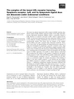
As shown in Fig 1A, the expression of
LOC101926975 was significantly reduced in HSCR
compared with the corresponding control tissues.
Numerous studies have shown lncRNAs also can act
as biomarkers of diseases. Thus, we used ROC curve
to assess the capacity of LOC101926975 distinguishing
HSCR from normal tissues (Fig 1B). The area under
the ROC curve was 0.900 with the cut off value of
0.1162 and 0.1288. The result shows that
LOC101926975 has the potential diagnostic value.
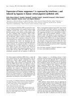
LOC101926975 knockdown inhibits cell
proliferation and causes G1 arrest
To investigate the function of LOC101926975 in
vitro, we used short interfering RNAs (siRNAs) to
reduce the expression of LOC101926975 in
SK-N-BE(2) cells. The siRNA could effectively reduce
the LOC101926975 expression level (Fig 2A). The
phenotype changes induced by LOC101926975
knockdown indicated that the low expression of
LOC101926975 significantly suppressed the cell
proliferation compared with the control cells (Fig 2B).
Meanwhile, flow cytometry analysis revealed that
LOC101926975 downregulation blocked the G0/G1 to
S phase transition (Fig 2C). However, no influence
was found on cell migration and apoptosis with the
siRNA treatment (Fig 2D, E).
LOC101926975 may regulate the expression of
FGF1
To explore the potential mechanism of
LOC101926975 regulating biological process, we
focused on FGF1 due to its near location on
chromosome. FGF1 is a member of the fibroblast
growth factor family, which plays key roles in cell
proliferation and embryonic development [9]. We
found that the expression of FGF1 was also low in
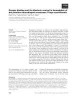
HSCR cases (Fig 3A). The correlation analysis showed
that the association between FGF1 and LOC101926975
was evident in both controls and cases with the r
value of 0.9844 and 0.9804 respectively and p value
<0.0001 (Fig 3B, C). And the expression of FGF1 in
LOC101926975 knockdown cells was lower than the
control according to the results of qRT-PCR (Fig 3D).
All above, hinted that LOC101926975 might regulate
the expression of FGF1 and thus participated in
HSCR.
Figure 1. Expression of LOC101926975 in HSCR. A. LOC101926975 was significantly downregulated in HSCR tissues compared control samples. B. Receiver
Operating Characteristic (ROC) curve for the LOC101926975 to distinguish HSCR cases from controls. * indicates significant difference (p<0.05)
Int. J. Med. Sci. 2016, Vol. 13
295
Figure 3 Relationship between FGF1 and LOC101926975. The expression of FGF1 was lower in HSCR tissues (A) and was correlated with the expression
of LOC101926975 in control samples (B), HSCR tissues (C) and cells (D). * indicates significant difference (p<0.05)
Figure 2 Function of LOC101926975 in vitro. A. LOC101926975 was effectively knocked down in SK-N-BE(2) cells. Downregulation of LOC101926975
suppressed cell proliferation (B) and caused cell cycle arrest (C) without impact on cell apoptosis (D) or cell migration. Pictures were captured under a light
microscope with the magnification, x20 (E). * indicates significant difference (p<0.05)
Int. J. Med. Sci. 2016, Vol. 13
Discussion
HSCR characterized by the absence of enteric
neurons in the distal gut is one of the most common
digestive diseases in the newborn. The main clinical
symptoms are abdominal distension and constipation.
Untreated HSCR is a fatal disease especially with
enterocolitis [10]. Roughly estimated initial costs for
neonatal with HSCR is $100,000 in the United States
[11]. However, we still cannot interpret clearly the
genetic factors or environmental underpinnings of
HSCR less to say clinical application of replacing
enteric nervous system [12]. Thus, it is important to
explore the pathogenesis of HSCR.
LncRNAs have been demonstrated to play key
roles in numerous biological processes and diseases
[13-15]. In this study, we investigate the functional
performance
of
LOC101926975
in
HSCR.
LOC101926975 is significantly downregulated in
HSCR tissues with FGF1 which is near to this lncRNA
on chromosome. Results in vitro show that
LOC101926975 impacts cell proliferation and cell
cycle without influencing cell migration or apoptosis.
FGF1 is a well characterized member of fibroblast
growth factor family. Dysregulation of FGF1 is
involved in cell proliferation, migration, cell arrest
and apoptosis by interacting with FGF receptors
[16-19]. LncRNAs have been reported to affect the
expression of neighboring genes positively or
negatively namely cis regulation[5]. And several
known lncRNAs such as Xist and Air can regulate
nearby or and distantly located genes by interacting
with histone modification complexes [20, 21].
Therefore, we wonder that if LOC101926975 can
regulate FGF1 expression. And we find that the
expression of FGF1 is correlated with LOC101926975
in both cells and population samples. However, there
is no difference in migration or apoptosis when FGF1
is knocked down in our study. It seems inconsistent
with previous studies, which hints LOC101926975
may also affect other genes in addition to FGF1. And
further validation is needed to confirm the specific
regulation mechanism of LOC101926975 for FGF1.
LncRNAs also can act as biomarkers in
numerous diseases especially cancers[22]. For
instance,
RP11–160H22.5,
XLOC_014172
and
LOC149086 are related to hepatocellular carcinoma
[23]. Traditional biomarkers are mostly blood-based,
which may influence the stability and sensitivities of
results [24]. In this study, we attempt to evaluate the
diagnostic value of LOC101926975 in tissue samples.
And LOC101926975 is significantly downregulated in
HSCR tissues with the AUC of 0.900, which implies
that LOC101926975 can effectively distinguish HSCR
cases from control samples.
296
In
conclusion,
we
demonstrate
that
LOC101926975 expression is downregulated in HSCR
tissues. Dysregulation of LOC101926975 can impact
cell proliferation as well as cell cycle and serve as
biomarker for HSCR. However, further study is still
needed to confirm the result and explain the
molecular mechanisms.
Abbreviations
HSCR: Hirschsprung disease; LncRNA: Long
non-coding RNA; ROC: Receiver operating
characteristic; NCCs: neural crest cells.
Acknowledgements
We thank Dr. Jie Zhang, HuanChen and
Changgui Lu (Nanjing Children’s Hospital Affiliated
to Nanjing Medical University) for sample collection.
This study was supported by Natural Science
Foundation of China (NSFC 81370473), Natural
Science Foundation of China (NSFC 81400574),
Natural
Science
Foundation
of
China
(NSFC 81570467), Natural Science Foundation of
Jiangsu Province of China (BK20131388), and Priority
Academic Program Development of Jiangsu Higher
Education Institutions (PAPD). Competing Interests:
the authors have no competing interests.
Competing Interests
The authors have declared that no competing
interest exists.
References
1.
2.
3.
4.
5.
6.
7.
8.
9.
10.
11.
12.
13.
Amiel J, Sproat-Emison E, Garcia-Barcelo M, Lantieri F, Burzynski G, Borrego
S, et al. Hirschsprung disease, associated syndromes and genetics: a review. J
Med Genet. 2008; 45: 1-14.
Lake JI, Heuckeroth RO. Enteric nervous system development: migration,
differentiation, and disease. Am J Physiol Gastrointest Liver Physiol. 2013; 305:
G1-24.
Wallace AS, Anderson RB. Genetic interactions and modifier genes in
Hirschsprung's disease. World J Gastroenterol. 2011; 17: 4937-44.
Kaikkonen MU, Lam MT, Glass CK. Non-coding RNAs as regulators of gene
expression and epigenetics. Cardiovasc Res. 2011; 90: 430-40.
Batista PJ, Chang HY. Long noncoding RNAs: cellular address codes in
development and disease. Cell. 2013; 152: 1298-307.
Geisler S, Coller J. RNA in unexpected places: long non-coding RNA functions
in diverse cellular contexts. Nat Rev Mol Cell Biol. 2013; 14: 699-712.
Ponting CP, Oliver PL, Reik W. Evolution and functions of long noncoding
RNAs. Cell. 2009; 136: 629-41.
Xie H, Zhu D, Xu C, Zhu H, Chen P, Li H, et al. Long none coding RNA
HOTTIP/HOXA13 act as synergistic role by decreasing cell migration and
proliferation in Hirschsprung disease. Biochem Biophys Res Commun. 2015;
463: 569-74.
Raju R, Palapetta SM, Sandhya VK, Sahu A, Alipoor A, Balakrishnan L, et al. A
Network Map of FGF-1/FGFR Signaling System. J Signal Transduct. 2014;
2014: 962962.
Momoh JT. Hirschsprung's disease: problems of diagnosis and treatment. Ann
Trop Paediatr. 1982; 2: 31-5.
Shinall MC, Jr., Koehler E, Shyr Y, Lovvorn HN, 3rd. Comparing cost and
complications of primary and staged surgical repair of neonatally diagnosed
Hirschsprung's disease. J Pediatr Surg. 2008; 43: 2220-5.
Hotta R, Natarajan D, Thapar N. Potential of cell therapy to treat pediatric
motility disorders. Semin Pediatr Surg. 2009; 18: 263-73.
Ellis BC, Molloy PL, Graham LD. CRNDE: A Long Non-Coding RNA
Involved in CanceR, Neurobiology, and DEvelopment. Front Genet. 2012; 3:
270.
Int. J. Med. Sci. 2016, Vol. 13
297
14. Lin N, Chang KY, Li Z, Gates K, Rana ZA, Dang J, et al. An evolutionarily
conserved long noncoding RNA TUNA controls pluripotency and neural
lineage commitment. Mol Cell. 2014; 53: 1005-19.
15. Yang M, Zhai X, Xia B, Wang Y, Lou G. Long noncoding RNA CCHE1
promotes cervical cancer cell proliferation via upregulating PCNA. Tumour
Biol. 2015.
16. Hossain WA, Morest DK. Fibroblast growth factors (FGF-1, FGF-2) promote
migration and neurite growth of mouse cochlear ganglion cells in vitro:
immunohistochemistry and antibody perturbation. J Neurosci Res. 2000; 62:
40-55.
17. Murphy M, Drago J, Bartlett PF. Fibroblast growth factor stimulates the
proliferation and differentiation of neural precursor cells in vitro. J Neurosci
Res. 1990; 25: 463-75.
18. Cassina P, Pehar M, Vargas MR, Castellanos R, Barbeito AG, Estevez AG, et al.
Astrocyte activation by fibroblast growth factor-1 and motor neuron
apoptosis: implications for amyotrophic lateral sclerosis. J Neurochem. 2005;
93: 38-46.
19. Browaeys-Poly E, Perdereau D, Lescuyer A, Burnol AF, Cailliau K. Akt
interaction with PLC(gamma) regulates the G(2)/M transition triggered by
FGF receptors from MDA-MB-231 breast cancer cells. Anticancer Res. 2009; 29:
4965-9.
20. Zhao J, Ohsumi TK, Kung JT, Ogawa Y, Grau DJ, Sarma K, et al. Genome-wide
identification of polycomb-associated RNAs by RIP-seq. Mol Cell. 2010; 40:
939-53.
21. Nagano T, Mitchell JA, Sanz LA, Pauler FM, Ferguson-Smith AC, Feil R, et al.
The Air noncoding RNA epigenetically silences transcription by targeting G9a
to chromatin. Science. 2008; 322: 1717-20.
22. Yang Y, Shao Y, Zhu M, Li Q, Yang F, Lu X, et al. Using gastric juice
lncRNA-ABHD11-AS1 as a novel type of biomarker in the screening of gastric
cancer. Tumour Biol. 2015.
23. Tang J, Jiang R, Deng L, Zhang X, Wang K, Sun B. Circulation long non-coding
RNAs act as biomarkers for predicting tumorigenesis and metastasis in
hepatocellular carcinoma. Oncotarget. 2015; 6: 4505-15.
24. Liu HS, Xiao HS. MicroRNAs as potential biomarkers for gastric cancer. World
J Gastroenterol. 2014; 20: 12007-17.