Synergistic antitumor effect of sorafenib in combination with ATM inhibitor in hepatocellular carcinoma cells
Bạn đang xem bản rút gọn của tài liệu. Xem và tải ngay bản đầy đủ của tài liệu tại đây (1.58 MB, 7 trang )
Int. J. Med. Sci. 2017, Vol. 14
Ivyspring
International Publisher
523
International Journal of Medical Sciences
2017; 14(6): 523-529. doi: 10.7150/ijms.19033
Research Paper
Synergistic Antitumor Effect of Sorafenib in
Combination with ATM Inhibitor in Hepatocellular
Carcinoma Cells
Jianhua Liu1, Yahui Liu1, Lingyu Meng1, Bai Ji1, Daqing Yang2
1.
2.
Department of Hepatobiliary and Pancreatic Surgery, the First Hospital of Jilin University, Changchun 130021, China;
The Hormel Institute, University of Minnesota, Austin, MN 55912, USA.
Corresponding author: Bai Ji, MD, Department of Hepatobiliary and Pancreatic Surgery, the First Hospital of Jilin University, Changchun 130021, China (Tel:
86-431-81875160; Email:)
© Ivyspring International Publisher. This is an open access article distributed under the terms of the Creative Commons Attribution (CC BY-NC) license
( See for full terms and conditions.
Received: 2017.01.03; Accepted: 2017.03.05; Published: 2017.04.09
Abstract
Background: Currently, sorafenib is the only systemic chemotherapy drug for advanced stage
Hepatocellular carcinoma (HCC). However, emerging data from some clinical HCC patients
indicate that sorafenib alone has only moderate antitumor efficacy, and could not inhibit disease
metastasis and progression. KU-55933 is a specific ATM inhibitor, which has pro-apoptotic effect
on tumor cells. In this study, we analyzed the synergistic effect of sorafenib and KU-55933 on the
proliferation of HCC cell lines.
Methods: Three HCC cell lines were treated with sorafenib and KU-55933 alone or combination
in vitro to investigate inhibitory effect by MTT and wound healing assay. Epithelial to mesenchymal
transition (EMT) phenotype change was investigated after sorafenib and KU-55933 treatment by
microscopy. Akt signaling pathway proteins including p-Akt, p-mTOR and p-p70S6K were
examined by western blot. In addition, cleaved PARP and autophage-related proteins LC3A/B
were detected by western blot.
Results: KU-55933 can enhance the effect of sorafenib in inhibiting cell proliferation and
migration, overcoming EMT, inducing cell apoptosis via inactivating Akt signaling pathway and
inducing autophage. The combination treatment with sorafenib and KU-55933 resulted in a strong
synergistic effect in vitro.
Conclusion: Our results demonstrate that sorafenib combined with KU-55933 treatment does
effectively inhibit proliferation of HCC cell lines synergistically. These data suggests that KU-55933
may be a promising chemosensitizer to sorafenib in the treatment of HCC.
Key words: Hepatocellular carcinoma; sorafenib; KU-55933; EMT; migration; autophage.
Introduction
Hepatocellular carcinoma (HCC) is the third
leading cause of cancer-related death worldwide,
with an increasing incidence in the United States and
China [1, 2]. In China, HCC commonly arises in
patients with chronic liver diseases. Only early stage
HCC patients are applicable to potentially curative
therapies, such as surgical resection and liver
transplantation. Today, the multi-kinase inhibitor
sorafenib is the only systemic therapy to improve
survival in those patients with advanced HCC [3, 4].
However, some patients show nature or acquired
resistance to it. Therefore, prognosis of advanced
HCC remains poor, and new effective therapeutic
strategies are urgently needed. To find efficient
targets, a number of large-scale molecular studies
have been conducted in HCC, including Akt [5].
The AKT/mTOR signaling pathway is a
promising target with respect to its frequent
Int. J. Med. Sci. 2017, Vol. 14
dysregulation in HCC and its key role in regulating
cell
proliferation,
migration,
survival
and
angiogenesis [6, 7]. Aberrant Akt signaling has been
detected in nearly half of hepatocellular carcinoma,
and a correlation between poor outcome and Akt
signaling activation has been shown [8]. ATM, a
protein deficient in patients with ataxia-telangiectasia
disease, functions as a signal transducer in response to
DNA damage [9]. It has recently been shown that
ATM is also a cytoplasmic protein that mediates the
full activation of Akt in response to insulin [10]. Li Y,
et al [11] reported that a specific ATM inhibitor,
KU-55933, blocks the phosphorylation of Akt induced
by insulin in cancer cells that exhibit abnormal Akt
activity. Moreover, KU-55933 inhibits cancer cell
proliferation by inducing G1 cell cycle arrest In
addition, KU-55933 treatment during serum
starvation triggers apoptosis in these cancer cells.
Furthermore, Li et al reported that combination of
KU-55933 and rapamycin not only induces apoptosis,
which is not seen in cancer cells treated only with
rapamycin, but also shows better efficacy in inhibiting
cancer cell proliferation than each drug alone. Based
on this data, we hypothesize KU-55933 can enhance
the effect of sorafenib.
Currently, sorafenib plays a critical role in
treating patients with advanced stage HCC,
contributing to an improved overall survival in
treated patients in clinical trials [12, 13].
Unfortunately, some patients don’t benefit from the
treatment. Therefore, it is imperative to investigate the
potential molecular mechanisms which lead to low
survival benefits to help develop potential strategies
aimed at increasing its efficacy against HCC. In this
study, we show that ATM inhibitor can enhance
sorafenib-induced apoptosis through downregulation
of p-Akt (Thr308), p-mTOR and p-p70S6K and
upregulation of cleaved PARP and LC3A/B II. In
addition, they present a synergistic effect in inhibiting
migration and EMT. These results suggest that
KU-55933 may be a novel chemosensitizer to increase
chemotherapeutic sensitivity of sorafenib on HCC
cells.
Materials and methods
Chemicals and antibodies
Sorafenib purchased from Santa Cruz Co.
KU-55933 was purchased from Calbiochem. They
were both dissolved in DMSO to prepare the stock
solution of 20mM and stored in aliquots at -20℃.
Antibodies against PARP, LC3A/B, phospho-Akt
(Thr308), phospho-mTOR, phospho-p70s6k (Thr 389),
and β-actin were purchased from cell signaling
Technology.
524
Cell lines and culture conditions
Hepatocellular carcinoma cell lines, HepG2,
Huh7 and Hep3B purchased from ATCC were
cultured in DMEM supplemented (Hyclone, Logan,
UT, USA) with 10% FBS (Hyclone, Logan, UT, USA)
and 1% of penicillin-streptomycin at 37℃, in
humidified air containing 5% CO2.
MTT Cell Proliferation Assay
Cells were seeded in a 48-well plate and
incubated overnight. Following treatment with
sorafenib and/or KU-55933, the viable cells in each
well
were
determined
using
a
CellTiter
Nonradioactive cell proliferation assay kit (Promega)
following the manufacturer’s instructions. Briefly,
MTS dye solution in the kit was added to each well
and incubated at 37℃ for 4 h. The absorbance at
490nm was recorded by a microplate reader.
Western blot
Cells were lysed with TGN lysis buffer
containing protease inhibitor cocktails (Roche). The
protein concentration was measured by the Lowry
method. Equal amounts of protein were subjected to
SDS-PAGE and then transferred to a nitrocellulose
membrane. Primary antibody was added in milk and
allowed to incubate overnight at 4℃, washed with
TBST for 3 times (5 min per time) before the secondary
antibody was added and then incubated for an hour
at room temperature. The membrane was again
washed 3 times before adding SuperSignal West Pico
Chemiluminescent Substrate (Thermo Scientific, IL,
USA) and then immediately developed by
chemiluminescence.
Cell migration
A total of 200000 cells were seeded onto a
six-well plate and allowed to reach full confluence.
The monolayer was wounded using a 200µL tip. Cells
were incubated with medium containing sorafenib
and KU-55933 alone or combination. Digital images
were taken at times of 0h and 48h. The results are
representative of three individual experiments.
EMT phenotype change
A total of 40000 cells were seeded onto a six-well
plate and incubated overnight. Following TGF-β1,
KU-55933 and sorafenib treatment, digital images
were taken at times of 48h. The results are
representative of three individual experiments.
Statistical analysis
All data were presented as mean±SD. Student’s
t-Test (unpaired, 2-tailed) was used for comparison
between two groups. One-way ANOVA was used to
Int. J. Med. Sci. 2017, Vol. 14
525
compare difference of multiple groups. P value less
than 0.05 was considered statistically significant.
KU-55933 inhibit cell proliferation in a synergistic
manner (Figure 2).
Results
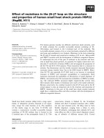
Sorafenib inhibits HCC cell lines proliferation
In order to determine the 50% of inhibitory
concentration (IC50) of sorafenib on three HCC cell
lines, we analyzed the effects of the sorafenib on the
HCC cell lines using MTT approach. The cell lines
were exposed to sorafenib (0 µM, 2.5 µM, 5 µM, 10
µM, 20 µM), and cell viability was determined by MTS
solution after 72h. In the experiment, we found that
sorafenib led to a dose-dependent inhibition on cell
proliferation (Figure 1), and IC50 was shown in
Table 1.
Figure 1. MTT proliferation assays. After treatment with sorafenib at
concentrations ranging from 0 to 20 µM on HCC cell lines HepG2, Huh7 and
Hep3B for 72h, cell viability was determined using MTS dye solution. Results are
presented as the median of 3 independent experiments.
Table 1. Inhibitory concentration 50% (IC50) of sorafenib
cells
IC50(μM)
HepG2
7.42
Huh7
5.97
Hep3B
3.31
KU-55933 and sorafenib inhibits hepatocellular
carcinoma cell lines proliferation
synergistically
In order to examine synergistic effect of
KU-55933 and sorafenib in vitro, we analyzed the
effects of two drugs on three different HCC cell lines
using MTS approach. The cell lines were grown in
24-well plate and was exposed to KU-55933(10
µmol/L), sorafenib (5 µmol/L) and combination
respectively. 72h later, cell viability was examined by
MTS. In the experiment we found that sorafenib and
Figure 2. KU-55933 and sorafenib inhibit the proliferation of HepG2, Huh7
and Hep3B cells in a synergistic manner. Cells were seeded in a 24-well plate
and were then treated with KU-55933 (10 µmol/L) and sorafenib (5 µmol/L)
alone or combination for 3 d. The cell proliferation rate in each well was
determined with the CellTiter 96 MTT cell proliferation assay kit (Promega)
following the manufacturer’s instructions. Columns, mean of absorbance from
three separate experiments (*,p<0.05;**, p<0.01; ***, p<0.001);bars , SD.
Int. J. Med. Sci. 2017, Vol. 14
Scratch wound healing assay
Cell scratch assay was used to analyze whether
KU-55933 and sorafenib inhibit the migration of HCC
cells. As shown in Figure 3, cell free area of the
combination treatment was wider than that of
526
single-drug treatment, and treatment groups were
wider than that of control in three cell lines at 48
hours. This demonstrates that KU-55933 and
sorafenib inhibit migration of three HCC cell lines in a
synergistic manner.
Figure 3. Sorafenib (5 µmol/L) and KU-55933 (10 µmol/L) showed synergistic effect in inhibiting migration in wound healing assays. Images show the gap change of
the scratched region of the different groups. Magnification of 25x.
Int. J. Med. Sci. 2017, Vol. 14
33 and sorafenib reverse EMT phenotype
change induced by TGF-β1 in HCC cell lines
TGF treatment alone led to phenotype change
from epithelium-like morphology to mesenchymal
phenotype and grew separately in a more aggressive
manner, which demonstrates that cells present
metastatic and invasive characteristics. Sorafenib and
KU-55933 combination can reverse EMT change more
obviously than single treatment, and re-acquire the
epithelium-like phenotype and proliferation in
clusters (Figure 4). This revealed that sorafenib and
KU-55933 can inhibit HCC cells metastasis and
invasion synergistically. The results are representative
of three individual experiments.
Combination of KU-55933 and sorafenib
induces stronger apoptosis in HCC cells by
inhibiting PI3K/Akt pathway activation and
inducing autophage
All three HCC cell lines were incubated with
KU-55933 (10 µmol/L) and sorafenib (5 µmol/L)
alone or combination for 24h, and the levels of p-Akt,
p-mTOR and p-p70S6k which is a downstream target
of Akt and cleaved PARP were detected. Our data
527
showed that KU-55933 or sorafenib treatment not only
resulted in reduced Akt phosphorylation at Thr308
and p70S6K phosphorylation, but also caused a
dramatic increase in cleaved PARP levels (Fig. 5A). In
addition, results in Fig. 5 indicate that inhibition of
Akt and p70S6 kinase phosporylation caused by
sorafenib in HCC cells was completely stronger than
that is induced by KU-55933. And cells with
combination treatment showed the lowest level of
p-Akt, p-mTOR and p-p70S6K, which demonstrates
that KU-55933 can work as chemosensitizer to
sorafenib. Furthermore, although KU-55933 alone
failed to induce apoptosis of Huh7 cells, cells treated
with KU-55933 plus sorafenib present increased
apoptosis by upregulation the cleaved PARP
compared with sorafenib alone treatment. These
results were also confirmed by an autophage related
protein detection. Here, western blotting analysis of
the expression of key autophagic proteins showed
that both KU-55933 and sorafenib increased, and in
combination further increased the expression of
LC3A/B-II in three HCC cell lines. All these data
suggest a synergistic effect of KU-55933 and sorafenib.
Figure 4. The effect of sorafenib and KU-55933 treatment on cell phenotype and marker change.cell phenotype changes resulting from different treatment with
TGF-β1 (10 ng/ml), sorafenib (5 µmol/L) and KU-55933 (10 µmol/L). Magnification of 100x. Con, control; T, TGF-β1; KU, KU-55933; sora, sorafenib. The results are
representative of three individual experiments.
Int. J. Med. Sci. 2017, Vol. 14
528
Figure 5. Sorafenib and KU-55933 inhibit cell proliferation and induce apoptosis by inactivating Akt signaling pathway in a synergistic manner. Both floating and
attached cells were collected after treatment. Cells were lysed by TGN lysis buffer, and cell lysates were subjected to SDS-PAGE and immunoblotting. PARP, cleaved
PARP, phospho-Akt at Thr308, phosph-mTOR, phospho-p70S6K, LC3A/B and β-actin were detected. A.B.C represent results of HepG2, Huh7 and Hep3B cell lines
respectively treated by sorafenib (5 µmol/L) and KU-55933(10 µmol/L) alone or combination for 24h, β-actin was detected as a control. The results in A to C are
representative of three individual experiments.
Discussion
Hepatocellular carcinoma is the third leading
cause of cancer -related mortality worldwide. Surgical
resection may provide curative treatment for patients
with early stage HCC. Once the cancer becomes the
advanced stage, sorafenib is the only systemic
chemotherapeutic drug to postpone survival time
because patients have lost the opportunity for
curative therapies [3, 4]. However, studies of HCC cell
lines have revealed that it is not fully effective in
preventing recurrence and progression because of
resistance [14]. Therefore, many research groups focus
on the molecular mechanisms in sorafenib resistance
in search of the established therapeutic agents that can
overcome the resistance of HCC cells, and help
develop potential strategies aimed at increasing its
efficacy against HCC [15, 16].
Akt is a major component of the
phosphoinositide 3-kinase (PI3K) signaling pathway.
In normal cells, Akt acts as a single transducer of PI3K
and promotes cell proliferation and cell survival [17].
However, upregulation of Akt leads to the
development of cancer [18]. Therefore, it is a
significant target in search for drugs that can be used
as chemotherapeutic agents for cancer [19]. In our
work, treatment with sorafenib and KU-55933 alone
or combination results in decreased expression of
p-Akt and downstream proteins, p-mTOR and
p-p70S6K.
KU-55933 is a specific inhibitor of the ATM
kinase [20]. Li et al [11] found that it can inhibit cell
proliferation and induce apoptosis via preventing the
activation of Akt and block the function of its
downstream substrates. However, there is no report
regarding its effect on HCC cells. In this study, we
showed that KU-55933 alone treatment had only a
minor effect on proliferation of three HCC cell lines,
and sorafenib alone treatment had a moderate
inhibition effect. However, combined sorafenib with
KU-55933 treatment led to a strong synergistic effect
on proliferation of HCC cells. Moreover, the same
synergistic effect was shown on apoptosis, migration
and EMT, which plays a key role in cancer
progression and resistance to different therapeutic
approaches. EMT data demonstrated that sorafenib
and KU-55933 treatment revert mesenchymal change
induced by TGF-β1 to epithelium cells. Further, The
mechanism of synergistic effect of sorafenib and
ku55933 in HCC cells was analyzed by western blot
and the results showed that the expression of cleaved
PARP and LC3A/B II were increased while p-Akt,
p-p70S6K and p-mTOR were strongly decreased after
treated with sorafenib combined with KU-55933, and
facilitate the sorafenib-induced apoptosis in HCC cell
lines. These data demonstrate that sorafenib and
KU-55933 combination treatment exerts anticancer
effect on HCC via inhibiting Akt signaling pathway
and inducing autophage. We also found that
sorafenib led to a dose-dependent cell apoptosis.
Therefore, we hypothesize that KU-55933 may be a
chemosensitizer to sorafenib for advanced HCC
patients. This study has therefore provided a
framework for the development of sorafenib-based
combination therapies for HCC.
Int. J. Med. Sci. 2017, Vol. 14
Conclusion
In conclusion, the results of this study
demonstrate that combining sorafenib and KU-55933
shows a synergistic effect in HCC cell lines. Therefore,
this combination treatment strategy may be a
promising treatment option for patients with
advanced HCC since KU-55933 is already being tested
in clinical trials and reported to be well tolerated.
529
20. Hickson I, Zhao Y, Richardson CJ, et al. Identification and characterization of a
novel and specific inhibitor of the ataxia-telangiectasia mutated kinase ATM.
Cancer Res. 2004; 64: 9152-9.
Acknowledgments
This work was in part supported by grants from
Foundation of Jilin Provincial Development and
Reform Commission (KY20160002, No. 3J115AJ73428 )
and Jilin University Research Fund for Excellent
Young teachers (No.419080500355 ).
Competing Interests
The authors have declared that no competing
interest exists.
References
1.
2
3.
4.
5.
6.
7.
8.
9.
10.
11.
12.
13.
14.
15.
16.
17.
18.
19.
Wang S, Sun H, Xie Z, et al. Improved survival of patients with hepatocellular
carcinoma and disparities by age, race, and socioeconomic status by decade,
1983-2012. Oncotarget. 2016 , doi: 10.18632/oncotarget.10930.
Altekruse SF1, McGlynn KA, Reichman ME. Hepatocellular carcinoma
incidence, mortality, and survival trends in the United States from 1975 to
2005. J Clin Oncol. 2009; 27:1485-91.
Finn RS, Poon RT, Yau T, et al. Phase I study of everolimus in combination
with sorafenib in patients with advanced hepatocellular carcinoma (HCC). J
Clin Oncol. 2011; 29(15_suppl): 4074.
Llovet JM, Hernandez-Gea V. Hepatocellular carcinoma: reasons for phase III
failure and novel perspectives on trial design. Clin Cancer Res. 2014;20:2072-9.
Grabinski N, Ewald F, Hofmann BT, et al. Combined targeting of AKT and
mTOR synergistically inhibits proliferation of hepatocellular carcinoma cells.
Mol Cancer. 2012;11:85.
Engelman JA. Targeting PI3K signaling in cancer: opportunities, challenges
and limitations. Nat Rev Cancer. 2009;9:550-62.
Baik SH, Lee J, Lee YS, et al. ANT2 shRNA downregulates miR-19a and
miR-96 through the PI3K/Akt pathway and suppresses tumor growth in
hepatocellular carcinoma cells. Exp Mol Med. 2016;48:e222.
Villanueva A, Chiang DY, Newell P, et al. Pivotal role of mTOR signaling in
hepatocellular carcinoma. Gastroenterology. 2008; 135:1972-83. .
Shiloh Y, Kastan MB.ATM: genome stability, neuronal development, and
cancer cross paths. Adv Cancer Res. 2001;83:209-54.
Viniegra JG1, Martínez N, Modirassari P, et al. Full activation of PKB/Akt in
response to insulin or ionizing radiation is mediated through ATM. J Biol
Chem. 2005;280:4029-36.
Li Y, Yang DQ. The ATM inhibitor KU-55933 suppresses cell proliferation and
induces apoptosis by blocking Akt incancer cells with overactivated Akt. Mol
Cancer Ther. 2010; 9:113-25.
Llovet JM, Ricci S, Mazzaferro V, et al. Sorafenib in advanced hepatocellular
carcinoma. N Engl J Med. 2008 ;359:378-90
Cheng AL, Kang YK, Chen Z, et al. Efficacy and safety of sorafenib in patients
in the Asia-Pacific region with advanced hepatocellular carcinoma: a phase III
randomised, double-blind, placebo-controlled trial. Lancet oncol. 2009;
10:25-34.
Liu J, Cui X, Qu L, et al. Overexpression of DLX2 is associated with poor
prognosis and sorafenib resistance in hepatocellular carcinoma. Exp Mol
Pathol. 2016;101:58-65
Tang S, Tan G, Jiang X, et al. An artificial lncRNA targeting multiple miRNAs
overcomes sorafenib resistance in hepatocellular carcinoma cells.
Oncotarget. 2016; doi: 10.18632/oncotarget.12304.
Nishida N, Kitano M, Sakurai T, et al. Molecular Mechanism and Prediction
of Sorafenib Chemoresistance in Human Hepatocellular Carcinoma. Dig
Dis. 2015; 33:771-9.
West KA, Castillo SS, Dennis PA. Activation of the PI3K/Akt pathway and
chemotherapeutic resistance. Drug Resist Updat. 2002;5:234-48
Nicholson KM1, Anderson NG. The protein kinase B/Akt signalling pathway
in human malignancy. Cell Signal. 2002;14:381-95.
Wang XJ, Feng CW, Li M. ADAM17 mediates hypoxia-induced drug
resistance in hepatocellular carcinoma cells through activation of
EGFR/PI3K/Akt pathway. Mol Cell Biochem. 2013; 380(1-2):57-66.