The relationship between follicle-stimulating hormone and bone health: Alternative explanation for bone loss beyond oestrogen
Bạn đang xem bản rút gọn của tài liệu. Xem và tải ngay bản đầy đủ của tài liệu tại đây (496.4 KB, 11 trang )
Int. J. Med. Sci. 2018, Vol. 15
Ivyspring
International Publisher
1373
International Journal of Medical Sciences
2018; 15(12): 1373-1383. doi: 10.7150/ijms.26571
Review
The Relationship between Follicle-stimulating Hormone
and Bone Health: Alternative Explanation for Bone Loss
beyond Oestrogen?
Kok-Yong Chin
Department of Pharmacology, Faculty of Medicine, Universiti Kebangsaan Malaysia Medical Centre, Malaysia.
Corresponding author: Department of Pharmacology, Level 17, Preclinical Building, Universiti Kebangsaan Malaysia Medical Centre, Jalan Yaacob Latif,
Bandar Tun Razak, 56000 Cheras, Kuala Lumpur, Malaysia. Tel: +603 9145 9573; Fax: +603 9145 9547; Email:
© Ivyspring International Publisher. This is an open access article distributed under the terms of the Creative Commons Attribution (CC BY-NC) license
( See for full terms and conditions.
Received: 2018.04.08; Accepted: 2018.08.27; Published: 2018.09.07
Abstract
Bone loss in women commences before the onset of menopause and oestrogen deficiency. The
increase of follicle-stimulating hormone (FSH) precedes oestrogen decline and may be a cause for
bone loss before menopause. This review summarizes the current evidence on the relationship
between FSH and bone derived from cellular, animal and human studies. Cellular studies found that
FSH receptor (FSHR) was present on osteoclasts, osteoclast precursors and mesenchymal stem
cells but not osteoblasts. FSH promoted osteoclast differentiation, activity and survival but exerted
negligible effects on osteoblasts. Transgenic FSHR or FSH knockout rodents showed heterogenous
skeletal phenotypes. Supplementation of FSH enhanced bone deterioration and blocking of FSH
action protected bone of rodents. Human epidemiological studies revealed a negative relationship
between FSH and bone health in perimenopausal women and elderly men but the association was
attenuated in postmenopausal women. In conclusion, FSH may have a direct action on bone health
independent of oestrogen by enhancing bone resorption. Its effects may be attenuated in the
presence of overt sex hormone deficiency. More longitudinal studies pertaining to the effects of FSH
on bone health, especially on fracture risk, should be conducted to validate this speculation.
Key words: follicotropin; gonadotropins; menopause; osteopenia; osteoporosis; skeleton.
Introduction
Accelerated bone loss in women during
menopausal transition is often attributed to oestrogen
deficiency. However, the Study of Women’s Health
Across the Nation (SWAN) involving women from
various ethnic groups showed negligible changes in
the bone mineral density (BMD) in pre- and early
perimenopausal women. Significant bone loss was
observed in late perimenopausal women (0.018 and
0.010 g/cm2 yearly at the spine and hip) and the rate
increased in postmenopausal women (0.022 and 0.013
g/cm2 yearly at the spine and hip) [1]. On the other
hand, the decline of oestrogen level transpires very
late during perimenopause but the gonadotropin
levels, especially follicle-stimulating hormone (FSH),
gradually increase 5-6 years before menopause [2, 3].
Thus, oestrogen deficiency alone may not explain the
accelerated bone loss during this period.
Although ovariectomy in female rodents
invariably causes a reduction in bone mass, studies
that inhibit the function or knockout the oestrogen
receptors (ERs) in rodents showed contradictory
results. Ogawa et al. (2000) generated a rat model with
a dominant negative ERα which inhibited both ERα
and ERβ. The transgenic rats showed similar BMD
with the wildtypes. After ovariectomy, the transgenic
rats also showed a similar degree of bone loss
compared with the wildtype, and the condition could
not be reversed with oestrogen replacement [4]. In
another study, ERβ knockout female mice showed
increased bone mineral content at the cortical bone
Int. J. Med. Sci. 2018, Vol. 15
compared to the wildtype at 11 weeks old, as well as
increased trabecular BMD and bone volume at 1 year
old. The ERβ knockout male mice showed similar
skeletal phenotypes as the wildtype [5, 6]. The
importance of oestrogen in maintaining bone health
was further complicated by the fact that
hypophysectomy inhibited high bone turnover
induced by ovariectomy in rats [7]. Subsequently, Sun
et al. (2006) observed that FSH receptor (FSHR)
knockout mice maintained their bone health despite
being hypogonadal [8]. Despite receiving criticisms on
the model used, their research raised the question
whether FSH plays a more vital role in regulating
bone loss among women during menopause
transition period.
The controversy on the role of FSH on bone
metabolism
remains
to-date.
The
studies
aforementioned hint a negative impact of high FSH on
bone health. However, sex hormone deprivation
therapy using gonadotropin agonists for the
treatment of prostate cancer has been shown to induce
bone loss in animals and humans [9-11]. Most
importantly, oestrogen-centric therapies, such as
hormone replacement therapy and selective oestrogen
receptor modulator, have been successful in treating
postmenopausal osteoporosis and preventing
fractures [12-14]. Considering the complexity of this
issue, this review aims to summarize the current
evidence on the skeletal effects of FSH from cellular,
animal and human studies. This issue is relevant
because it can potentially shift the paradigm of an
oestrogen-centric explanation for bone loss during
menopause transition period. It may also offer a new
avenue for the treatment of postmenopausal bone
loss.
Mechanism of FSH action on bone cells
Protein and mRNA expression of FSHR have
been detected in human monocytic cells (sharing the
same lineage with osteoclast), osteoclasts and
mesenchymal stem cells at a concentration lower than
in ovarian samples [8, 15]. However, they were absent
in human osteoblasts [15]. The FSHR expressed in
these cells belonged to the type-2 FSHR isoform and
the expression level was not influenced by sex
hormones [15]. The blocking of FSHR with mono- or
polyclonal antibodies abolished the formation of
osteoclast-like cells from bone marrow macrophages
from mice [16]. Similarly, the promoting effects of
FSH on osteoclast formation was impaired in bone
marrow macrophages from FSHR knockout mice [16].
These studies showed that FSHR is essential for the
action of FSH in promoting osteoclast formation.
Sun et al. (2006) showed that FSH increased
osteoclast differentiation in human peripheral blood
1374
macrophages, mouse bone marrow culture and RAW
cells but did not influence the proliferation of
osteoclast precursors directly [8]. On the other hand,
FSH induced the production of tumour necrosis alpha
(TNFα) in monocytes and bone marrow macrophages
from mice [15, 17], which in turn promoted the
proliferation of osteoclast precursor cells as illustrated
in cellular and in silico studies [17]. Several pathways
related to osteoclast formation in monocytes were
activated by FSH, including osteoclast differentiation
(toll-like receptor and interleukin-1 receptorassociated
kinases),
cell
adhesion,
survival
(anti-apoptotic
TNFs/nuclear
factor-κB/B-cell
lymphoma 2 (BCL-2)) and cytoskeletal remodelling
[15]. FSH promoted the formation of tartrate-resistant
acid
phosphatase
(TRAP)
positive
cells
(osteoclast-like cells) from various types of
macrophages (RAW 264.7 cells, RAW c3 cells, bone
marrow macrophages from mouse) through FSHR
[18]. This process was mediated by the ability of FSH
to activate pathways essential to osteoclastogenesis,
such as phosphorylation of protein kinase B (Akt) and
extracellular-signal-regulated kinase (Erk), and
nuclear translation of c-fos [18]. FSH also increased
the formation of resorption pits and action ring of
osteoclast-like cells, as well as promoted their survival
[18]. This corroborated with the findings of Robinson
et al. (2010) [15]. In short, FSH increases the
proliferation of osteoclast precursors indirectly
through inflammatory cytokines, as well as their
differentiation into mature osteoclasts through direct
interaction with the signalling pathways involved.
Furthermore, it also promotes the bone resorption
activity of osteoclasts.
Apart from TNFα, osteoclast formation also
requires the interaction between receptor activator of
nuclear factor κ-Β (RANK) on osteoclast surface and
RANK ligand (RANKL) secreted by osteoblast.
Cannon et al. (2011) showed that FSH at 50 mIU/ml
(physiological FSH level in perimenopausal women)
promoted the expression of RANK on human CD14+
monocytes [19]. However, at 100 mIU/ml
(physiological FSH level in postmenopausal women),
the effect of FSH was attenuated [19]. Similarly, Wang
et al. (2015) found that with increasing concentration
of FSH, the mRNA expression of RANK increased
concurrently with other markers of osteoclast
differentiation (TRAP, MMP-9 and cathepsin K) in
RAW 264.7 cells [20]. Thus, FSH-induced
osteoclastogenesis may be a result of increased
RANKL-RANK interaction.
Conversely, negative results on the effects of
FSH on osteoclast formation have also been reported.
Ritter et al. (2008) showed that FSH did not affect the
resorption pit area and formation of osteoclasts from
Int. J. Med. Sci. 2018, Vol. 15
human mononuclear cells and RAW cells [21].
However, at 3 µg/ml, FSH decreased the formation of
multinucleated TRAP-positive cells [21]. FSH also did
not affect the gene expression of osteoclast markers,
such as TRAP, calcitonin, MMP-9, aquaporin 9,
V0ATPase, TRAF6 and FSHR. The concentration
difference could contribute to the discrepancy of this
study with the previous ones [21].
Sun et al. (2006) showed that FSH did not
influence the formation of mineralized nodules by
colony forming units in mice bone marrow culture. It
also did not affect the synthesis of RANKL [8]. This
was not surprising since FSHR was absent in
osteoblasts. Since mesenchymal stem cells displayed
FSHR and the differentiation of osteoclasts required
soluble factors from other cells, the indirect action of
FSH in promoting osteoclastogenesis was tested. Sun
et al. (2006) found that coculturing stromal cells,
which was supposed to produce factors stimulating
osteoclast formation, with FSHR-/- macrophages in
the addition of FSH did not stimulate
osteoclastogenesis [8]. Considering all evidence
above, the effects of FSH on osteoclast formation is
direct, without the involvement of osteoblasts or
stromal cells.
Despite the absence of FSHR and the lack of
effects in osteoblasts, FSH could enhance the
1375
osteogenic potential of mouse embryonic fibroblasts,
indicated by increased bone morphogenetic protein 9
(BMP-9) and alkaline phosphatase activity [22].
Combination of FSH and BMP-9 transfection
increased the protein and mRNA expression of
osteoblast markers (osteopontin and osteocalcin) and
matrix mineralization in embryonic fibroblasts [22].
This was mediated by increased phosphorylation
Smad1/5/8 and expression of transcription factors
osterix and runt-related factor-2 essential in osteoblast
formation [22]. When the transfected cells were
injected into the flank of nude mice, they formed a
bony mass [22]. Although being an innovative
therapeutic approach, the use of genetically
manipulated fibroblasts prevents the interpretation of
FSH action on bone formation in normal physiology.
Therefore, the FSH seems to exert a direct effect
on osteoclasts by promoting their formation,
resorption activity and survival through FSHR. FSH
enhances the osteogenic potential of pluripotent stem
cells but its action on osteoblasts remains unclear due
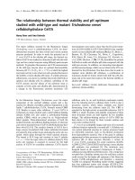
to the absence of FSHR in osteoblasts (Figure 1).
Animal studies
Sun et al. (2006) piloted the study on the skeletal
effects of FSH using FSHR knockout (FSHR-/-) female
mice [8]. These mice were hypogonadal but their bone
Figure 1. The direct effects of FSH on bone cells. FSH increases the expression of RANK and production of TNFα by osteoclast precursors. It also enhanced pathways leading
to osteoclast differentiation. Formation of actin ring and resorption pits increase with FSH. It also prevents the apoptosis of osteoclasts. The effects of FSH on osteoblasts are
not clear. Abbreviation: Akt=protein kinase B; c-FOS=Fos proto-oncogene; Erk=extracellular-signal-regulated kinase; FSH=follicle-stimulating hormone; MMP-9=matrix
metallopeptidase 9; OPG=osteoprotegerin; RANK=receptor activator of nuclear kactor κ B; RANKL= RANK ligand
Int. J. Med. Sci. 2018, Vol. 15
status in terms of areal BMD, femoral trabecular
microstructure and bone remodelling markers were
similar with the wildtype mice [8]. This showed that
eliminating the interaction between FSH and bone
protected bone health in these hypogonadal mice.
Culture of calvarial bone extracted from mice showed
that FSH and RANKL enhanced formation of
TRAP-positive surface in wildtype samples but not in
FSHR-/- samples [8]. This observation was in
accordance with the in vitro osteoclast generation
assays using macrophages. FSHβ knockout mice
(FSHβ-/-) shared similar skeletal features with FSHR-/mice. On the other hand, FSHβ haploinsufficient
(FSHβ+/-) female mice, which were eugonadal, had a
higher femoral BMD but similar lumbar spine,
femoral neck and tibial BMD compared with the
wildtype and FSHβ-/- mice [8]. Femoral cortical
thickness and trabecular structural indices were
higher in the FSHβ+/- mice compared to the wildtype
[8]. This was expected since the presence of oestrogen
and the attenuated FSH interaction provided
protections to the bone of these mice. TRAP-labelled
resorption surfaces and serum TRAP level were
reduced in the FSHβ+/- mice but mineralising surface
and mineral apposition rate were similar in both the
FSHβ+/- mice and the wildtype mice [8]. mRNA
expression of TRAP, cathepsin K and RANK were
reduced in the bone marrow of FSHβ+/- mice
compared to wildtype [8]. This reflects a reduced
osteoclast differentiation in the bone of these mice.
This study received criticism for overlooking the fact
that FSHR-/- female mice had very high testosterone
level, which could be accountable for the observed
bone-sparing phenomenon [23]. The increased
testosterone level was due to the double negative
feedback actions, whereby the pituitary synthesised
higher LH level in the absence of FSH, which in turn
increased the production of testosterone by theca cells
in the ovaries. As a result, raised testosterone level
and uterine degeneration had been observed in
FSHR-/- mice [23-25]. Hence, the use of transgenic
animals cannot fully resolve the skeletal action of FSH
due to changes in the hormonal milieu.
By contrast, Gao et al. (2007) showed that
FSHR-/- mice demonstrated lower femoral and
lumbar spine BMD values starting from month three
of age compared to wildtype [26]. The trabecular bone
volume, osteoblast number, bone formation rate and
mineral apposition rate were also lower in these mice
compared to the wildtype [26]. Concurrently,
osteoclast number, RANKL/OPG number were
significantly higher in FSHR-/- mice compared to the
wildtype [26]. The cause of these degenerative bone
changes was oestrogen deficiency, as ovarian
transplantation prevented the decline in BMD [26].
1376
The high testosterone level apparently did not
prevent bone loss in the FSHR-/- mice. Ovariectomy
reduced BMD and trabecular bone volume, as well as
increased osteoclast surface and RANKL/OPG ratio
in both FSHR-/- and wildtype mice, but only
osteoblast surface, mineralizing surface and bone
formation rate increased in wildtype mice, indicating
a higher bone turnover level [26]. The lack of
osteoblastic response in FSHR-/- mice might suggest
the uncoupling of bone remodelling process, although
previous studies had established that FSH might not
possess direct effects on osteoblasts [8]. Ovariectomy
also eliminated the high circulating testosterone level
in the mice [26]. Blocking the effects of testosterone
using flutamide did not reduce the BMD in FSHR-/mice, but blocking the conversion of testosterone to
oestrogen using letrozole, an aromatase inhibitor, did
[26]. Flutamide did reduce the trabecular bone
volume and increase osteoclast surface in the mice,
while letrozole increased osteoclast surface and
reduced osteoblast surface and bone formation rate
[26]. This illustrated that oestrogen might be more
important than testosterone in determining the bone
health of FSHR-/- mice [26].
By using transgenic mice expressing human FSH
independent of the pituitary (TgFSH) with or without
hypogonadism, Allan et al. (2010) showed that higher
FSH level was associated with higher tibial and
vertebral bone volume regardless of gonadal status
[27]. This observation was different from the previous
study that pointed to the skeletal degenerative effects
of FSH. The phenomenon might be contributed by the
high testosterone and inhibin A level in
FSH-high-expression mice [27]. Very high FSH also
caused the formation of woven bone in marrow space
and increased osteoblast surface [27]. This was not
shown in TgFSH mice with moderate FSH level [27].
The results suggest that the skeletal effects of FSH, at
least on bone formation, were concentration
dependent. However, the osteoclast surface was
similar across high FSH, moderate FSH and wildtype
mice [27]. Hypogonadal TgFSH mice showed reduced
N-terminal propeptide of type I procollagen (PINP)
and increased TRAP, indicative of an imbalanced
bone remodelling towards bone resorption [27].
Non-hypogonadal TgFSH mice expressing high FSH
also showed higher TRAP level [27]. Ovariectomy
abolished the skeletal protective effects of FSH in the
hypogonadal group, indicated by reduced bone
volume, reduced TRAP and PINP [27]. This showed
that an intact ovary was needed for the skeletal action
of FSH.
Apart from genetically modified mice,
supplementation of FSH on normal rodents has also
been used to examine the effects the hormone on
Int. J. Med. Sci. 2018, Vol. 15
bone. Liu et al. (2010) supplemented 3 µg/kg FSH for
two weeks to ovariectomized rats aged 3-4 months
with periodontitis [28]. FSH increased alveolar bone
loss in ovariectomized rats with periodontitis, as
evidenced by
increased
distanced
between
amelocemental junction to alveolar crest, compared to
untreated rats [28]. The osteoclast number at the bone
crest of furcation region was increased in rats with
periodontitis treated with FSH compared to untreated
rats [28]. Immunohistological staining showed that
the number of FSHR positive cells correlated
positively with alveolar bone loss area [28]. Therefore,
this study shows that high FSH aggravates bone loss
in rats with pre-existing inflammatory condition.
Lukefahr et al. (2012) used 4-vinylcyclohexene
diepoxide (VCD) to establish a hormonal milieu
similar with premenopausal women (high FSH,
normal oestrogen) in rats [29]. Distal femoral BMD
was lower in VCD-treated rats starting from two
months and 11 months after treatment of 160 and 80
mg/kg/day VCD was initiated [29]. This
corresponded to the changes in their hormonal milieu,
whereby FSH increased consistently two months after
treatment initiation in VCD-treated rats [29]. Their
oestrogen level was similar with the untreated rats
[29]. Correlation studies revealed a negative
relationship between distal femoral BMD and FSH
[29]. Therefore, this study validates that high FSH is
detrimental to bone health in the presence of normal
oestrogen level. However, the model used in this
study might not resemble postmenopausal women
entirely because the circulating inhibin A level was
suppressed in VCD-treated rats but it did not happen
in women undergoing menopausal transition [29].
The skeletal effects of FSH could be also
illustrated by blocking its action using an antibody.
Geng et al. (2013) immunized three-month-old
ovariectomized rats with FSHβ antibody [30]. Three
months later, they found that femoral BMD,
trabecular structural indices (bone volume, thickness
and number) and biomechanical indices (maximum
load, stiffness, Young’s modulus and stress) were
significantly higher in the immunized ovariectomized
rats than untreated rats [30]. Therefore, blocking the
effects of FSH could partially eliminate some of the
negative skeletal changes of hypogonadism in these
rats.
Only one supplementation study revealed a
negligible association between FSH and bone health.
Ritter et al. (2008) supplemented 16-week-old male
mice with 6 or 60 µg/kg/day FSH intermittently or 6
µg/kg/day continuously via osmotic pump for one
month. FSH did not alter the femoral BMD or any
trabecular indices in the mice [21]. It is unclear
whether the skeleton of normal male mice is less
1377
responsive of the effects of high FSH compared to
female mice.
Human studies
Premenopausal Women
Many observational studies on the relationship
between FSH and bone health among premenopausal
women have been performed. Among 68
spontaneously menstruating women aged 45-55
years, Garton et al. (1996) showed that those with FSH
level at the highest tertile (>35 IU/l) had the lowest
lumbar spine and femoral BMD, lowest forearm
trabecular bone density assessed by peripheral
quantitative computed tomography (pQCT), and the
highest serum phosphate, pyridinoline (PYD) and
deoxypyridinoline (DPD) level [31]. Similarly,
Cannon et al. (2010) demonstrated that FSH was the
significant negative predictor of total BMD and
lumbar spine BMD among 36 women aged 20-50
years with normal menstrual cycles, after adjustment
for confounding factor such as oestrogenic hormones,
inhibin-B, age, body anthropometry and leisure time
physical activity [32]. Both studies were limited by
their small sample size. Using the data from SWAN, a
large multiethnicity (Caucasian, African American,
Japanese, Chinese) involving 2336 women aged 42-52
years, Sowers et al. (2003) found that the relationships
between FSH and femoral neck, total hip and lumbar
spine BMD were negative, independent of ethnicity,
physical activity and BMI of the subjects [33]. In the
subsequent analysis, Sowers et al. (2003) found that a
higher FSH was associated with a higher N-terminal
telopeptide (NTX) level and a lower osteocalcin level.
Other sex hormones were not associated with the
variation in bone remodelling markers [34]. The
relationship between FSH and BMD at three different
phases of menses (ovulatory, anovulatory and
ovulatory disturbance) was also re-examined in a
subset of SWAN subjects consisting only of African
American and Caucasian women (n=643, aged 43-53
years). Urinary FSH was negatively and significantly
associated with lumbar spine BMD at all three phases
[35]. Therefore, higher FSH is associated with poorer
bone health indicated by BMD and higher bone
resorption indicated by bone markers, among
premenopausal women as evidenced in these studies.
The association between bone health and FSH
suggested by cross-sectional studies is hypothetical at
best because the causal relationship cannot be
assessed. Therefore, longitudinal studies were
performed to validate this relationship. Among 130
non-Hispanic Caucasian women aged 31-50 years
followed up for 1-9 years, Hui et al. (2002) revealed
that those who lost bone faster (>1% BMD reduction
Int. J. Med. Sci. 2018, Vol. 15
per year) had significant higher FSH and LH, and
lower oestradiol compared to those who lost bone
slower [36]. The rate of bone loss was inversely
associated with FSH level in all subjects regardless of
BMD value [36]. The study was restricted by its
sample size and the wide variation in follow-up
period. Data from SWAN (n=2311) showed that the
degree of bone loss over 4 years was related with the
baseline FSH level in the subjects [37]. Those with a
baseline FSH < 25 mIU/ml lost 0.05 g/cm2 lumbar
spine BMD when their follow up FSH raised to 40-70
mIU/ml. Those with a higher baseline FSH (35-45
mIU/ml) lost similar amount of BMD when their
follow-up FSH raised to 40-50 mIU/ml. The greatest
lumbar spine BMD loss (0.069 g/cm2) occurred when
the follow up FSH level was 70-75 mIU/ml [37]. At
15-year follow-up, Sowers et al. (2010) divided the
subjects from SWAN (n=629 women aged 24-44 years
at baseline) based on four FSH stages (stage 1=FSH
<15, 2=15-33, 3=34-54 and 4≥54 mIU/ml) [38]. They
observed that annual spinal BMD loss was the highest
for those in stage 3 and 4. The BMD for those at stage 4
was 6.4% lower at the spine, and 5% lower at the
femoral neck compared to those at stage 1. A higher
BMI could attenuate the degree of bone loss [38]. The
study also showed that the annual bone loss in
women two years before menopause was 1.7%, which
indicated a significant bone loss even before oestrogen
production ceased [38]. Therefore, the longitudinal
studies validate that premenopausal women with
higher FSH level have higher rate of bone loss.
Women Across Menopausal Stages
The relationship between FSH and bone may be
dependent on menopausal status. An early study by
Ebeling et al. (1996) showed that the negative
relationship between FSH and femoral neck and
lumbar spine BMD among 281 women aged 45-57
years diminished when menopausal status was
adjusted [39]. Data from the third US National Health
and Nutrition Examination Survey (3247 women aged
42-60 years) showed an inverse association between
femoral BMD and FSH among perimenopausal
women with high BMI and postmenopausal women
with low BMI [40]. Elevated FSH level was also
associated with increased risk for osteoporosis (odds
ratio: 2.59, 95% confidence interval: 1.49-4.49) after
adjustment for multiple risk factors [40]. There were a
number of cross-sectional studies among Asian
population pertaining to this issue as well. Yasui et al.
(2006) showed that spinal BMD correlated negatively
with FSH and positively with oestradiol among 193
Japanese women aged 39-66 years from a university
hospital [41]. Desai et al. (2007) observed that FSH
was the lowest in Indian women (n=365, aged 20-70
1378
years) who belonged to the highest quartiles of
lumbar spine and femoral BMD [42]. Similarly, Xu et
al. (2009) showed that FSH correlated with BMD at
spine, total hip and distal forearm in 699 healthy
Chinese women aged 20-82 years [43]. The prevalence
of osteoporosis at 3rd and 4th quartile of FSH was
27.1±8.90% and 30.9±9.89% [43]. However, analysis of
these three studies lacked proper adjustment for
potential confounding factors. The FSH level might be
a surrogate for menopausal status in these studies.
Wu et al. (2013) estimated the BMD decrease rate
among 368 healthy adult Chinese women aged 35-60
years based on the difference between measured BMD
of the subjects with the reference peak BMD [44].
Lumbar spine and femoral neck BMD correlated
negatively with FSH level after adjustment for age
and BMI [44]. In the multivariate model including
FSH, LH and oestradiol, FSH was the most important
negative predictors of BMD decrease rate, explaining
18.2%, 33.3% and 29.9% of the variation in the rate at
femoral neck, lumbar spine and ultradistal radius and
ulna [44]. Therefore, it can be concluded that FSH is
an important determinant of BMD in women across
menopausal stages.
Cross-sectional evaluation of the association
between FSH and bone remodelling markers
indicated heterogenous results. Ebeling et al. (1996)
noted that FSH correlated positively with bone
resorption markers (urinary DPD, total PYD, NTX)
and bone formation markers (alkaline phosphatase
(BAP)) in pre, peri and postmenopausal Australian
women [39]. Yasui et al. (2006) also showed that FSH
correlated positively with intact and uncarboxylated
osteocalcin in the Japanese women but they did not
adjust for vitamin K status [41]. On the other hand,
data from Rochester Epidemiology Study involving
188 Caucasian women aged 21-85 years demonstrated
that FSH was not associated with any bone
remodelling markers (AP, BAP, PYD, DPD) in preand postmenopausal women. C-terminal telopeptide
was
correlated
positively
with
FSH
in
postmenopausal women before adjustment [45].
Instead, inhibin A was the best predictor for bone
formation markers and oestradiol was the best
predictor for bone resorption markers in these
postmenopausal women [45]. Despite some
inconsistencies, these studies show that high FSH is
associated with increased bone remodelling
characterized serum/urinary markers.
Crandall et al. (2013) followed a group of
pre/perimenopausal women (aged 42-52 years) from
SWAN for 10 years and examined their bone changes
at before, during and after transmenopausal period
[46]. During pretransmenopausal period, every
doubling of FSH level was associated with a loss of
Int. J. Med. Sci. 2018, Vol. 15
0.28% in the lumbar spine BMD of the subjects (vs
0.10% slower BMD loss contributed by doubling of
oestrogen) [46]. In the multivariate model adjusted for
oestradiol, testosterone and sex-hormone binding
globulin level, only FSH was positively associated
with increased lumbar spine BMD loss of -0.32%
annually [46]. During transmenopausal period, every
doubling of FSH was associated with an annual
-0.33% BMD change at lumbar spine (vs -0.38%
caused by doubling of SHBG) [46]. In the adjusted
multivariate model, FSH was associated with a
reduction of 0.25% BMD annually at femoral neck
[46]. In late postmenopausal period, lumbar spine
BMD loss was associated with oestrogen and SHBG
level but not with FSH. No hormone was predictive of
femoral neck BMD loss in this period [46]. This
highlighted the significant role of FSH in bone loss
during pre/perimenopausal period, but not
postmenopausal when the effects of oestrogen
deficiency are prominent.
Postmenopausal women
Several studies scrutinized the skeletal effects of
FSH in the postmenopausal population to validate the
speculation aforementioned. In 111 communitydwelling multi-ethnic postmenopausal women aged
50-64 years, Gourlay et al. (2011) indicated that both
FSH and oestradiol were not significantly associated
with BMD at lumbar spine, femoral neck, total hip
and distal radius in adjusted multivariate models [47].
However, it was a significant negative predictor for
trabecular volumetric BMD assessed by pQCT in
these women [47]. In the subsequent analysis (94
postmenopausal
women
aged
50-64
years)
considering body composition, FSH was significantly
and negatively associated with lean mass and bone
mass but not BMD [48]. Since Bonferroni adjustment,
a very conservative approach, was performed in both
studies, type II error (false negative) might be inflated.
Wang et al. (2015) found a negative correlation
between forearm BMD and FSH level in 248 Chinese
women aged > 50 years (128 were osteoporotic and
120 had normal bone health) but the analysis was not
adjusted for confounding factors [20]. The
osteoporotic subjects were shown to have a higher
FSH and lower oestradiol level in each age group [20].
From these studies, it is observed that the relationship
between FSH and bone health is between negative to
negligible in postmenopausal women. However,
causality cannot be inferred because no longitudinal
studies on the association between FSH and bone in
postmenopausal women have been published.
A gene polymorphism study among 289
postmenopausal women aged 50-75 years showed
that BAP and CTX-1 levels were higher, and femoral
1379
neck and total body BMD were lower in
postmenopausal women with AA (Asn680/Asn680)
rs6166 compared with those with GG (Ser680/Ser680)
rs6166 FSHR genotype [49]. Women with AG
(Ser680/Asn680) genotype also showed significantly
lower femoral neck and total body BMD and
quantitative ultrasound stiffness index compared to
those with GG genotype [49]. Multiple regression
analysis confirmed that women with AA genotype
had increased risk for osteoporosis (odds ratio: 1.87,
95% CI: 1.18-2.70) and osteopenia (odds ratio: 1.75,
95% CI: 1.25-2.26) compared to GG genotype after
adjustment for various confounding factors. Besides,
more subjects with the AA genotype experienced at
least one clinical fracture compared to GG genotype
[49]. This showed that polymorphism of the FSH gene
could influence the bone health of women beyond
menopausal age.
Men
Osteoporosis is traditionally linked to the
gradual decline of testosterone due to age [50, 51].
Two independent studies have examined the
relationship between FSH and bone in men. In a case
control study by Karim et al. (2008) involving 63
community-dwelling osteoporotic and 93 normal men
in UK (aged 57.7±13.7 years), FSH was a significant
negative predictor of BMD at lumbar spine, femoral
neck and hip in an adjusted multivariate model [52].
The relationship persisted when case and control
were analysed separately [52]. Hsu et al. (2015)
analysed the data from the Concord Health and
Ageing in Men Project, which followed 1705 men
aged > 70 years for 5 years [53]. The baseline FSH
level was negatively associated with BMD change in
univariate and multivariate analysis adjusted for age,
BMI, smoking status, physical activity and number of
comorbidities [53]. High FSH was also associated with
a higher risk for all types of fracture and hip fracture
in univariate model but after adjustment in
multivariate model, the association was rendered not
significant [53]. The authors suggested that since
testosterone was not associated with BMD of the
subjects, the relationship between bone and FSH
could be independent of the androgenic status in
these men [53]. Despite the limited number of studies
compared to women, the current evidence suggests a
negative association between bone health and FSH
level in men.
Experiment by nature or human
Hyper- and hypogonadotropic conditions
induced by diseases and drugs provide an
opportunity to study the relationship between FSH
and bone in humans. Devleta et al. (2004) studied the
Int. J. Med. Sci. 2018, Vol. 15
spinal and femoral BMD of hypergonadotropic
(FSH>40 IU/l; n=7; aged 37.43±3.10 years) and
hypogonadotropic (FSH< 40 IU/l; n=15; aged
29.8±5.71 years) amenorrhoeic and eumenorrheic
women (n=12; aged 33.81±5.89 years) [54]. As
expected, the amenorrhoeic women had lower lumbar
spine T-score compared to eumenorrheic women [54].
Despite the difference in oestradiol level was not
statistically significant, hypergonadotropic women
were found to have a significant lower lumbar spine
BMD than hypogonadotropic women [54]. In a group
of women aged 45.9±5.5 years diagnosed with breast
cancer and receiving cancer chemotherapy for at least
one year, FSH was associated with the degree of BMD
loss at lumbar spine and femoral neck since treatment
initiation [55]. The rate of bone loss at lumbar spine
was the highest in the highest tertile of FSH [55]. In
addition, BMD at femoral neck and hip, CTX, PINP
and osteocalcin were the lowest in the highest tertile
of FSH [55]. However, the use of tamoxifen, a known
agent that increases BMD, was not adjusted in this
study [55]. These studies show that alteration of FSH
level due to diseases or drugs could also influence
bone health in humans.
The bone remodelling markers and BMD of
adolescent women with Kallman syndrome
(hypogonadotropic;
n=8),
Turner
syndrome
(hypergonadotropic; n=11) and pure gonadal
dygenesia (hypergonadotropic; n=11) were compared
[56]. Women with Kallman syndrome had the lowest
lumbar spine and hip BMD compared to women with
1380
the other two conditions, although the NTX was not
significantly different among them [56]. There was a
significant negative relationship between FSH and
spinal BMD in unadjusted correlation test [56]. After
adjustment for growth hormone therapy, the
association was lost [56]. In another study, no
correlation was found between FSH and total or
lumbar spine BMD among 76 long-term survivors
treated for paediatric cancer (43 men and 33 women,
aged 24.1±3.5 years) [57]. Due to the small sample size
and heterogeneity of the conditions and treatments in
both studies, it is difficult to interpret the relationship
between FSH level and bone health in the subjects.
In a clinical trial, post-menopausal women were
randomized into leuprolide (7.5 mg i.m. every 28
days; n=21 aged 67.4±1.2 years) and placebo group
(n=20 aged 66.1±1.3 years) [58]. Both group received
letrozole, an aromatase inhibitor to prevent
exogenous synthesis of oestradiol [58]. At the end of
the experiment, both group experienced a significant
increase in CTX and TRAP5b level [58]. Only the
leuprolide group showed increased PINP level [58].
Therefore, the inhibition of FSH through leuprolide
did not prevent high bone remodelling, but rather
enhanced it. Since these women were menopausal, the
effects of FSH might be different from women in other
stages of life.
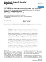
The epidemiological studies regarding the
relationship between FSH and bone health in humans
are summarized in Table 1.
Table 1. The relationship between FSH and bone health in humans.
Authors
Study design
Premenopausal Women
Garton et al. 1996 [31]
68 spontaneously menstruating women aged 45–55 years. The subjects
were divided based on tertiles of FSH level (<10 U/l; 10–35 U/l; >35 U/l).
Sowers et al. 2003 [33] 2336 women aged 42– 52 years (pre and peri menopause) from the Study of
Women’s Health Across the Nation (SWAN). Composition of the subjects
were 28.2% African-American, 49.9% Caucasian, 10.5% Japanese or 11.4%
Chinese.
Sowers et al. 2003 [34] 2,375 pre- and early perimenopausal women from SWAN, aged 42-52
years. Multiethnicities.
Grewal et al. 2006 [35] 643 pre- and perimenopausal women, aged 43-53 years from SWAN. BMD
at lumbar spine and femoral hip was measured.
Cannon et al. 2010 [32] 36 women aged 20-50 years with normal natural menstrual cycles.
Vural et al. 2005 [59]
87 healthy volunteers from the community aged 35-50 years.
Hui et al. 2002 [36]
130 non-Hispanic white women aged 31–50 years. Followed up at least 3
times for 1-9 years.
Sowers et al. 2006 [37] 4-year longitudinal study of the SWAN cohort. 2311 premenopausal or
early perimenopausal African-American, Caucasian, Chinese, and
Japanese women.
Sowers et al. 2010 [38] 629 women aged 24 – 44 years at baseline were followed up for 15 years.
Subjects were divided into FSH stages 1-4: 1=<15, 2=15-33, 3=34-54, 4=>54
mlU/ml.
Crandall et al. 2013 [46] A 10-year follow up of 720 women in SWAN cohort. Subjects aged 42–52
(mean 46.2) years at baseline.
Women across menopausal stages
Ebeling et al. 1996 [39] 281 women aged 45-57 years (pre, peri and postmenopausal groups)
selected from a larger randomized urban population cohort (Melbourne
Relationship with variables
BMD
Bone remodelling
markers
Negative
pQCT
Fracture
Serum phosphate, PYD,
DYD: positive
Negative
NTX: positive
Osteocalcin: negative
Negative
Negative
Not significant NTX: positive
Osteocalcin: not
significant
Negative
Negative
Negative
Negative
Negative
Not significant uDPD, total PYD, NTX,
after
BAP: positive
Int. J. Med. Sci. 2018, Vol. 15
Authors
1381
Study design
Relationship with variables
BMD
Bone remodelling
pQCT
markers
Women's Midlife Health Project).
adjustment
Perrien et al. 2006 [45] 188 pre- and postmenopausal women not using oral contraceptives or
CTX: positive
hormone replacement therapy (age, 21-85 yr) from Rochester
AP, BAP, PYD, DPD: Not
Epidemiology Project. Only 2 subjects were non-Caucasians.
significant
Yasui et al. 2006 [41]
Cross-sectional study. 193 female outpatients of a Japanese university
Negative
Osteocalcin (intact and
hospital aged 39-66 years. 40 were premenopause, 47 were perimenopause,
uncarboxylated): Positive
106 were postmenopause stage. Serum biochemical markers measured
included uncarboxylated osteocalcin, intact osteocalcin, bone alkaline
phosphatase, urinary N-telopeptide, LH, FSH, oestradiol, estrone.
Desai et al. 2007 [42]
365 Indian women aged 20–70 years from a community-based clinic.
Negative
Xu et al. 2009 [43]
Cross-sectional study. 699 healthy Chinese women aged 20-82 years.
Negative
Serum LH, FSH measured. BMD measured at posteroanterior spine, lateral
spine, TH and distal forearm.
Gallagher et al. 2010
3247 peri- and postmenopausal women aged 42-60 years from US National Negative
[40]
Health and Nutrition Examination Survey (NHANESIII).
Wu et al. 2013 [44]
Cross-sectional study. 368 healthy adult Chinese women (155
Negative
premenopausal women, 63 perimenopausal, 150 postmenopausal women),
aged 35-60 years.
Post-menopausal Women
Gourlay et al. 2011 [47] 111 community-dwelling postmenopausal women aged 50–64 years (mean Negative but
57.5 ± 3.7) from various ethnicities.
lost after
adjustment
Gourlay et al. 2012 [48] 94 younger (aged 50 to 64 years, mean 57.5 years) community dwelling
Negative
postmenopausal women not using HRT.
Wang et al. 2015 [20]
248 postmenopausal Chinese women aged 50 years or above (128
Negative
osteoporotic and 120 normal bone health)
Men
Karim et al. 2008 [52]
Case-control study. 156 community-dwelling men in London UK aged 57.7 Negative
± 13.7 years. 63 osteoporotic men, 93 normal control.
Hsu et al. 2015 [53]
1705 men aged 70 years and older from the Concord Health and Ageing in Negative
Men Project were followed up for 5 years.
Experimental by nature or human
Kawai et al. 2004 [60]
A retrospective study on 125 women undergoing hormone replacement
Negative
therapy. Sequential measurement of hormone was performed before, at 12
and 24 months after starting hormone replacement therapy.
Devleta et al. 2004 [54] 7 hypergonadotropic (FSH>40 IU/l; aged 37.43 ± 3.10), 15
Negative
hypogonadotropic (FSH<40 IU/l; aged 29.8 ± 5.71) amenorrhoeic and 12
eumenorrheic women (aged 33.81 ± 5.89) were recruited.
Castelo-Branco et al.
8 adolescent women with Kallman syndrome (hypogonadotropic); 11 with Not significant
2008 [56]
Turner syndrome (hypergonadotropic); 11 with pure gonadal dysgenesia after
(hypergonadotropic).
adjustment
Drake et al. 2010 [58]
Post-menopausal women were randomized into two groups. One group
High bone turnover not
(n=21, aged 67.4 ± 1.2) received leuprolide (7.5 mg i.m. every 28 d) and the
inhibited.
other group (n=20, aged 66.1±1.3 years) received placebo. Both groups
received aromatase inhibitor (letrozole, 2.5 mg/d) to prevent exogenous
synthesis of oestradiol.
Latoch et al. 2015 [57]
76 long-term survivors (43 men and 33 women) treated for paediatric
Not significant
cancer. 38% leukaemia, 36% lymphoma, 26% solid tumours. Age at the
study was 24.1 ±3.5 years
Tabatabai et al. 2016
206 women (64% white) age ≤ 55 (mean 45.9 ±5.5) years at breast cancer
Negative
CTX, PINP, osteocalcin:
[55]
diagnosis receiving adjuvant cancer chemotherapy and at least 1 year after
positive
diagnosis.
AP, NTX: Not significant
Fracture
Not significant
Abbreviation:
AP=alkaline phosphatase; BAP=bone-specific alkaline phosphatase; BMD=bone mineral density; DPD=deoxypyridinoline; CTX=C-terminal telopeptide of type I collagen;
FSH=follicle-stimulating hormone; NTX=N-terminal telopeptide of type I collagen; PINP=N-terminal propeptide of type I procollagen; pQCT=peripheral quantitative
computed tomography; PYD=pyridinoline
Conclusion
FSH has a direct effect on bone resorption
mediated by FSHR receptors found on osteoclasts and
their precursors. The effects of FSH on osteoblasts
could be negligible since they do not express FSHR.
Transgenic rodent model showed heterogenous
results on the skeletal effects of FSH, which seem to be
dependent on the ovarian production of testosterone
in rodents lacking FSH and FSHR. Supplementing
FSH in rats has been shown to be detrimental to the
bone, while blocking its activity seems to be beneficial
to the skeleton. The human studies generally reveal a
significant and negative relationship between FSH
level and bone health but the relationship diminishes
after menopause, when the effects of oestrogen
deficiency are dominant. Thus, FSH may partially
explain bone loss during perimenopausal period.
Skeletal
deterioration
in
hypogonadotropic
hypogonadism may occur because the influence of sex
hormone deficiency is greater than FSH deficiency.
Similar negative relationship between FSH and bone
health is observed in men.
Several research gaps need to be bridged to
validate the relationship between FSH and bone
health. The current evidence is predominantly from
Int. J. Med. Sci. 2018, Vol. 15
cross-sectional
studies
which
prevents
the
interpretation of causality. More longitudinal
investigations on the effects of FSH on bone health,
especially fracture risk in women, should be made.
More studies on men should also be performed
because their FSH level and fracture risk also increase
gradually with age. In addition to that, post-fracture
mortality rate of men is higher than women, which
necessitates a better understanding of male
osteoporosis and its predictors like FSH. Since the
hormonal changes across life stages is complex, there
is a need to understand the influence of not just FSH
alone, but also other related hormone factors, or the
whole hormonal milieu alternation on bone health.
While the use of anti-FSH antibody to stop bone loss is
tempting, there is insufficient evidence currently, to
support that blocking the effects of FSH during
perimenopause period exerts skeletal beneficial in
humans. We hope that more enlightening discoveries
in the future will lead to a better understanding of the
involvement of FSH in the pathogenesis of
osteoporosis in aging women and men. Hopefully,
this will spark more innovative and safer
interventions to halt bone loss by manipulating the
hormonal milieu.
Acknowledgements
1382
9.
10.
11.
12.
13.
14.
15.
16.
17.
18.
19.
20.
21.
22.
The author wishes to acknowledge Universiti
Kebangsaan Malaysia for funding his studies via
grants GUP-2017-060 and AP-2017-009/1. He also
thanks Miss Shu Shen Tay for proofreading the
manuscript.
25.
Competing Interests
26.
The authors have declared that no competing
interest exists.
27.
References
28.
1.
29.
2.
3.
4.
5.
6.
7.
8.
Finkelstein JS, Brockwell SE, Mehta V, Greendale GA, Sowers MR, Ettinger B,
et al. Bone mineral density changes during the menopause transition in a
multiethnic cohort of women. J Clin Endocrinol Metab. 2008; 93: 861-8.
Rannevik G, Jeppsson S, Johnell O, Bjerre B, Laurell-Borulf Y, Svanberg L. A
longitudinal study of the perimenopausal transition: altered profiles of steroid
and pituitary hormones, SHBG and bone mineral density. Maturitas. 1995; 21:
103-13.
Randolph JF, Jr., Zheng H, Sowers MR, Crandall C, Crawford S, Gold EB, et al.
Change in follicle-stimulating hormone and estradiol across the menopausal
transition: effect of age at the final menstrual period. J Clin Endocrinol Metab.
2011; 96: 746-54.
Ogawa S, Fujita M, Ishii Y, Tsurukami H, Hirabayashi M, Ikeda K, et al.
Impaired Estrogen Sensitivity in Bone by Inhibiting Both Estrogen Receptor α
and β Pathways. J Biol Chem. 2000; 275: 21372-9.
Windahl SH, Hollberg K, Vidal O, Gustafsson JA, Ohlsson C, Andersson G.
Female estrogen receptor beta-/- mice are partially protected against
age-related trabecular bone loss. J Bone Miner Res. 2001; 16: 1388-98.
Windahl SH, Vidal O, Andersson G, Gustafsson JA, Ohlsson C. Increased
cortical bone mineral content but unchanged trabecular bone mineral density
in female ERbeta(-/-) mice. J Clin Invest. 1999; 104: 895-901.
Yeh JK, Chen MM, Aloia JF. Ovariectomy-induced high turnover in cortical
bone is dependent on pituitary hormone in rats. Bone. 1996; 18: 443-50.
Sun L, Peng Y, Sharrow AC, Iqbal J, Zhang Z, Papachristou DJ, et al. FSH
directly regulates bone mass. Cell. 2006; 125: 247-60.
23.
24.
30.
31.
32.
33.
34.
35.
36.
Mohamad NV, Soelaiman IN, Chin KY. Effect of Androgen Deprivation
Therapy (ADT) on Bone Health Status in Men with Prostate Cancer. Endocr
Metab Immune Disord Drug Targets. 2017; 17: 276 - 84.
Mohamad N-V, Zulkepli MAAC, Theseira KM, Zulkifli N, Shahrom
NQNAMR, Jamil NA, et al. Establishing an Animal Model of Secondary
Osteoporosis by Using a Gonadotropin-releasing Hormone Agonist. Int J Med
Sci. 2018; 15: 300-8.
Mohamad
NV,
Ima-Nirwana
S,
Chin
KY.
The
effects
of
gonadotropin-releasing hormone agonist (buserelin) and orchidectomy on
bone turnover markers and histomorphometry in rats. Aging Male. 2018: in
press.
Levin VA, Jiang X, Kagan R. Estrogen therapy for osteoporosis in the modern
era. Osteoporos Int. 2018: in press.
Peng L, Luo Q, Lu H. Efficacy and safety of bazedoxifene in postmenopausal
women with osteoporosis: A systematic review and meta-analysis. Medicine
(Baltimore). 2017; 96: e8659.
Gambacciani M, Levancini M. Management of postmenopausal osteoporosis
and the prevention of fractures. Panminerva Med. 2014; 56: 115-31.
Robinson LJ, Tourkova I, Wang Y, Sharrow AC, Landau MS, Yaroslavskiy BB,
et al. FSH-Receptor Isoforms and FSH-dependent Gene Transcription in
Human Monocytes and Osteoclasts. Biochemical and biophysical research
communications. 2010; 394: 12-7.
Zhu L-L, Tourkova I, Yuen T, Robinson LJ, Bian Z, Zaidi M, et al. Blocking
FSH action attenuates osteoclastogenesis. Biochemical and Biophysical
Research Communications. 2012; 422: 54-8.
Iqbal J, Sun L, Kumar TR, Blair HC, Zaidi M. Follicle-stimulating hormone
stimulates TNF production from immune cells to enhance osteoblast and
osteoclast formation. Proc Natl Acad Sci U S A. 2006; 103: 14925-30.
Sun L, Zhang Z, Zhu L-L, Peng Y, Liu X, Li J, et al. Further evidence for direct
pro-resorptive actions of FSH. Biochemical and Biophysical Research
Communications. 2010; 394: 6-11.
Cannon JG, Kraj B, Sloan G. Follicle-stimulating hormone promotes RANK
expression on human monocytes. Cytokine. 2011; 53: 141-4.
Wang J, Zhang W, Yu C, Zhang X, Zhang H, Guan Q, et al. Follicle-Stimulating
Hormone Increases the Risk of Postmenopausal Osteoporosis by Stimulating
Osteoclast Differentiation. PLoS One. 2015; 10: e0134986.
Ritter V, Thuering B, Saint Mezard P, Luong-Nguyen NH, Seltenmeyer Y,
Junker U, et al. Follicle-stimulating hormone does not impact male bone mass
in vivo or human male osteoclasts in vitro. Calcif Tissue Int. 2008; 82: 383-91.
Su XY, Zou X, Chen QZ, Zeng YH, Shao Y, He BC, et al. Follicle-Stimulating
Hormone beta-Subunit Potentiates Bone Morphogenetic Protein 9-Induced
Osteogenic Differentiation in Mouse Embryonic Fibroblasts. J Cell Biochem.
2017; 118: 1792-802.
Seibel MJ, Dunstan CR, Zhou H, Allan CM, Handelsman DJ. Sex steroids, not
FSH, influence bone mass. Cell. 2006; 127: 1079.
Abel MH, Huhtaniemi I, Pakarinen P, Kumar TR, Charlton HM. Age-related
uterine and ovarian hypertrophy in FSH receptor knockout and FSHbeta
subunit knockout mice. Reproduction. 2003; 125: 165-73.
Balla A, Danilovich N, Yang Y, Sairam MR. Dynamics of ovarian development
in the FORKO immature mouse: structural and functional implications for
ovarian reserve. Biol Reprod. 2003; 69: 1281-93.
Gao J, Tiwari-Pandey R, Samadfam R, Yang Y, Miao D, Karaplis AC, et al.
Altered ovarian function affects skeletal homeostasis independent of the
action of follicle-stimulating hormone. Endocrinology. 2007; 148: 2613-21.
Allan CM, Kalak R, Dunstan CR, McTavish KJ, Zhou H, Handelsman DJ, et al.
Follicle-stimulating hormone increases bone mass in female mice. Proc Natl
Acad Sci U S A. 2010; 107: 22629-34.
Liu S, Cheng Y, Fan M, Chen D, Bian Z. FSH aggravates periodontitis-related
bone loss in ovariectomized rats. J Dent Res. 2010; 89: 366-71.
Lukefahr AL, Frye JB, Wright LE, Marion SL, Hoyer PB, Funk JL. Decreased
bone mineral density in rats rendered follicle-deplete by an ovotoxic chemical
correlates with changes in follicle-stimulating hormone and inhibin A. Calcif
Tissue Int. 2012; 90: 239-49.
Geng W, Yan X, Du H, Cui J, Li L, Chen F. Immunization with FSHbeta fusion
protein antigen prevents bone loss in a rat ovariectomy-induced osteoporosis
model. Biochem Biophys Res Commun. 2013; 434: 280-6.
Garton M, Martin J, New S, Lee S, Loveridge N, Milne J, et al. Bone mass and
metabolism in women aged 45-55. Clin Endocrinol (Oxf). 1996; 44: 563-70.
Cannon JG, Cortez-Cooper M, Meaders E, Stallings J, Haddow S, Kraj B, et al.
Follicle-stimulating hormone, interleukin-1, and bone density in adult women.
Am J Physiol Regul Integr Comp Physiol. 2010; 298: R790-8.
Sowers MR, Finkelstein JS, Ettinger B, Bondarenko I, Neer RM, Cauley JA, et
al. The association of endogenous hormone concentrations and bone mineral
density measures in pre- and perimenopausal women of four ethnic groups:
SWAN. Osteoporos Int. 2003; 14: 44-52.
Sowers MR, Greendale GA, Bondarenko I, Finkelstein JS, Cauley JA, Neer RM,
et al. Endogenous hormones and bone turnover markers in pre- and
perimenopausal women: SWAN. Osteoporos Int. 2003; 14: 191-7.
Grewal J, Sowers MR, Randolph JF, Jr., Harlow SD, Lin X. Low bone mineral
density in the early menopausal transition: role for ovulatory function. J Clin
Endocrinol Metab. 2006; 91: 3780-5.
Hui SL, Perkins AJ, Zhou L, Longcope C, Econs MJ, Peacock M, et al. Bone loss
at the femoral neck in premenopausal white women: effects of weight change
and sex-hormone levels. J Clin Endocrinol Metab. 2002; 87: 1539-43.
Int. J. Med. Sci. 2018, Vol. 15
1383
37. Sowers MR, Jannausch M, McConnell D, Little R, Greendale GA, Finkelstein
JS, et al. Hormone predictors of bone mineral density changes during the
menopausal transition. J Clin Endocrinol Metab. 2006; 91: 1261-7.
38. Sowers MR, Zheng H, Jannausch ML, McConnell D, Nan B, Harlow S, et al.
Amount of bone loss in relation to time around the final menstrual period and
follicle-stimulating hormone staging of the transmenopause. J Clin Endocrinol
Metab. 2010; 95: 2155-62.
39. Ebeling PR, Atley LM, Guthrie JR, Burger HG, Dennerstein L, Hopper JL, et al.
Bone turnover markers and bone density across the menopausal transition. J
Clin Endocrinol Metab. 1996; 81: 3366-71.
40. Gallagher CM, Moonga BS, Kovach JS. Cadmium, follicle-stimulating
hormone, and effects on bone in women age 42-60 years, NHANES III.
Environ Res. 2010; 110: 105-11.
41. Yasui T, Uemura H, Tomita J, Miyatani Y, Yamada M, Miura M, et al.
Association of serum undercarboxylated osteocalcin with serum estradiol in
pre-, peri- and early post-menopausal women. J Endocrinol Invest. 2006; 29:
913-8.
42. Desai Meena P, Khatkhatay MI, Bhanu Prakash KV, Savardekar LS, Shah RS,
Ansari Z. Hormonal profiles and biochemical indices of bone turnover in
Indian women. Osteoporos Int. 2007; 18: 923-9.
43. Xu ZR, Wang AH, Wu XP, Zhang H, Sheng ZF, Wu XY, et al. Relationship of
age-related concentrations of serum FSH and LH with bone mineral density,
prevalence of osteoporosis in native Chinese women. Clin Chim Acta. 2009;
400: 8-13.
44. Wu XY, Yu SJ, Zhang H, Xie H, Luo XH, Peng YQ, et al. Early bone mineral
density decrease is associated with FSH and LH, not estrogen. Clin Chim Acta.
2013; 415: 69-73.
45. Perrien DS, Achenbach SJ, Bledsoe SE, Walser B, Suva LJ, Khosla S, et al. Bone
turnover across the menopause transition: correlations with inhibins and
follicle-stimulating hormone. J Clin Endocrinol Metab. 2006; 91: 1848-54.
46. Crandall CJ, Tseng CH, Karlamangla AS, Finkelstein JS, Randolph JF, Jr.,
Thurston RC, et al. Serum sex steroid levels and longitudinal changes in bone
density in relation to the final menstrual period. J Clin Endocrinol Metab. 2013;
98: E654-63.
47. Gourlay ML, Preisser JS, Hammett-Stabler CA, Renner JB, Rubin J.
Follicle-stimulating hormone and bioavailable estradiol are less important
than weight and race in determining bone density in younger postmenopausal
women. Osteoporos Int. 2011; 22: 2699-708.
48. Gourlay ML, Specker BL, Li C, Hammett-Stabler CA, Renner JB, Rubin JE.
Follicle-stimulating hormone is independently associated with lean mass but
not BMD in younger postmenopausal women. Bone. 2012; 50: 311-6.
49. Rendina D, Gianfrancesco F, De Filippo G, Merlotti D, Esposito T, Mingione A,
et al. FSHR gene polymorphisms influence bone mineral density and bone
turnover in postmenopausal women. Eur J Endocrinol. 2010; 163: 165-72.
50. Chin KY, Ima-Nirwana S. Sex steroids and bone health status in men. Int J
Endocrinol. 2012; 2012: 208719.
51. Mohamad NV, Soelaiman IN, Chin KY. A concise review of testosterone and
bone health. Clin Interv Aging. 2016; 11: 1317-24.
52. Karim N, MacDonald D, Dolan AL, Fogelman I, Wierzbicki AS, Hampson G.
The relationship between gonadotrophins, gonadal hormones and bone mass
in men. Clin Endocrinol (Oxf). 2008; 68: 94-101.
53. Hsu B, Cumming RG, Seibel MJ, Naganathan V, Blyth FM, Bleicher K, et al.
Reproductive Hormones and Longitudinal Change in Bone Mineral Density
and Incident Fracture Risk in Older Men: The Concord Health and Aging in
Men Project. J Bone Miner Res. 2015; 30: 1701-8.
54. Devleta B, Adem B, Senada S. Hypergonadotropic amenorrhea and bone
density: new approach to an old problem. J Bone Miner Metab. 2004; 22: 360-4.
55. Tabatabai LS, Bloom J, Stewart S, Sellmeyer DE. FSH Levels Predict Bone Loss
in Premenopausal Women Treated for Breast Cancer More Than One Year
After Treatment. J Clin Endocrinol Metab. 2016; 101: 1257-62.
56. Castelo-Branco C, Leon M, Duran M, Balasch J. Follicle-stimulating hormone
does not directly regulate bone mass in human beings: evidence from nature.
Fertil Steril. 2008; 90: 2211-6.
57. Latoch E, Muszynska-Roslan K, Panas A, Panasiuk A, Rutkowska-Zelazowska
B, Konstantynowicz J, et al. Bone mineral density, thyroid function, and
gonadal status in young adult survivors of childhood cancer. Contemp Oncol
(Pozn). 2015; 19: 142-7.
58. Drake MT, McCready LK, Hoey KA, Atkinson EJ, Khosla S. Effects of
suppression of follicle-stimulating hormone secretion on bone resorption
markers in postmenopausal women. J Clin Endocrinol Metab. 2010; 95: 5063-8.
59. Vural F, Vural B, Yucesoy I, Badur S. Ovarian aging and bone metabolism in
menstruating women aged 35-50 years. Maturitas. 2005; 52: 147-53.
60. Kawai H, Furuhashi M, Suganuma N. Serum follicle-stimulating hormone
level is a predictor of bone mineral density in patients with hormone
replacement therapy. Arch Gynecol Obstet. 2004; 269: 192-5.