fficacy and safety of nucleos(t)ide analogues to prevent hepatitis B virus mother-to-child transmission in pregnant women with high viremia: Real life practice from China
Bạn đang xem bản rút gọn của tài liệu. Xem và tải ngay bản đầy đủ của tài liệu tại đây (369.23 KB, 6 trang )
Int. J. Med. Sci. 2018, Vol. 15
Ivyspring
International Publisher
796
International Journal of Medical Sciences
2018; 15(8): 796-801. doi: 10.7150/ijms.25047
Research Paper
Efficacy and safety of nucleos(t)ide analogues to prevent
hepatitis B virus mother-to-child transmission in
pregnant women with high viremia: real life practice
from China
Qiuju Sheng1, Yang Ding1, Baijun Li2, Chao Han1, Yanwei Li1, Chong Zhang1, Han Bai1, Jingyan Wang1,
Lianrong Zhao1, Tingting Xia1, Ziying An1, Mingxiang Zhang2, Xiaoguang Dou1
1.
2.
Department of Infectious Disease, Shengjing Hospital, China Medical University, Shenyang 110022, China
The Sixth People’s Hospital of Shenyang, Shenyang 110006, China
Corresponding author: Dr. Xiaoguang Dou, Professor of Department of Infectious Diseases, Shengjing Hospital, China Medical University, No. 39 Huaxiang
Road, Tiexi District, Shenyang 110022, China. Phone: 86-18940251121; E-mail:
© Ivyspring International Publisher. This is an open access article distributed under the terms of the Creative Commons Attribution (CC BY-NC) license
( See for full terms and conditions.
Received: 2018.01.19; Accepted: 2018.04.27; Published: 2018.05.22
Abstract
Purpose: To evaluate the efficacy and safety of nucleos(t)ide analogues, especially telbivudine (LdT)
for the prevention of mother-to-child transmission (MTCT) of hepatitis B virus (HBV) in women
with high viremia.
Methods: We conducted a prospective, open-label, multicenter study of LdT for treating pregnant
women having high viral loads of hepatitis B virus (HBV DNA>5 log10 IU/mL) but normal levels of
alanine aminotransferase (ALT). Maternal HBV DNA, HBV serologic status and ALT were measured
at baseline, 4 weeks after therapy, before delivery, 4 weeks after delivery, and 12 weeks after
delivery. Infant HBV serologic status and HBV DNA levels were measured at 7 months. We
calculated the MTCT rate of LdT-treated and LdT-untreated groups and analyzed the efficacy and
safety of LdT.
Results: Ninety-one women (the treatment group) were treated with LdT, and twenty-one
patients (the observation group) did not undergo antiviral therapy. The baseline HBV DNA levels
were 8.15±0.82 log10 IU/mL in the treatment group, and 8.09±1.04 log10 IU/mL in the observation
group. The MTCT rate was 0% in the treatment group, and 9.5% in the observation group
(p=0.042). In the treatment group, HBV DNA levels were 5.02±0.74 log10 IU/mL at one month after
therapy, and 3.95±0.94 log10 IU/mL before delivery. Both groups had significant differences from
baseline levels in HBV DNA levels (p<0.001). In total, five patients had elevated ALT levels but
without evidence of decompensate liver function. No severe adverse events or complications were
observed in women or infants.
Conclusions: For pregnant women with HBV DNA greater than 5 log10IU/mL, LdT therapy was
effective in reducing HBV MTCT. If serum HBV DNA was detectable at delivery, discontinuation of
LdT immediately was found to be safe and rarely induced off-treatment hepatitis flare.
Key words: HBV; chronic hepatitis B; pregnancy; mother-to-child-transmission; antiviral therapy; nucleos(t)ide
analogues; telbivudine
Introduction
Hepatitis B virus (HBV) infection can cause
many severe diseases like cirrhosis, hepatic cellular
carcinoma (HCC) and liver failure. Worldwide, more
than 800,000 people die every year due to
complications from hepatitis B [1]. That is a heavy
burden to public health.
Int. J. Med. Sci. 2018, Vol. 15
HBV is transmitted through many routes [2, 3].
In China, mother-to-child transmission (MTCT) is the
most common [1, 4]. Importantly, HBV transmitted
through MTCT in the perinatal period often leads to
chronic infection [5]. Without intervention, 40%-95%
of infants born to hepatitis B surface antigen
(HBsAg)-positive women will acquire HBV infection
[6, 7]. Newborns inoculated with HBV vaccine and
hepatitis B immunoglobulin (HBIG) have reduced
MTCT rates ranging from 5-10% overall [8]. However,
immune prophylaxis failure still occurs. The most
important risk factor in MTCT is high maternal HBV
DNA levels [9-13]. In infants born to women with
HBV DNA levels of more than 6 log10 copies/ml, the
risk of MTCT may rise to 30% despite immune
prophylaxis [13-18]. Our previous studies in China
demonstrated that high viral loads are common in
women during the perinatal period [19, 20]. So
antiviral therapy, such as nucleos(t)ide analogues
(NAs) in late pregnancy to prevent MTCT is
recommended now.
According to the US Food and Drug
Administration (FDA) classification standard, all HBV
antiviral NAs are category C (teratogenic in animals,
but unknown in humans), except for telbivudine
(LdT) and tenofovir disoproxil fumarate (TDF), which
are category B drugs (no risk in animal studies, but
unknown in humans) [21]. In our study, we chose LdT
as our treatment agent.
The purposes of this study were to investigate
the efficacy of LdT therapy, when to stop the drug,
and the safety of drug discontinuation.
Materials and methods
Patients selected
This was a prospective, open-label, multicenter
study. Patients were from Shengjing Hospital of
China Medical University and the Sixth People’s
Hospital of Shenyang and were enrolled between Jan
2013 and Dec 2015. The trial was approved by the
ethics committee of Shengjing Hospital. All patients
signed informed consent forms before screening. The
inclusion criteria were as follows: 1. Women between
20 to 40 years of age, confirmed pregnancy, HBsAg
positive, alanine aminotransferase (ALT) below the
upper limit of normal (ULN) (40 IU/mL). 2. HBV
DNA>5 log10 IU/mL between 24 and 32 weeks of
pregnancy. The exclusion criteria included: 1.
Evidence of cirrhosis or hepatic cellular carcinoma
(HCC), co-infection with hepatitis A, C, D or E or
human immunodeficiency virus (HIV). 2. Use of
antiviral therapy before or during pregnancy. 3.
Combination use of other immune modulators,
steroids and cytotoxic drugs. 4. Evidence of
797
miscarriage or fetal deformity. Patients fulfilling the
inclusion and exclusion criteria were enrolled in our
study.
Treatment regime
Baseline serum HBV DNA levels were measured
between 24 and 32 weeks of pregnancy. Antiviral
therapy was initiated when the serum HBV DNA
levels exceeded 5 log10 IU/mL. Based on the patients’
choice, they were divided into treatment or
observation groups. Patients in the treatment group
were given oral LdT 600 mg daily. If HBV DNA levels
declined less than 2 log10 IU/mL at 4 weeks after
initiation of therapy (compared to baseline), LdT was
changed to TDF. If hepatitis flare occurred during
pregnancy, antiviral therapy was continued after
delivery. If there was a hepatitis flare after delivery,
patients were treated like other chronic hepatitis B
(CHB) patients without pregnancy. At the time of the
predelivery visit, if serum HBV DNA was detectable,
the agent was immediately discontinued after
delivery. Otherwise, the patients continued to take the
agent after delivery until reaching the drug
withdrawal criteria for CHB. Patients in the
observation group received no antiviral therapy.
All infants received HBIG 100 IU and
recombinant HBV vaccine 10 ug within 12 hours of
birth. The second and third does of recombinant HBV
vaccine were administered at 1 and 6 months of age,
respectively. Women could breastfeed their babies
after 1 week of agent cessation. Otherwise,
breastfeeding was forbidden with agent.
Detection indexes
ALT, aspartate aminotransferase (AST), total
bilirubin (TBIL), HBV DNA, HBV serologic status,
and creatine kinase (CK) levels were measured at
baseline for the patients in both groups, and at 4
weeks after therapy, before delivery, 4 weeks after
delivery and 12 weeks after delivery for women in the
treatment group. Infant HBV serologic status and
HBV DNA levels were measured at 7 months of age (1
month after final vaccine). We calculated the MTCT
rate between the two groups and analyzed the
efficacy and safety of the antiviral agent.
Biochemical and virologic assessments
HBV serologic markers, including HBsAg,
anti-HBs antibody, hepatitis B e antigen (HBeAg),
anti-HBe and anti-HBc antibodies titers were assayed
with a chemi-luminescent microparticle immunoassay using an automated Abbott AxSYM analyzer
(Abbott, USA). HBV DNA levels were measured by
real-time polymerase chain reaction (PCR) assay
using a COBAS AmpliPrep/COBAS TaqMan 48
analyzer (Roche Diagnostics, Switzerland).
Int. J. Med. Sci. 2018, Vol. 15
Definitions
MTCT was defined as detectable levels of HBV
DNA or HBsAg in peripheral serum samples of
infants at 7 months age. Hepatitis flare was defined as
ALT ≥ 2×ULN during or after treatment.
Statistical analysis
Baseline characteristics and laboratory results
were summarized by means of descriptive statistics,
including percentage, and means ± standard
deviation (SD). The t test was used for group
comparisons of quantitative variables. The chi-square
test was used to compare group differences of
categorical variables. Significance levels were set at
p<0.05. All data were analyzed by SPSS 16.0.
Results
General characteristics
During the three years of study, 127 chronic
hepatitis B infected pregnant women with normal
ALT levels were referred to the infectious disease
clinic. One-hundred and sixteen (116/127, 91.3%)
patients had high viral loads (>5 log10 IU/mL). Three
patients failed to attend the hepatology/infectious
disease clinic for treatment before 32 weeks of
pregnancy, and one patient underwent an abortion
for worrying about fetus safety. In total, 112 patients
were enrolled in this study; all had positive HBeAg
levels. Ninety-one (91/112, 80.5%) patients (treatment
group) accepted antiviral therapy. The remaining 21
patients were enrolled in the observation group. In
the treatment group, 2 (2/91, 2.2%) patients switched
to TDF due to HBV DNA levels decline of less than 2
log10 IU/mL after 4 weeks of therapy compared to
baseline. Four (4/91, 4.4%) patients continued to take
the antiviral agent after delivery. Of these four
patients, two had undetectable serum HBV DNA
levels before delivery, and the other two experienced
hepatitis flare during the pregnancy. Three patients
discontinued the agent at the time of delivery but
were retreated after delivery due to hepatitis flare.
Baseline characteristics
In the treatment group, the median age was 27
(range, 21-40) years, the baseline HBV DNA load was
8.15±0.82 log10 IU/mL (range, 5.54-9.53), the average
ALT level was 26.53±8.32 U/L (range, 6-40), and the
HBsAg and HBeAg levels were 4.34±0.33 log10 IU/mL
(range, 3.22-5.05) and 1179.14±371.09 s/co (range,
5.3-1842.5), respectively. The mean duration of
therapy was 13.62±2.12 weeks (range, 8-16). In the
observation group, the median age was 26 (range,
20-34) years, the baseline HBV DNA load was
8.09±1.04 log10 IU/mL (range, 5.38-9.72), the average
798
ALT was 23.62±6.51 U/L (range, 10-36), and the
HBsAg and HBeAg level was 4.22±0.30 log10 IU/mL
(range, 3.50-4.59) and 1294.94±329.29 s/co (range,
736.25-1867.35), respectively. There were no
differences of baseline values between the treatment
and observation groups (Table 1).
Table 1. Maternal baseline values of the two study groups
Parameter
Age (years)
ALT (U/L)
HBV DNA (log10 IU/ mL)
HBsAg (log10 IU/mL)
HBeAg (s/co)
Treatment group
(n=91)
27.8±4.17
26.53±8.32
8.15±0.82
4.34±0.33
1179.14±371.09
Observation group
(n=21)
26.8±3.66
23.62±6.51
8.09±1.04
4.22±0.30
1294.94±329.29
P value
0.442
0.934
0.410
0.690
0.448
ALT, alanine aminotransferase; HBV, hepatitis B virus; HBsAg, hepatitis B surface
antigen; HBeAg, hepatitis B e antigen.
MTCT rate
Ninety-two babies were born to the 91 women in
the treatment group. All of them were inoculated with
HBIG and HBV vaccine within 12 hours of birth, and
then completed the HBV vaccine series at 1 and 6
months. However, 13 infants were lost to follow up at
7 months of age. None of the other 79 infants (0/79,
0%) were HBsAg positive or had detectable serum
HBV DNA levels at 7 months of age. The MTCT rate
was 0 in our treatment group.
In contrast, in the observation group, there were
21 infants born to 21 women; they were followed up at
7 months. Even with standard immune prophylaxis,
two infants (2/21, 9.52%) were HBV infected. The
difference between the two study groups was
significant (p=0.042). One infant was born via vaginal
delivery, while the other was born by cesarean
section. Both infants infected with HBV were
breastfed.
Efficacy analysis
In the treatment group, the average HBV DNA
level at 4 weeks after therapy was 5.02±0.74 log10
IU/mL (range, 3.42-6.85); it declined by 3.13 log10
IU/mL compared to that of baseline (p<0.001). Before
delivery, the average HBV DNA level was 3.95±0.94
log10 IU/mL (range, 0-5.34), declining by 1.07 log10
IU/mL compared to that of 1-month therapy
(p<0.001); it declined by 4.20 log10 IU/mL compared
to that of baseline (P<0.001). At the 1 month visit after
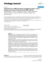
delivery, the serum HBV DNA levels rebounded to
7.75±1.68 log10 IU/mL (range, 0-8.95) (Fig. 1)
There was no significant change in HBsAg and
HBeAg levels. Two patients who had low levels of
HBsAg and HBeAg at baseline had undetectable
serum HBV DNA levels before delivery (Fig. 2 and
Fig. 3)
Int. J. Med. Sci. 2018, Vol. 15
Figure 1. HBV DNA loads at baseline, 1 month after therapy, before delivery
and 1 month after delivery for women undergoing antiviral therapy (n=91).
Figure 2. HBsAg levels at baseline, 1 month after therapy, before delivery and
1 month after delivery for women undergoing antiviral therapy (n=91).
799
prothrombin time (PT), without other evidence of
decompensate liver function. Three patients
experienced hepatitis flare. The ALT levels of two
patients rise to 4×ULN and 6×ULN at one month after
delivery separately. For the third patient, ALT went
up to 16×ULN at 3 months after delivery. All three
patients showed no evidence of decompensate liver
function; they accepted retreatment with antiviral
therapy and had normal ALT levels at the next visit.
The other 86 patients maintained normal ALT levels
not only during the therapy but also after stopping
the agent.
Among the treatment group, all but one patient
had normal CK values during the LdT therapy (1/91,
1.1%). This patient had mild CK elevation (1.45×ULN)
without any symptoms, levels which returned to
normal by the time of the next visit. One (1/91, 1.1%)
patient developed a rash during LdT therapy but
remitted after several days without agent
interruption. The two patients who switched from
LdT to TDF did not show renal impairment during
the therapy.
In the treatment group, 56 (56/91, 61.5%) women
chose cesarean section; in the observation group, 12
(12/21, 57.1%) women chose cesarean section. There
was no obvious difference between the two groups
(p=0.71). Among the 87 patients in the treatment
group who discontinued drug treatment at the time of
delivery, 30 (30/87, 34.4%) breastfed their infants. No
congenital malformations were identified. All
neonates had normal Apgar scores at birth and
developed normally.
Discussion
Figure 3. HBeAg levels at baseline, 1 month after therapy, before delivery and
1 month after delivery for women undergoing antiviral therapy (n=91).
Safety analysis
In the treatment group, only 2 (2/91, 2.2%)
patients had abnormal ALT levels during the therapy
before delivery. One patient’s ALT level increased to
3×ULN, and the other one increased to 2×ULN. They
continued to take LdT after delivery, and the ALT
returned to normal at 1 month after delivery. Both
patients
had
normal
TBIL,
albumin
and
Antiviral therapy is recommended to avoid
MTCT, as described in the guidelines of the European
Association for the Study of the Liver (EASL)[22], the
American Association for the Study of Liver Diseases
(AASLD)[23] and the Asian Pacific Association for the
Study of the Liver (APASL)[24]. Based on these
guidelines, antiviral therapy is recommended for
pregnant women with high viremia during the third
trimester. These recommendations are based on the
findings that high maternal HBV DNA levels and
high HBsAg titers are closely correlated to MTCT [9,
10, 25, 26], especially the former. We previously (2012)
investigated HBV-infected pregnant women. In those
earlier studies, there were 249 cases enrolled, and 167
(167/249, 67.07%) were HBeAg positive. We
measured their serum HBV DNA levels, and found
that 37 (37/167, 22.2%) in HBeAg positive cases had
high viral loads, greater than 7 log10 IU/mL. This
cohort has a high risk of immune prophylaxis failure
[19]. Furthermore, we still see MTCT occur in infants
born to women with lower HBV DNA levels between
Int. J. Med. Sci. 2018, Vol. 15
5-7 log10 IU/mL [11-13, 27-29]. The first trimester of
pregnancy is the most critical stage for organogenesis,
and long exposure to NAs in the HBV-immune
tolerant phase can easily cause HBV mutations.
Therefore, in our study, we set HBV DNA levels
greater than 5 log10 IU/mL as the threshold for
antiviral therapy, and we started antiviral treatment
between 24 and 32 weeks of pregnancy; the mean
duration of treatment was 13.6±2.1 weeks (range,
8-16). Before delivery, the average HBV DNA levels
declined to 3.95±0.94 log10 IU/mL. None of the infants
born to these women were infected with HBV. We
conclude that if maternal HBV DNA levels are less
than 4 log10 IU/mL and the course of treatment is
suitable, nearly 100% infants will not be infected with
HBV.
EASL 2017 guidelines recommended that all
pregnant women with high HBV DNA levels should
start antiviral prophylaxis with TDF at week 24–28 of
pregnancy [22]. AASLD 2016 guidelines suggested
LdT and TDF be used for prevention [23]. APASL
2016 guidelines recommended using either TDF or
LdT for those women with HBV DNA levels above
6–7 log10 IU/ml [24]. In China, LdT was approved by
Chinese FDA in 2007, and was included in the health
care list in 2009. TDF was approved by Chinese FDA
much later and was not included in the health care
list. Moreover, there was a study reported that
whole-body bone mineral content of TDF-exposed
infants born to HIV-infected women was lower than
for unexposed infants [30]. There are much more data
about the safety of LdT than for TDF in China [14, 31].
There are concerns about the primary resistance to
LdT. Studies from Zhuang H, et al. [32] found that
younger women with a high HBV DNA levels harbor
fewer NA mutations and that this population may
respond more readily to NA treatment for the
prevention of MTCT. We chose LdT as the antiviral
agent in our initial therapy. Only 2 patients
transferred from LdT to TDF due to HBV DNA
decline to less than 2 log10 IU/mL compared to
baseline at 4 weeks after therapy. After drug switch,
they both had HBV DNA levels less than 5 log10
IU/mL before delivery, and with no evidence of
MTCT. We conclude that LdT is effective for pregnant
women with high viremia and that LdT may be used
for HBV MTCT prophylaxis. In the rare cases where
LdT therapy efficacy is insufficient, we can switch the
therapy to TDF [33, 34].
Safety of antiviral therapy with LdT during the
third trimester of pregnancy has been reported [14, 31,
35]. For women, mild adverse events (AEs) could be
seen, such as headache, diarrhea, nausea, arthralgia,
dizziness, dyspepsia, abdominal pain, insomnia, and
ALT elevation. Asymptomatic mild CK elevation
800
(<2-3×ULN) was reported in 1.5% (4/263) of cases,
but without abnormal electrocardiography (EKG).
Levels were all normal after drug cessation [14]. In
our study, asymptomatic mild CK elevation
(1.45×ULN) was observed only in one woman, and all
women tolerated the agent well and rarely felt
uncomfortable. Infants were all with normal Apgar
scores and without congenital malformations.
However, more safety data about the infants’ growth
and development in future are needed.
The timing for therapy discontinuation is still
controversial. EASL 2017 guidelines suggest drug
withdrawal at 12 weeks after delivery [22]. Antiviral
therapy be discontinued at birth to 3 months
postpartum according to AASLD 2016 guidelines [23].
APASL 2016 guidelines recommend that NAs be
stopped at birth [24]. Early withdrawal of the antiviral
therapy at birth may shorten the use of NA, which
avoids resistance and allows earlier breastfeeding, in
accordance with the recommendations by WHO.
However, avoiding hepatitis flare is the main reason
for clinicians to stop NA at a later time. In our study,
we wanted patients to discontinue the antiviral drug
immediately at birth if the HBV DNA levels were
detectable before delivery. Otherwise, patients should
continue taking the agent after delivery until reaching
the CHB drug withdrawal criteria. The reasons are the
following. Patients who have a good response to NA
therapy with a rapid decline in HBV DNA levels
usually have active CHB, and are not carriers.
Therefore, if the patients have undetectable serum
HBV DNA levels after such a short duration, we do
not allow them to stop the agent. If not, the patient
may have a high possibility for hepatitis flare when
the drug is withdrawn. In our study, there were two
patients in the treatment group with undetectable
HBV DNA levels before delivery. We asked them to
continue taking LdT after delivery without
breastfeeding. Furthermore, there were only three
patients (3/87, 3.5%) in our study who had
off-treatment hepatitis flare, a much lower rate than in
other studies [14, 15, 36]. In Zhang’s study [14], 303
patients in the treatment group stopped antiviral
therapy at postpartum week 4. Among them, 5.3% (16
of 303) had off-treatment ALT elevations (range,
1.38-2.57×ULN) at postpartum week 8. Pan et al. [15]
observed the safety and efficacy of TDF in highly
viremic pregnant women. All the patients in the TDF
group received treatment from 30-32 weeks of
pregnancy until postpartum week 4. They found 45%
(44/97) had higher serum ALT elevations after the
TDF discontinuation (p=0.03). Therefore, our drug
discontinuation criteria seem much safer.
There are some limitations to this current study.
First, we discontinued NA drugs early after delivery,
Int. J. Med. Sci. 2018, Vol. 15
with generally positive results; however, we still need
more data to clarify the safety of early discontinuation
of NA. Second, HBV DNA levels of two patients in the
treatment group failed to decrease more than 2 log10
IU/mL. We regrettably did not sequence the HBV
DNA to identify possible mutations. Detection of the
sequence and any mutations of HBV DNA may
identify the mechanisms of ineffective treatments and
help to better prevent MTCT.
In conclusion, for women having HBV DNA
levels greater than 5 log10 IU/mL, LdT therapy from
24 weeks of pregnancy may effectively and safely
reduce HBV MTCT. If there are detectable serum HBV
DNA levels at delivery, patients may safely stop the
drug. Such discontinuation infrequently results in
off-treatment hepatitis flare.
Acknowledgements
This work was supported by grants from the
National Science and Technology Major Project
(2017ZX10201201, 2017ZX10202202, 2017ZX10202203),
Liaoning Provincial Science and Technology Major
Project for Liver Disease Control (2013-41), and
Outstanding Research Fund from Shengjing Hospital
of China Medical University (2011-02).
Competing Interests
The authors have declared that no competing
interest exists.
References
1.
[Internet] World Health Organization. Hepatitis B fact sheet, reviewed July
2017. />2. Custer B, Sullivan SD, Hazlet TK, et al. Global epidemiology of hepatitis B
virus. Journal of clinical gastroenterology. 2004; 38 (Suppl 3): S158-68.
3. Mohamed R, Desmond P, Suh DJ, et al. Practical difficulties in the
management of hepatitis B in the Asia-Pacific region. Journal of
gastroenterology and hepatology. 2004; 19: 958-69.
4. Komatsu H, Inui A, Fujisawa T, et al. Transmission route and genotype of
chronic hepatitis B virus infection in children in Japan between 1976 and 2010:
A retrospective, multicenter study. Hepatology research. 2015; 45: 629-37.
5. Alter MJ. Epidemiology of hepatitis B in Europe and worldwide. Journal of
hepatology. 2003; 39 (Suppl 1): S64-9.
6. Okada K, Kamiyama I, Inomata M, et al. E antigen and anti-e in the serum of
asymptomatic carrier mothers as indicators of positive and negative
transmission of hepatitis B virus to their infants. The New England journal of
medicine. 1976; 294: 746-9.
7. Centers for Disease Control and Prevention. Postvaccination serologic testing
results for infants aged United States, 2008-2011. Morbidity and mortality weekly report. 2012; 61:
768-71.
8. del Canho R, Grosheide PM, Mazel JA, et al. Ten-year neonatal hepatitis B
vaccination program, The Netherlands, 1982-1992: protective efficacy and
long-term immunogenicity. Vaccine. 1997; 15: 1624-30.
9. Lv N, Chu XD, Sun YH, et al. Analysis on the outcomes of hepatitis B virus
perinatal vertical transmission: nested case-control study. European journal of
gastroenterology & hepatology. 2014; 26: 1286-91.
10. Singh AE, Plitt SS, Osiowy C, et al. Factors associated with vaccine failure and
vertical transmission of hepatitis B among a cohort of Canadian mothers and
infants. Journal of viral hepatitis. 2011; 18: 468-73.
11. Chen Y, Wang L, Xu Y, et al. Role of maternal viremia and placental infection
in hepatitis B virus intrauterine transmission. Microbes and infection. 2013; 15:
409-15.
12. Sellier P, Maylin S, Amarsy R, et al. Untreated highly viraemic pregnant
women from Asia or sub-Saharan Africa often transmit hepatitis B virus
despite serovaccination to newborns. Liver international. 2015; 35: 409-16.
801
13. Zou H, Chen Y, Duan Z, et al. Virologic factors associated with failure to
passive-active immunoprophylaxis in infants born to HBsAg-positive
mothers. Journal of viral hepatitis. 2012; 19: e18-25.
14. Zhang H, Pan CQ, Pang Q, et al. Telbivudine or lamivudine use in late
pregnancy safely reduces perinatal transmission of hepatitis B virus in real-life
practice. Hepatology. 2014; 60: 468-76.
15. Pan CQ, Duan Z, Dai E, et al. Tenofovir to Prevent Hepatitis B Transmission in
Mothers with High Viral Load. The New England journal of medicine. 2016;
374: 2324-34.
16. Pan CQ, Han GR, Jiang HX, et al. Telbivudine prevents vertical transmission
from HBeAg-positive women with chronic hepatitis B. Clinical
gastroenterology and hepatology. 2012; 10: 520-6.
17. Xu WM, Cui YT, Wang L, et al. Lamivudine in late pregnancy to prevent
perinatal transmission of hepatitis B virus infection: a multicentre,
randomized, double-blind, placebo-controlled study. Journal of viral hepatitis.
2009; 16: 94-103.
18. del Canho R, Grosheide PM, Schalm SW, et al. Failure of neonatal hepatitis B
vaccination: the role of HBV-DNA levels in hepatitis B carrier mothers and
HLA antigens in neonates. Journal of hepatology. 1994; 20: 483-6.
19. Ding Y, Sheng Q, Ma L, et al. Chronic HBV infection among pregnant women
and their infants in Shenyang, China. Virology journal. 2013; 10: 17.
20. Sheng QJ, Ding Y, Li BJ, et al. Telbivudine for prevention of perinatal
transmission in pregnant women infected with hepatitis B virus in
immune-tolerant phase: a study of efficacy and safety of drug withdrawal.
Chinese journal of hepatology. 2016; 24: 258-64.
21. Giles M, Visvanathan K, Sasadeusz J. Antiviral therapy for hepatitis B
infection during pregnancy and breastfeeding. Antiviral therapy. 2011; 16:
621-8.
22. European Association for the Study of the Liver. EASL 2017 Clinical Practice
Guidelines on the management of hepatitis B virus infection. Journal of
hepatology. 2017; 67: 370-98.
23. Terrault NA, Bzowej NH, Chang KM, et al. AASLD guidelines for treatment of
chronic hepatitis B. Hepatology. 2016; 63: 261-83.
24. Sarin SK, Kumar M, Lau GK, et al. Asian-Pacific clinical practice guidelines on
the management of hepatitis B: a 2015 update. Hepatology international. 2016;
10: 1-98.
25. Wen WH, Huang CW, Chie WC, et al. Quantitative maternal hepatitis B
surface antigen predicts maternally transmitted hepatitis B virus infection.
Hepatology. 2016; 64: 1451-61.
26. Brown RSJ, McMahon BJ, Lok AS, et al. Antiviral therapy in chronic hepatitis B
viral infection during pregnancy: A systematic review and meta-analysis.
Hepatology. 2016; 63: 319-33.
27. Pan CQ, Duan ZP, Bhamidimarri KR, et al. An algorithm for risk assessment
and intervention of mother to child transmission of hepatitis B virus. Clinical
gastroenterology and hepatology. 2012; 10: 452-9.
28. Pan CQ, Lee HM. Antiviral therapy for chronic hepatitis B in pregnancy.
Seminars in liver disease. 2013; 33: 138-46.
29. Sarkar M, Terrault NA. Ending vertical transmission of hepatitis B: the third
trimester intervention. Hepatology. 2014; 60: 448-51.
30. Siberry GK, Jacobson DL, Kalkwarf HJ, et al. Lower Newborn Bone Mineral
Content Associated With Maternal Use of Tenofovir Disoproxil Fumarate
During Pregnancy. Clinical infectious diseases. 2015; 61: 996-1003.
31. Han GR, Cao MK, Zhao W, et al. A prospective and open-label study for the
efficacy and safety of telbivudine in pregnancy for the prevention of perinatal
transmission of hepatitis B virus infection. Journal of hepatology. 2011; 55:
1215-21.
32. Chen J, Yan L, Zhu FC, et al. Amino acid polymorphism in the reverse
transcriptase region of hepatitis B virus and the relationship with nucleos(t)ide
analogues treatment for preventing mother-to-infant transmission. Journal of
medical virology. 2014; 86: 1288-95.
33. Keeffe EB, Dieterich DT, Han SH, et al. A treatment algorithm for the
management of chronic hepatitis B virus infection in the United States: 2008
update. Clinical gastroenterology and hepatology. 2008; 6: 1315-41; quiz 1286.
34. Tong MJ, Pan CQ, Hann HW, et al. The management of chronic hepatitis B in
Asian Americans. Digestive diseases and sciences. 2011; 56: 3143-62.
35. Wu Q, Huang H, Sun X, et al. Telbivudine prevents vertical transmission of
hepatitis B virus from women with high viral loads: a prospective long-term
study. Clinical gastroenterology and hepatology. 2015; 13: 1170-6.
36. Liu J, Wang J, Jin D, et al. Hepatic flare after telbivudine withdrawal and
efficacy of postpartum antiviral therapy for pregnancies with chronic hepatitis
B virus. Journal of gastroenterology and hepatology. 2017; 32: 177-83.