Survey of serum thyroglobulin and anti - thyroglobulin concentration in differentiated thyroid cancer
Bạn đang xem bản rút gọn của tài liệu. Xem và tải ngay bản đầy đủ của tài liệu tại đây (118.67 KB, 6 trang )
Journal of military pharmaco-medicine no3-2019
SURVEY OF SERUM THYROGLOBULIN AND
ANTI-THYROGLOBULIN CONCENTRATION IN
DIFFERENTIATED THYROID CANCER
Tran Thi Doan1; Nguyen Vinh Quang1
SUMMARY
Objectives: To evaluate serum thyroglobulin and anti-thyroglobulin concentration and its relation
with some features of differentiated thyroid cancer patients. Subjects and methods: A crosssectional study was implemented on 168 differentiated thyroid cancer patients. All patients had been
quantified serum thyroglobulin and anti- thyroglobulin by radioimmunoassay methods. Results: The
mean serum thyroglobulin and anti- thyroglobulin concentration was 20.03 µg/L and 33.75 UI/mL,
respectively. There were 95.8% of patients who had increasing serum thyroglobulin concentration
and 42.9% of patients who had increasing serum anti-thyroglobulin concentration. The proportion of
patients with both increasing serum thyroglobulin and anti-thyroglobulin concentration was 41.1%.
In group of patients with age ≥ 40 years old, ≥ T2, N1 and medium and high MACIS scores,
serum thyroglobulin and anti-thyroglobulin concentration was significantly higher than those without
above features, p < 0.01. There was a significant positive correlation between serum thyroglobulin,
anti-thyroglobulin concentration and serum CRP concentration (p < 0.05). Conclusions: Increasing
serum thyroglobulin and anti-thyroglobulin concentration was common and related to some
characteristics of patients with differentiated thyroid cancer.
* Keywords: Differentiated thyroid cancer; Serum thyroglobulin; Anti-thyroglobulin; Nodal metastasis.
INTRODUCTION
Thyroid cancer is rare compared to
other types of cancer. In the United
States, in 2016, the number of patients
with thyroid cancer revealed nearly
64,000 patients which was equivalent to
1/4 of breast cancer patients and 1/2 of
colon cancer patients. In Vietnam, thyroid
cancer accounts for only 1% of all types
of cancer, but it is the most common
cancer in all types of endocrine cancers
[1, 2]. The incidence of this disease in
female is higher than in male (3 times).
In patients with thyroid cancer, depending
on the type of cancer, we can detect the
changing of some blood biomarkers such
as thyroglobulin (TG) and anti-TG. TG is
synthesized by thyroid follicular cells and
may be a marker for specific thyroid
cancer such as papillary and follicular
thyroid cancer. TG, which is used as a
marker to evaluate the efficacy of thyroid
cancer treatment and the recurrence of
differentiated thyroid cancer, is often tested
along with anti-TG antibodies. TG is
usuallyTG is usually prescribed prior to
surgery and periodically after thyroid
surgery to evaluate the therapeutic effect.
This also helps to determine whether
surgery has completely removed the
cancerous tissue or left cancerous tissue.
For these reasons,
1. National Hospital of Endocrinology
Correspongding author: Tran Thi Doan ()
Date received: 12/12/2018
Date accepted: 13/02/2019
126
Journal of military pharmaco-medicine no3-2019
we conducted this study with the aims:
Evaluation of serum TG, anti-TG concentration
and its relation with some features of
differentiated thyroid cancer patients.
- Patients were asked for past medical
history and present illness.
SUBJECTS AND METHODS
1. Subjects.
The study was conducted on 168
differentiated thyroid cancer patients who
were treated at National Hospital of
Endocrinology from March 2014 to March
2016.
* Inclusion criteria:
- Patients who were diagnosed with
differentiated thyroid cancer by small
needle aspiration.
- Age ≥ 18 years old.
- Agreed to participate in the research.
* Exclusion criteria:
- Patients had acute diseases such as
pneumonia, fever…
- Patients did not agree to participate
in the study.
2. Methods.
- Study design: A cross-sectional
descriptive study.
- Full bood count and some biochemical
tests were done.
- Patients were examinated to evaluate
the thyroid disease status.
- The MACIS scores of patients were
calculated based on metastatic status,
age, radical surgery, invasion and tumor
size. The MACIS scores were divided into
3 subgroups: low, moderate, high.
- Patients had been quantified serum
TG and anti-TG by the following
technique: All patients were taken blood
in the early morning. After that, serum
separation was performed. Finally, serum
TG and anti-TG were quantified by
radioimmunoassay method. The unit of
TG and anti-TG was μg/L and UI/mL
respectively. Diagnosis of increasing or
decreasing serum TG and anti-TG were
based on the laboratory reference ranges
(TG > 5.0 μg/L and anti-TG > 40 UI/mL).
- Data was processed by SPSS software
version 22.0.
RESULTS AND DISCUSSIONS
The study group had an average age of 41.25 years old and 87.5% of females.
There were 13.1% of patients on stage I, 69.6% on stage II and 17.3% on stage III of
thyroid cancer. According to the MACIS prediction, 83.3% of patients had low risk,
13.1% were at moderate risk and 3.6% at high risk.
Table 1: Characteristics of serum TG and anti-TG concentrations in patients with
thyroid carcinoma (n = 168).
Indices
TG (µg/L)
Number of patients
Percentage (%)
Increased
161
95.8
Normal
7
4.2
Median (interquartile)
Anti-TG (UI/mL)
20.03 (10.60 - 47.37)
Increased
72
42.9
Normal
96
57.1
Median (interquartile)
33.75 (20.02 - 84.92)
127
Journal of military pharmaco-medicine no3-2019
Mean values of both TG and anti-TG concentrations were within the normal range.
However, up to 95.8% of patients had elevated TG concentration, the remaining 4.2%
of patients had TG concentration in normal range. Meanwhile, 42.9% of patients had
increased anti-TG concentration, the remaining 57.1% of patients had anti-TG
concentration in normal range. Our results were consistent with Mai Trong Khoa’s [1]
and Li C’s findings [9]. Indrasena B.S’s [6] findings also highlighted the use of TG as a
marker for thyroid cancer. In patients with thyroid cancer, there was a proliferation of
cancer cells, so TG level usually increased in serum. The use of TG as a screening
and diagnostic tool for thyroid cancer was shown. The sensitivity and specificity of this
test were 70% and 80%, respectively for follicular thyroid cancer.
Table 2: The proportion of patients with increased TG and anti-TG concentration
(n = 168).
Features
Number of patients
Percentage (%)
Increased TG
161
95.8
Increased anti-TG
72
42.9
Increased both TG and anti-TG
69
41.1
Up to 41.1% of the patients had both increased TG and anti-TG concentration.
Although TG is a marker for diagnosis, prognosis and evaluation of treatment response
of thyroid cancer, especially papillary and follicular cancer, however, if TG and anti-TG
levels are not determined, which will sometimes lead to a misjudgment of the patient's
condition. It has been found that, in the presence of anti-TG in the patient's blood,
some patients have low artificial TG levels. This may be due to the association
between TG and anti-TG. In fact, this compound is often not quantified, so it is not
known how accurate the concentration of TG and anti-TG is. High levels of anti-TG will
reduce TG levels. However, in our study, we still had 41.1% of patients with both TG
and anti-TG elevations. Thus, in patients with differentiated thyroid cancer, there was a
simultaneous increase in these biomarkers which showed that both TG and anti-TG
played an independent roles in thyroid cancer [10].
Table 3: Relation between TG, anti-TG and ages.
Indices
< 40 years old (n = 82)
≥ 40 years old (n = 86)
p
TG (µg/L) median (interquartile)
17.75 (9.89 - 25.91)
32.46 (12.97 - 78.65)
< 0.01
26.95 (20 - 51.5)
50.1 (22.4 - 110.7)
< 0.001
Anti-TG (UI/mL) median
(interquartile)
Our results indicated that TG and anti-TG levels were significantly different between
the older and younger groups (p < 0.001), which may be related to the severity of
patients with thyroid cancer. Our results were in contrast with Mai Trong Khoa’s
findings [1]. This might due to the selected sample characteristics in our study differrent
from those of above authors.
128
Journal of military pharmaco-medicine no3-2019
Table 4: Relation between TG, anti-TG and the characteristics of thyroid mass on ultrasound.
Indices
T1 (n = 22)
T2-4 (n = 146)
p
TG (µg/L) median (interquartile)
12.5 (7.33 - 15.51)
23.93 (11.28 - 51.18)
< 0.01
Anti-TG (UI/mL) median
(interquartile)
22.7 (14.85 - 36.02)
37.65 (21.47 - 99.36)
< 0.001
Patients with T2-4 thyroid tumors had higher TG and anti-TG levels than patients
with T1 thyroid tumors with p < 0.01. Lee EK et al (2012) [7] also assessed the
relationship between tumor size, tumor characteristics and concentration of some
biological markers in 88 patients with thyroid disease, 35 patients with thyroid tumor
and 41 patients with papillary thyroid tumor. The results showed that patients with
thyroid cancer had a larger tumor size than patients with benign tumors. In addition,
serum TG levels were related to the size of the tumor mass [10].
Table 5: Relation between TG, anti-TG and the nodal characteristics on ultrasound.
Indices
TG (µg/L) median (interquartile)
Anti-TG (UI/mL) median
(interquartile)
N0 (n = 143)
N1 (n = 25)
p
18.5 (10.2 - 38.64)
51.7 (14.5 - 91.41)
< 0.01
31 (20 - 72.7)
101.59 (28.3 - 131.85)
< 0.01
Group of patients with N1 had higher TG and anti-TG levels than patients with N0
nodal characteristics (p < 0.01). Characteristics of lymph node metastasis are that
thyroid cancer tissue will metastasize and proliferate in lymphoid tissue [5]. With largesized lymph nodes, the result indicates that a large number of thyroid epithelial cells
appear, so does an increased secretion of biomarkers such as TG and anti-TG into the
blood. The levels of TG and anti-TG in lymph node metastasis patients are higher than
those without lymph node metastases.
Table 6: Relation between TG, anti-TG and the MACIS prediction.
Indices
TG (µg/L) median (interquartile)
Anti-TG (UI/mL) median (interquartile)
Low MACIS (n = 140)
Moderate and high
MACIS (n = 28)
p
18.25 (10.2 - 38.48)
38.41 (13.95 - 77.52)
< 0.05
31 (20 - 76.75)
69.5 (26.12 - 110)
< 0.05
Thyroid cancer patients with moderate and high MACIS score had higher TG and
anti-TG levels than those with low MACIS score (p < 0.05). The correlation with the
MACIS score also confirmed the association of increased TG and anti-TG with tumor size,
metastasis status and the age of thyroid cancer patients.
129
Journal of military pharmaco-medicine no3-2019
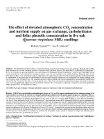
TG = 8.883*CRP + 0.042
200
TG (µg/l)
150
100
50
0
0
2
4
6
8
10
12
14
CRP (mg/l)
Chart 1: Correlation between TG and CRP (n = 168).
TG had a moderate possitive correlation with serum CRP (r = 0.596, p < 0.001).
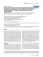
AntiTG = 14.103* CRP + 1.47
AntiTG (UI/ml)
250
200
150
100
50
0
0
2
4
6
8
10
12
14
CRP (mg/l)
Chart 2: Correlation between anti-TG and CRP (n = 168).
Anti-TG had a moderate possitive correlation with serum CRP (r = 0.693, p < 0.001).
The association between inflammation
and cancer was also reported in Lee S et
al’s study in 2011 [8]. They compared
hs-CRP levels in 80,781 patients, of whom
729 were diagnosed with primary cancer.
The results revealed that the mean hs-CRP
level of cancer patients was 2.9 mg/L,
130
which was significantly higher than that
of non-cancer patients (1.4 mg/L).
Inflammation is also considered as a
risk factor for thyroid cancer [3, 4].
Pro-inflammatory cytokines that arise in
chronic inflammation are thought to
trigger or promote cancer or may affect
Journal of military pharmaco-medicine no3-2019
the progression of this disease. In thyroid
cancer, histologic studies had confirmed the
penetration of white blood cell components
such as CD8+, CD4+, regulated T cells into
tumor tissue.
CONCLUSIONS
Study on serum TG and anti-TG
concentration in 168 patients with
differentiated thyroid cancer, we had
some comments:
- The median serum TG and anti-TG
concentration was 20.03 µg/L and
33.75 UI/mL, respectively. There was
95.8% of patients who had increasing
serum TG concentration and 42.9% of
patients who had increasing serum anti-TG
concentration. The proportion of patients
with both increasing serum TG and anti-TG
concentration was 41.1%.
- In group of patients ≥ 40 years old,
≥ T2, N1 and medium and high MACIS
scores, serum TG and anti-TG concentration
was significantly higher than those wthout
above features (p < 0.01). There was a
significant positive correlation between
serum TG, anti-TG concentration and
serum CRP concentration (p < 0.05).
REFFERENCES
1. Mai Trong Khoa. Quantification of serum
thyroglobulin in patients with thyroid cancer
treated with radioactive iodine-I131. Journal of
Practical Medicine. Ministry of Health. 2013,
869 (5), pp.101-104.
2. Ministry of Health. Treatment of thyroid
cancer by I131. Guidelines for the Diagnosis
and Treatment of Diseases with Nuclear
Medicine. 2014, pp.13-21.
3. Fugazzola L, Colombo C, Perrino M,
Muzza M. Papillary thyroid carcinoma and
inflammation. Front Endocrin. 2011, 2, p.88.
/>4. Ghoshal A, Garmo H, Arthur R et al.
Thyroid cancer risk in the Swedish AMORIS
study: The role of inflammatory biomarkers in
serum. Oncotarget. 2017 Dec. 4, 9 (1),
pp.774-782.
5. Holmes B.J, Sokoll L.J, Li Q.K.
Measurement of fine-needle aspiration
thyroglobulin levels increases the detection of
metastatic papillary thyroid carcinoma in cystic
neck lesions. Cancer Cytopathol. 2014, 122,
pp.521-526.
6. Indrasena B.S. Use of thyroglobulin as a
tumour marker. World J Biol Chem. 2017, Feb
26, 8 (1), pp.81-85.
7. Lee E.K, Chung K.W, Min H.S et al.
Preoperative serum thyroglobulin as a useful
predictive marker to differentiate thyroid
cancer from benign nodules in indeterminate
nodules. J Korean Med Sci. 2012, Sep, 27 (9),
pp.1014-1018.
8. Lee S, Choe J.W, Kim H.K, Sung J.
High-sensitivity C-reactive protein and cancer.
J Epidemiol. 2011, 21 (3), pp.161-168.
9. Li C, Yu W, Fan J et al. Thyroid functional
parameters and correlative autoantibodies as
prognostic factors for differentiated thyroid
cancers..Oncotarget. 2016, Aug 2, 7 (31),
pp.49930-49938.
10. Martins-Costa M.C, Maciel RMB,
Kasamatsu T.S et al. Clinical impact of
thyroglobulin (Tg) and Tg autoantibody (TgAb)
measurements in needle washouts of neck
lymph node biopsies in the management of
patients with papillary thyroid carcinoma. Arch
Endocrinol Metab. 2017, Mar-Apr, 61 (2),
pp.108-114.
131