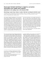
Semicircular canal anatomy as seen in microdissection
Bạn đang xem bản rút gọn của tài liệu. Xem và tải ngay bản đầy đủ của tài liệu tại đây (241.08 KB, 8 trang )
Journal of military pharmaco-medicine n06-2018
SEMICIRCULAR CANAL ANATOMY AS SEEN IN
MICRODISSECTION
Nguyen Thanh Vinh*; Tran Ngọc Anh**
Nguyen Hoang Vu***; Le Gia Vinh****; Pham Ngoc Chat*****
SUMMARY
Objectives: To investigate semicircular canal anatomy as seen in microdissection; evaluate
osseous semicircular canal, membranous semicircular canal and the relationship of semicircular
canal with the adjacent anatomical structures. Subjects and methods: Samples of 9 human
corpse heads, 18 ears were selected belonging to Department of Anatomy, University of Medicine
and Pharmacy, Hochiminh City. The semicircular canal was explored with transmastoid and
cranial fossa approach. Results: 9 human corpse heads, 18 ears were selected with average
age 61.5 (53 - 70). Horizontal, superior osseous semicircular canal and posterior membranous
semicircular canal was clearly identified with cranial fossa approach. Horizontal, posterior osseous
and membranous semicircular canals were clearly identified with transmastoid approach.
Membranous semicircular canal was situated the outer edge of the osseous semicircular canal.
Conclusions: All of these approaches can be used to clearly identify the semicircular canal
anatomical structure.
* Keywords: Osseous semicircular canal; Membranous semicircular canal; Cranial fossa approach;
Transmastoid approach.
INTRODUCTION
The semicircular canals system is a
component of the vestibular system,
contributing significantly to the body's
balance function. Anatomically, there are
three semicircular canals: horizontal (lateral),
superior and posterior semicircular canals.
They are very small structures, in different
planes, lied within the earlobe and buried
deep in the temporal bones. When studying,
researching or teaching, people has to
depend on images printed in textbooks or
models, which causes a lot of difficulties
to understand clearly, especially related
specialties such as: anatomy, neurosurgery
and ENT.
In the world, there are many books
have been written on temporal bone
surgery, but the presentation of the
approach to this system is still unclear
and specific. In Vietnam, there have also
been reports of semicircular canals, there
are images and clinical applications in the
diagnosis and treatment of the disease.
* ENT Hospital, Hochiminh City
** Vietnam Military Medical University
*** Medicine and Pharmacy University
**** Vietnam Medical Asociation
Corresponding author: Nguyen Thanh Vinh ()
Date received: 26/04/20181
Date accepted: 29/06/2018
121
Journal of military pharmaco-medicine n06-2018
In fact, when participating in the temporal
bone surgery courses, it is always difficult
to study the anatomy of semicircular canals
and requires intensive means as well as
the experience of surgeons performing
surgery. In order to solve this problem, we
need to have a specific approach that can
help physicians and practitioners to see
and understand correctly the anatomy of
semicircular canal system. Therefore, we
investigate “Labyrinth anatomy as seen in
microdissection and evaluate osseous
labyrinth, membranous labyrinth and the
relationship of labyrinth with the adjacent
anatomical structures”.
SUBJECTS AND METHODS
1. Subjects.
Vietnamese adults human corpse heads
were selected belonging to Department of
Anatomy, University of Medicine and
Pharmacy, Hochiminh City.
* Selection criteria:
- Vietnamese adults.
- Corpse heads were selected belong
to Department of Anatomy, University of
Medicine and Pharmacy of Hochiminh City.
- Normal temporal bone in anatomy.
* Exclusion criteria:
- Age < 18.
- Having ear problems.
- Interventions for ear surgery.
- Congenital malformations of the head
and neck.
- Traumatic in head or temporal region.
2. Methods.
Case series report.
* Research location: Department of
Anatomy, University of Medicine and
Pharmacy, Hochiminh City.
122
* Research facilities:
- Temporal bone dissection instruments.
- Semicircular canal microsurgery
instruments.
- Electric drilling machine.
- Carving drill bits and sharping drill bits,
sizes ranging from 4 mm to 0.5 mm.
- Aspirator machine, suction, syringe.
- Karz Zeiss microscope.
- Camera.
- Computers to save images.
* Microdissection with cranial fossa
approach:
- Cut the skull forming oval shape,
across the edge of the ear on both sides.
- Cut the brain stem, revealing the entire
base of the skull.
- Determine the Arch convex (prominence
of lateral semicircular canal).
- Three straight lines are perpendicular
to the petromastoid bones, 1 through the
center of the convex, 1 tangent to the
upper edge of the convex and 1 tangent
to the lower edge of the convex.
- The line is perpendicular to the three
lines above, tangent to the outer edge of
the Arch convex.
- Use electric drill machine, 3 or 4 mm
carving drill pit, drill the bone along the
outer edge of the tangent line outside the
Arch convex, reveal the mucosal layer of
posterior atrium ceiling.
- Use the microsurgical knife cut the
mucosal layer of posterior atrium ceiling.
- Identify short process of incus and
lateral semicircular canal.
- Determine the superior osseous
labyrinth from the Arch convex to the front
of lateral osseous labyrinth.
Journal of military pharmaco-medicine n06-2018
- Use the 1 - 2 mm carving drill pit, drill
out mastoid cells of surrounding vestibule
group of the superior osseous labyrinth,
exposing the entire superior osseous
labyrinth.
- Drill the mastoid cells around the
lateral and posterior semicircular canal.
- Use the 3 mm carving drill pit, drill the
cranial fossa bone surrounding the superior
osseous labyrinth.
- Grind the edge of 3 semicircular canals.
* Microdissection with transmastoid
approach:
- Make a postaural incision with No.15
or No. 20 scalpel blade, until the temporal
bone.
- Detach the musculoperiosteal flap
posteriorly and anteriorly to the external
auditory canal.
- The self-retaining retractor is utilized to
pull up the flap, expose the mastoid cortex.
- Using a large cutting burr (3 - 4 mm),
drilling is started along the temporal line,
then along the posterior wall of the external
auditory canal. Finally, a third line is drilled
perpendicular to the temporal line, through
the mastoid tip, to create a triangle.
- Continue to drill the mastoid cells to
open the antrum.
- Drill the tegmental mastoid cells, expose
the middle cranial fossa.
- Using a small cutting burr (1 - 2 mm),
drill the anterior and posterior signal cells
to expose the sinus and the Citelli’s angle.
- Using a 0.5 - 1 mm diamond burr, drill
the mastoid cells around the semicircular
canal, until no mucosa left.
- Identify the subarcuate artery, near the
center of 3 semicircular canals.
- Using a 0.5 - 1 mm diamond burr,
grind the bone surface of the mastoid
segment of CN VII, near posterior and
lateral semicircular canal, the second
genu of CN VII.
* Microdissect the semicircular canals:
- Through cranial fossa approach, use a
0.5 mm diamond burr to drill along the
medial side of the superior semicircular
canal, from the conjunction between the
superior semicircular canal and the lateral
semicircular canal to the conjuction between
the superior semicircular canal to the
posterior semicircular canal.
- Drill the bony semicircular canal until
the mucosa of the membranous semicircular
canal can be seen; from there, continue to
drill the superior semicircular canal to
expose totally the membranous superior
semicircular canal, from the ampulla to
the crus commune.
- Drill posteriorly to the tip cells, expose
the bone around the digastric muscle.
- Use the 0.5 - 1 mm diamond burr to
drill along the medial side of the lateral
semicircular canal, from the connection
with the superior semicircular canal to the
conjunction between the lateral semicircular
canal to the posterior semicircular canal.
- Drill to open the aditus, until the incus
can be identified. The lateral semicircular
canal can be seen.
- Drill the bony lateral semicircular canal
until the mucosa of the membranous
lateral semicircular canal can be seen;
- Continue to drill the perifacial cells,
expose the third segment of facial nerve.
123
Journal of military pharmaco-medicine n06-2018
continue to drill carefully the bony lateral
semicircular canal to expose totally the
membranous lateral semicircular canal.
- Left the bony part between the bony
superior semicircular canal and the lateral
semicircular canal, to distinguish the border
line between 2 membranous semicircular
canals and the ampulla of the superior
and lateral semicircular canal.
- Use 0.5 - 1 mm diamond burr, drill the
bony posterior semicircular canal, from
the crus commune to the opening of the
ampulla into the utricle. Continue to drill to
expose the mucosa of the membranous
posterior semicircular canal, then expose
totally the posterior semicircular canal.
- Use 0.5 mm diamond burr to drill the
bone between the superior semicircular
canal and the lateral semicircular canal.
- Use 0.5 mm diamond burr, drill the
crus commune of the superior semicircular
canal and the lateral semicircular canal.
RESULT
Through observation in 9 human corps
(18 ears), including 5 men and 4 women.
* Age: The youngest was 53, the oldest
was 71, mean age 61.5.
* Gender: Males 5 cases (55.5%); females:
4 cases (44.5%).
* Mastoid cells around the semicircular
canals: Well-developed: 11 cases (61.1%);
moderate developed: 6 cases (33.3%);
underdeveloped: 1 case (5.6%).
* Lateral bony semicircular canal:
Very clear: 4 cases (22.2%); clear: 12 cases
(66.7%); not clear: 2 cases (11.1%).
124
Table 1: Relative structure.
Relative structure
Number
of ears
Ratio
Tympanic segment of CN VII
18
100
Mastoid segment of CN VII
18
100
Second genu of CN VII
18
100
Short process of incus
18
100
Table 2: Bony semicircular canal.
Bony
semicircul
ar canal
Cranial fossa
approach
Postauricular
approach
Superior
Very clear
Quite clear
Lateral
Very clear
Very clear
Posterial
Quite clear
Very clear
Journal of military pharmaco-medicine n06-2018
Table 3: Membranous semicircular canal.
Membranous
Cranial fossa Postauricular
semicircular canal approach
approach
Superior
Not clear
Quite clear
Lateral
Quite clear
Very clear
Posterial
Very clear
Very clear
Table 4: Position of the membranous
semicircular canal in bony semicircular canal.
Semicircular
canal
Superior
Lateral
Posterior
Anterior wall
0
5
1
Lateral wall
16
12
15
Medial wall
2
1
2
Posterior wall
0
0
0
* Abnormalities:
- The lateral membranous semicircular
canal is concave downward in 1 case.
- Absence of the crus commune in 1 case.
DISCUSSION
1. Bony semicircular canal.
In terms of morphology, all three bony
semicircular canals are in the same
position as being described in books, in
which the lateral bony semicircular canal
had a higher rate to be seen clearly than
other bony semicircular canals in both
2 approaches (cranial and postauricular);
only two ears were not really visible
because of the extensive development of
the mastoid air cells, which surrounded
the lateral bony semicircular canal, so it
was difficult to see. Due to the relatively
vertical position, it was more difficult to
recognize superior bony semicircular canal
by the postauricular approach than cranial
approach. On the other hand, on the base
of the skull base, it was possible to see the
protrusion of the lateral bony semicircular
canal, which was easier to define. If mastoid
air cell is well-developed, there will be an
air cell between the superior semicircular
canal and the base of the skull; so it is
more easily recognized due to protrusion
of the lateral bony semicircular canal.
Particularly, because the posterior
semicircular canal was on the horizontal
position and lower than the superior and
lateral semicircular canal, it was buried
deep inside the otic capsule and was
difficult to recognize. On the other hand,
in mastoid bone with a well-developed air
cell, many layers of air cell covered the
lateral side of the posterior semicircular
canal. Thus, it is more difficult to detect
posterior semicircular canal.
125
Journal of military pharmaco-medicine n06-2018
Through the postauricular approach,
to see clearly the semicircular canal,
we need to extend the way up to the
epitympanum through the mastoid antrum
to the malleus-incudal joint. If the mastoid
air cells are good, mastoid antrum will be
large and the distance from the mastoid
antrum to the epitympanic roof will be
farther; so the approach was much easier
as the lateral semicircular canal was
easily recognized. If the mastoid air cell
was poorly developed, it was more
difficult to access the lateral bony
semicircular canal. Because semicircular
canal are located inside otic capsule, the
lateral semicircular canal plays an important
role in guiding to identify the remaining
semicircular canals. Thus, rely on an air
cells, identification of lateral semicircular
canal can be easy or difficult.
In autopsy bony semicircular canal,
we noted the connection of the bony
semicircular canals, particularly the
perilymph between the superior and
lateral semicircular canal in ampullae of
membranous semicircular canal position.
Therefore, perilymph connect to all 3
semicircular canals.
2. Membranous semicircular canal.
Membranous semicircular canal was
inside the bony semicircular canal. It had
been noted that membranous semicircular
canal was in lateral margin of semicircular
canal. In fact, we noted that most
membranous semicircular canal were
located on the outer edge of the bony
semicircular canal. It make the surgeons
open the bony semicircular canal carefully,
otherwise it will damage the membranous
semicircular canal.
126
Ampulle of superior membranous
semicircular canal is located near ampulle
of lateral membranous semicircular canal
in position of utricle.
3. Adjacent structures.
In the procedure, we noted that there
were structures associated with bony
semicircular canal, which will help
surgeons to identify semicircular canals in
difficult cases.
- Short limb of incus: The most nearby
anatomical landmark, slightly deviated
from the posterior branch of the lateral
bony semicircular canal. The more visible
the lateral semicircular canal is, the
shorter this distance is. In case of much
mastoid air cells, the air cells inserted
between the lateral bony semicircular
canal and the short limb of incus, making
this distance farther.
- Segment 2 of the nerve VII usually
position anterio-inferior to lateral bony
semicircular canal, bony covered of the
second segment can be defective,
revealing nerve VII. Due to the location
and defection of the bone, this anatomy
landmark is less mentioned although it is
considered to be related to the semicircular
canal system.
- Segment 2 of facial nerve: Usually is
located under short limb of incus,
surrounded by bone, covered above by
air cells. Thus, it is more difficult to
identify than short limb of incus. If mastoid
has less air cells, segment 2 of facial
nerve are located near the bony lateral
semicircular canal rather than short limb
of incus. If mastoid has more air cells,
segment 2 will be far away from lateral
bony semicircular canal.
Journal of military pharmaco-medicine n06-2018
- Segment 3 of the facial nerve is more
related to the posterior bony semicircular
canal, in the position of the ampulle
poured into the utricle. Normally, ampulle
of the lateral bony semicircular canal is
located just below - segment 3 of the
facial nerve, at the moment of passing of
facial nerve. If mastoid air cell is welldeveloped, more mastoid air cells insertion
between posterior bony semicircular canal
and segment 3 of the facial nerve,
widening the distance between ampulle
posterior bony semicircular canal and
segment 3 of the facial nerve.
4. Autopsy approach.
For ENT specialists, the postauricular
approach is a common since this way is
easy to learn and easy to apply to the
surgery. The semicircular canal approach
is to drill half of the bony semicircular canal
to clearly observe membranous semicircular
canal. The postauricular approach helps
accessing easily in the following order:
Lateral semicircular canal, posterior
semicircular canal and lastly superior
semicircular canal. Lateral semicircular
canal is easy to approach because the
direction of the microscope is straight,
and the lateral semicircular canal protrudes
more distal than other semicircular canal.
On the other hand, superior semicircular
canal, due to the vertical position, is higher
than the lateral semicircular canal and
contact with the skull bone; so this
semicircular canal is partially hidden,
difficult to dislocate. Moreover, the posterior
semicircular canal is usually more accessible
than superior semicircular canal because
it is only covered by well-developed air
cell on the surface. Surgeons, after drilling
these air cells, can recognize posterior
semicircular canal.
As membranous semicircular canal,
the postauricular approach helps surgeon
see anatomy structures clearly in the
following order: Posterior membranous
semicircular canal, lateral membranous
semicircular canal, and finally superior
semicircular canal. If only doing surgery to
see the anatomy structures, approach
type is not a big matter. However, as we
do research, our autopsy approach will
be more accurate than other approach.
Furthermore, for our application in surgery,
our autopsy approach is much more
applicable in real surgery for disease
treatment.
Cranial approach is easier to manipulate
with the superior semicircular canal,
easier to observe the lateral membranous
semicircular canal than the postauricular
approach. On the other hand, this approach
is wider than the postauricular one, which
can manipulate on all three semicircular
canals. However, this approach is only used
in autopsy, not applied in real surgery.
CONCLUSION
Through a combination of two approaches:
The postauricular approach and cranial
approach for accessing to bony membranous semicircular canal, with
9 corpses, 18 ears, we can conclude:
* Postauricular approach:
- Helping to approach bony semicircular
canal well in following order: Lateral,
posterior, and finally superior.
- For membranous semicircular canal,
the order is posterior, lateral, and superior.
127
Journal of military pharmaco-medicine n06-2018
* Cranial approach:
- Helping to approach bony semicircular
canal well in following order: Superior,
lateral and posterior.
- For membranous semicircular canal,
the order is lateral, posterior, superior.
Therefore, all two autopsy approaches
support each other, providing good access
to all three bony and membranous
semicircular canals.
REFERENCES
1. J.V Beek-King. Retrolabyrithine approach.
Operative Techniques in Otolaryngology. 2013,
Vol 24, pp.169-171.
2. J Bowman. The translabyrinthine approach.
Operative Techniques in Otolaryngology. 2013,
Vol 24, pp.149-156.
3. C. Cremers, J Mulder. Total labyrinthectomy.
Temporal Bone Dissection Manua. Kluger
Publication, Amsterdam, Netherlands. 2011,
pp.33-36.
4. C. Cremers, J Mulder. Translabyrinthine
approach of the internal acoustic canal.
Temporal Bone Dissection Manual. Kluger
Publication, Amsterdam, Netherlands. 2011,
pp.37-40.
128
5. H.W Francis, J.K Niparko. Laryrinthectomy.
th
Tymporal Bone Dissection Guide. 2 Ed,
Thieme, New York. 2016, pp.49-52.
6. H.W Francis, J.K Niparko. Translabyrinthine
exposure of the internal audistory canal.
th
Tymporal Bone Dissection Guide. 2 Ed,
Thieme, New York. 2016, pp.53-54.
7. A.J Gulya. Surgical anatomy of the
temporal bone and dissection guide. Surgery
th
of the Ear. 6 Ed, People Medical Publishing
House-USA, Connecticut. 2010, pp.771-790.
8. M Goycoolea. Temporal bone dissection.
Atlas of Otologic Surgery and Magic Otology.
Jaypee Brothers Medical Publicshers, London.
2012, Vol 1, pp.141-198.
9. M Sanna, T. Khrais. Transmastoid
th
approaches. The Temporal Bone. 1 Edition,
Thieme, New York. 2006, pp.22-54.
10. M Sanna, T. Khrais. Transotic approach.
th
The Temporal Bone. 1
Edition, Thieme,
New York. 2006, pp.98-105.
11. S.A Shanna, M Eid. Dehiscences of
the semicircular canals as discrete third
window lesions of inner ear. The Egyptian
Journal of Radiology and Nuclear Medicine.
2013, Vol 44, pp.15-21.
12. C Wijaya, A Dias. Superior semicircular
canal occlussion - transmastoid approach.
International Journal of Surgery Case Reports.
2012, Vol 3, pp.42-44.