Ebook Swanton’s cardiology (6/E): Part 1
Bạn đang xem bản rút gọn của tài liệu. Xem và tải ngay bản đầy đủ của tài liệu tại đây (5.42 MB, 320 trang )
Swanton’s
Cardiology
Swanton’s Cardiology: A concise guide to clinical practice Sixth Edition By R. H. Swanton and S. Banerjee
© 2008 R H Swanton and S Banerjee. ISBN: 978-1-405-17819-8
To Lindsay and Robin: with all our thanks for their patience, and for keeping
our tummies full when our minds were empty.
Swanton’s
Cardiology
A concise guide to clinical practice
R.H. Swanton
MA, MD, FRCP,
FESC, FACC
Consultant Cardiologist
The Heart Hospital
University College London Hospitals
Westmoreland Street
London W1G 8PH
S. Banerjee
MBChB, MD, MRCP
Consultant Cardiologist
East Surrey Hospital
Surrey and Sussex NHS Healthcare Trust
Canada Avenue, Redhill
Surrey RH1 5RH
The Heart Hospital
University College London Hospitals
Westmoreland Street
London W1G 8PH
SIXTH EDITION
© 2008 R H Swanton and S Banerjee
© 1984, 1989, 1994, 1998, 2003 by Blackwell Science Ltd
Published by Blackwell Publishing
Blackwell Publishing, Inc., 350 Main Street, Malden, Massachusetts 02148-5020, USA
Blackwell Publishing Ltd, 9600 Garsington Road, Oxford OX4 2DQ, UK
Blackwell Publishing Asia Pty Ltd, 550 Swanston Street, Carlton, Victoria 3053, Australia
The right of the Author to be identified as the Author of this Work has been asserted in accordance
with the Copyright, Designs and Patents Act 1988.
All rights reserved. No part of this publication may be reproduced, stored in a retrieval system, or
transmitted, in any form or by any means, electronic, mechanical, photocopying, recording or
otherwise, except as permitted by the UK Copyright, Designs and Patents Act 1988, without the prior
permission of the publisher.
First published 1984
Italian edition 1986
Spanish edition 1986
Second edition 1989
Yugoslav edition 1990
1
Italian edition 1991
German edition 1994
Third edition 1994
Polish edition 1994
Fourth edition 1998
Polish edition 1998
Fifth edition 2003
Italian edition 2006
Russian edition 2008
Sixth edition 2008
2008
Library of Congress Cataloging-in-Publication Data
Swanton, R. H.
Swanton’s cardiology : a concise guide to clinical practice/ by R.H. Swanton, S. Banerjee. – 6th ed.
p. ; cm.
Rev. ed. of: Cardiology / R.H. Swanton. 5th ed. c2003.
Includes bibliographical references and index.
ISBN 978-1-4051-7819-8
1. Cardiology–Handbooks, manuals, etc. I. Banerjee, S. (Shrilla) II. Swanton, R.H.
Cardiology. III. Title.
[DNLM: 1. Heart Diseases–Handbooks. WG 39 S972c 2008]
RC682.S837 2008
616.1′2–dc22
2007037459
ISBN: 978-1-4051-7819-8
A catalogue record for this title is available from the British Library
Set in 9.5/12 pt Palatino by SNP Best-set Typesetter Ltd., Hong Kong
Printed and bound in Singapore by Markono Print Media Pte Ltd
Commissioning Editor: Gina Almond
Development Editor: Victoria Pittman
Editorial Assistant: Jamie Hartmann-Boyce
Production Controller: Debbie Wyer
For further information on Blackwell Publishing, visit our website:
The publisher’s policy is to use permanent paper from mills that operate a sustainable forestry policy,
and which has been manufactured from pulp processed using acid-free and elementary chlorine-free
practices. Furthermore, the publisher ensures that the text paper and cover board used have met
acceptable environmental accreditation standards.
Designations used by companies to distinguish their products are often claimed as trademarks. All
brand names and product names used in this book are trade names, service marks, trademarks or registered trademarks of their respective owners. The Publisher is not associated with any product or
vendor mentioned in this book.
The contents of this work are intended to further general scientific research, understanding, and discussion only and are not intended and should not be relied upon as recommending or promoting a
specific method, diagnosis, or treatment by physicians for any particular patient. The publisher and the
author make no representations or warranties with respect to the accuracy or completeness of the contents of this work and specifically disclaim all warranties, including without limitation any implied
warranties of fitness for a particular purpose. In view of ongoing research, equipment modifications,
changes in governmental regulations, and the constant flow of information relating to the use of medicines, equipment, and devices, the reader is urged to review and evaluate the information provided in
the package insert or instructions for each medicine, equipment, or device for, among other things, any
changes in the instructions or indication of usage and for added warnings and precautions. Readers
should consult with a specialist where appropriate. The fact that an organization or Website is referred
to in this work as a citation and/or a potential source of further information does not mean that the
author or the publisher endorses the information the organization or Website may provide or recommendations it may make. Further, readers should be aware that Internet Websites listed in this work
may have changed or disappeared between when this work was written and when it is read. No warranty may be created or extended by any promotional statements for this work. Neither the publisher
nor the author shall be liable for any damages arising herefrom.
Contents
Preface, vii
Acknowledgements, ix
1 Cardiac Symptoms and Physical Signs, 1
2 Congenital Heart Disease, 17
3 Valve Disease, 71
4 The Cardiomyopathies, 130
5 Coronary Artery Disease, 159
6 Cardiac Failure, 255
7 Disturbances of Cardiac Rhythm: Bradycardias, Pacing, the ICD,
Biventricular Pacing for Heart Failure, 310
8 Disturbances of Cardiac Rhythm: Tachycardias and Ablation, 365
9 Infective Endocarditis, 419
10 Pericardial Disease, 448
11 The Heart in Systemic Disease, 460
12 Systemic Hypertension, 483
13 Pulmonary Hypertension and Pulmonary Embolism, 495
14 Diseases of the Aorta, 512
v
vi
Contents
15 Pregnancy and Heart Disease, 526
16 Cardiac Investigations, 545
17 Echocardiography, 594
Appendices
1 Nomogram for Body Size, 633
2 Rate Conversion Chart, 634
3 Further Reading, 635
4 References of Important Trials or Papers Quoted in the Text, 637
5 Useful Addresses and Hyperlinks, 646
6 Driving and Cardiovascular Disease in the UK, 648
7 List of Abbreviations, 653
Index, 659
Preface
I am delighted and very grateful that Dr Shrilla Banerjee has agreed to become
a co-author of the sixth edition of this cardiology handbook. Her enthusiasm,
knowledge and ideas have proved invaluable.
It is hoped that the book will be of practical help to doctors, nurses and
cardiac scientific officers confronted by typical management problems in the
cardiac patient. As a practical guide it is necessarily dogmatic and much
information is given in list format or in tables, especially in the sections
dealing with drug therapy.
Some subjects in cardiology are often not well covered in clinical training
and it is hoped that some sections will help fill any gaps in doctors’ or nurses’
clinical course, e.g. sections on congenital heart disease, pacing and cardiac
investigations. In addition, scientific officers and technical staff should find
that the clinical side of cardiology covered here complements their technical
training. And we hope that anaesthetists and intensive care unit physicians
will find the book of value.
Since the publication of the fifth edition 4 years ago there have been enormous advances in many aspects of cardiology and we have tried to highlight
these. Many sections have been extensively revised, and in particular those on
the cardiomyopathies, coronary disease, heart failure, echocardiography and
the heart in systemic disease. For ease of access the book now has 17 chapters.
The rhythm section has been split into two: bradycardias, pacing, implantable
cardioverter defibrillators and pacing for heart failure are dealt with in one
chapter, and tachycardias and ablation in another. There is a new, badly
needed chapter on pregnancy in patients with heart disease. A summary of the
At a Glance Guide for driving in the UK for patients with heart disease is now
included in appendix 6 by kind agreement of the DVLA. It should be remembered that the full guidance is updated on their website every 6 months.
In response to suggestions we are now able to include many more figures
and illustrations and we hope that these will increase the appeal of the book
without significantly increasing its bulk or expense. With regret we have still
decided not to have a separate section on nuclear cardiology but have included
its use in diagnosis where relevant.
vii
viii Preface
Practical procedures such as cardiac catheterization cannot be learnt from
a book. However, interpretation of catheter laboratory data is discussed and
it is hoped that the book will be helpful to the doctor learning invasive cardiology or the scientific officer monitoring it. Echocardiography is very much
a ‘hands-on’ technique and cannot be covered in depth in a book of this size.
However, this section has been considerably expanded with many more
illustrations.
Of all the specialities in medicine cardiology is right at the front in evidencebased practice. There are literally hundreds of trials to guide us in our dayto-day management decisions. Most of the trials have acronyms, which have
now become part of the language of cardiology. We have referred to the most
important trials in the text with the reference section expanded in Appendix
4. To save space we have used abbreviations liberally – but only those that
are in common use in everyday cardiology practice. The list of abbreviations
in Appendix 7 should cover these.
Drug names are changing. We have switched where appropriate from the
British Approved Name (BAN) to the Recommended Non-proprietary Name
(rINN) for medicinal substances. Adrenaline and noradrenaline remain
unchanged, however.
Finally, we are very grateful to colleagues who have suggested improvements or the inclusion of new material and would encourage the reader to
contact us with suggestions of subjects that are not covered at all or dealt with
inadequately.
R.H. Swanton
Acknowledgements
The work of a large number of authors has contributed to the body of
knowledge in this book and it would be impossible to thank them individually or provide detailed references to their work. In the list of trials, references
and further reading we have been able to incorporate their work and our
thanks to them all. We are very grateful to many cardiology colleagues, registrars and cardiac technical staff for their enthusiastic help in providing so
many ECG pressure tracings and echocardiograms.
Our thanks also to Ms Kalaiarasi Janagarajan, Mr Justin O’Leary, Ms Vivienne Palmer-White and Dr Stavros Kounas and all our colleagues who have
made suggestions for new material or alterations.
We are indebted to Dr Richard Sutton and Medtronic Ltd for permission to
modify their pacing code diagrams, to Dr Simon Horner for his diagram on
VT provocation, to Dr PE Gower for permission to include the nomogram for
body surface area, to Dr Diana Holdright for the illustrations on septal ablation pressure measurement, to Dr James Moon and Dr Sanjay Prasad for their
MRI pictures and to Dr Denis Pellerin for help with the echocardiography
section. Thanks to Fiona England for her patient acquisition of angiograms
and CT scans. Our thanks to Medtronic for permission to include the coronary
stent diagram and to Boston Scientific Ltd for the picture of the rotablator and
the Taxus stent. Our thanks to Cheryl Friedland for her invaluable and patient
tuition on ICDs and to Rhian Davies for her tireless help with drug queries.
A particular thanks to Dr Ewa Dzielicka from Krakow who has been of great
help in bringing several sections up to date.
Finally, and last but not least, we would like to thank Gina Almond and Vicky
Pittman from Wiley-Blackwell who have been towers of strength and encouragement. We are grateful to them for their ideas, their patience and their gentle
but regular persistence without which we would never have got this far.
R.H. Swanton
S. Banerjee
ix
x
Acknowledgements
Special Thanks
A big thank you to Lindsay and Tracy Harvey for their work in preparing the
early manuscript and to Jo Goddard for her tireless help sorting out numerous
emailed illustrations and references.
RHS
My thanks to my parents, Robin and family, and special thanks to my son,
Arun Lalit George – for being as inspiring as his namesakes.
SB
CHAPTER 1
1
Cardiac Symptoms and
Physical Signs
1.1 Common Cardiac Symptoms
Angina
Typical angina presents as a chest tightness or heaviness brought on by effort
and relieved by rest. The sensation starts in the retrosternal region and radiates across the chest. Frequently it is associated with a leaden feeling in the
arms. Occasionally it may present in more unusual sites, e.g. pain in the jaw
or teeth on effort, without pain in the chest. It may be confused with oesophageal pain, or may present as epigastric or even hypochondrial pain. The most
important feature is its relationship to effort. Unilateral chest pain (submammary) is not usually cardiac pain, which is generally symmetrical in
distribution.
Angina is typically exacerbated by heavy meals, cold weather (just breathing in cold air is enough) and emotional disturbances. Arguments with colleagues or family and watching exciting television are typical precipitating
factors.
Stable Angina
This is angina induced by effort and relieved by rest. It does not increase in
frequency or severity, and is predictable in nature. It is associated with STsegment depression on ECG.
Decubitus Angina
This is angina induced by lying down at night or during sleep. It may be
caused by an increase in LVEDV (and hence wall stress) on lying flat, associated with dreaming or getting between cold sheets. Coronary spasm may
occur in REM sleep. It may respond to a diuretic, calcium antagonist or nitrate
taken in the evening.
Swanton’s Cardiology: A concise guide to clinical practice Sixth Edition By R. H. Swanton and S. Banerjee
© 2008 R H Swanton and S Banerjee. ISBN: 978-1-405-17819-8
1
2
Chapter 1
Unstable (Crescendo) Angina
This is angina of increasing frequency and severity. Not only is it induced by
effort but it comes on unpredictably at rest. It may progress to myocardial
infarction.
Variant Angina (Prinzmetal’s Angina)
This is angina occurring unpredictably at rest associated with transient STsegment elevation on the ECG. It is not common, and is associated with coronary spasm often in the presence of additional arteriosclerotic lesions.
Other Types of Retrosternal Pain
• Pericardial pain: described in Section 10.1. It is usually retrosternal or epigastric, lasts much longer than angina and is often stabbing in quality. It is
related to respiration and posture (relieved by sitting forward). Diaphragmatic pericardial pain may be referred to the left shoulder.
• Aortic pain (Section 14.2): acute dissection produces a sudden tearing
intense pain, retrosternally radiating to the back. Its radiation depends on the
vessels involved. Aortic aneurysms produce chronic pain especially if rib or
vertebral column erosion occurs.
• Non-cardiac pain: may be oesophageal or mediastinal with similar distribution to cardiac pain but not provoked by effort. Oesophageal pain may be provoked by ergonovine, making it a useless test for coronary spasm. Oesophageal
spasm causes intense central chest pain, which may be relieved by drinking
cold water. Chest wall pain is usually unilateral. Stomach and gallbladder pain
may be epigastric and lower sternal, and be confused with cardiac pain.
Dyspnoea
This is an abnormal sensation of breathlessness on effort or at rest. With
increasing disability, orthopnoea and PND occur. Pulmonary oedema is not
the only cause of waking breathless at night: it may occur in non-cardiac
asthma. A dry nocturnal cough is often a sign of impending PND. With acute
pulmonary oedema, pink frothy sputum and streaky haemoptysis occur. With
poor LV function Cheyne–Stokes ventilation makes the patient feel dyspnoeic
in the fast cycle phase.
Effort tolerance is graded by New York Heart Association (NYHA) criteria
as follows.
Class I
Patients with cardiac disease but no resulting limitations of physical activity.
Ordinary physical activity does not cause undue fatigue, palpitation or
angina.
Class II
Patients with cardiac disease resulting in slight limitation of physical activity.
They are comfortable at rest. Ordinary physical activity results in fatigue,
Cardiac Symptoms and Physical Signs 3
palpitation, dyspnoea or angina (e.g. walking up two flights of stairs, carrying
shopping basket, making beds). By limiting physical activity, patients can still
lead a normal social life.
Class III
Patients with cardiac disease resulting in marked limitation of physical activity. They are comfortable at rest, but even mild physical activity causes fatigue,
palpitation, dyspnoea or angina (e.g. walking slowly on the flat). Patients
cannot do any shopping or housework.
Class IV
Patients with cardiac disease who are unable to do any physical activity
without symptoms. Angina or heart failure may be present at rest. They are
virtually confined to bed or a chair and are totally incapacitated.
Syncope
Syncope may be caused by several conditions:
• Vasovagal (vasomotor, simple faint): the most common cause. Sudden dilatation of venous capacitance vessels associated with vagally induced bradycardia. Induced by pain, fear and emotion.
• Postural hypotension: this is usually drug-induced (by vasodilators). May
occur in true salt depletion (by diuretics) or hypovolaemia.
• Carotid sinus syncope: a rare condition with hypersensitive carotid
sinus stimulation (e.g. by tight collars) inducing severe bradycardia (see
Section 7.6).
• Cardiac dysrhythmias: most common causes are sinus arrest, complete AV
block and ventricular tachycardia; 24-hour ECG monitoring is necessary.
• Obstructing lesions: aortic or pulmonary stenosis, left atrial myxoma or ballvalve thrombus, HCM, massive pulmonary embolism. Effort syncope is commonly secondary to aortic valve or subvalve stenosis in adults and
Fallot’s tetralogy in children.
• Cerebral causes: sudden hypoxia, transient cerebral arterial obstruction,
spasm or embolism.
• Cough syncope: this may result from temporarily obstructed cerebral
venous return. Profound bradycardia can be the cause mediated via the
vagus.
• Micturition syncope: this often occurs at night, and sometimes in men with
prostatic symptoms. It may result partly from vagal overactivity and partly
from postural hypotension.
The most common differential diagnosis needed is sudden syncope in an
adult with no apparent cause. Stokes–Adams attacks and epilepsy are the
main contenders (Table 1.1).
A prolonged Stokes–Adams episode may produce an epileptiform attack
from cerebral hypoxia. It is not always possible to distinguish the two
clinically.
4
Chapter 1
Table 1.1 Differentiation of Stokes-Adams attacks from epilepsy
Stokes-Adams attacks
Epilepsy
No aura or warning
Transient unconsciousness (often only a few seconds)
Very pale during attack
Rapid recovery
Hot flush on recovery
Aura often present
More prolonged unconsciousness
Tonic–clonic phases
Prolonged recovery; very drowsy
Absent
Cyanosis
Central cyanosis should be detectable when arterial saturation is <85% and
when there is >5 g reduced haemoglobin present. It is more difficult to detect
if the patient is also anaemic. Cardiac cyanosis may be caused by poor pulmonary blood flow (e.g. pulmonary atresia), right-to-left shunting (e.g.
Fallot’s tetralogy) or common mixing situations with high pulmonary blood
flow (e.g. TAPVD).
Cyanosis from pulmonary causes should be improved by increasing the
Fio2. The child breathes 100% O2 for 5 min. The arterial Po2 should increase
to >21 kPa (160 mmHg) if the cyanosis is pulmonary in origin. Cyanosis caused
by right-to-left shunting should change little in response to 100% O2 and certainly <21 kPa (160 mmHg).
Peripheral cyanosis in the absence of central cyanosis may be the result of
peripheral vasoconstriction, poor cardiac output or peripheral sludging of red
cells (e.g. polycythaemia).
Embolism
Both systemic and pulmonary embolisms are common in cardiac disease.
Predisposing factors in cardiology are shown in Table 1.2.
Table 1.2 Predisposing factors to pulmonary and systemic emboli
Pulmonary emboli
Systemic emboli
Either or both
Prolonged bed rest
High venous pressure
Central lines
Femoral vein catheterization
Pelvic disease (tumour,
inflammation)
Tricuspid endocarditis
Deep vein thrombosis
Atrial fibrillation
Aortic stenosis (calcium)
Mitral stenosis AF > SR
Infective endocarditis
LA myxoma
HCM
Prosthetic aortic or mitral
valves
Floppy mitral valve
Closed mitral valvotomy or
valvuloplasty
Mitral annulus calcification
Myocardial infarction
Dilated cardiomyopathy
CCF
Polycythaemia
Diuretics
Procoagulable state
Eosinophilic heart disease
Cardiac Symptoms and Physical Signs 5
Oedema
Factors important in cardiac disease are: elevated venous pressure (CCF pericardial constriction), increased extracellular volume (salt and water retention), secondary hyperaldosteronism, hypoalbuminaemia (liver congestion,
anorexia and poor diet), venous disease and secondary renal failure.
Acute oedema and ascites may develop in pericardial constriction. Proteinlosing enteropathy can occur, with a prolonged high venous pressure exacerbating the oedema.
Other Symptoms
These are discussed in the relevant chapter:
• Palpitation: principles of paroxysmal tachycardia diagnosis – see
Section 8.1
• Haemoptysis: mitral stenosis – see Section 3.2
• Cyanotic attack: catheter complications – see Section 16.3.
1.2 Physical Examination
Hands
It is important to check for the following:
• Dilated hand veins with CO2 retention
• Temperature (?cool periphery with poor flows, hyperdynamic
circulation)
• Peripheral cyanosis
• Clubbing: cyanotic congenital heart disease, infective endocarditis
• Capillary pulsation, aortic regurgitation, PDA
• Osler’s nodes, Janeway’s lesions, splinter haemorrhages (Figure 1.1), infective endocarditis
• Nail-fold telangiectases: collagen vascular disease
• Arachnodactyly: Marfan syndrome (see Figure 14.12)
Figure 1.1 Splinter haemorrhages in a man
with prosthetic valve endocarditis.
6
Chapter 1
Figure 1.2 Hypercholesterolaemia: knuckle xanthomas.
Figure 1.3 Familial hypercholesterolaemia: large xanthomas; serum cholesterol 14.1 mmol/l.
• Polydactyly, syndactyly, triphalangeal thumbs: ASD
• Tendon xanthomas: hypercholesterolaemia (Figures 1.2–1.5)
• Peripheral digital infarcts: hyperviscosity, cryoglobulinaemia (Figure 1.6).
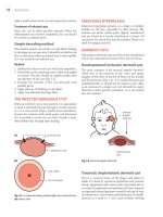
Facial and General Appearance
• Down syndrome (AV canal)
• Elf-like facies (supravalvar aortic stenosis)
• Turner syndrome (coarctation, AS)
• Moon-like plump facies (pulmonary stenosis)
• Noonan syndrome (pulmonary stenosis, peripheral pulmonary artery
stenosis)
• Mitral facies with pulmonary hypertension
Cardiac Symptoms and Physical Signs 7
Figure 1.4 Xanthelasma.
Figure 1.5 Tendon xanthomas: severe familial hypercholesterolaemia with massive cholesterol
deposition in Achilles’ tendon.
Figure 1.6 Peripheral digital infarcts: cryoglobulinaemia.
8
Chapter 1
Figure 1.7 Ear-lobe crease: in a young
patient may be a sign of coronary disease.
• Central cyanosis
• Differential cyanosis in PDA + pulmonary hypertension or interrupted
aortic arch
• Xanthelasma (see Figure 1.4)
• Ear-lobe crease in the young patient (Figure 1.7) association with coronary
disease
• Teeth: must be checked as part of general CVS examination
• Dyspnoea at rest. ?Accessory muscles of respiration.
The Jugular Venous Pulse
Waveform examples are shown in Figure 1.8. The JVP should fall on inspiration. Inspiratory filling of the neck veins occurs in pericardial constriction
(Kussmaul’s sign). The waves produced are as follows:
• ‘a’ wave: atrial systole. It occurs just before the carotid pulse and is lost in
AF. Large ‘a’ waves indicate a raised RVEDP (e.g. PS, PHT). Cannon ‘a’ waves
occur in: junctional tachycardia, complete AV block, ventricular ectopics
(atrial systole against a closed tricuspid valve).
• ‘c’ wave: not visible with the naked eye. Effect of tricuspid valve closure
on atrial pressure.
• ‘x’ descent: fall in atrial pressure during ventricular systole caused by
downward movement of the base of the heart.
Cardiac Symptoms and Physical Signs 9
Figure 1.8 Examples of waveforms seen on jugular venous pulse.
10 Chapter 1
• ‘v’ wave: atrial filling against a closed tricuspid valve.
• ‘y’ descent: diastolic collapse after opening of the tricuspid valve. Slow ‘y’
descent in patients with tricuspid stenosis or mechanical tricuspid valve
replacements.
• ‘s’ wave occurs in tricuspid regurgitation. Fusion of ‘x’ descent and ‘v’
wave into a large systolic pulsation can occur with rapid ‘y’ descent.
The normal range of JVP is –7 to +3 mmHg. The patient sits at 45° and the
sternal angle is used as a reference point.
Distinction between the JVP and the Carotid Pulse
Distinction of the JVP from the carotid pulse involves the following five
features:
1 Timing
2 The ability to compress the JVP
3 The ability to obliterate the JVP
4 The demonstration of hepatojugular reflux, the alteration of the JVP with
position
5 The site of the pulsation itself.
Although transient pressure on the liver is classically used to augment the
JVP, pressure anywhere on the abdomen will have the same effect. The congested liver is often tender and is pulsatile in severe tricuspid regurgitation.
Transient obliteration of the JVP to confirm that a pulse is venous is not
easy. The internal jugular vein is wide at the base of the neck and using the
point of a finger to obliterate it is often unsuccessful and thereby misleading.
Use the whole of the side of the index finger pushed firmly and briefly against
the side of the base of the neck. In addition the fact that a pulse is palpable
does not necessarily mean that it is arterial. Strong venous pulsations are also
palpable.
Using the external jugular vein to decide on the height of the JVP is not
always reliable. In some patients there may be a slight positional kink between
the junction of the external jugular vein with the subclavian vein. The external
jugular vein may thus appear full when the JVP (taken from the internal
jugular vein) is in fact normal.
The Carotid Pulse
Waveform examples are shown in Figure 1.9. There are three components to
the carotid pulse: percussion wave, tidal wave and dicrotic notch.
Percussion Wave
This is a shock wave transmitted up the elastic walls of the arteries.
Tidal Wave
This is reflection of the percussion wave with a forward-moving column
of blood. It follows the percussion wave and is not usually palpable
separately.
Cardiac Symptoms and Physical Signs 11
Figure 1.9 Examples of carotid pulse waveforms.
12 Chapter 1
Dicrotic Notch
This is timed with aortic valve closure.
All the pulses are felt, radials and femorals simultaneously (coarctation).
Any pulse may disappear with dissection of the aorta. Right arm and carotid
pulses are stronger than left in supravalvar aortic stenosis (see Section 3.4).
An absent radial pulse may occur:
• after a peripheral embolus
• after a Blalock shunt on that side
• after brachial artery catheterization with poor technique on that side
• after a radial artery line for pressure monitoring, or after the use of the
radial artery for cardiac catheterization
• with subclavian artery stenosis.
Palpation
This checks for: thrills, apex beat, abnormal pulsation and palpable sounds.
Systolic thrill in the aortic area suggests aortic stenosis. Feel for thrills in other
sites as follows.
• Left sternal edge: VSD or HCM
• Apex: ruptured mitral chordae
• Pulmonary area: pulmonary stenosis
• Subclavicular area: subclavian artery stenosis.
Diastolic thrills are less common: feel for apical diastolic thrill in mitral
stenosis with patient lying on left side and breath held in expiration. A left
sternal edge diastolic thrill is occasionally felt in aortic regurgitation.
Apex beat and cardiac pulsations
Heart is displaced, not enlarged (e.g. scoliosis, pectus excavatum?). Normal
apex beat is in the fifth left intercostal space in the midclavicular line. It is
palpable but does not lift the finger off the chest. In abnormal states
distinguish:
• normal site but thrusting, e.g. HCM, pure aortic stenosis, hypertension, all
with good LV
• laterally displaced and hyperdynamic, e.g. mitral and/or aortic regurgitation, VSD
• laterally displaced but diffuse, e.g. DCM, LV failure
• high dyskinetic apex, e.g. LV aneurysm
• double apex (enhanced by ‘a’ wave), in HCM, hypertension
• left parasternal heave; RV hypertrophy, e.g. pulmonary stenosis, cor pulmonale, ASD
• dextrocardia with apex in fifth right intercostal space.
Abnormal pulsations are very variable, e.g. ascending aortic aneurysm
pulsating in aortic area, RVOT aneurysm in pulmonary area, collateral pulsation round the back in coarctation, pulsatile RVOT in ASD, pulsatile liver
(felt in the epigastrium and right hypochondrium) in severe tricuspid
regurgitation.
Cardiac Symptoms and Physical Signs 13
Palpable heart sounds represent forceful valve closure, or valve situated
close to the chest wall, e.g. palpable S1 (mitral closure) in mitral stenosis, P2
in pulmonary hypertension, A2 in transposition, or both S1 and S2 in thin
patients with tachycardia.
1.3 Auscultation
Heart Sounds
First and second heart sounds are produced by valve closure. Mitral (M1) and
aortic (A2) are louder than and precede tricuspid (T1) and pulmonary (P2)
heart sounds. Inspiration widens the split.
A widely split second sound in mitral regurgitation and VSD is the result
of early ventricular emptying and consequent early aortic valve closure.
However, the widely split sound is rarely heard because the loud pansystolic
murmur usually obscures it. A summary is shown in Table 1.3.
Third Sound (S3)
This is pathological over the age of 30 years. It is thought to be produced by
rapid LV filling, but the exact source is still debated. Loud S3 occurs in a
dilated LV with rapid early filling (mitral regurgitation, VSD) and is followed
Table 1.3 The first and second heart sounds
First sound (S1) = M1 + T1
Loud
Soft
Variable
Widely split
Short PR interval
Tachycardia
Mitral stenosis
Long PR interval
Heart block
Delayed
ventricular
contraction (e.g.
AS, infarction)
Third-degree AV block
AF
Nodal tachycardia or VT
VT
RBBB
LBBB
VPBs
Second sound (S2) = A2 + P2
Loud A2
Widely split
Reversed split
Single
Tachycardia
Hypertension
Transposition
RBBB
PS (soft P2)
Deep inspiration
Mitral regurgitation
VSD
Hypertension
LBBB
Aortic stenosis
PDA
RV pacing
Fallot’s tetralogy
Severe PS
Pulmonary atresia
Eisenmenger
syndrome
Large VSD
Loud P2
Fixed split
PHT
ASD
14 Chapter 1
by a flow murmur. It also occurs in a dilated LV with high LVEDP and poor
function (post-infarction, DCM). A higher-pitched early S3 occurs in restrictive
cardiomyopathy and pericardial constriction.
Fourth Sound (S4)
The atrial sound is not normally audible but is produced at end-diastole (just
before S1) with a high end-diastolic pressure or with a long PR interval. It
disappears in AF. It is most common in systemic hypertension, aortic stenosis,
HCM (LV S4), pulmonary stenosis (RV S4) or after an acute MI.
Triple Rhythm
A triple/gallop rhythm is normal in children and young adults but is usually
pathological over the age of 30 years. S3 and S4 are summated in SR with a
tachycardia.
S3 and S4 are low-pitched sounds. Use the bell of the stethoscope and touch
the chest lightly.
Added Sounds
• Ejection sound: in bicuspid aortic or pulmonary valve (not calcified), i.e.
young patients
• Midsystolic click: mitral leaflet prolapse
• Opening snap, mitral: rarely tricuspid (TS, ASD, Ebstein’s anomaly)
• Pericardial clicks (related to posture).
Innocent Murmurs
Probably 30% of healthy young children have a heart murmur but <1% will
have congenital heart disease. This is usually the result of a pulmonary flow
murmur heard best at the left sternal edge radiating into the pulmonary
area.
Characteristics of Innocent Murmur
• Ejection systolic: diastolic or pansystolic murmurs are pathological. The
only exceptions are a venous hum or mammary soufflé.
• No palpable thrill.
• No added sounds (e.g. ejection click).
• No signs of cardiac enlargement.
• Left sternal edge to pulmonary area. May be heard at the apex.
• Normal femoral pulses.
• Normal ECG: chest radiograph or echocardiogram may be necessary for
confirmation.
The venous hum is a continuous murmur, common in children, reduced by
neck vein compression, turning the head laterally, bending the elbows back
or lying down. It is at its loudest in the neck and around the clavicles. It may
reappear in pregnancy.