Ebook Clinical atlas of head and neck anatomy: Part 2
Bạn đang xem bản rút gọn của tài liệu. Xem và tải ngay bản đầy đủ của tài liệu tại đây (8.36 MB, 102 trang )
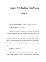
TI{E MOUTH, PALATE AND PHARYNX
The roof and floor of the mouth and the salivary
glands
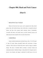
B The floor of the mouth (with the tongue
rernoved, together with the gingiva on the kft)
A The left half of the roof of the mouth, mandible
and parotid gland in a horizontal sectionofthe
head
45 Epiglottis
46 Vallecula
47 Bodv
I Dorsal root ganglion
I of second
;;;i;;i;"*"
3
?:;i?li:s,
I
4 Spinal root ofaccessorynerve
5 Lateral massof atlas
6 Dens of axis
7 Superiorconstrictorofpharynx
8 Nasal part of pharynx
9 Soft palate
l0 Hard palate
ll Palatalraphe
12 Alveolar processof maxilla
13 Vestibuleof mouth
14 Labial glands
15 Buccinator
16 Facial artery
17 Buccalfat pad
18 Masseter
19 Ramusof mandible
20 Lingual nerve
2l lnferior alveolarnerve
22 Inferior alveolarartery
23 Medial pterygoid
24 Styloglossus
25 Stylopharyngeus
nerve
26 Glossopharyngeal
27 lnternal carotid artery
28 Hypoglossalnerve
29 Superiorcervicalsympatheticganglion
30 Vertebral artery
3l Transverseprocessof atlas
32 Vagus nerve
33 Internal jugular vein
34 Stylohyoidligament
35 Stylohyoid
36 Posteriorauricularartery
37 External carotid artery
38 Retromandibularvein
39 Parotid gland
40 A zygomatic brdnch of facial nerve
4l Posteriorbelly of digastric
42 Accessorynerve
43 Occipital artery
44 Sternocleidomastoid
O The submandibularduct is 5 cm long. It emergesfrom the
superficialpart of the gland near the posteriorborder of
mylohyoid and passesforward betweenmylohyoidand
hyoglossusand then betweenthe sublingualglandand
genioglossus.It opensin the floor of the mouth on the
sublingualpapilla at the sideof the frenulum of the tongue.
'l
ii 6iJit"' t'o- J of hvoidbone
49
50
5l
$2
53
54
55
56
57
58
Hyoglossus
Geniohyoid
Mylohyoid
Genioglossus
Edentulous body of mandible
Frenulumoftongue
Sublingual papilla
Sublingual fold
Sublingual gland
Submandibularduct
c
Isolated left parotid gland and the mandible'
from the medial side (For the lateral surfaceof
the gland seepage 112)
D Isolated right sublingual and submandibular
glands and the mandible, from the medial side
59 Condylar processof mandible
60 Maxillary artery
61 Parotid gland
o
63
g
65
ffi
67
68
69
70
7l
72
73
74
75
External carotid artery
Great auricular nerve
Posterior $ivpjon ] of retromandibularvein
Anterror cllvrsron )
Ramus of mandible
Accessoryparotid gland
Parotid duct
Lower secondmolar tooth
Sublingual gland
Submandibularduct
Mylohyoid line of body of mandible
gland
of submandibular
Ylil
part )J
Deep tlll
Facial arterv
O The submandibulargland hasa largesuperficialand small
deep part, continuousround the posteriorborder of
mylohyoid.
O The superficialpart lies in the digastrictriangle. Important
relations include:
below - skin, platysma,the investinglayer of deepcervical
fascia,the facial vein, the cervicalbranchof the facial
nerve, submandibularlymph nodes.
lateially - the submandibularfossaof the mandible(below
the mylohyoid line), the insertionof the medialpterygoid,
the facial artery. .
medially - the mylohyoid and vessels,the lingual nerveand
submandibularganglion,the hypoglossalnerve, the deep
lingual vein, the hyoglossus.
O The deeppart of the gland lies on hyoglossuswith the
lingual nerve above, and the hypoglossalnerveand the
submandibularduct below.
sst'
*,fft'r,:.'
:,.o --..*
Ft"
"*#'
ru
"rdf
\' {' g :
&.
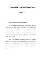
TIIE MOUTH, PALATE AND PHARYNX
The inside of the mouth and the hard and soft
palates
A Therighthalf of themouth, fromthelcft(with
skull dissectionto show the trigeminal,
ptery gopalatine and otic ganglia)
B The left half of the roof of the mouth, from
below (in a horizontal sectionthrough the head
below the level of the hard palate)
C The right half of the soft palate, from behind
(in a deepdissectionwith adjacentstructures
after removal of much of thepharynx)
I
2
3
4
5
6
7
8
9
l0
11
12
13
14
15
16
17
lt
19
20
2l
22
23
A
25
Xi
27
2t
29
30
31
32
33
34
35
36
37
38
39
40
4l
42
43
44
45
45
Sphenoidalsinus
Maxillary nerve
Sphenopalatineforamen and artery
Pterygopalatineganglion
Greater palatine nerve
Nerve of pterygoid canal
Tensor veli palatini
Nerve to tensor veli palatini
Nerve to medial pterygoid
Lingual nerve
Otic ganglion
Mandibular nerve
Greater petrosal nerve
Trigeminal ganglion
Internal carotid artery
Chorda tympani
Auriculotemporal nerve
Middle meningealartery
Maxillary artery
Inferior alveolar nerve
Medial pterygoid
Occipital artery
Posterior belly of digastric
External carotid artery
Facial artery
Deep part of submandibulargland
Tendon of digastric
Stylohyoid
Hypoglossalnerve
Stylohyoid ligament
Middle constrictor of pharynx
Epiglottis
Vallecula
Lingual artery
Hyoglossus
Vena comitans of hypoglossalnerve
Geniohyoid
Mylohyoid
Submandibularduct
Submandibularganglion
Nerve to mylohyoid
Superior constrictor of pharynx
Pterygomandibularraphe
Buccinator
Pterygoid hamulus
Palatopharyngeus
47 Soft palate
48 Dens of axis
49 Lateralmass of atlas
50 Nasal part of pharynx
5l Uwla
52 Tonsil
53 Stylopharyngeus
54 Vagus nerve
55 Internal jugular vein
56 Stylohyoid
57 Styloglossus
5t Parotid gland
59 Masseter
60 Ramus of mandible
6l Palatal glands
62 Hard palate
63 Vestibule of mouth
64 Base of styloid process
65 Intra-articular discof temporomandibularjoint
66 Lateral pterygoid
67 Inferior alveolar artery
6t Posterior part of submandibulargland
69 Superior thyroid artery
70 Superior laryngeal artery
7l Inferior constrictor of pharynx
72 Lamina of thyroid cartilage
73 Piriform fossa
74 Aryepiglottic fold
75 Internal laryngealnerve
76 Thyrohyoid
77 Thyrohyoid membrane
78 Greater horn of hyoid bone
79 Glossopharyngealnerve
t0 Palatine aponeurosis
81 Levator veli palatini
82 Musculus uvulae
83 Cartilaginous part of auditory tube
E4 Longus capitis
85 Posterior nasal aperture (choana)
E6 Nasal septum (vomer)
a All the musclesof the palateare suppliedby the
pharyngealplexusexcepttensorveli palatini which is supplied
by the nerve to the medial pterygoid (mandibularnerve).
O The mucousmembraneof the palateis suppliedby the
nasopalatine,greaterand lesserpalatineand glossopharyngeal
O The surfaceof the tonsil is pitted by downgrowthsof the
epithelium to form the tonsillar crypts.
O A deepcrypt-like structurenear the upper pole of the
tonsil is the intratonsillar cleft, and representsthe proximal
end of the embryonicsecondpharyngealpouch.
O The mucousmembraneon the surfaceof the tonsil is
and lesserpalatinenerves.
suppliedby the glossopharyngeal
a After entering the oral cavity beneath the lower border of
the superior constrictor of the pharynx, the lingual nerve lies in
contact with the periosteum of the mandible immediately
below and behind the third molar tooth.
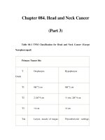
THE MOUTH, PALATE AND PHARYNX
The pharynx-external and internal surfaces
A The external surface; from the right (after deep
dissectionof the right inlraternporal fossa and
neck)
I Roots of auriculotemporalnerve
2 Middle meningealartery
3 Mandibular nerve
4 Lateral pterygoid plate
5 Maxillary artery entering pterygomaxillary
fissure
6 Chorda tympani
7 Lingual nerve
8 Tensor veli palatini
9 Levator veli palatini
10 Pharyngobasilarfascia
l l Superior constrictorof pharynxand ascending
palatine artery
12 Stylopharyngeusand glossopharyngealnerve
13 Styloglossus
14 Pterygomandibularraphe
15 Parotid duct
16 Buccinator
17 Molar glands
lE Facial artery
19 Mucoperiosteum of mandible
20 Sublingual gland
2l Submandibularduct
22 Geniohyoid
23 Mylohyoid
Z Nerve to geniohyoid
25 Hypoglossalnerve
26 Hyoglossus
27 Stylohyoid ligament
28 Middle constrictor of pharynx
29 Lingual artery
30 Greater horn of hyoid bone
3l Internal laryngealnerve
32 Superior hom of thyroid cartilage
33 Thyrohyoid membrane
34 Body of hyoid bone
35 Thyrohyoid
36 Superiorbelly of omohyoid
37 Sternohyoid
3E Stemothyroid
39 External laryngeal nerve
40 Inferior constrictor of pharynx
41 Cricothyroid
42 Arch of cricoid cartilage
43 Cricotracheal ligament
4 Truchea
45 Recurrent laryngealnerve
45 Inferior laryngeal artery
47 Inferior thyroid artery
48 Middle cervical sympatheticganglion
49 Vagus nerve
50 Scalenusanterior
5l Ventral ramus of fourth cervical nerve
52 Sympathetictrunk
53
54
55
56
57
5t
59
60
61
62
63
Ascending pharyngealartery
Superior laryngeal nerve
Superior root of ansacervicalis
Occipital artery
Transverseprocessof atlas
Accessorynerve
Posterior auricular artery
Internal jugular vein
Stylohyoid
Styloid process
Longus capitis
B The right internal surface (after removal of the
mucousrnembraneandpharyngobasilarfasci.a.
The tongueand uvula havebeendisplaced
forwards, and the epiglottis backwards)
64 Sphenoidalsinus
65 Vomer (posterior part of nasalseptum)
66 Tensor veli palatini
6l Cartllaginous part of auditory tube
68 Levator veli palatini
69 Soft palate
70 Uvula
71 Palatopharyngeus
72 Salpingopharyngeus
73 Superior constrictor
74 Longus capitis
75 Attachment of pharyngealrapheto pharyngeal
tubercle
76 Middle constrictor
77 Inferior constrictor
78 Piriform fossa
79 Larnina of cricoid cartilage
t0 Epiglottis
El Pharyngealwall overlying superior horn of
thyroid cartilage
E2 Greater horn of hyoid bone
83 Stylohyoid ligament
t4 Glossopharyngealnerve
85 Postsulcalpart ofdorsum oftongue
E5 Palatoglossus
joining it)
O Palatopharyngeus(with salpingopharyngeus
passesdownwardsinternal to the superiorconstrictor.
O Stylopharyngeuspassesdownwardsbetweenthe superior
and middle constrictors.
reach
a Fibresfrom palatopharyngeusand stylopharyngeus
the posterior border of the laminaof the thyroid cartilage,
and together with the inferior constrictorof the pharynxare
important in helping to elevatethe larynx during swallowing.
O All the musclesof the pharynxare suppliedby the
which is
pharyngealplexusexceptthe stylopharyngeus
supplied by the muscularbranchof the glossopharyngeal
nerve. The cricopharyngealpart of the inferior constrictor
an additional supplyfrom the externallaryngeal
leceives
O Passingsuperficialto hyglossus:the lingual nerve,
submandibularduct and hypoglossalnerve.
O Passingdeepto the posterior border of hyoglossus:the
glossopharyngealnerve, stylohyoidligamentand lingual
artery.
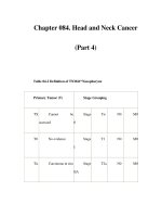
THE MOUTH, PALATE AND PHARYNX
The pharynx from behind
A From behind (with the sympathetictrunk and
part of the intemal carotid artery removedon
the right)
B The left half, from behind (after removal of the
left part of thepharyngobasilar fascia and parts
of the middle and inferi.or constrictors)
I Attachment of pharyngealraphe to pharyngeal
tubercle
2 Pharyngobasilarfascia
3 Ascending pharyngealartery
4 Internal carotid artery
5 Vagus nerve
6 Glossopharyngealnerve
7 Accessorynerve
E Hypoglossalnerve
9 Inferior ganglion of vagusnerve
10 Posterior meningealartery
ll Stylopharyngeus
12 Pharyngealbranch of glossopharyngeal
nerve
13 Pharyngealbranchofvagus nerve
14 Vagal branch to carotid body
15 Superior laryngealbranch of vagusnerve
16 Carotid sinus
'tip
17
ofgreater horn ofhyoid bone
lE Internal laryngealnerve
19 Superior thyroid artery
20 External laryngealnerve
2l Common carotid artery
22 lnternal jugular vein
23 Lateral lobe of thyroid gland
Z Cricopharyngealpart \ of inferior
25 Thyropharyngeal part I constrictor
26 Sympathetictrunk
27 Upper border of inferior constrictor
2E Superior cervicalsympatheticganglion
29 Middle constrictor
30 Upper border of middle constrictor
3l Superior constrictor
32 Upper border of superior constrictor
33 Pharyngealveins
34 Levator veli palatini
35 Tensor veli palatini
36 Ascending palatine artery
37 Medial pterygoid
3E Posterior border of lamina of thvroid
cartilage
a The pharynx extendsfrom the baseof the skull to the level
of the sixth cervicalvertebra, a distanceof about 12cm.
O The nasalpart (nasopharynx)extendsasfar down asthe
lower border of the soft palate. It containsthe openingof the
auditory tube and the pharyngealrecesslaterally,the
pharyngealtonsil on the posteriorwall, and opensanteriorly
into the nasalcavity through the posteriornasalapertures
(choanae).
the soft palateand
O The oral part (oropharyni
the upper border of the epiglottis,containsthe (palatine)
tonsil and palatopharyngealarch in its lateralwall, and opens
anteriorly into the mouth through the oropharyngealisthmus
(palatoglossalarches).
O The laryngealpart (laryngopharynx)extendsfrom the
upper border of the epiglottisto the lower border of the
cricoid cartilage,and is continuousbelow with the oesophagus.
The larynx projectsbackwardsinto it, with the piriform fossae
on either sideof the laryngealinlet.
O The pharyngobasilarfasciais the thickenedsubmucosaof
the pharynx that extendsbetweenthe upper border of the
superior constrictor and the baseof the skull.
O The buccopharyngealfascia(which is very much thinneq
than the pharyngobasilarfascia)lies on the externalsurfaceof
the pharyngealconstrictors,and is continuedanteriorly on to
the outer surfaceof the buccinator.
O Someof the uppermostfibresof the superiorconstrictor
and of the palatopharyngeusform a muscularbandthat during
ridge) on
swallowingraisesa transverseridge (Passavant's
the posterior pharyngealwall which, togetherwith elevationof
the soft palate, closesoff the nasalpart of the pharynxfrom
the oral part.
O The pharyngealplexuses(of nervesand of veins)are
situated mainly on the posterior surfaceof the middle
constrictor.
O The pharyngealplexusof nervesis formed by the
and vagus
pharyngealbranchesof the glossopharyngeal
componentis afferentonly; the
nerves.The glossopharyngeal
vagal componentis motor to the pharynx and palateaswell as
containing afferent fibres.
O
Glossopharyngealnerveparalysis:
No detectablemotor disability, asthe nervesupplies
only one small muscle,stylopharyngeus.
Loss of taste from the posteriorone-third of the tongue,
with anaesthesiain the sameareaand in part of the
pharyngealmucousmembrane.
a
Vagus and cranial accessorynerveparalysis:
Paralysisof the soft palateon the affectedside(the
palate is pulled towardsthe unaffectedsideon saying
'Ah').
Dysphagia(diffrculty in swallowing)due to paralysisof
pharyngealmuscles.
Hoarsenessof voice due to paralysisof laryngeal
muscles.
O
Spinalaccessorynerveparalysis:
Paralysisof sternocleidomastoidand trapezius.
O
Hypoglossalnerve paralysis:
Paralysisofthe tongueon the affectedside(with
deviation towardsthe affectedsideon protrusion,due
to the unopposedaction of the intact side).
The Ear
The external ear
A Right auricle, from the lateral side
B Right auricular cartilage, from the lateral side
C Rigbt auricular cartilage, from the medial side
I Helix
2 Scaphoid fossa
3 Upper crus of
antihelix
4 Lower crus of
antihelix
5 Triangular fossa
6 Crus of helix
7 Cvmba conchae
16
17
18
19
20
21
22
23
Z
25
26
E Concha
9 Cavum conchae
l0 External acoustic
meatus
1l Tragus
12 Intertragic notch
13 Antitragus
14 Lobule
15 Antihelix
Position of auricular tubercle (if present)
Spine of helix
Terminal notch
Tail of helix
Antitragohelicine notch
Cartilageof external acousticmeatus
Scaphoid eminence
Triangular eminence
Transverseantihelicine groove
Conchal eminence
Ponticulus
The middle and internal ear
D Dissectionthrough the right mastoid process
(in a dried skull) from the right
E Dissectionthrough the right mastoid process,
from the right and behind
F Section through the right temporal bone
showing the lateral wall of the middle ear, from
the left (with a black bristle indicartng the
chordatympani)
G Section through the right temporal bone
showing the medial wall of the middle ear, from
the right
H The left auditory tube and the lateral wall of the
middle ear, and the nasal part of the pharynx,
ftom the right (magnifiedx1.5)
27
28
29
30
31
32
33
34
35
Anterior I
Lateral
I semicircularcanal
Posterior I
Canal for fdcial nerve
External acousticmeatus
Tympanic part of temporal bone
Postglenoidtubercle
Mastoid air cells
Mastoid foramen
Dura mater of middle cranial fossa
Head of malleus in epitympanicrecess
Chorda tympani
Tympanic membrane
Facial nerve
Sheath of styloid process
Styloid process
Occipital condyle
Dura mater of sigmoid sinus
Tegmen tympani
Incudomallear joint
Body of incus
Epitympanic recess
Aditus to mastoid antrum
Mastoid antrum
Stylomastoidforamen
Semicanalfor auditory tube
Semicanalfor tensor tympani
Arcuate eminence(overlying anterior
semicircularcanal)
55 Oval window (fenestravestibuli)
56 Promontory
57 Trochleariform (cochleariform) process
58 Position of opening of auditory tube
59 Carotid canal
60 Jugular bulb
61 Round window (fenestracochleae)
62 Incudostapedialjoint
63 Handle of malleus
64 Tendon of tensor tympani and 57
65 Medial lamina \ of cartilaginouspart of
66 Lateral lamina J auditory tube
67 Opening of auditory tube
68 Inferior nasalconcha
69 Soft palate
36
37
3t
39
40
41
42
43
44
45
46
47
48
49
50
51
52
53
54
O The external ear consistsof the auricle (pinna) and the
external acousticmeatus,at the medial end of which lies the
tympanic membrane,separatingthe externalear from the
middle ear.
O The middle ear (tympaniccavity) is an irregularspacein
the temporal bone, lined with mucousmembrane,containing
the auditory ossicles(malleus,incusand stapes)and filled with
air that communicateswith the nasopharynxthrough the
auditory tube (Eustachiantube).
O The tympaniccavity consistsof the cavity proper and the
epitympanic recess.
O The epitympanicrecessis the part of the tympaniccavity
that projects upwardsabovethe tympanicmembrane,and
lodgesthe headof the malleusand the body of the incus.It
leadsbackwardsthrough the aditusihto the mastoidantrum,
which is an enlargedmastoidair cell.
149
!{;i
4,,u
;t....
Hil,
*.
qry
W
ffi;*"-
A
.c
, a
q
THE EAR
Horizontal sectionthrough the external, middle
and internal ear
A The lower surface of a sectionthrough a left
ear, fromabove
B The upper surface of the samesection,from
below (The two sectioru thus resembleadjacent
pages in a book that has beenopencd up)
C The central area of B (magnified x4)
D The upper surface of a sectionthrough a right
ear, from brllow (magnified x4) (This seaion is
at a slightly lower level than the sectionin
B and C)
I
2
3
4
Sigmoid sinus
Mastoid air cells
External acousticmeatus
Intra-articular disc of temporomandibular
joint
5 Superficial temporal artery
6 Zygomatic arch
7 Temporalis
E Maxillary artery
9 Maxillary sinus
l0 Pterygopalatinefossa
1 l Sphenoidal sinus
12 Cavernoussinus
13 Semicanalwith tensor tympani
t4 Internal carotid artery in carotid canal
15 Opening of auditory tube
t6 Cavity of middle ear
17 Tympanic membrane
18 Cochlea
19 Floor of intemal acousticmeatus
20 Promontory
2 l Facial nerve
22 Posteriorsemicircularcanal
23 Vestibular part of osseouslabyrinth
A Vestibular part \ of vestibulocochlearnerve
25 Cochlear part J in internal acousticmeatus
26 Labyrinthine artery
27 Internal carotid artery in foramen lacerum
28 Tendon of tensor tympani and trochleariform
(cochleariform) process
29 Chorda tympani
30 Long limb of incus
31 Pyramid
32 Stapedius
33 Stapes
34 Osseousspiral lamina
35 Basilar membrane
36 Scala tympani
37 Scala vestibuli
3E Modiolus
39 Handle of malleus
40 Incudostapedialjoint
E The disarticulated auditory ossiclesof the right
ear (magnifiedx4)
4l
42
43
44
45
45
47
4E
49
50
5l
Head
I
Neck
I
Anterior process I of malleus
Lateralprocess
I
Handle
l
Body
I
Short limb
I ".rncus
or
Long limb
I
Lenticular process I
Head
I
Posteriorlimb I
of staPes
ii n"","ri- rlit"u I
53 Base(footplate) J
O
O
For the genicularganglionof the facial nerveseepage166.
Featuresof the walls of the middle ear:
Lateral wall - the tympanicmembrane,part of the
petrotympanicfissure,the anterior and posterior
canaliculifor the chorda tympani.
Medial wall (from abovedownwards)- the prominencedue
to the lateral semicircularcanal, the prominencedue to
the canal for the facial nerve, the promontory (due to the
first turn of the cochlea),with the oval window (fenestra
vestibuli) occupiedby the footplate of the stapesabove
and behind the promontory, and the round window
(fenestracochleae)occupiedby the secondarytympanic
membranebelow and behindthe promontory.
Roof - the tegmentympani (part of the petrouspart of the
temporalbone).
Floor - above the superiorbulb of the internal jugular vein,
with the canaliculusfor the tympanicbranchof the
glossopharyngealnerve.
Anterior wall - the carotid canalwith (laterally) the
openingsof the semicanalsfor the tensortympani and the
auditorytube.
Posterior wall - the aditus to the mastoidantrum, the
pyramid (with stapediusemerging)in front of the vertical
part of the canalfor the facial nerve, and the fossafor the
lncus.
O The intemal ear consistsof the osseouslabyrinth and the
membranouslabyrinth.
O The osseouslabyrinth (within the temporalbone) consists
of the vestibule.the semicircularcanalsand the cochlea,
O The membranouslabyrinth is insidethe bony labyrinth
and consistsof the utricle and saccule(within the vestibule),
the semicircularducts (within the semicircularcanals),and the
duct of the cochlea(within the cochlea).
O The membranouslabyrinth containsendolymphand is
separatedfrom the bony labyrinth by perilymph.Thesetwo
fluids do not communicatewith one another,but the perilymph
probably communicateswith the cerebrospinalfluid in the
subarachnoidspacevia the cochlearbanaliculus.
The Larynx
The hyoid bone and cartilages of the tarynx
The cricoid and arytenoid cartilages
The hyoid bone
F From behind, with attachments
G From the left, with attachments
A From above and in front, with attachments
B From b€hind, with attachments
I
2
3
4
5
6
7
E
9
10
11
12
13
14
15
16
17
Greater horn
Lesser horn
Body
Stylohyoid ligament
Genioglossus
Geniohyoid
Mylohyoid
Sternohvoid
Omohvoid
Stylohyoid
Hyoglossus
Middle constrictor
Thyrohyoid
Thyrohyoid membrane
Hyoepiglottic ligament
Bursa
Chondroglossus
36 Apex
I
37 Muscular process I
of arytenoid cartilage
3E Articular surface II
)
39 Vocal process
40 Transvene arytenoid
41 Oblique arytenoid
42 Posterior crico-aMenoid
43 Corniculate cartilige
44 Cuneiform cartilage
45 Lamina of cricoid cartilage
46 Articular surface for arytenoid cartilage
47 Tendon ofoesophagus
48 Articular surfacefor inferior horn of thvroid
cartilage
49 Arch
50 Lateral crico-arytenoid
51 Cricothyroid ligament
52 Quadrangular membrane
The epiglottic cartilage
H From behind
The thyroid cartilage
C Fromthefront
D From the left, with attachments
E From behind, with attachments
18 Superior horn
19 Lamina
20 lnferior horn
2l Thyroid notch
22 Laryngeal prominence (Adam's apple)
;i iJ,?:il?'
) ,uu",.r"
25
26
27
28
29
30
31
32
33
34
35
Oblique line
Inferior constrictor
Sternothyroid
Thyrohyoid
Cricothvroid
Thyro-epiglottic ligament
Thyro-epiglottic muscle
Thyro-arytenoid
Vocal ligament
Conus elasticus
Stylopharyngeusand palatopharyngeus
O The hyoid bone consistsof a body with greater and lesser
homs on eachside.
O The thyroid cartilageconsistsof two laminaeunited
anteriorly and with superiorand inferior hornsposteriorly,
The gap above the united laminaeis the thyroid notch which is
bounded below by the laryngealprominence(Adam's apple).
The angle betweenthe laminaeis more acutein malesthan in
females,in whom the prominenceis lessobvious.
O The cricoid cartilageis shapedlike a signetring, with an
arch anteriorly and a liaminaat the back.
a The paired arytenoid cartilages have the shapeof a threesided pyramid, with at the basean (anterior) vocalprocdssto
which the vocal ligament is attachedand a (lateral) muscular
processto which the posterior and lateral crico-arytenoid
musclesare attached.
O The thyroid, cricoid and alrtrostall of the arytenoid
cartilagesare composedof hyalinecartilageand may undergo
some degree of calcification (becoming visible on radiographs).
O The apex of the arytenoid cartilage is composedof elastic
fibro-cartilage, like the epiglottic cartilage(which is
leaf-shapedwith numerouspits or perforations)and the
corniculate and cuneiform cartilages (which are like small
pips or rice grains). The triticeal cartilages are very small
nodulesthat are often found in the posterior marginofthe
thyrohyoid membrane.
THE LARYI\X
The larynx with the pharynx, hyoid bone and
trachea
A From the right (with the lateral lobe of the
thyroid gland displacedslightly backwards)
B After removal of the thyroid gland and part
of the inferior constrictor
C From the ftont and the right after removal of
muscles
(For a view of the inlet (aditus)of thelarynx
seepage 139)
O The intrinsic musclesof the larynx are suppliedby the
recurrent laryngealnerve, exceptthe cricothyroidwhich is
suppliedby the external laryngealnerve.
O The mucousmembraneof the larynx abovethe level of the
vocal folds is suppliedby the internal laryngealnerve,and
below the vocalfolds by the recurrentlaryngealnerve.
O The internal laryngealnerveentersthepharynxby
piercing the thyrohoid membrane,and from there fibres
spreadinto the larynx.
O The recurrent laryngealnerve lies immediatelybehindthe
cricothyroid joint, and entersthe larynx by passingdeepto the
lower border of the inferior constrictorof the oharvnx.
I
2
3
4
Lingual artery
Tip of greater horn of hyoid bone
Hyoglossus
O ln completeparalysisof the recurrentlaryngealnerve,
there is permanenthoarsenesS
of the voice,and the affected
Hypoglossalnerve
vocal cord assumesthe 'cadaveric'position,midwaybetween
S Suprahyoid artery
full abduction and adduction.
6 Nerve to thyrohyoid
7 Tendon of digastric
O ln incompleteparalysisof the recurrentlaryngealnerve,
E Digastric sling
the affectedcord takesup the adductedposition.
9 Body of hyoid bone
a In paralysisof the extemal laryngealnervethere may be
10 Sternohyoid
no detectableabnormality. If there is any, there is some
1 1 Superior belly of omohyoid
hoarsenessdue to lossof tensionin the affectedcord from the
12 Thyrohyoid
paralysedcricothyroid, but the hoarseness
will disappeardue
to hypertrophy of the oppositecricothyroid.
13 Lamina of thyroid cartilage
14 Laryngeal prominence
15 Sternothyroid
l6 External laryngeal nerve
t7 Inferior constrictor
18 Tendinousband
t9 Cricothyroid (straight part)
l\r*rlYtrrnsa
20 Lateral lobe of thyroid gland
2l Trachea
x2 Inferior laryngeal artery
23 Recurrent laryngeal nerve
24 Oesophagus
25 Inferior thyroid artery
26 Posterior pharyngealwall
27 Superior thyroid artery
28 Superior laryngealartery
29 Thyrohyoid membrane
30 Internal laryngealnerve
3l Cricothyroid (oblique part)
32 Arch of cricoid cartilage
33 Inferior horn of tfuyroidcartilage
v Cricothyroid joint
35 Epiglottis
36 Lesser horn of hyoid bone
37 Aperture for internal laryngealnerve and
superior laryngealartery
38 Conus elasticus(central part of cricothyroid
membrane)
39 Cricothyroid membrane(lateral part)
N Cricotracheal ligament
4l First tracheal ring (unusuallylarge)
)
-tt
$r^t,haa,^^l^,
ut^As/
[.0d4- +
Cru"ophU6{/1
Lt^6r/flt\
4
**' t^{^^rc\Avrql.w
0ed^^10,
J
qt k*d ,a^a^^brc..tae,
THE LARYNX
The muscles, ligaments and membranes
A From behind
B From the left (after reflecting the thyroid lamina
forwards)
C The internal surface of the right half (after
removal of most of the cricothyroi.dligament
and the overlyingmucous membrane)
D From the left (after resectingmost of the left
thyroid lamina)
E From behind (after removal of musclesin an
asymmetric specimen)
I Epiglottis
2 Vestibule
3 Aryepiglottic fold
'
4 Piriform fossa
5 Transversearytenoid
6 Oblique arytenoid
7 Posterior crico-arytenoid
E Lamina of cricoid cartilage
9 Site of attachmentof oesophagealtendon
10 Recurrent laryngealnerve
11 Cricothyroid joint
12 Inferior horn I
13 Lamina
I of thyroid cartilage
14 Superior horn )
15 Greater horn of hyoid bone
16 Vallecula
17 Aryepiglottic muscle
18 Thyro-epiglottic muscle
19 Superior thyro-arytenoid
20 Thyro-arytenoid
2l Lateral crico-arytenoid
22 First tracheal ring
23 Cricotracheal ligament
A Arch of cricoid cartilage
25 Cricothyroid
26 Internal laryngeal nerve
27 Vestibule and mucousmembraneoverlying
quadrangularmembrane
28 Vestibular fold
29 Ventricle of larynx
30 Vocal processof arytenoid cartilage
31 Vocalis part of thyro-arytenoid
32 Vocal ligament
33 Thyro-epiglottic ligament
v Body of hyoid bone
35 Hyo-epiglottic ligament
36 Thyrohyoid membrane
37 Quadrangular membrane
38 Cuneiform cartilage
39 Corniculate cartilage
& Muscular processof arytenoid cartilage
4l Cricothyroid ligament
a Crico-arytenoid joint
O The central part of the cricothyroid ligamentis usually
known as the conuselasticus(althoughsometextsusethis
term for the whole ligament). The lateral part of the
cricothyroid ligamentis sometimesknown asthe cricovocal
membrane.
O The upper (free) margin of the cricothyroid ligamentis
slightly thickened to form the vocal ligament.Coveredby
mucousmembraneit becomesthe vocal fold (vocalcord)' and
is attachedanteriorly to the laminaof the thyroid cartilage
adjacent to the midline, and posteriorly to the vocal processof
the arytenoid cartilage.
O The lower margin of the cricothyroid ligarnentis not free
but attachedto the upper border of the laminaand arch of the
cricoid cartilage.
O The quadrangularmembrane(a very thin sheetof
connectivetissuewhich hasbeenartificially thickenedfor
emphasisin D) passesbetweenthe lateral sideof the arytenoid
cariilage (which is relatively short) to the lateral edgeof the
epiglottic cartilage (which is relatively long). The membrane
is thus an irregular quadrilateralin shapeand not rectangular.
O The upper (free) margin of the quadrangularmembraneis
covered by mucousmembraneto form the aryepiglotticfold.
O The lower (free) margin of the quadrangularmembraneis
covered by mucousmembraneto form the vestibularfold
(false vocal cord).
O The slit-like spacebetweenthe vestibularand vocal folds
is the ventricle (or sinus)of the larynx, and is continuouswith
the saccule,a small pouch of mucousmembranethat extends
upwardsfor a few millimetres at the anterior part of the
vintricle betweenthe vestibularfold and the inner surfaceof
the thyroid lamina. Mucous secretionfrom glandsin the
sacculelubricatesthe vocal folds.
a The posterior crico-arytenoidis commonlyacceptedto be
the one musclethat can abductthe vocal fold (openthe
glottis).
O The lateral crico-arytenoidand the transverseand oblique
arytenoidsadduct the vocal fold (closethe glottis).
a The cricothyroid lengthens(and may increasetensionin)
the vocal fold.
V0= '+ r\s4v(
E , L5,,^,.
o+
t (-(a can^"
The Cranial Cavity
1-!.
.-
. f
: i l
.3-:::
l<::
li
.:-.
158
THE CRANIAL CAVITY
The cranial cavity, brain and meninges
The right half of a sagittal section,slightly to the
left of the midline
I Vault of skull
2 Superior sagittal sinus
3 Aperture of a superior cerebral vein
4 Falx cerebri
5 Corpus callosum
6 Septum pellucidum
7 Body of fornix
8 Choroid plexus of third ventricle
9 Thalamus and third ventricle
10 Midbrain
11 Aqueduct of midbrain
12 Inferior colliculus
13 Superior colliculus
14 Pineal body
15 Great cerebralvein
16 Basal vein
17 Straight sinus
18 Tentorium cerebelli
19 Falx cerebelli
20 External occipital protuberance
2l Posterior margin of foramen magnum
22 Cerebellum
23 Fourth ventricle
24 Choroid plexus of fourth ventricle
25 Pons
26 Medulla oblongata
27 Filamentsof arachnoidmaterin
cerebellomedullarycistern (cisternamagna)
28 Posterior atlanto-occipitalmembraneand
overlying dura mater
29 Posterior arch of atlas
30 Spinal cord (spinal medulla)
'l
3l Dorsal rootlets
^
nerves
32 Ventral rootlets I or spmal
33 Spinal subarachnoidspace
34 Body of axis
35 Dens of axis (left side)
36 Transverseligament of atlas
37 Alar ligament
3E Dura mater
39 Tectorial membrane
40 Superior longitudinal band of cruciform
ligament
41 Apical ligament
42 Anterior atlanto-occipitalmembrane
43 Anterior arch of atlas
44 Longus capitis
45 Posterior pharyngeal wall
46 Vertebral artery
47 Basilar artery
4t Basilar sinus
49 Sphenoidalsinus
50 Pituitary gland
51 Pituitary stalk
52 Dorsum sellae
53 Superior cerebellar artery
54 Posterior cerebral artery
55 Oculomotor nerve
56 Mamillary body'
57 Hypothaliamus
5E Optic chiasma
59 Anterior cerebral artery
60 Arachnoid mater overlying medial surfaceof
cerebral hemisphere
61 Crista galli
62 Lower border of falx cerebri and inferior
sagittal sinus
O The dura mater is sometimesknown asthe pachymeninx;
the arachnoidand pia mater togetherconstitutethe
leptomeninges.
12
i.F. '
4b
-s
. 4
i11
q'%
!
t
,'
',
''l
"'1,
:
-\.
tf
' - .-...,,'.*.-'.,";iS*=d
THE CRANIAL CAVITY
The cranial cavity and its coverings, from above
A Layered dissection, from above
B Cerebral dura mater, from the fight (The doued
circle indicates the position of pteri.on)
I Skin and densesubcutaneoustissue
2 Epicranial aponeurosis(galeaaponeurotica)
3 occipita.lbelly
) of occipitofrontalis
4 Frontal bellv
)
5 Branchesof superficialtemporal artery
6 Branchesof supra-orbitalnerve
7 Loose connectivetissueand pericranium
8 Bone of cranialvault
9 Sagittal suture
10 Coronal suture
11 Frontal (metopic) suture
12 Dura mater
13 Arachnoid mater
14 Cerebral hemispherecoveredby pia mater
15 Subarachnoidspace
16 Frontal branch I ot^mrddlemenmgealartery
17 parietal branch i
l8 Scalp
19 Arachnoid granulation
O
The scalpconsistsof
five layers:
the skin
denseconnectivetissue
the epicranialaponeurosisand the occipitofrontalis
muscle
looseconnectivetissue
the pericranium(periosteumon the outer surfaceof the
cranialvault)
O The meninges comprise the dura mater, arachnoid mater
and pia mater.
O The dura mater has cerebral and spinal parts.
O The cerebral part of the dura mater lines the inside of the
skull and consists of an outer endosteal layer (corresponding to
periosteum) which ends at the foramen magnum, and an inner
meningeal layer which forms sheathsfor the cranial nerves as
they pass out through skull foramina, and also forms four
processes- the falx cerebri, tentorium cerebelli, falx cerebelli
and diaphragma sellae.
O The venous sinusesof the dura mater lie between the
endosteal and meningeal layers and can be divided into two
groups:
Antero-inferior
Posterosuperior
Cavernous (paired)
Superiorsagittal
Intercavernous
Inferior sagittal
Sphenoparietal (paired)
Straight
Superior petrosal (paired)
Transverse(paired)
Inferior petrosal (paired)
Sigmoid(paired)
(paired)
Petrosquamous
Occipital
Basilar
Middle meningealveins
(paired)
to the
O The spinalpart of the dura matercorresponds
meningeallayerof the cerebralpart and formsa sheathfor the
spinalcord within the vertebralcanal.
a The arachnoidmaterliesinsidethe duramaterseparated
from it by the subduralspacewhichis merelya capillary
interval.
O The pia materadheresintimatelyto the surfaceof the
brain and spinalcord, and is separatedfrom the arachnoid
spacewhichcontainsthe
materby the subarachnoid
fluid. The pia materformsthe denticulate
cerebrospinal
septum.
ligament,filum terminaleand subarachnoid
a The middlemeningealarterydoesnot supplythe brain;it
lies betweenthe dura materandthe skull.
161
'.
"l!'-
6
*;:,, ,
''*
.h,
i.1$r
.,
tt
,'
t . .","j
N
*s-,
TIIE
CRANIAL
CAVITY
The brain in situ
From the left, (after removal of part of the skull and
duia mater)
I
2
3
4
5
6
7
8
9
10
11
12
Scalp
Cranial vault
Superior sagittal sinus
Openings of superior cerebralveins
Arachnoid granulations
Vesselsand arachnoidmater overlying
cerebral hemisphere
External occipital protuberance
Transversesinus
Cerebellar hemisphere
Sigmoid sinus
Mastoid air cells
External acousticmeatus
o\
q
t
r,
'tr
,*,