The effectiveness of silibinin on the change in some blood biochemical indices and liver histopathology of rabbit experimentally poisoned with amanita virosa mushroom
Bạn đang xem bản rút gọn của tài liệu. Xem và tải ngay bản đầy đủ của tài liệu tại đây (667.14 KB, 9 trang )
Journal of military pharmaco-medicine n06-2018
THE EFFECTIVENESS OF SILIBININ ON THE CHANGE IN SOME
BLOOD BIOCHEMICAL INDICES AND LIVER HISTOPATHOLOGY
OF RABBIT EXPERIMENTALLY POISONED WITH
AMANITA VIROSA MUSHROOM
Ngo Thi Thanh Hai*; Nguyen Thanh Binh*; Tran Van Tung*; Be Hong Thu**
SUMMARY
Objectives: To evaluate the effectiveness of silibinin on the change in some blood biochemical
indices and liver histopathology of rabbit poisoned with Amanita virosa mushroom in order to
get evidence of protective effect of the drug against the toxicity of amatoxin. Subjects and
methods: Rabbits were randomly grouped then poisoned with Amanita virosa mushroom.
12 hour after the intoxication, the rabbits were given silibinin then blood samples were collected
for the testing of GOT, GPT, GGT, urea, glucose, total bilirubin and direct bilirubin on a
automated system. Liver histology was processed and examined followed standard HE staining
procedure. Results: GOT, GPT, GGT concentrations in blood of rabbits poisoned with Amanita
rd
th
virosa in the group with silibinin treatment decreased on the 3 and 5 days after poisoning
compared to the group without treatment. Silibinin treatment also decreased hepatocellular
damages caused by Amanita virosa mushroom poisoning. Conclusion: Silibinin has protective
effect against the toxicity of Amanita virosa mushroom.
* Keywords: Silibinin; Amatoxin; Amanita virosa; Blood biochemical indices; Liver histopathology;
Rabbit.
INTRODUCTION
According to statistics from the Poison
Control Center of Vietnam Military Medical
University from 2004 to 2011, in Backan
province there were 28 incidences of
mushroom poisoning with the total number
of 94 infected people and 14 deaths.
The results of investigation showed that
the deaths from poisoning of poisonous
mushrooms in Backan province were
caused by Amanita virosa [1].
Amanita virosa is pure white, fleshy,
very beautiful and attractive. Amanita
virosa contains amanitines that cause
slow and sustainable poisoning to heat
and has a very high toxicity. Characteristics
of these toxins are to cause hepatocellular
necrosis leading to liver failure and death
[3, 4, 5].
The principle of treatment Amanita
virosa poisoning includes the limited
absorption and increased excretion of
amatoxin, and use liver protection drugs
as soon as possible accompanied by
maintaining vital functions and symptomatic
treatment [5, 6].
* Vietnam Military Medical University
** Bachmai Hospital
Corresponding author: Ngo Thi Thanh Hai ()
Date received: 18/05/2018
Date accepted: 20/06/2018
146
Journal of military pharmaco-medicine n06-2018
Silibinin dihemisusccinate prevents
amatoxin from entering hepatocytes,
reducing amatoxin in the intestinal and
hepatic circulation, thereby increasing
amatoxin excretion in bile and increasing
the synthesis of ARN polymerase 1 to
reduce hepatocellular injury [4 ,6, 7]. It is
recommended that this drug should be
used as an antidote to amantoxin. This study
was conducted to: Investigate the role of
silibinin in the change of biochemical indices
of rabbits poisoned with of amanita virosa
to provide the experimental evidence on
the ability of hepatocellular protection of
silibinin against toxic effect of amatoxin.
SUBJECTS AND METHODS
1. Subjects.
- Amanita virosa mushroom samples
were collected in Backan province. After
collection, the mushroom samples were
weighed, then preserved in ethanol till
next experiment.
- Rabbit: 30 rabbits, regardless of male
or female, healthy, weight: 2.0 ± 0.2 kg.
The rabbits were raised in the same mode
during the experimental period.
®
- Legalon SIL (silibinin dihemisusccinate)
528.5 mg.
2. Research methods.
* Method of poisoning on rabbits:
Amanita virosa preserved samples
were processed to evaporate ethanol.
The ethanol free samples were then
homogenized with a tissue homogenizer.
Water was then added to dilute the
sample then filtered with a filter paper.
The homogenizing and filtering procedure
were repeated three times to remove fiber
and particulate material from the mushroom.
The filtered solution was then administered
orally into rabbit stomach by specialized
equipment with a dosage equal to 2/3 of
the minimum lethal dose (LDmin) which
was pre-determined.
* Method of conducting the indicators:
- 30 rabbits were divided into 3 groups,
each group had 10 rabbits marked separately.
- Collect blood from vein in rabbit ears
before and after poisoning, on the first,
third and fifth days to test biochemical and
hematological indicators.
- Group 2 is not treated by drug after
poisoning.
- Group 3: After poisoning 1 day, inject
silibinin into rabbit vein with a maintenance
dose of 60 mg/kg/day for 4 days.
- Biochemical and hematological indicators
include: GOT, GPT, GGT, and urea
concentrations; creatinine, electrolytes,
blood sugar, total and bilirubin, etc. are
directly performed on automatic biochemical
analyzer.
- Liver histology was processed and
examined followed standard HE staining
procedure at Department of Pathology,
103 Military Hospital.
- Evaluation of clinical criteria: Skip meals,
diarrhea, mischievous level, etc.
* Statistical processing method:
The data is averaged (X), standard
deviation (SD) and compare 2 average
values. Calculate p before and after
poisoning, compare between the experimental
group and control group by t-test.
147
Journal of military pharmaco-medicine n06-2018
RESULTS
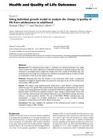
1. Changes in GOT, GPT and GGT activities in rabbits poisoned with Aminata
virosa treated or untreated with silibinin.
Group 1
UI/L
Group 2
800
Group 3
748.51
700
600
500
351.27
400
300
200
100
0
44.29
51.48
49.93
42.9
44.02
153.71
47.72
182.13
46.79
Time
43.21
Before poisoning
After 12h
3rd days after
5th days after
Figure 1: Changes in GOT activity.
GOT activity in serum of rabbits poisoned with Aminata virosa increased highly on the 3rd
and 5th days after poisoning. However, GOT activity in serum of poisoned rabbits untreated
with sibilinin was higher than that in poisoned rabbits treated with sibilinin (p < 0.05).
Group 1
UI/L
Group 2
Group 3
700
598.33
600
500
333.75
400
300
200
100
54.56
60.32
62.94
59.19
63.1
70.39
101.64
66.3
54.14
0
Before poisoning
181.42
After 12h
3rd days after
5th days after
Time
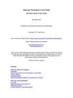
Figure 2: Changes in GPT activity.
GPT activity in serum of rabbits poisoned with Aminata virosa increased highly on
the 3rd and 5th days after poisoning. However, GPT activity in serum of rabbits untreated
with sibilinin was higher than that of rabbits treated with sibilinin (p < 0.05).
148
Journal of military pharmaco-medicine n06-2018
Before poisoning
UI/L
After 12h
3rd days after
192.5
5th days after
200
180
167.14
160
138.83
140
126.05
120
100
80
44.05
60
40
24.29
27.86
30.29
11.46
11.49
20
12.31 11.68
0
Group 1
Group 2
Group 3
Group
Figure 3: Changes in GGT activity in 3 groups.
GGT activity in non-poisoned groups did not differ between time points. In the
untreated group (group 2), GGT activity increased significantly from day 3. In the treated
group (group 3), GGT activity also increased from day 3 after poisoning. However,
GGT activity in group 3 was significantly lower than group 1.
2. Changes on CPK, glucose and urea levels in serum of rabbits poisoned with
Aminata virosa treated and untreated with silibinin.
UI/L
Group 1
Group 2
Group 3
1400
1167
1200
1000
716.8
800
600
266
400
200
635.5
287.8
284.8
271.4
491.7
0
Before poisoning
After 12h
3rd days after
5th days after
Figure 4: Changes in CPK level in 3 groups.
Before poisoning and after poisoning 1 day, CPK level did not differ between groups
with p > 0.05. CPK level increased at 3 days after poisoning and CPK in group 1 increased
significantly higher than group 2 with p < 0.05.
149
Journal of military pharmaco-medicine n06-2018
mmol/L
Before poisoning
After 12h
7.6
8
7
3rd days after
7.2
7.1
6.5
6.3 6.1
6
5.7
6
6.2
5th days after
6.1
6
6.4
5
4
3
2
1
0
Group 1
Group 2
Group 3
Group
Figure 5: Changes in glucose level.
Average blood glucose level in groups 2 and 3 increased slightly at 12 hours after
poisoning compared with before poisoning. Plasma glucose concentrations on days 3
and 5 after poisoning did not differ between three groups.
mmol/L
Group 1
Group 2
Group 3
9
8
7
6
5
4
3
2
1
0
Before poisoning
After 12h
3rd days after
5th days after Time
Figure 6: Changes in urea concentration.
The plasma concentrations of uremia in rabbit blood at the time before and after
poisoning were not statistically significant.
150
Journal of military pharmaco-medicine n06-2018
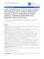
3. Changes in liver histopathology of rabbits poisoned with Aminata virosa
treated and untreated with silibinin.
A
B
C
Figure 7: Liver histopathology of rabbit normal (A, H.Ex.400),
rabbit poisoned with Amanita virosa untreated with silibinin (B, H.Ex.400),
rabbit poisoned with Amanita virosa treated with silibinin (C, H.Ex400)
(The liver of rabbit poisoned with Aminata virosa untreated with silibilin (B) showed
acute lesions: The central vein and vasculature in the liver were swollen, enlarged,
hepatocellular degeneration and necrosis. The infection by polymorphonuclear leukocytes
and lymphocytes into the portal and hepatic lobules. The liver of rabbit poisoned with
Aminata virosa used intravenous silibinin (C) showed a significant reduction in edema
and necrosis and moderate hepatocellular degeneration)
DISCUSSION
1. The effect of Amanita virosa on
some indicators of liver function.
AST and ALT are the most abundant
enzymes in the hepatocytes and play the
role in transporting amines in cell metabolism.
AST and ALT activities are usually relatively
stable in cytoplasm of hepatocytes. In
hepatocytes, AST is mostly in cytoplasm
and about 35 - 40% in plastid, while ALT is
only in the cytoplasm. When the hepatocytes
are injured, the permeability of the cell
membrane changes, causing AST and
ALT escape from cytoplasm into blood,
resulting AST and ALT of serum increase.
The more severe the hepatocytes are injured,
the more the AST, ALT activities in serum
increase, especially in cases of hepatocellular
necrosis. γ-GT is an enzyme of hepatocytes.
The activity of this enzyme increases
along with hepatocellular injury, especially
the injury caused by poisoning.
The research results showed that:
AST, ALT, γ-GT concentrations in serum
of rabbits poisoned with Amanita virosa
increased significantly on the 3rd and 5th
days compared to those before poisoning,
and AST activity increased higher than ALT
activity. This proved that the hepatocytes
were severely injured with hepatocellular
necrosis. In the event of hepatocellular
necrosis, the plastid is destroyed causing
151
Journal of military pharmaco-medicine n06-2018
AST from plastid escape into blood, plus
AST contained in cytoplasm leading AST
in serum increase higher than ALT. Our
research results were also consistent with
foreign authors’, that the liver of patients
poisoned with mushrooms containing
amatoxin such as Amanita virosa, Amanita
verna, Amanita phaloides, etc. is usually
injured severely. Patients with hepatic
failure due to poisonous mushrooms are
mostly fatal and these patients can only
be rescued thank to liver transplantation.
Hoang Cong Minh’s research (2009) on
rabbits poisoned with Amanita verna
containing amatoxin showed that AST
activity on the 5th day increased 25 times,
ALT activity increased 12 times higher
than before poisoning [2].
According to Tamas R Peredy (2015),
silibinin that is given intravenously was
effective in preventing liver injury and
should be used as soon as possible.
When silibinin is not available for intravenous
delivery, it should drain gallbladder
through the skin. The results in the group
treated by intravenous silibinin on the 3rd
and 5th days after poisoning, GOT, GPT and
GGT concentrations decreased markedly
with p < 0.005
2. The effect of Amanita virosa on
urea concentration in rabbit blood.
The research results showed that the
urea concentration in rabbits poisoned with
Amanita virosa changed insignificantly
compared to before poisoning. According
to Tamas (2015) and Timothy (2015), the
toxins of Amanita virosa in later stage will
cause renal cell injury. Urea is the major
degredation product of protein and
synthesized in the liver. Creatinine is a
152
decomposition product of phosphocreatinerich organisms, especially in muscle fibers.
Urea and creatinine are excreted through
the kidney and by monitoring the concentration
of these two substances in blood, we can
assess the functions and level of kidney
injury. A number of foreign authors studying
the effects of amatoxin on animals also
found that amatoxin causes damage to
the renal tubules. In this research, urea
concentration did not change much at the
research times, probably because the
rabbits were only poisoned with a dosage
equal to 2/3 of the minimum and determined
lethal dose.
3. The effect of Amanita virosa on
glucose metabolism function.
Glucose is the primary energy source
of organs, especially brain and muscles.
Glucose in blood is one of the indicators
to assess the glucose metabolism in the
body. The research results showed that
glucose concentration in blood increased
slightly on the first day and was changed
unclearly on the days after poisoning and
there was no difference in 2 groups with
and without treatment. Research results
of Floersheim G.L (1987); Chang A.K (2007);
Bivins et al (1985) showed that glucose
concentration in blood decreased in cases
of poisoning with species of mushroom
containing amatoxin, and in some cases,
blood glucose dropped to very low.
4. The effect of Amanita virosa on
CPK concentration in blood of poisoned
rabbit.
CPK usually increases in cases of
severe poisoning and acute destruction
of striated muscle cells. In this research,
CPK concentration increased significantly
Journal of military pharmaco-medicine n06-2018
on the 3rd and 5th days after poisoning with
p < 0.05. After treatment with silibinin,
CPK concentration decreased significantly
in group being treated with silibinin compared
to the group without treatment.
5. Changes on liver histopathology
of rabbits poisoned with Aminata virosa
treated and untreated with silibinin.
Liver of rabbit poisoned with Aminata
virosa untreated (figure 7A) showed acute
lesions: The central vein and vasculature
in the liver were swollen, enlarged,
hepatocellular degeneration and necrosis.
In contrast, after treatment by intravenous
injection of silibinin, we found the significant
reduction in degree of edema, necrosis
and moderate hepatocellular degeneration
in the liver of rabbit poisoned with Aminata
virosa (figure 7B).
CONCLUSION
The blood biochemical indices and
liver histopathology suggest that silibinin
has protective effect against the toxicity of
Amanita virosa mushroom.
REFERENCES
1. Ngo Thi Thanh Hai, Hoang Cong Minh,
Be Hong Thu. Situation of poisoning of
poisonous mushrooms in Backan province for
8 years (2004 - 2011). Journal of Military
Pharmaco-Medicine. 2012, 37 (7), pp.89-93.
2. Hoang Cong Minh. Study the effects of
Amanita verna extract on some biochemical
indicators in rabbits. Practical Medicine Magazine.
2009, 4 (656), pp.14-16.
3. Trinh Tam Kiet. List of large mushrooms
in Vietnam. Hanoi Agriculture Publishing House.
1996, pp.62-80.
4. Tamas R Peredy. Amatoxin-containing
mushroom poisoning including ingestion of
Amanita phalloides. Uptodate. 2015.
5. Timothy J Wiegand. Clinical manifestations
and evaluation of mushroom poisoning. Uptodate.
2015.
6. New Zealand National Poisons Centre.
Amatoxin. 2015.
7. Linsay Murray. Approaches to mushrooms
poisoning. Toxicology Handbook. Second edition,
Churchill Livingstone. 2011, pp.44-49.
153
Journal of military pharmaco-medicine n06-2018
CURRENT SITUATION OF HEALTHCARE RESOURCES
AT COMMUNE LEVEL IN BORDER AREAS OF
TAY NGUYEN FROM THE YEAR 2014 - 2016
Nguyen Minh Hung*; Trinh Thanh Hung*; Nguyen Van Chuyen**
Nguyen Van Ba***; Nguyen Dinh Thanh***; Le Bach Quang**
SUMMARY
Objectives: To study the status of healthcare resources at commune level in border areas of
Tay Nguyen (2014 - 2016). Subject and methods: A cross-sectional study, along with data
retrospective method to describe healthcare situation in 28 commune-level health stations
border areas of Tay Nguyen. Results: Commune health system have enough health staff;
however, they lack a structure of professional qualifications. 92.86% of commune health system
have doctors, of which 88.46% were general practitioners, only 3.85% were traditional medicine
doctors and 7.69% were pediatricians. The percentage of unskilled doctors remained high,
accounting for 57.69%. All commune health system had their own facilities; most of them did not
have enough functional departments as prescribed. Their equipment was still limited compared
to regualtions, especially basic equipment for medical examination and special treatment.
Conclusion: There was a lack of human resources and facilities in healthcare systems at commune
level in border areas of Tay Nguyen.
* Keywords: Healthcare resources; Commune health system; Border areas; Tay Nguyen.
INTRODUCTION
There are 28 communes and 12 districts
of Kontum, Gialai, Daklak, Daknong
sharing a border with Laos, Cambodia,
including 530 km of border line, of which
Laos has 142 km, Cambodia has 388 km.
This area has the lowest socioeconomic
status and the poorest transportation
system in Tay Nguyen. Health care for
people in the border area of Tay Nguyen
is still heavily dependent on grassroots
health care, especially at commune and
village levels. However, this area encounters
many difficulties in human resources, medical
infrastructure and equipment. The medical
management in some aspects is limited;
the quality of health care is still inadequate.
Therefore, many health indicators such as
health care services and others of Tay
Nguyen are slowly improved compared
with other regions and with general level
of the country. Hence, a research on the
status of health resources at the commune
level in Tay Nguyen is very necessary.
This is a scientific basis to develop solutions
to improve the medical examination and
treatment capacity of communes.
Objectives: Research on the status of
healthcare resources at commune level in
border areas of Tay Nguyen (2014 - 2016).
* Ministry of Science and Technology
** Vietnam Military Medical University
*** 103 Military Hospital
Corresponding author: Nguyen Minh Hung ()
Date received: 10/04/2018
Date accepted: 20/06/2018
154