Ebook Diabetes in childhood and adolescence (Vol 10): Part 2
Bạn đang xem bản rút gọn của tài liệu. Xem và tải ngay bản đầy đủ của tài liệu tại đây (2.73 MB, 202 trang )
Chiarelli F, Dahl-Jørgensen K, Kiess W (eds): Diabetes in Childhood and Adolescence.
Pediatr Adolesc Med. Basel, Karger, 2005, vol 10, pp 181–189
Sports and Physical Activity in Children
and Adolescents with Type 1 Diabetes
mellitus
K. Raile, A. Galler, T.M. Kapellen, V. Noelle, W. Kiess
Universitätsklinik und Poliklinik für Kinder und Jugendliche,
Universität Leipzig, Leipzig, Deutschland
Physical exercise has been one of the basic principles in the management
of diabetes, even before the introduction of insulin therapy. Nowadays, all levels
of exercise, including leisure activities, recreational sports and competitive performance can be managed by people with type 1 diabetes. Any kind of physical
activity is to be highly valued, because exercise improves the known risk factors
for macrovascular disease, in particular lipoprotein profile, blood pressure,
obesity and cardiovascular fitness. This chapter focuses on first, the rating of
physical activity in children and adolescents with type 1 diabetes. Second, the
physiology and pathophysiology of muscular activity in type 1 diabetes. Third,
how sports and exercise interact with diabetes acute and late complications.
Finally, practical guidelines at any level of physical activity are provided.
Olympic Gold and Himalayas with Diabetes
Nowadays, all levels of physical activity can be performed by individuals
with type 1 diabetes [1]. Athletes with type 1 diabetes have managed to win
Olympic gold medals, like Steve Redgrave, British champion rower, or Karsten
Fischer, player in the national German hockey team. These two athletes and
many others are organized in the ‘Diabetes Exercise and Sports Association’
DESA (former IDAA). Their main targets are to educate people with diabetes,
to enhance self-care and self-management skills, and to provide a forum to
exchange information, experience, and resources (www.diabetes-exercise.org).
Also, extreme altitude mountaineering on Himalayas’ summits has been
managed by climbers with type 1 diabetes. These extreme sports challenge
not only man but also the technique of glucose monitoring and insulin application [2].
Knowing that people with type 1 diabetes manage these extreme physical
boundaries helps some children, adolescents and families with type 1 diabetes
to trust again in their own physical opportunities. Diabetes care teams should
support any kind of sports, especially if children are motivated to start a particular sport. Sports performed before diabetes manifestation should be continued
and treatment regimens to keep the performance level should be worked out. If
diabetic retinopathy or nephropathy is present, special monitoring is required
and exercise levels should be selected with care.
Physical Activity in Childhood and Youth
Some aspects on exercise in children and adolescents with diabetes shall
be reviewed here. In a cohort study, we interviewed 142 children with type 1
diabetes of school age (6–18 years) and 97 healthy siblings of similar age and
BMI as controls. We used a structured questionnaire and recorded time spent
on physical activity and sports at school, in competitive sports and in general.
We asked for favorite sports in general and in competitive sports. Age, weight,
height and body mass index were obtained from both groups. In the diabetes
group, duration of diabetes, average daily carbohydrate intake, number of
insulin injections and daily insulin dose was documented.
The groups did not differ in terms of time spent for sports at school and in
competitive sports. In their spare time, boys and girls with diabetes reported
significantly more physical activity (table 1). Interestingly, their favorite sports
in general did not differ between the diabetes and control groups, but it was
remarkably different between boys and girls (table 2).
Within the diabetes group (total n ϭ 142), those boys and girls who regularly participated in competitive sports (n ϭ 42) were significantly more active
during the rest of their spare time, while the mean BMI, daily insulin dose and
HbA1c were only slightly higher in the group that reported no competitive
sports activity (n ϭ 98; table 3) [3].
Thus, diabetes does not seem to restrict children and adolescents from
spending time with sports and to select their favorite sporting disciplines.
The higher sporting activity in girls and boys with diabetes is of special interest as it might be a compensating social behavior and a help for assimilation
within their peer group. Also, the request for perceived physical fitness and
Raile/Galler/Kapellen/Noelle/Kiess
182
Table 1. Time spent for sports in children with type 1 diabetes mellitus
and healthy siblings
Exercise and sports
h/week
Diabetes mellitus
(n ϭ 142) mean
(SD)
Healthy siblings
(n ϭ 97) mean
(SD)
p value
Spare time
Competitive sports
School
6.80 (4.20)
1.79 (2.47)
2.49 (0.88)
4.60 (4.51)
2.02 (2.47)
2.36 (0.78)
0.001
0.40
0.17
Table 2. Ranking list of the favorite sports of girls and boys with diabetes mellitus and
healthy siblings
Girls
Boys
Diabetes
Siblings
Diabetes
Siblings
Biking (28%)
Swimming (16%)
Inline skating (13%)
Biking (28%)
Swimming (15%)
Inline skating (15%)
Biking (27%)
Soccer (20%)
Inline skating (13%)
Biking (32%)
Soccer (24%)
Inline skating (11%)
Sports are % of all nominated sports.
Table 3. Impact of competitive sports on diabetes treatment in children and adolescents with type 1 diabetes mellitus
Age, years (average)
Duration of diabetes, years
BMI, kg/m2
Injections/day
Daily insulin dose, IU/kg/day
Carbohydrates per day, g
HbA1c
Sport in spare time, h/week
No competitive sports
(n ϭ 98)
Competitive sports
(n ϭ 44)
p value
12.5 (3.3)
5.1 (3.7)
19.8 (3.9)
3.2 (0.91)
0.93 (0.34)
203.1 (45.6)
7.62 (1.40)
6.15 (4.15)
12.7 (2.7)
5.3 (3.0)
19.7 (3.8)
3.1 (0.95)
0.90 (0.36)
217.2 (49.2)
7.30 (1.05)
8.23 (4.00)
NS
NS
NS
NS
NS
NS
NS
0.006
Data are means (SD). HbA1c represents the mean of HbA1c values within the preceding
year.
Sports and Physical Activity in Children and Adolescents
183
Frequency of sport
participation
Perceived health
status
ϩ
Ϫ
Tobacco
consumption
Ϫ
Ϫ
Alcohol
consumption
Ϫ
Ϫ
Feelings of
anxiety
Ϫ
Ϫ
Feelings of
depression
Ϫ
ϩ
Perceived
physical fitness
ϩ
Fig. 1. Model of sports and perceived health according to Pastor et al. [19].
health might explain the higher physical activity in children with diabetes
(fig. 1).
Physiology and Pathophysiology of Muscular Activity
Muscular activity increases insulin sensitivity. This principle was already
used as the first treatment in severely insulin-deficient patients with type 1
diabetes. With their poor insulin secretion, the increase of insulin sensitivity
even prolonged their survival. Nowadays, physical activity is an established
treatment for type 2 diabetes. Insulin sensitivity is increased and hyperinsulinemia is reduced. It is known for more than 30 years that contracting muscle increases its own glucose uptake [4, 5]. More recent research highlighted
the biochemical aspects. As part of the increased muscular glucose uptake,
GLUT4 glucose transporters are up-regulated to the cell surface by insulin
but also independently by muscular contraction [6, 7]. In insulin-resistant
patients with type 2 diabetes only insulin-induced not exercise-induced
GLUT4 regulation is impaired [8]. There is increasing evidence that
AMP-activated protein kinase (AMPK) is stimulated by high AMP-to-ATP
and creatine-to-phosphocreatine ratios. Thus, muscular contraction, leading to
low intracellular phospho-energy stores, activates AMPK independently
of insulin. AMPK activation results in acute up-regulation of GLUT4 glucose transporters and in an increased glucose uptake, in addition to insulinstimulated effects [9, 10].
Raile/Galler/Kapellen/Noelle/Kiess
184
These new, biochemical aspects explain why insulin and physical activity
lower blood glucose independently and synergistically. Insulin has a much
stronger effect during and after muscular exercise and high insulin levels combined with physical activity can lead to life-threatening hypoglycemia.
Acute Complications: Hypoglycemia and Ketoacidosis
Hypoglycemia is a classical complication during and after physical activity because insulin effects are enhanced and hypoglycemia awareness might be
reduced. Nevertheless, there is no link between either physical fitness or physical activity and the incidence of severe hypoglycemia [3, 11, 12]. The experience of an acute hypoglycemic attack might induce fear and anxiety in parents
of children with type 1 diabetes [13]. Fears of hypoglycemia might be a burden to start sports even at school. Severe hypoglycemia is the most feared
acute complication of physical exercise by parents, teachers, or team coaches,
and education, information materials and in some severe cases psychological
intervention might be considered necessary to overcome these fears and enable
regular sports participation [14].
Severe ketoacidosis could develop if muscular activity starts at insulin
levels that are too low to block ketogenesis. So if glucose levels are high
before exercise, urine should be tested for ketone bodies [see chapter by
Brink, Management Recommendations, pp. 94–121]. In case of ketonuria,
severe activity should be avoided, short-acting insulin should be injected and
ketonuria tested until glucose levels and ketonuria decrease. The safest way
to avoid unexpected and severe hypoglycemia or ketoacidosis is frequent
blood testing, adjustment of insulin dose and intake of carbohydrates at short
intervals. Practical skills must be trained in diabetes education and diabetes
camps.
Late Complications: Sports and Risk Factors
Since the DCCT or other major studies investigating the development of
diabetic retinopathy and nephropathy, HbA1c levels are the dominant surrogate
marker to estimate an individual risk to develop late complications [15, 16].
Austin et al. [17] investigated VO2max levels by progressive bicycle ergometry
to assess physical fitness in 28 boys and 31 girls with type 1 diabetes. They
found an inverse correlation of VO2max and HbA1c, Lp(a), and LDL-cholesterol
and concluded that physical fitness might thus reduce the risk for cardiovascular disease. Furthermore, lower HbA1c levels might account for a lower risk
Sports and Physical Activity in Children and Adolescents
185
for diabetes late complications. Similar results have already been found by
Huttunen et al. [11] in 1984 and by Campaigne et al. [12] 1984. Campaigne
et al. [12] evaluated a physical activity program in younger children and found
lower HbA1 levels and higher cardiovascular fitness in those attending a structured physical activity program. In contrast, we found no significant decrease
of HbA1c levels in those children, attending competitive sports [3]. But average HbA1c levels have been constantly lower than in the studies by Austin,
Huttunen and Campaigne. Nevertheless, until now no longitudinal study proved
a clear benefit of physical activity on the development of late complications in
type 1 diabetes.
Sports and Perceived Health
Among the most significant psychosocial issues affecting children with
chronic disease is sports participation next to self-esteem and school functioning [18]. Chronically ill children and adolescents struggle with their competence and desire to be accepted by their peers. Physical activity and successful
sports participation therefore is not only a desired goal but also has many direct
and indirect goods by itself.
To participate in any kind of physical activity improves perceived physical
fitness and reduces ‘negative’ feelings like depression and anxiety. In a recent
study, Pastor et al. [19] examined the direct and indirect effects of participation
in sports on perceived health in 528 girls and 510 boys aged between 15 and 18
years. They applied two different models investigating smoking, alcohol use, as
well as feelings of anxiety and depression. An extended model investigated the
effect of perceived physical fitness on these variables.
Most interestingly, they clearly found in both models that sport participation
affected perceived health directly and indirectly by less smoking, less alcohol
consumption and by decreasing feelings of depression and anxiety. In addition,
perceived physical fitness explained approximately 10% of the variance (fig. 1).
In children and adolescents with diabetes, a high-perceived health status
should be a leading goal. First, because the above-mentioned links act also vice
versa. High-perceived health and physical fitness reduce alcohol and tobacco
consumption as well as the negative feelings depression and anxiety. Tobacco
consumption is a major risk factor for diabetic cardiovascular and renal disease. Depression and anxiety contribute to a lower perceived health status and
a reduced adherence to medical recommendations and instructions of diabetes
care providers. Therefore, physical activity could improve emotional well-being
and contribute to disease-related perceived health in adolescents with type 1
diabetes. Second, perceived ‘diabetes health’ could determine the responses to
Raile/Galler/Kapellen/Noelle/Kiess
186
Perceived
diabetes health
ϩ
Motivation to follow
treatment regimens
ϩ
Improved glycemic control
Lower feelings
of depression
and anxiety
Fig. 2. Impact of perceived diabetes health status on responses to diabetes in terms of
following treatment regimens. Adapted from Skinner [20].
diabetes in terms of diabetes treatment regimen, dietary self-care and glycemic
control (fig. 2) [20].
Physical Exercise – Management Recommendations
Physical exercise and insulin therapy has three main aspects. First, glucose
uptake into muscle is increased by exercise. Therefore, insulin must be reduced
or more carbohydrates should be given. Second, insulin absorption is increased
from injection site. This is further enhanced if the injection site is involved into
muscular activity, like the thigh in running. Third, during or after exercise,
hypoglycemia awareness might be decreased. Hypoglycemia might develop
rapidly and unexpected.
Diabetes education should focus on special characteristics of exercise and
insulin treatment. Insulin demands during and after exercise might differ substantially and first of all individual experience must be collected. Therefore,
detailed documentation in a diabetes log book is helpful and enables the diabetes
team to work out detailed regimens [21, 22]. The following recommendations
are made to start with:
• Insulin shots should be taken at least 1–2 h before starting exercise.
Otherwise the strongest glucose lowering effect of insulin might take place
within the start of exercise.
• Check blood glucose before exercise. If low (Ͻ5–6 mmol/l), eat additional
fast acting carbohydrates (dextrose, juice, banana).
• If high (Ͼ15 mmol/l) check urine for ketones. In case of ketonuria, wait
2 h, no sports, use rapid acting insulin to correct hyperglycemia. Retest
thereafter.
• If exercise is longer than 30 min check blood glucose during exercise, eat
additional carbohydrates during exercise.
Sports and Physical Activity in Children and Adolescents
187
•
•
•
Reduce insulin: Decrease insulin dose prior to exercise (premeal and basal)
and following exercise (premeal, following night-time insulin).
Reduce insulin dose dependently on increase and duration of activity compared to normal.
Document blood glucose values, meals and insulin adjustments. Work on
your individual ‘exercise rules’.
Insulin Pump Therapy
Insulin pump therapy is now being used increasingly in children and
adolescents. If insulin pump therapy is new, blood glucose levels should be
monitored carefully. A major difference to insulin injection therapy is the
danger of ketoacidosis, because subcutaneous insulin ‘deposits’ are small and
especially if the insulin pump is disconnected, ketoacidosis can rapidly
develop. For exercise up to 2 h, the insulin pump can be disconnected during
exercise. If some insulin deposit is needed, this should be given as a bolus
before disconnection. Disconnecting the pump is most practical for any kinds
of water sports like swimming or diving. If the duration of the sports exceeds
2 h, the insulin pump should not be disconnected to avoid insulin deficiency
and following ketoacidosis. The basal rate should be decreased by 20–80%,
depending on the level of exercise. Following sports, meal time boli should
be decreased by 30–50% and the following night-time basal rate by 10–40%.
The main advantage of insulin pump therapy is the continuous insulin delivery at exactly the rate insulin is needed during exercise. This plays an important role during competitions or during long-distance exercise like bicycle
races. Finally, insulin pump therapy offers many opportunities to adapt insulin
to specific demands and therefore is frequently used among athletes at high
performance levels.
References
1
2
3
4
Zinman B, Ruderman N, Campaigne BN, Devlin JT, Schneider SH: American Diabetes
Association: Physical activity/exercise and diabetes. Diab Care 2004;27:58–62.
Pavan P, Sarto P, Merlo L, Casara D, Ponchia A, Biasin R, Noventa D, Avogaro A: Extreme altitude mountaineering and type 1 diabetes: The Cho Oyu alpinisti in Alta Quota expedition. Diab
Care 2003;26:3196–3197.
Raile K, Kapellen T, Schweiger A, Hunkert F, Nietzschmann U, Dost A, Kiess W: Physical activity and competitive sports in children and adolescents with type 1 diabetes. Diab Care 1999;22:
1904–1905.
Gould MK, Chaudry IH: The action of insulin on glucose uptake by isolated rat soleus muscle:
Effects of cations. Biochim Biophys Acta 1970;215:249–257.
Raile/Galler/Kapellen/Noelle/Kiess
188
5
6
7
8
9
10
11
12
13
14
15
16
17
18
19
20
21
22
Ploug T, Galbo H, Richter EA: Increased muscle glucose uptake during contractions: No need for
insulin. Am J Physiol 1984;247:E726–E731.
Hayashi T, Wojtaszewski JF, Goodyear LJ: Exercise regulation of glucose transport in skeletal
muscle. Am J Physiol 1997;273:E1039–E1051.
Holloszy JO, Hansen PA: Regulation of glucose transport into skeletal muscle. Rev Physiol
Biochem Pharmacol 1996;128:99–193.
Kennedy JW, Hirshman MF, Gervino EV, Ocel JV, Forse RA, Hoenig SJ, Aronson D, Goodyear LJ,
Horton ES: Acute exercise induces GLUT4 translocation in skeletal muscle of normal human subjects and subjects with type 2 diabetes. Diabetes 1999;48:1192–1197.
Musi N, Fujii N, Hirshman MF, Ekberg I, Froberg S, Ljungqvist O, Thorell A, Goodyear LJ:
AMP-activated protein kinase (AMPK) is activated in muscle of subjects with type 2 diabetes
during exercise. Diabetes 2001;50:921–927.
Jessen N, Pold R, Buhl ES, Jensen LS, Schmitz O, Lund S: Effects of AICAR and exercise on
insulin-stimulated glucose uptake, signaling, and GLUT-4 content in rat muscles. J Appl Physiol.
2003;94:1373–1379.
Huttunen NP, Kaar ML, Knip M, Mustonen A, Puukka R, Akerblom HK: Physical fitness of children and adolescents with insulin-dependent diabetes mellitus. Ann Clin Res 1984;16:1–5.
Campaigne BN, Gilliam TB, Spencer ML, Lampman RM, Schork MA: Effects of a physical activity program on metabolic control and cardiovascular fitness in children with insulin-dependent
diabetes mellitus. Diab Care 1984;7:57–62.
Clarke WL, Gonder-Frederick A, Snyder AL, Cox DJ: Maternal fear of hypoglycemia in their
children with insulin dependent diabetes mellitus. J Pediatr Endocrinol Metab 1998;11:189–194.
Nordfeldt S, Johansson C, Carlsson E, Hammersjo JA: Prevention of severe hypoglycaemia in
type I diabetes: A randomised controlled population study. Arch Dis Child 2003;88:240–245.
Brink SJ: How to apply the experience from the diabetes control and complications trial to children and adolescents? Ann Med 1997;29:425–438.
Danne T, Weber B, Hartmann R, Enders I, Burger W, Hovener G: Long-term glycemic control has
a nonlinear association to the frequency of background retinopathy in adolescents with diabetes.
Follow-up of the Berlin Retinopathy Study. Diab Care 1994;17:1390–1396.
Austin A, Warty V, Janosky J, Arslanian S: The relationship of physical fitness to lipid and lipoprotein(a) levels in adolescents with IDDM. Diab Care 1993;16:421–425.
Vitulano LA: Psychosocial issues for children and adolescents with chronic illness: Self-esteem,
school functioning and sports participation. Child Adolesc Psychiatr Clin N Am 2003;12:
585–592.
Pastor Y, Balaguer I, Pons D, Garcia-Merita M: Testing direct and indirect effects of sports participation on perceived health in Spanish adolescents between 15 and 18 years of age. J Adolesc
2003;26:717–730.
Skinner TC, Hampson SE: Personal models of diabetes in relation to self-care, well-being, and
glycemic control: A prospective study in adolescence. Diab Care 2001;24:828–833.
Swift PGF (ed.): International Society for Pediatric and Adolescent Diabetes Consensus
Guidelines 2000. Zeist, Medical Forum, 2000.
Dorchy H, Poortmans J: Sport and the diabetic child. Sports Med 1989;7:248–262.
Dr. K. Raile
Universitätsklinik und Poliklinik für Kinder und Jugendliche, Universität Leipzig
Oststrasse 21–25, DE–04317 Leipzig (Germany)
Tel. ϩ49 341 97 26 068, Fax ϩ49 341 97 26 117
Sports and Physical Activity in Children and Adolescents
189
Chiarelli F, Dahl-Jørgensen K, Kiess W (eds): Diabetes in Childhood and Adolescence.
Pediatr Adolesc Med. Basel, Karger, 2005, vol 10, pp 190–201
Invasive and Noninvasive
Means of Diabetes
Self-Management
Dorothee Deiss, Reinhard Hartmann, Olga Kordonouri
Clinic of General Pediatrics, Otto-Heubner-Centrum,
Charité, Campus Virchow-Klinikum, Humboldt University,
Berlin, Germany
Historical Background
‘If one wants to dose the insulin exactly, one must determine the blood
sugar level.’
Already in 1926, Richard Wagner of the University Hospital in Vienna
observed that the most important goal of every rational diabetes treatment to
achieve normal blood glucose levels is not easy to be obtained in everyday life,
due to the need for permanent blood glucose determination. At the same time,
the pediatrician Karl Stolte developed an ‘intensive insulin therapy’, which was
checked by means of metabolic self-control ‘three times per day via a urine test
which was performed directly prior to the insulin injection’. However, in the
following decades, this liberal viewpoint was suppressed by strict and conventional principles of therapy and nutrition, caused, in part, by minimal possibilities of metabolic self-control.
Up to the 1970s, one had to be satisfied with the indirect estimation of
blood glucose concentration by means of semiquantitative testing of urine glucose. With the introduction of high specific and economic enzymatic methods
utilizing glucose dehydrogenase, hexokinase, or glucokinase in conjunction
with colormetric, photometric or electrochemical detection devices, the urine
glucose determination was gradually replaced by blood glucose measurements
[1]. Through small inexpensive hand-held meters, the era of home glucose
monitoring based on capillary blood had begun and ‘the path towards intensive
forms of insulin therapy was open’ [2].
Means of Diabetes Self-Management
Urine Glucose Testing
Semiquantitative test-strip methods using specific reactions for glucose
are recommended for the limited application of urine glucose determination.
Most commercial strips are based on glucose oxidase reaction [3] and use a
color chart with which the test strip color is compared. The measurement of
urine glucose has become less important due to very different renal thresholds
for glucosuria and because the correlation between urinary and blood glucose
is subjected to considerable inter- and intraindividual fluctuations [4].
Furthermore, it is not possible to assess glucose concentration in the normo- or
hypoglycemic range by urine testing of glucose. Since the urine measurements
are not invasive and provide an overview of a certain time interval, they still
play a role in the self-monitoring of pediatric patients with non-insulin dependent diabetes like dietary-treated type 2 diabetes or MODY [3, 5].
Blood Glucose Testing
Routine monitoring by blood glucose measurements firstly became possible
since 1975 by using strips impregnated with glucose oxidase to estimate the
blood glucose concentration by comparison with a color scale [6]. The disadvantages of this method are sources of error in improper application, changes
in hematocrit and possible interfering with drugs. In the meantime, these
methods have been almost completely replaced by electrochemical methods of
blood glucose measurements based on electrical signals generated by glucose
oxidase reaction. The advantages of these meter devices are small sample
volume requirements (minimal 0.3 l), rapid measurements even within 5s, and
the ability to store up to several hundred results that can be downloaded for
analysis.
An additional simplification of diabetes self-management is being initiated with the development of a blood glucose monitor, which automatically
sends test results wireless by radio frequency to an insulin pump [7].
Ketone Testing
Due to the importance of testing ketone during hyperglycemia, urine tests
are still used. The principal ketone bodies, -hydroxybutyrate and acetoacetate
are usually present in approximately equimolar amounts; however, in diabetic
ketoacidosis -hydroxybutyrate increases more than 6-fold than acetoacetate.
The semiquantitative test of urine ketone bodies is basically a reaction with
acetoacetate, none of the tests detect -hydroxybutyrate. Urine testing is very
unpopular in children and adolescents and is not performed even in impending
Means of Diabetes Self-Management
191
ketoacidosis. Recently, inexpensive quantitative tests for -hydroxybutyrate
(-OHB) concentration have become available for use with small blood
samples in a hand-held meter which is also able to measure blood glucose
(MediSense Xtra®). The diagnosis of ketosis can be obtained with fingerstick
determinations of -OHB levels more than 60 min earlier than with urine testing [8]. In this way, patients would have an earlier warning mechanism for
detecting the development of metabolic deterioration, for example by interruption of insulin infusion in pump therapy. Thus, they immediately can take selfmeasures for adjustment in time in their home setting to prevent ketoacidosis
and hospital admission. On the other side, during recovery from ketoacidosis,
ketone bodies in urine may be persisting long after blood concentrations have
been normalized [8] leading to overdosed and prolonged insulin therapy.
Hemoglobin A1c Testing
Glycated hemoglobin (GHb) describes a series of stable minor hemoglobin
components formed slowly and nonenzymatically from hemoglobin and
glucose. In the late 1970s, it became clear that the minor hemoglobin fraction
HbA1c resulted from a posttranslational modification of HbA and that there
was a linear correlation with average glycemia of the proceeding 6–12 weeks
[9]. The different HbA1c assays can be divided into two major categories:
methods based on charge differences between GHb and non-GHb like cationexchange chromatography, electrophoresis, and isoelectric focusing and methods based on structural characteristics of glycogroups of hemoglobin like
affinity chromatography and immunoassay [10, 11]. The widely used method
for HbA1c determination is the high-performance liquid chromatography
(HPLC) method, which has been used since 1985 in important long-term studies like the Diabetes Control and Complications Trial (DCCT) [12] and in
routine patient care. The analysis is bound to a clinical laboratory and offers
only a delayed overview of glycemic control to patient and physician. With
the introduction of the DCA2000 Analyzer (Bayer Diagnostics, Germany),
the HbA1c value is available within 6 min during the patient’s visit at the
outpatient clinic. Thus, therapy adjustments can be discussed directly and realized faster. The most recent development is a potentially home self-monitoring
method with a single-use test for HbA1c (A1cNow®, Metrika, Sunnyvale,
Calif., USA) [13].
Up to now, there are many different commercial methods available for
measuring HbA1c, but without international standardization. However, national
initiatives for the harmonization of HbA1c results did important steps toward
improvement of methods comparability and the future basis for international
standardization may be a reference system developed by the IFCC Working
Group on HbA1c Standardization [14].
Deiss/Hartmann/Kordonouri
192
Clinical Relevance of Means for Diabetes
Self-Management
Capillary Blood Glucose Measurements
In the past years, it became increasingly apparent that glycemic control
before and during puberty is of great importance concerning the development
of microvascular complications in young patients with type 1 diabetes [15–17].
To achieve near-normoglycemia is considerably more difficult in children than
in adults. In addition to intensified insulin management, frequent blood glucose
self-measurements are required to improve metabolic control [12]. The frequency of self-monitoring blood glucose (SMBG) has been shown to be predictive
for HbA1c concentration. Increased frequency of SMBG testing corresponded
with lower HbA1c [18, 19]. Performing capillary finger sticks is for many
children and adolescents much more cumbersome than insulin injections.
Moreover, despite of frequent capillary blood glucose monitoring, a high
number and even prolonged hypo- and hyperglycemic episodes may remain
undetected, because information on blood glucose concentration between the
single-pointed self-measurements is lacking. Therefore, already in the 1970s
and 1980s, two parameters for the estimation of 24-hour glucose profiles by
means of repeated capillary self-measurements were proposed:
• MAGE (mean amplitude glycemic excursions) to determine within-day
blood glucose swings [20].
• MODD (mean of daily differences) to determine day-to-day variation as a
measure of diabetic instability [21].
HbA1c
Up to now, HbA1c has been the primary measure of diabetes treatment
efficacy and the best parameter to extrapolate the individual’s risk for the
development of late complications [22]. The relationship of glycemic exposure (HbA1c) to the risk of development and progression of retinopathy
and nephropathy was clearly demonstrated in the DCCT [23]. In the
Berlin Retinopathy Study, the risk of background retinopathy has been
shown to be mainly influenced by long-term HbA1c in pediatric patients.
However, it remained unclear why, in individual cases, HbA1c was a poor
predictor [24].
During the past years, the limitations of HbA1c as the golden standard for
measuring glycemic control and diabetes treatment success became more and
more apparent. High and low glucose fluctuations are masked in a mean value
of HbA1c. Low HbA1c values can be achieved with frequent hypoglycemic
episodes despite of glycemic excursions.
Means of Diabetes Self-Management
193
Means of Continuous Glucose Monitoring
The need of more sophisticated methods and parameters for the evaluation
of metabolic control was increasing. The concept of continuous glucose
monitoring was already developed in the mid-1970s [25]. However, even the
mobile version of the Biostator device (artificial pancreas) could hardly be
regarded as a glucose home-monitor.
In recent years, significant efforts have been directed toward the development of technologies providing minimal invasive approaches for continuous
glucose monitoring which should allow ambulatory monitoring of patients.
The glucose sensors must fulfill the accuracy and safety conditions
required for any clinically usable device and the specific requirements of longterm stability and high reactivity in glucose measurement [26]. Although
various approaches in glucose sensing have been and are still being investigated,
only a limited number can presently fulfill the requirements for clinical use.
Minimally Invasive Enzymatic Glucose Sensors
Enzymatic sensors using glucose oxidase still remain the most clinically
usable approach for glucose sensing. The generated electrical signal is proportional to the glucose concentration in the sensor environment [26]. However,
altered stability of signal can impair sensor accuracy. Efforts to improve accuracy and stability of enzymatic sensors continue.
The Continuous Glucose Monitoring System (CGMS®, MedtronicMinimed,
Northridge, Calif., USA), is a needle-type sensor, implanted in the subcutaneous tissue, which has been approved by FDA for clinical use in 1999 and
received a CE marking in 2000. The sensor provides measurement of interstitial
glucose concentration between 40 and 400 mg/dl. It is connected by a cable to
a portable pager-size monitor that records the sensor signals every 5 min for at
least 3 days. Real-time data are not given, but downloaded to a computer for
retrospective analysis, presented as a continuous glucose curve and statistical
data. The sensor signal must be calibrated against capillary blood glucose at
least four times a day [27]. The delay between the blood glucose level and
sensor signal, which corresponds to glucose concentration in interstitial fluid, is
around 4 min, indicating a good reactivity [28]. There is some literature to
suggest that CGMS suffers from accuracy problems in the hypoglycemic range
[29]. In recent studies, it could be demonstrated that subcutaneous sensor
glucose values are closely parallel to blood glucose during insulin-induced
hypoglycemia [30, 31]. In clinical practice, the quality of generated data
depends on the comprehensiveness of instructions given to the patient on
handling the CGMS [32].
Deiss/Hartmann/Kordonouri
194
The Guardian®RT (MedtronicMinimed, Northridge, Calif., USA) representing a cableless version of the CGMS with real-time display and hyper-/
hypoglycemic alerts needs two capillary blood glucose measurements per day
for calibration. The device is CE marked since 2004.
The GlucoWatch G2 biographer® (Cygnus, Redwood City, Calif., USA) is
based upon the principle of reverse iontophoresis for glucose recovery. An
electric current of low intensity applied on intact skin extracts interstitial fluid,
in which glucose is measured by glucose oxidase reaction [33]. Several limitations of the technique such as a warm-up phase for several hours, an average
time lag of sensor data behind blood glucose of 10 minutes [34], local skin
irritations at the site of electrodes, and false low glucose readings have
been reported. The GlucoWatch is CE marked and FDA approved for children
since 2002.
The GlucoDay® (A. Menarini Diagnostics, Basel, Switzerland) uses a
microdialysis system with a subcutaneous probe. Calibration of sensor data is
performed against one capillary blood glucose measurement once the dialysis
system is in steady state [36]. However, sufficient data presented in real-time
are lacking so far, especially in children, for whom the system may be to large
and uncomfortable to use. The device is CE marked, but not yet FDA approved
for children.
Minimally-Invasive Non-Enzymatic Glucose Sensors
GlucOnline® (Roche Diagnostics, Basel, Switzerland) is also using microdialysis with a viscometric method [37]. Reported clinical data are few and a
possible long-term side effect of the concanavalin A used in the glucose sensor
has still to be proven (pending FDA submission).
Noninvasive Nonenzymatic Glucose Sensors
Pendra® (Pendragon Medical, Florence, Italy) with an attractive appearance of a wristwatch uses impedance spectroscopy and electrolytic changes
related to glucose fluctuations measured through the skin [38]. There are not yet
sufficient studies about data accuracy and reliability under usual life conditions
(CE marked, not yet FDA approved).
Clinical Relevance of Continuous Glucose Monitoring
Since CGMS and GlucoWatch G2 biographer were the first devices of
continuous glucose monitoring approved for children, most experiences about
feasibility and applicability of continuous glucose monitoring in children are
based on studies with these devices.
Means of Diabetes Self-Management
195
Detection of Hypoglycemic Episodes
Asymptomatic and nocturnal hypoglycemia is a common problem in
pediatric patients with type 1 diabetes. Prevalence rates up to 70% in children
and 50% in adolescents are reported [39, 40]. Failure to recognize hypoglycemia may cause defective counter-regulatory responses resulting in hypoglycemia unawareness [40], which could then increase the risk of subsequent
prolonged and severe hypoglycemia. The results of the DCCT show that a
history of one or more episodes of severe hypoglycemia may predict further
hypoglycemic episodes [12]. Nocturnal hypoglycemia has been suggested to
contribute to fasting and post-meal hyperglycemia during the morning due to
long-lasting post-hypoglycemic insulin resistance [41].
The detection of hypoglycemic episodes may be difficult. Particularly in
children, asymptomatic and nocturnal hypoglycemia may often remain undetected in spite of frequent blood glucose monitoring by finger pricks [35, 39,
42–44]. Moreover, the treatment of type 1 diabetes is often complicated by the
presence of the dawn phenomenon, i.e. early morning hyperglycemia, particularly in children and adolescents during puberty [45]. Continuous glucose
monitoring is a useful tool to diagnose asymptomatic hypoglycemia, which
often remain undetected although lasting up to eight hours [39, 46]. With
CGMS, hypoglycemic events were diagnosed in more than 70% of toddlers and
preschool children with type 1 diabetes, but less than 30% were detected by finger pricks [39]. Many children and adolescents are not aware of hypoglycemia
and cannot react by adequate supply of carbohydrates – the consequence of
which is uncontrolled glucose fluctuations. Thus, continuous glucose monitoring is a great help for patients with reduced awareness of hypoglycemia which
mostly can be improved by appropriate education. Furthermore, without performing finger pricks, continuous glucose monitoring allows glucose measurements and, thereby, changes in attitude and therapy adjustment in patients with
an increased risk of hypoglycemia under daily life conditions like during sport.
Monitoring of Postprandial Hyperglycemia
Rapid and marked glycemic excursions after the meals often remain undetected. Despite excellent HbA1c and target preprandial glucose levels, profound
postprandial hyperglycemia could be detected in children using continuous glucose monitoring [46]. Therefore, there are controversial discussions whether
fasting or postprandial glucose values have more impact on metabolic control
[47]. In a small number of children changing to insulin pump therapy (continuous subcutaneous insulin infusion, CSII), an improvement of HbA1c could be
demonstrated as a result of reduced postprandial glycemic excursions according
to the evaluation of CGMS data [48]. Similarly, in 50 pediatric patients starting
with CSII in our center, the improvement of HbA1c was mainly related to an
Deiss/Hartmann/Kordonouri
196
overall hyperglycemic decrease [49]. Not only bolus but also correction insulin
dose is assumed to be fitted more exactly and individually by the diagnostic possibilities of continuous glucose monitoring. Moreover, usage of continuous glucose monitoring may provide more insight into different glycemic effects of
meals and kind of food in patients with type 1 diabetes.
Monitoring of Therapy Changes
Changing insulin therapy, it seems very helpful to evaluate a continuous
glucose curve over some days. Before changing from multiple daily injections
(MDI) to CSII, the primary bolus and basal doses can be individually determined
and tailored for CSII by means of CGMS measurements. During pump therapy,
CGMS facilitates to optimize the basal rate. Conventional basal tests are often
unpopular in adolescents and parents of younger children, whereas the application of CGMS could be superior for realizing this monitoring. After change to
CSII, glycemic control improves for a short time period in most patients, but
HbA1c values increase up to previous levels after a few months. Possible causes
such as poor compliance concerning the performance of recommended blood
glucose measurements and omission of meal related insulin boluses could be
identified by using read-out memory from pumps and information of CGMS
[50]. Information from CGMS can be used to identify underlying problems and
may be helpful for the patient’s consulting and compliance.
Correlation between CGMS Data and HbA1c
HbA1c reflects the average glycemic control over a period up to 3 months,
while the current methods of continuous glucose monitoring provide information about metabolic conditions over 12 h (GlucoWatch G2 biographer) or
3 days (CGMS). With continuous glucose monitoring, the association between
HbA1c and several new metabolic parameters, as measured by CGMS, can be
assessed. The area under the glucose curve (AUC) is a measure for hypo- and
hyperglycemic amount offering more extensive information than the number of
hypo- and hyperglycemic events documented by SMBG. In pediatric patients
with CSII, we found a strong correlation of HbA1c with AUC Ͼ180 mg/dl and
AUC/24 h, particularly at day [49]. In another cohort of 145 children and adolescents treated with MDI or CSII, the glucose AUCϾ180 mg/dl was the most
predictive independent factor of HbA1c (fig. 1).
Conclusion
Intensive diabetes self-management, particularly by means of frequent
SMBG, is the condition to achieve good metabolic control in patients with type 1
Means of Diabetes Self-Management
197
Glucose AUCϾ 180 (mg/dL · 24 h)
120
100
80
60
40
20
0
Nϭ
55
Ͻ8.0%
51
8.0 –8.9%
39
Ն9.0%
HbA1c
Fig. 1. Relationship between glycemic control (HbA1c) and area under the curve
(AUC) of glucose values above 180 mg/dl и24 h as measured by CGMS (continuous glucose
monitoring system) in 145 children with type 1 diabetes. AUC values are represented by
box-and-whisker plots with median (line in the box), interquartile range (box), 95th
percentile range (whisker), and outlier (circle).
diabetes. For this purpose, glucose meter devices offering rapid measurements
and using very small amounts of capillary blood are available. However, frequent SMBG is a painful procedure leading to a poor compliance, particularly
in young patients with diabetes. New systems enabling accurate continuous
measurement of interstitial glucose concentrations with good correlation to
blood glucose levels have been developed recently offering new possibilities
for diabetes management both in patients and diabetes specialists. Patients are
faced with devices which are able to continuously measure glucose levels, to
detect and assess rapid fluctuations and unmask otherwise undetected glycemic
situations. Furthermore, waiting for new systems with real-time display, they
are hoping to get better diabetes self-management with fewer invasive and
painful procedures like conventional finger sticks. On the other hand, diabetes
health care providers are getting the opportunity to better assess metabolic
control of their patients analyzing a variety of data retrospectively or even
prospectively with real-time devices. To our opinion, a great challenge will be
the use of continuous real-time glucose monitoring in clinical application of
new insulin preparations and treatment. These are all steps striking the goal that
the external and internal closed-loop system of continuous glucose monitoring
and insulin delivery systems will be available for daily use in diabetes patients
in the near future.
Deiss/Hartmann/Kordonouri
198
References
1
2
3
4
5
6
7
8
9
10
11
12
13
14
15
16
17
18
19
20
21
Tattersall RB: Home blood glucose monitoring. Diabetologia 1979;16:71–74.
Hürter P: Diabetes bei Kindern und Jugendlichen, ed 5. Berlin, Springer, 1997.
Sacks DB, Bruns DE, Goldstein DE, Maclaren NK, McDonald JM, Parrott M: Guidelines and
recommendations for laboratory analysis in the diagnosis and management of diabetes mellitus.
Clin Chem 2002;48:436–472.
Tattersall R, Walford S, Peacock I, Gale E, Allison S: A critical evaluation of methods of monitoring diabetic control. Diabetes Care 1980;3:150–154.
American Diabetes Association: Standards of medical care in diabetes (position statement).
Diabetes Care 2004;27(suppl 1):S15–S35.
Christensen SE, Jorgensen OL, Moller N, Andersen KJ, Moller J, Orskov H: A test strip method
for visual and reflectometric reading of blood glucose. Diabet Med 1985;2:272–273.
Halvorson MJ KF, Carpenter SD, Cooper K, Kolopp M, Mueller J: The Medtronic Minimed
Paradigm 522 continuous glucose monitoring system for patient use: Real-time sensor glucose
values. Diabetes 2004;53(suppl 2):3-LB.
Guerci B, Drouin P, Grange V, Bougneres P, Fontaine P, Kerlan V, et al: Self-monitoring of blood
glucose significantly improves metabolic control in patients with type 2 diabetes mellitus: The
Auto-Surveillance Intervention Active (ASIA) study. Diabetes Metab 2003;29:587–594.
Koenig RJ, Peterson CM, Jones RL, Saudek C, Lehrman M, Cerami A: Correlation of glucose
regulation and hemoglobin AIc in diabetes mellitus. N Engl J Med 1976;295:417–420.
Benjamin RJ, Sacks DB: Glycated protein update: Implications of recent studies, including the
diabetes control and complications trial. Clin Chem 1994;40:683–687.
Goldstein DE, Little RR, Lorenz RA, Malone JI, Nathan D, Peterson CM, et al: Tests of glycemia
in diabetes. Diabetes Care 2004;27:1761–1773.
Diabetes Control and Complications Trial Research Group: Effect of intensive diabetes treatment on the development and progression of long-term complications in adolescents with
insulin-dependent diabetes mellitus: Diabetes Control and Complications Trial. J Pediatr 1994;
125:177–188.
Stivers CR, Baddam SR, Clark AL, Ammirati EB, Irvin BR, Blatt JM: A miniaturized selfcontained single-use disposable quantitative test for hemoglobin A1c in blood at the point of care.
Diabetes Technol Ther 2000;2:517–526.
Hoelzel W WC, Jeppsson JO, Miedema K, Barr JR, Goodall I, Hoshino T, John WG, Kobold U,
Little R, Mosca A, Mauri P, Paroni R, Susanto F, Takei I, Thienpont L, Umemoto M, Wiedmeyer
HM, IFCC Working Group on HbA1c Standardization: IFCC reference system for measurement
of hemoglobin A1c in human blood and the national standardization schemes in the United States,
Japan, and Sweden: A method-comparison study. Clin Chem 2004;50:166–174.
Donaghue KC, Fung AT, Hing S, Fairchild J, King J, Chan A, et al: The effect of prepubertal
diabetes duration on diabetes. Microvascular complications in early and late adolescence.
Diabetes Care 1997;20:77–80.
Svensson M, Eriksson JW, Dahlquist G: Early glycemic control, age at onset, and development of
microvascular complications in childhood-onset type 1 diabetes: A population-based study in
northern Sweden. Diabetes Care 2004;27:955–962.
Kordonouri O, Danne T, Enders I, Weber B: Does the long-term clinical course of type 1 diabetes
mellitus differ in patients with prepubertal and pubertal onset? Results of the Berlin Retinopathy
Study. Eur J Pediatr 1998;157:202–207.
Haller MJ SM, Silverstein JH: Predictors of control of diabetes: Monitoring may be the key.
J Pediatr 2004;144:660–661.
Levine BS, Anderson BJ, Butler DA, Antisdel JE, Brackett J, Laffel LM: Predictors of glycemic
control and short-term adverse outcomes in youth with type 1 diabetes. J Pediatr 2001;139:197–203.
Service FJ, Molnar GD, Rosevear JW, Ackerman E, Gatewood LC, Taylor WF: Mean amplitude
of glycemic excursions, a measure of diabetic instability. Diabetes 1970;19:644–655.
Molnar GD, Taylor WF, Langworthy AL: Plasma immunoreactive insulin patterns in insulin-treated
diabetics. Studies during continuous blood glucose monitoring. Mayo Clin Proc 1972;47:709–719.
Means of Diabetes Self-Management
199
22
23
24
25
26
27
28
29
30
31
32
33
34
35
36
37
38
39
40
41
42
43
Diabetes Control and Complications Trial Research Group: The effect of intensive treatment of
diabetes on the development and progression of long-term complications in insulin-dependent
diabetes mellitus. N Engl J Med 1993;329:977–986.
The relationship of glycemic exposure (HbA1c) to the risk of development and progression of
retinopathy in the diabetes control and complications trial. Diabetes 1995;44:968–983.
Danne T, Weber B, Hartmann R, Enders I, Burger W, Hovener G: Long-term glycemic control has
a nonlinear association to the frequency of background retinopathy in adolescents with diabetes.
Follow-up of the Berlin Retinopathy Study. Diabetes Care 1994;17:1390–1396.
Pfeiffer EF: On the way to the automated (blood) glucose regulation in diabetes: The dark past,
the gray present and the rosy future. XII Congress of the International Diabetes Federation,
Madrid, 22–28 September 1985. Diabetologia 1987;30:51–65.
Gough DA, Armour JC: Development of the implantable glucose sensor. What are the prospects
and why is it taking so long? Diabetes 1995;44:1005–1009.
Mastrototaro J: The MiniMed Continuous Glucose Monitoring System (CGMS). J Pediatr
Endocrinol Metab 1999;12(suppl 3):751–758.
Rebrin K, Steil GM, van Antwerp WP, Mastrototaro JJ: Subcutaneous glucose predicts plasma
glucose independent of insulin: Implications for continuous monitoring. Am J Physiol 1999;277:
E561–E571.
The accuracy of the CGMS in children with type 1 diabetes: Results of the diabetes research in
children network (DirecNet) accuracy study. Diabetes Technol Ther 2003;5:781–789.
Caplin NJ, O’Leary P, Bulsara M, Davis EA, Jones TW: Subcutaneous glucose sensor values
closely parallel blood glucose during insulin-induced hypoglycemia. Diabet Med 2003;20:
238–241.
Monsod TP, Flanagan DE, Rife F, Saenz R, Caprio S, Sherwin RS, et al: Do sensor glucose levels
accurately predict plasma glucose concentrations during hypoglycemia and hyperinsulinemia?
Diabetes Care 2002;25:889–893.
Melki V, Hanaire-Broutin H: Indication of CGMS (Continuous Glucose Monitoring System) in
the functional investigations of adult type 1 diabetic patients. Diabetes Metab 2001;27:618–623.
Garg SK, Potts RO, Ackerman NR, Fermi SJ, Tamada JA, Chase HP: Correlation of fingerstick
blood glucose measurements with GlucoWatch biographer glucose results in young subjects with
type 1 diabetes. Diabetes Care 1999;22:1708–1714.
Tamada JA, Garg S, Jovanovic L, Pitzer KR, Fermi S, Potts RO: Noninvasive glucose monitoring:
Comprehensive clinical results. Cygnus Research Team. JAMA 1999;282:1839–1844.
Chase HP, Roberts MD, Wightman C, Klingensmith G, Garg SK, Van Wyhe M, et al: Use of the
GlucoWatch biographer in children with type 1 diabetes. Pediatrics 2003;111:90–94.
Maran A, Crepaldi C, Tiengo A, Grassi G, Vitali E, Pagano G, et al: Continuous subcutaneous
glucose monitoring in diabetic patients: A multicenter analysis. Diabetes Care 2002;25:347–352.
Beyer U, Schafer D, Thomas A, Aulich H, Haueter U, Reihl B, et al: Recording of subcutaneous
glucose dynamics by a viscometric affinity sensor. Diabetologia 2001;44:416–423.
Caduff A, Hirt E, Feldman Y, Ali Z, Heinemann L: First human experiments with a novel noninvasive, non-optical continuous glucose monitoring system. Biosens Bioelectron 2003;19:209–217.
Deiss D, Kordonouri O, Meyer K, Danne T: Long hypoglycaemic periods detected by subcutaneous continuous glucose monitoring in toddlers and pre-school children with diabetes mellitus.
Diabet Med 2001;18:337–338.
Matyka KA, Wigg L, Pramming S, Stores G, Dunger DB: Cognitive function and mood after
profound nocturnal hypoglycaemia in prepubertal children with conventional insulin treatment for
diabetes. Arch Dis Child 1999;81:138–142.
Fowelin J, Attvall S, von Schenck H, Smith U, Lager I: Postprandial hyperglycaemia following a
morning hypoglycaemia in type 1 diabetes mellitus. Diabet Med 1990;7:156–161.
Schiaffini R, Ciampalini P, Fierabracci A, Spera S, Borrelli P, Bottazzo GF, et al: The Continuous
Glucose Monitoring System (CGMS) in type 1 diabetic children is the way to reduce hypoglycemic risk. Diabetes Metab Res Rev 2002;18:324–329.
Amin R, Ross K, Acerini CL, Edge JA, Warner J, Dunger DB: Hypoglycemia prevalence in prepubertal children with type 1 diabetes on standard insulin regimen: Use of continuous glucose
monitoring system. Diabetes Care 2003;26:662–667.
Deiss/Hartmann/Kordonouri
200
44
45
46
47
48
49
50
Kaufman FR, Austin J, Neinstein A, Jeng L, Halvorson M, Devoe DJ, et al: Nocturnal hypoglycemia detected with the Continuous Glucose Monitoring System in pediatric patients with type
1 diabetes. J Pediatr 2002;141:625–630.
Danne T, Deiss D, Hopfenmuller W, von Schutz W, Kordonouri O: Experience with insulin
analogues in children. Horm Res 2002;57(suppl 1):46–53.
Boland E, Monsod T, Delucia M, Brandt CA, Fernando S, Tamborlane WV: Limitations of conventional methods of self-monitoring of blood glucose: Lessons learned from 3 days of continuous glucose sensing in pediatric patients with type 1 diabetes. Diabetes Care 2001;24:1858–1862.
Bastyr EJ 3rd, Stuart CA, Brodows RG, Schwartz S, Graf CJ, Zagar A, et al: Therapy focused on
lowering postprandial glucose, not fasting glucose, may be superior for lowering HbA1c. IOEZ
Study Group. Diabetes Care 2000;23:1236–1241.
Heptulla RA, Allen HF, Gross TM, Reiter EO: Continuous glucose monitoring in children with
type 1 diabetes: Before and after insulin pump therapy. Pediatr Diabetes 2004;5:10–15.
Deiss D, Hartmann R, Hoeffe J, Kordonouri O: Assessment of glycemic control by continuous
glucose monitoring system (CGMS) in 50 children with type 1 diabetes starting on insulin pump
therapy. Pediatr Diabetes 2004;5:117–121.
Burdick J, Chase HP, Slover RH, Knievel K, Scrimgeour L, Maniatis AK, et al: Missed insulin
meal boluses and elevated hemoglobin A1c levels in children receiving insulin pump therapy.
Pediatrics 2004;113:e221–e224.
Olga Kordonouri, MD
Klinik für Allgemeine Pädiatrie
Otto-Heubner-Zentrum für Kinder- und Jugendmedizin
Charité Universitätsmedizin Berlin, Campus Virchow-Klinikum
Augustenburger Platz 1, DE–13353 Berlin (Germany)
Tel. ϩ49 30 450 566181, Fax ϩ49 30 450 566916, E-Mail
Means of Diabetes Self-Management
201
Chiarelli F, Dahl-Jørgensen K, Kiess W (eds): Diabetes in Childhood and Adolescence.
Pediatr Adolesc Med. Basel, Karger, 2005, vol 10, pp 202–224
Adolescence
David B. Dunger, Carlo L. Acerini, Marion L. Ahmed
Department of Paediatrics, University of Cambridge,
Addenbrooke’s Hospital, Cambridge, UK
Adolescence is a period of transition from physical immaturity to maturity
and from parental dependency to independence. It is a period of rapid change,
and for the young person with type 1 diabetes mellitus (T1DM) striving for independence, the daily ritual of injections, blood testing and awareness of diet represent additional burdens. Thus, although it may be frustrating for those trying
to care for these young people, their occasional disinterest and poor compliance
are predictable. However, whereas the focus is often on behaviour and issues of
compliance, the transition through puberty also poses considerable challenges
in providing appropriate insulin replacement, and improving glycaemic control
whilst avoiding hypoglycaemia and excess weight gain. These problems were
highlighted by the Diabetes Care and Complications Trial (DCCT), where for
the intensively treated adolescents, glycated haemoglobin indices (HbA1c)
were on average 1% higher than those in adults, and achieving similar benefits
from blood glucose control in terms of complications outcome came at the
expense of an increased frequency of hypoglycaemia and obesity [1]. It is
unlikely that these differences were related to poor compliance but rather reflect
the inherent difficulties in diabetes management during adolescence (table 1).
Pubertal Growth and Development
The age at onset of puberty is rarely delayed in subjects with T1DM and
the sequence of events is identical to that observed in normal children [2].
Some investigators have reported a degree of disassociation between adrenarche and gonadarche with reduced levels of adrenal androgens during early
puberty in boys [3]. In contrast, features of both ovarian hyperandrogenism and
polycystic ovarian syndrome may be evident during late puberty in girls [4].
Table 1. Comparison of efficacy and safety of intensive treatment between
adolescents and adults
Mean HbA1c, %
Intensive
Conventional
Decreased risk, %
Retinopathy
Microalbuminuria
All severe
hypoglycaemia
Rate/100 PYR
Coma/seizure
Rate/100 PYR
Adults
Adolescents
p
8.06 Ϯ 0.13
9.76 Ϯ 0.12
7.12 Ϯ 0.03
9.02 Ϯ 0.05
Ͻ0.001
Ͻ0.001
61
35
63
45
0.802
0.886
85.7
56.9
0.004
26.7
14.4
0.001
Adapted and reprinted from [99]. Copyright 1994, with permission from
Elsevier.
During puberty there is a rapid increase in statural growth and marked
changes in body composition. An adolescent will gain around 16% of their
mature height, around 45% of their adult weight and experience a near doubling of lean body mass as they pass through puberty. Predictably, insulin
requirements increase during this time, but in addition, pubertal development is
characterised by increasing insulin resistance.
Insulin Resistance
In young people without diabetes, although plasma glucose levels are
maintained within a very narrow range through puberty, fasting insulin levels
increase, returning to pre-pubertal levels only in early adult life [5, 6]. Maximal
fasting plasma insulin levels are observed around Tanner Stage 3–4 and a
consistent finding has been that the fasting insulin levels tend to be slightly
higher in females than in males [6, 7]. There is a strong relationship between
fasting plasma insulin concentrations and height velocity in normal children
[8] and the higher levels in girls may reflect their earlier maturation and pubertal growth, although similar findings have been reported in prepubertal
subjects. Stimulated insulin concentrations following oral or intravenous
glucose are also greater during puberty and are accompanied by parallel
changes in C-peptide levels [9, 10]. Stephanie Amiel and colleagues were the
first to demonstrate that puberty was associated with alterations in insulin
stimulated glucose metabolism that could be reduced by 34 to 40% during midpuberty [11]. These changes largely relate to reduced peripheral glucose uptake
Adolescence
203
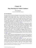
Insulin-stimulated glucose metabolism
(mg/m2/min)
400
**pϽ0.01
Tanner I
Tanner II–IV
Tanner V
300
*p Ͻ0.05
200
100
0
Non-diabetic
Type 1 DM
Fig. 1. The impact of puberty on insulin stimulated peripheral glucose uptake.
Comparison between healthy controls and subjects with type 1 diabetes. Puberty according
to Tanner Stage [Copyright 1986 Massachusetts Medical Society. All rights reserved.
Reproduced with permission from [15]].
rather than changes in hepatic glucose production [12]. Insulin resistance is
associated with compensatory hyperinsulinaemia that leads to progressive falls
in fasting free fatty acids and branch chain amino acids levels, suggesting an
inhibition of lipid and protein breakdown [13]. Furthermore, hyperinsulinaemia
also leads to consistent falls in levels of the inhibitory insulin-like growth factor
(IGF) binding protein-1 (IGFBP-1) through puberty suggesting that the insulin
resistance of puberty may play a physiological role in pubertal growth and
development [14]. Adolescents with T1DM show the same pattern of change in
insulin sensitivity during puberty, but at all stages they are more insulin resistant
than control subjects without diabetes [15] (fig. 1).
The Growth Hormone/IGF-I Axis
Amiel et al. [15] identified a correlation between insulin sensitivity during puberty and mean 24-hour plasma growth hormone (GH) levels. The
insulin antagonistic effects of GH have been well characterised and have been
shown to be due to reductions in peripheral glucose metabolism and, to a
lesser extent, to enhancements in hepatic glucose production [12, 16, 17]. GH
may be acting directly through its own receptor (interacting with post receptor
insulin signalling), but may also be acting indirectly through mobilisation of
non-esterified free fatty acids (NEFAs) from adipose tissue. NEFAs have
suppressive effects on peripheral glucose metabolism [18, 19] and have been
implicated in regulating hepatic glucose metabolism [20, 21]. The characteristics of the GH pulses produced overnight are thought to be important determinants of the metabolic actions of GH, and increases in GH pulse amplitude
Dunger/Acerini/Ahmed
204
lead to sustained changes in insulin sensitivity [12]. There is compelling
evidence to suggest that the accentuated insulin resistance in T1DM results
from GH hypersecretion; the overnight pattern of GH secretion leading to the
‘Dawn Phenomenon’ of increasing insulin requirements during the early hours
of the morning [22].
Abnormalities of the GH/ IGF-I axis have been consistently reported in
adolescents with T1DM. Compared to healthy controls they have increased
nocturnal GH concentrations and GH pulses are characterised by increases in
both pulse amplitude and baseline concentrations [23]; there is some evidence
that GH clearance may also be delayed [24] and deconvolution analysis suggest
that there may be decreases in GH pulse periodicity and increases in overall
GH secretion rate [25].
The GH hypersecretion seen in T1DM results from an increased feedback
drive at the level of hypothalamus/pituitary secondary to the presence of paradoxically low circulating IGF-I levels. Circulating IGF-I levels are frequently
observed to be low, or in the low-normal range, as are those of the principal
IGF-binding protein, IGFBP-3 [26, 27]. These abnormalities are thought to arise
because of partial insensitivity to GH at the level of the hepatic GH receptor and
are largely explained by the central role of insulin in the regulation of the GH/
IGF-I axis. Insulin enhances IGF-I production either by direct regulation of
the hepatic GH receptor, or by way of permissive effects on post-GH receptor
signalling [28].
Insulin also has an important role in regulating IGF bioavailability and
bioactivity through regulation of circulating concentrations of IGFBP-1.
IGFBP-1 is a potent inhibitor of IGF-I action and its production by the liver is
inversely regulated by insulin [29]. Raised serum IGFBP-1 levels may, by
mopping up ‘free’ or ‘unbound’ IGF-I within the circulation, be directly implicated in the development of the ‘Dawn Phenomenon’ [30]. Reduced IGF-I
levels and bioavailability may also have direct effects on insulin sensitivity.
IGF-I exhibits a high degree of structural homology (42–50%) with both proinsulin and insulin and has been shown to exert metabolic effects through its own
receptor that are distinct from those of insulin [31].
Therefore, insulin, or rather portal insulin, concentrations play a pivotal
role in the regulation of GH/IGF-I axis, and the low IGF-I levels in T1DM
reflect the peripheral rather than portal route of insulin delivery [32]. In T1DM,
the GH hypersecretion, reduced IGF-I bioactivity and increased IGFBP-1 levels
are linked to deteriorating glycaemic control [33].
Height and Weight Gain
Historically, T1DM was associated with quite considerable growth impairment as exemplified by the case reports from Mauriac [34] in the 1930s.
Adolescence
205