Gerard J. Tortora-Principles of Anatomy and Physiology 14th Edition- 2
Bạn đang xem bản rút gọn của tài liệu. Xem và tải ngay bản đầy đủ của tài liệu tại đây (39.61 MB, 733 trang )
The Brain and
Cranial Nerves
The brain, cranial nerves, and homeostasis
Your brain contributes to homeostasis by receiving sensory input, integrating new and stored
information, making decisions, and executing responses through motor activities.
Solving an equation, feeling hungry, laughing—the neural processes needed for each of these activities occur in different
regions of the brain, that portion of the central nervous system contained within the cranium. About 85 billion neurons and
10 trillion to 50 trillion neuroglia make up the brain, which has a mass of about 1300 g (almost 3 lb) in adults. On average,
each neuron forms 1000 synapses with other neurons. Thus, the total number of synapses, about a thousand trillion or 1015, is
larger than the number of stars in our galaxy.
The brain
center for registering sensations, correlating them with
braiin is the control
c
one
on
ne another and with stored information, making decisions, and taking
actions. It also is the center for the intellect, emotions, behavior, and
memory. But the brain encompasses yet a larger domain: It directs
mem
our behavior toward others. With ideas that excite, artistry that
dazzles,
or rhetoric that mesmerizes, one person’s thoughts
d
and
a actions may influence and shape the lives of many others.
As you will see shortly, different regions of the brain are
specialized for different functions. Different parts of the brain
aalso work together to accomplish certain shared functions. This
chapter
explores how the brain is protected and nourished, what
c
functions occur in the major regions of the brain, and how the
fu
spinal cord and the 12 pairs of cranial nerves connect with the
spin
brain to form the control center of the human body.
Dr. P.
M
Did you ever wonder how cerebrovascular accidents
(strokes) occur and how they are treated
473
araz
zi/
Sc
i en
ce
S
ou
rc
e
474
CHAPTER 14
• THE BRAIN AND CRANIAL NERVES
14.1 Brain Organization,
Protection, and Blood Supply
OBJECTIVES
• Identify the major parts of the brain.
• Describe how the brain is protected.
• Describe the blood supply of the brain.
In order to understand the terminology used for the principal
parts of the adult brain, it will be helpful to know how the brain
develops. The brain and spinal cord develop from the ectodermal neural tube (see Figure 14.27). The anterior part of the
neural tube expands, along with the associated neural crest tissue. Constrictions in this expanded tube soon appear, creating
three regions called primary brain vesicles: prosencephalon,
mesencephalon, and rhombencephalon (see Figure 14.28). Both
the prosencephalon and rhombencephalon subdivide further,
forming secondary brain vesicles. The prosencephalon (PROSen-sefЈ-a-lon), or forebrain, gives rise to the telencephalon and
diencephalon, and the rhombencephalon (ROM-ben-sefЈ-a-lon),
or hindbrain, develops into the metencephalon and myelencephalon. The various brain vesicles give rise to the following adult
structures:
• The telencephalon (telЈ-en-SEF-a-lon; tel- ϭ distant;
-encephalon ϭ brain) develops into the cerebrum and lateral
ventricles.
• The diencephalon (dı¯Ј-en-SEF-a-lon) forms the thalamus,
hypothalamus, epithalamus, and third ventricle.
• The mesencephalon (mesЈ-en-SEF-a-lon (mes- ϭ middle)), or
midbrain, gives rise to the midbrain and aqueduct of the midbrain
(cerebral aqueduct).
• The metencephalon (metЈ-en-SEF-a-lon; met- ϭ after) becomes
the pons, cerebellum, and upper part of the fourth ventricle.
• The myelencephalon (mı¯-el-en-SEF-a-lon; myel- ϭ marrow)
forms the medulla oblongata and lower part of the fourth
ventricle.
The walls of these brain regions develop into nervous tissue,
while the hollow interior of the tube is transformed into its various
ventricles (fluid-filled spaces). The expanded neural crest tissue
becomes prominent in head development. Most of the protective
structures of the brain—that is, most of the bones of the skull,
associated connective tissues, and meningeal membranes—arise
from this expanded neural crest tissue.
These relationships are summarized in Table 14.1.
Major Parts of the Brain
The adult brain consists of four major parts: brain stem, cerebellum, diencephalon, and cerebrum (Figure 14.1). The brain stem
is continuous with the spinal cord and consists of the medulla
oblongata, pons, and midbrain. Posterior to the brain stem is the
cerebellum (serЈ-e-BEL-um ϭ little brain). Superior to the brain
stem is the diencephalon (di- ϭ through), which consists of the
thalamus, hypothalamus, and epithalamus. Supported on the
diencephalon and brain stem is the cerebrum (se-RE¯-brum ϭ
brain), the largest part of the brain.
TABLE 14.1
Development of the Brain
Five secondary
brain vesicles
Three primary
brain vesicles
Wall
Walls
Walls
Cavities
TELENCEPHALON
Cerebrum
Lateral ventricles
DIENCEPHALON
Thalamus,
hypothalamus,
and epithalamus
Third ventricle
Midbrain
Aqueduct
of the midbrain
Cavity
PROSENCEPHALON
(FOREBRAIN)
MESENCEPHALON
(MIDBRAIN)
MESENCEPHALON
Pons
METENCEPHALON
RHOMBENCEPHALON
(HINDBRAIN)
Three- to fourweek embryo
Adult structures
derived from:
Cerebellum
Upper part of
fourth ventricle
Medulla oblongata Lower part of
fourth ventricle
MYELENCEPHALON
Five-week
embryo
Five-week
embryo
14.1 BRAIN ORGANIZATION, PROTECTION, AND BLOOD SUPPLY
475
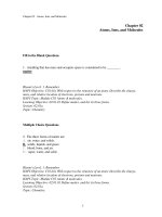
Figure 14.1 The brain. The pituitary gland is discussed with the endocrine system in Chapter 18.
The four principal parts of the brain are the brain stem, cerebellum, diencephalon, and cerebrum.
Sagittal
plane
CEREBRUM
DIENCEPHALON:
Thalamus
Hypothalamus
View
Pineal gland
(part of epithalamus)
BRAIN STEM:
Midbrain
Pons
Medulla oblongata
Pituitary gland
CEREBELLUM
(a) Sagittal section, medial view
CEREBRUM
DIENCEPHALON:
Thalamus
Hypothalamus
BRAIN STEM:
Midbrain
CEREBELLUM
Pons
Medulla oblongata
Spinal cord
(b) Sagittal section, medial view
Which part of the brain is the largest?
C H A P T E R
ANTERIOR
POSTERIOR
14
Spinal cord
476
CHAPTER 14
• THE BRAIN AND CRANIAL NERVES
Protective Coverings of the Brain
The cranium (see Figure 7.4) and the cranial meninges surround
and protect the brain. The cranial meninges (me-NIN-je¯z) are
continuous with the spinal meninges, have the same basic structure,
ˉ -ter),
and bear the same names: the outer dura mater (DOO-ra MA
the middle arachnoid mater (a-RAK-noyd), and the inner pia
mater (PE¯-a or PI¯ -a) (Figure 14.2). However, the cranial dura
Figure 14.2 The protective coverings of the brain.
Cranial bones and cranial meninges protect the brain.
Superior
sagittal sinus
Frontal plane
Skin
Parietal bone
Periosteal
layer
CRANIAL MENINGES:
Dura mater
Meningeal
layer
Arachnoid mater
Pia mater
Subarachnoid
space
Arachnoid villus
Cerebral cortex
Falx cerebri
(a) Anterior view of frontal section through skull showing the cranial meninges
Dura mater
Falx cerebri
Frontal bone
Parietal bone
Superior sagittal
sinus
Inferior sagittal
sinus
Tentorium
cerebelli
Straight sinus
Transverse sinus
Sphenoid
bone
Falx cerebelli
Occipital bone
(b) Sagittal section of extensions of the dura mater
What are the three layers of the cranial meninges, from superficial to deep?
14.2 CEREBROSPINAL FLUID
Blood flows to the brain mainly via the internal carotid and vertebral arteries (see Figure 21.19); the dural venous sinuses drain
into the internal jugular veins to return blood from the head to the
heart (see Figure 21.24).
In an adult, the brain represents only 2% of total body weight,
but it consumes about 20% of the oxygen and glucose used by the
body, even when you are resting. Neurons synthesize ATP almost
exclusively from glucose via reactions that use oxygen. When the
activity of neurons and neuroglia increases in a particular region
of the brain, blood flow to that area also increases. Even a brief
slowing of brain blood flow may cause disorientation or a lack of
consciousness, such as when you stand up too quickly after sitting
for a long period of time. Typically, an interruption in blood flow
for 1 or 2 minutes impairs neuronal function, and total deprivation
of oxygen for about 4 minutes causes permanent injury. Because
virtually no glucose is stored in the brain, the supply of glucose
also must be continuous. If blood entering the brain has a low
level of glucose, mental confusion, dizziness, convulsions, and
loss of consciousness may occur. People with diabetes must be
vigilant about their blood sugar levels because these levels can
drop quickly, leading to diabetic shock, which is characterized by
seizure, coma, and possibly death.
The blood–brain barrier (BBB) consists mainly of tight junctions that seal together the endothelial cells of brain blood capillaries and a thick basement membrane that surrounds the capillaries. As you learned in Chapter 12, astrocytes are one type of
neuroglia; the processes of many astrocytes press up against the
capillaries and secrete chemicals that maintain the permeability
characteristics of the tight junctions. A few water-soluble substances, such as glucose, cross the BBB by active transport. Other
substances, such as creatinine, urea, and most ions, cross the BBB
very slowly. Still other substances—proteins and most antibiotic
drugs—do not pass at all from the blood into brain tissue. However, lipid-soluble substances, such as oxygen, carbon dioxide,
alcohol, and most anesthetic agents, are able to access brain tissue
freely. Trauma, certain toxins, and inflammation can cause a
breakdown of the blood–brain barrier.
Breaching the Blood–
Brain Barrier
Because it is so effective, the blood–brain barrier prevents
the passage of helpful substances as well as those that are
potentially harmful. Researchers are exploring ways to move drugs
that could be therapeutic for brain cancer or other CNS disorders past
the BBB. In one method, the drug is injected in a concentrated sugar
solution. The high osmotic pressure of the sugar solution causes the
endothelial cells of the capillaries to shrink, which opens gaps between
their tight junctions, making the BBB more leaky and allowing the
drug to enter the brain tissue. •
CHECKPOINT
1. Compare the sizes and locations of the cerebrum and
cerebellum.
2. Describe the locations of the cranial meninges.
3. Explain the blood supply to the brain and the importance
of the blood–brain barrier.
14.2 Cerebrospinal Fluid
OBJECTIVE
• Explain the formation and circulation of cerebrospinal
fluid.
Cerebrospinal fluid (CSF) is a clear, colorless liquid composed
primarily of water that protects the brain and spinal cord from
chemical and physical injuries. It also carries small amounts of
oxygen, glucose, and other needed chemicals from the blood to
neurons and neuroglia. CSF continuously circulates through cavities in the brain and spinal cord and around the brain and spinal
cord in the subarachnoid space (the space between the arachnoid
mater and pia mater). The total volume of CSF is 80 to 150 mL (3
to 5 oz) in an adult. CSF contains small amounts of glucose, proteins, lactic acid, urea, cations (Naϩ, Kϩ, Ca2ϩ, Mg2ϩ), and anions
(Cl– and HCO3–); it also contains some white blood cells.
Figure 14.3 shows the four CSF-filled cavities within the brain,
which are called ventricles (VEN-tri-kuls ϭ little cavities). There
is one lateral ventricle in each hemisphere of the cerebrum.
(Think of them as ventricles 1 and 2.) Anteriorly, the lateral ventricles are separated by a thin membrane, the septum pellucidum
(SEP-tum pe-LOO-si-dum; pellucid ϭ transparent). The third
ventricle is a narrow slitlike cavity along the midline superior to
the hypothalamus and between the right and left halves of the
thalamus. The fourth ventricle lies between the brain stem and
the cerebellum.
Functions of CSF
The CSF has three basic functions:
1. Mechanical protection. CSF serves as a shock-absorbing medium that protects the delicate tissues of the brain and spinal
14
Brain Blood Flow and the Blood–Brain Barrier
CLIN ICA L CON N ECTI O N |
C H A P T E R
mater has two layers; the spinal dura mater has only one. The two
dural layers are called the periosteal layer (which is external)
and the meningeal layer (which is internal). The dural layers
around the brain are fused together except where they separate to
enclose the dural venous sinuses (endothelial-lined venous channels) that drain venous blood from the brain and deliver it into
the internal jugular veins. Also, there is no epidural space around
the brain. Blood vessels that enter brain tissue pass along the
surface of the brain, and as they penetrate inward, they are
sheathed by a loose-fitting sleeve of pia mater. Three extensions
of the dura mater separate parts of the brain: (1) The falx cerebri
(FALKS ser-i-BRE¯; falx ϭ sickle-shaped) separates the two
hemispheres (sides) of the cerebrum. (2) The falx cerebelli (serЈe-BEL-ı¯) separates the two hemispheres of the cerebellum.
(3) The tentorium cerebelli (ten-TO¯-re¯-um ϭ tent) separates the
cerebrum from the cerebellum.
477
478
CHAPTER 14
• THE BRAIN AND CRANIAL NERVES
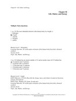
Figure 14.3 Locations of ventricles within a “transparent” brain. One interventricular foramen on each side connects a lateral
ventricle to the third ventricle, and the aqueduct of the midbrain connects the third ventricle to the fourth ventricle.
Ventricles are cavities within the brain that are filled with cerebrospinal fluid.
POSTERIOR
ANTERIOR
Cerebrum
LATERAL VENTRICLES
INTERVENTRICULAR FORAMEN
FOURTH VENTRICLE
THIRD VENTRICLE
LATERAL APERTURE
AQUEDUCT OF THE MIDBRAIN
(CEREBRAL AQUEDUCT)
Cerebellum
Pons
MEDIAN APERTURE
Medulla oblongata
CENTRAL CANAL
Spinal cord
Right lateral view of brain
Which brain region is anterior to the fourth ventricle? Which is posterior to it?
cord from jolts that would otherwise cause them to hit the
bony walls of the cranial cavity and vertebral canal. The fluid
also buoys the brain so that it “floats” in the cranial cavity.
2. Homeostatic function. The pH of the CSF affects pulmonary
ventilation and cerebral blood flow, which is important in maintaining homeostatic controls for brain tissue. CSF also serves as
a transport system for polypeptide hormones secreted by hypothalamic neurons that act at remote sites in the brain.
3. Circulation. CSF is a medium for minor exchange of nutrients
and waste products between the blood and adjacent nervous
tissue.
Because of the tight junctions between ependymal cells, materials
entering CSF from choroid capillaries cannot leak between these
cells; instead, they must pass through the ependymal cells. This
blood–cerebrospinal fluid barrier permits certain substances to
enter the CSF but excludes others, protecting the brain and spinal
cord from potentially harmful blood-borne substances. In contrast
to the blood–brain barrier, which is formed mainly by tight junctions of brain capillary endothelial cells, the blood–cerebrospinal
fluid barrier is formed by tight junctions of ependymal cells.
Formation of CSF in the Ventricles
The CSF formed in the choroid plexuses of each lateral ventricle
flows into the third ventricle through two narrow, oval openings,
the interventricular foramina (inЈ-ter-ven-TRIK-uˉ-lar; singular
is foramen; Figure 14.4b). More CSF is added by the choroid
plexus in the roof of the third ventricle. The fluid then flows
through the aqueduct of the midbrain (cerebral aqueduct)
(AK-we-dukt), which passes through the midbrain, into the
fourth ventricle. The choroid plexus of the fourth ventricle
contributes more fluid. CSF enters the subarachnoid space
through three openings in the roof of the fourth ventricle: a single
median aperture (AP-er-chur) and paired lateral apertures,
The majority of CSF production is from the choroid plexuses
(KO¯-royd ϭ membranelike), networks of blood capillaries in the
walls of the ventricles (Figure 14.4a). Ependymal cells joined by
tight junctions cover the capillaries of the choroid plexuses. Selected substances (mostly water) from the blood plasma, which
are filtered from the capillaries, are secreted by the ependymal
cells to produce the cerebrospinal fluid. This secretory capacity is
bidirectional and accounts for continuous production of CSF and
transport of metabolites from the nervous tissue back to the blood.
Circulation of CSF
14.2 CEREBROSPINAL FLUID
479
CLINICAL CONNECTION | Hydrocephalus
Abnormalities in the brain—tumors, inflammation, or developmental malformations—can interfere with the circulation
of CSF from the ventricles into the subarachnoid space.
When excess CSF accumulates in the ventricles, the CSF pressure
rises. Elevated CSF pressure causes a condition called hydrocephalus
(hı¯Ј-dro¯ -SEF-a-lus; hydro- ϭ water; -cephal- ϭ head). The abnormal
accumulation of CSF may be due to an obstruction to CSF flow or
an abnormal rate of CSF production and/or reabsorption. In a baby
whose fontanels have not yet closed, the head bulges due to the
increased pressure. If the condition persists, the fluid buildup compresses and damages the delicate nervous tissue. Hydrocephalus is
relieved by draining the excess CSF. In one procedure, called endoscopic third ventriculostomy (ETV), a neurosurgeon makes a hole
in the floor of the third ventricle and the CSF drains directly into
the subarachnoid space. In adults, hydrocephalus may occur after
head injury, meningitis, or subarachnoid hemorrhage. Because the
adult skull bones are fused together, this condition can quickly
become life-threatening and requires immediate intervention. •
one on each side. CSF then circulates in the central canal of
the spinal cord and in the subarachnoid space around the surface of the brain and spinal cord.
CSF is gradually reabsorbed into the blood through arachnoid
villi, fingerlike extensions of the arachnoid mater that project into
the dural venous sinuses, especially the superior sagittal sinus
(see Figure 14.2). (A cluster of arachnoid villi is called an arachnoid granulation.) Normally, CSF is reabsorbed as rapidly as it
is formed by the choroid plexuses, at a rate of about 20 mL/hr
(480 mL/day). Because the rates of formation and reabsorption
are the same, the pressure of CSF normally is constant. For the
same reason, the volume of CSF remains constant. Figure 14.4d
summarizes the production and flow of CSF.
CHECKPOINT
14
4. What structures produce CSF, and where are they located?
5. What is the difference between the blood–brain barrier
and the blood–cerebrospinal fluid barrier?
C H A P T E R
Figure 14.4 Pathways of circulating cerebrospinal fluid.
CSF is formed from blood plasma by ependymal cells that cover the choroid plexuses of the ventricles.
ANTERIOR
View
Ependymal
cell
Falx cerebri
Blood
capillary of
CHOROID
PLEXUS
Cerebrum
LATERAL
VENTRICLE
Transverse
plane
Tight
junction
Septum
pellucidum
CSF
CHOROID
PLEXUS
Ventricle
Details of a section through
a choroid plexus (arrow indicates
direction of filtration from blood
to CSF)
Falx cerebri
Superior sagittal
sinus
POSTERIOR
(a) Superior view of transverse section of brain showing choroid plexuses
F I G U R E 14. 4
CONTINUES
480
CHAPTER 14
F I G U R E 14.4
• THE BRAIN AND CRANIAL NERVES
CONTINUED
POSTERIOR
ANTERIOR
CHOROID PLEXUS OF
LATERAL VENTRICLE
Superior cerebral vein
CHOROID PLEXUS OF
THIRD VENTRICLE
ARACHNOID VILLUS
Cerebrum
Interthalamic
adhesion of
thalamus
SUBARACHNOID SPACE
SUPERIOR SAGITTAL
SINUS
Posterior
commissure
Corpus callosum
Great cerebral
vein
LATERAL VENTRICLE
Straight sinus
INTERVENTRICULAR
FORAMEN
Anterior commissure
THIRD VENTRICLE
AQUEDUCT OF THE
MIDBRAIN (CEREBRAL
AQUEDUCT)
Midbrain
Hypothalamus
Pons
Cerebellum
Cranial meninges:
LATERAL APERTURE
CHOROID PLEXUS OF
FOURTH VENTRICLE
Pia mater
FOURTH VENTRICLE
MEDIAN APERTURE
Arachnoid mater
Dura mater
Medulla oblongata
Spinal cord
CENTRAL CANAL
Path of:
CSF
Sagittal
plane
SUBARACHNOID SPACE
View
Filum terminale
(b) Sagittal section of brain and spinal cord
Venous blood
481
14.2 CEREBROSPINAL FLUID
Superior sagittal sinus
ARACHNOID VILLUS
Frontal
plane
Falx cerebri
Corpus callosum
LATERAL VENTRICLE
View
Septum pellucidum
CHOROID PLEXUS
THIRD VENTRICLE
Cerebrum
SUBARACHNOID SPACE
(surrounding brain)
AQUEDUCT OF
THE MIDBRAIN
(CEREBRAL AQUEDUCT)
Tentorium cerebelli
Cerebellum
LATERAL APERTURE
FOURTH VENTRICLE
MEDIAN APERTURE
Spinal cord
Lateral ventricle's
choroid plexuses
CSF
Lateral ventricles
Through
interventricular
foramina
Third ventricle's
choroid plexus
CSF
Third ventricle
Through aqueduct
of the midbrain
(cerebral aqueduct)
(c) Frontal section of brain and spinal cord
Fourth ventricle's
choroid plexus
CSF
Fourth ventricle
Through
lateral and median
apertures
Subarachnoid space
Arachnoid villi of dural
venous sinuses
Arterial blood
Venous blood
Heart and lungs
(d) Summary of the formation, circulation, and
absorption of cerebrospinal fluid (CSF)
Where is CSF reabsorbed?
C H A P T E R
14
SUBARACHNOID SPACE
(surrounding spinal cord)
482
CHAPTER 14
• THE BRAIN AND CRANIAL NERVES
14.3 The Brain Stem and
Reticular Formation
OBJECTIVE
• Describe the structures and functions of the brain stem
and reticular formation.
The brain stem is the part of the brain between the spinal cord and
the diencephalon. It consists of three structures: (1) medulla oblongata, (2) pons, and (3) midbrain. Extending through the brain
stem is the reticular formation, a netlike region of interspersed
gray and white matter.
Medulla Oblongata
The medulla oblongata (me-DOOL-la obЈ-long-GA-ta), or more
simply the medulla, is continuous with the superior part of the
spinal cord; it forms the inferior part of the brain stem (Figure 14.5; see also Figure 14.1). The medulla begins at the foramen
magnum and extends to the inferior border of the pons, a distance
of about 3 cm (1.2 in.).
The medulla’s white matter contains all sensory (ascending)
tracts and motor (descending) tracts that extend between the
spinal cord and other parts of the brain. Some of the white
matter forms bulges on the anterior aspect of the medulla.
These protrusions, called the pyramids (Figure 14.6; see also
Figure 14.5), are formed by the large corticospinal tracts that
pass from the cerebrum to the spinal cord. The corticospinal
tracts control voluntary movements of the limbs and trunk (see
Figure 16.10). Just superior to the junction of the medulla with
the spinal cord, 90% of the axons in the left pyramid cross to
the right side, and 90% of the axons in the right pyramid cross
to the left side. This crossing is called the decussation of pyrˉ -shun; decuss ϭ crossing) and explains why
amids (de¯Ј-ku-SA
each side of the brain controls voluntary movements on the
opposite side of the body.
The medulla also contains several nuclei. (Recall that a nucleus is a collection of neuronal cell bodies within the CNS.)
Some of these nuclei control vital body functions. Examples of
nuclei in the medulla that regulate vital activities include the
cardiovascular center and the medullary rhythmicity center. The
cardiovascular center regulates the rate and force of the heartbeat and the diameter of blood vessels (see Figure 21.13). The
medullary respiratory center adjusts the basic rhythm of breathing (see Figure 23.23).
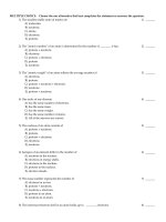
Figure 14.5 Medulla oblongata in relation to the rest of the brain stem.
The brain stem consists of the medulla oblongata, pons, and midbrain.
ANTERIOR
View
Cerebrum
Olfactory bulb
View
Olfactory tract
Pituitary gland
Optic tract
CEREBRAL PEDUNCLE
OF MIDBRAIN
Mammillary body
PONS
Cerebellar
peduncles
MEDULLA
OBLONGATA
Olive
Pyramids
Spinal nerve C1
Spinal cord
Cerebellum
POSTERIOR
Inferior aspect of brain
What part of the brain stem contains the pyramids? The cerebral peduncles? Literally means “bridge”?
instructions that the cerebellum uses to make adjustments to muscle activity as you learn new motor skills.
Nuclei associated with sensations of touch, pressure, vibration,
and conscious proprioception are located in the posterior part of
the medulla. These nuclei are the right and left gracile nucleus
¯ -ne¯-aˉt ϭ wedge).
(GRAS-il ϭ slender) and cuneate nucleus (KU
Ascending sensory axons of the gracile fasciculus (fa-SIK-uˉ-lus)
and the cuneate fasciculus, which are two tracts in the posterior
columns of the spinal cord, form synapses in these nuclei (see
Figure 16.5). Postsynaptic neurons then relay the sensory information to the thalamus on the opposite side of the brain. The axons ascend to the thalamus in a band of white matter called the
medial lemniscus (lem-NIS-kus ϭ ribbon), which extends through
the medulla, pons, and midbrain (see Figure 14.7b). The tracts of
the posterior columns and the axons of the medial lemniscus are
collectively known as the posterior column–medial lemniscus
pathway.
The medulla also contains nuclei that are components of
sensory pathways for gustation (taste), audition (hearing), and
equilibrium (balance). The gustatory nucleus (GUS-ta-toˉЈ-re¯)
of the medulla is part of the gustatory pathway from the tongue
to the brain; it receives gustatory input from the taste buds of
the tongue (see Figure 17.3e). The cochlear nuclei (KOKle¯-ar) of the medulla are part of the auditory pathway from the
Figure 14.6 Internal anatomy of the medulla oblongata.
The pyramids of the medulla contain the large motor tracts that run from the cerebrum to the spinal cord.
Choroid plexus
Fourth ventricle
VAGUS NUCLEUS
(dorsal motor)
View
Transverse plane
HYPOGLOSSAL
NUCLEUS
INFERIOR OLIVARY
NUCLEUS
Vagus (X) nerve
OLIVE
Hypoglossal (XII)
nerve
PYRAMIDS
DECUSSATION
OF PYRAMIDS
Lateral corticospinal tract axons
Spinal nerve C1
Anterior corticospinal tract axons
Transverse section and anterior surface of medulla oblongata
What does decussation mean? What is the functional consequence of decussation of the pyramids?
Spinal cord
14
Besides regulating heartbeat, blood vessel diameter, and the
normal breathing rhythm, nuclei in the medulla also control reflexes for vomiting, swallowing, sneezing, coughing, and hiccupping. The vomiting center of the medulla causes vomiting, the
forcible expulsion of the contents of the upper gastrointestinal
(GI) tract through the mouth (see Section 24.9). The deglutition
center (de¯-gloo-TISH-un) of the medulla promotes deglutition
(swallowing) of a mass of food that has moved from the oral cavity of the mouth into the pharynx (throat) (see Section 24.8).
Sneezing involves spasmodic contraction of breathing muscles
that forcefully expel air through the nose and mouth. Coughing
involves a long-drawn and deep inhalation and then a strong exhalation that suddenly sends a blast of air through the upper respiratory passages. Hiccupping is caused by spasmodic contractions
of the diaphragm (a muscle of breathing) that ultimately result in
the production of a sharp sound on inhalation. Sneezing, coughing, and hiccupping are described in more detail in Table 23.2.
Just lateral to each pyramid is an oval-shaped swelling called
an olive (see Figures 14.5, 14.6). Within the olive is the inferior
olivary nucleus, which receives input from the cerebral cortex,
red nucleus of the midbrain, and spinal cord. Neurons of the inferior olivary nucleus extend their axons into the cerebellum, where
they regulate the activity of cerebellar neurons. By influencing
cerebellar neuron activity, the inferior olivary nucleus provides
483
C H A P T E R
14.3 THE BRAIN STEM AND RETICULAR FORMATION
484
CHAPTER 14
• THE BRAIN AND CRANIAL NERVES
inner ear to the brain; they receive auditory input from the cochlea of the inner ear (see Figure 17.23). The vestibular nuclei
(ves-TIB-uˉ-lar) of the medulla and pons are components of the
equilibrium pathway from the inner ear to the brain; they receive sensory information associated with equilibrium from
proprioceptors in the vestibular apparatus of the inner ear (see
Figure 17.26).
Finally, the medulla contains nuclei associated with the following five pairs of cranial nerves (see Figure 14.5):
1. Vestibulocochlear (VIII) nerves. Several nuclei in the medulla receive sensory input from and provide motor output to
the cochlea of the internal ear via the vestibulocochlear nerves.
These nerves convey impulses related to hearing.
2. Glossopharyngeal (IX) nerves. Nuclei in the medulla relay
sensory and motor impulses related to taste, swallowing, and
salivation via the glossopharyngeal nerves.
3. Vagus (X) nerves. Nuclei in the medulla receive sensory impulses from and provide motor impulses to the pharynx and
larynx and many thoracic and abdominal viscera via the vagus
nerves.
4. Accessory (XI) nerves (cranial portion). These fibers are
actually part of the vagus (X) nerves. Nuclei in the medulla are
the origin for nerve impulses that control swallowing via the
vagus nerves (cranial portion of the accessory nerves).
5. Hypoglossal (XII) nerves. Nuclei in the medulla are the origin for nerve impulses that control tongue movements during
speech and swallowing via the hypoglossal nerves.
CLINICAL CONNECTION | Injury to the Medulla
Given the vital activities controlled by the medulla, it is not
surprising that injury to the medulla from a hard blow to
the back of the head or upper neck such as falling back on ice
can be fatal. Damage to the medullary respiratory center is particularly serious and can rapidly lead to death. Symptoms of nonfatal
injury to the medulla may include cranial nerve malfunctions on the
same side of the body as the injury, paralysis and loss of sensation on
the opposite side of the body, and irregularities in breathing or heart
rhythm. Alcohol overdose also suppresses the medullary rhythmicity
center and may result in death. •
Pons
The pons (ϭ bridge) lies directly superior to the medulla and anterior to the cerebellum and is about 2.5 cm (1 in.) long (see Figures 14.1, 14.5). Like the medulla, the pons consists of both nuclei and tracts. As its name implies, the pons is a bridge that
connects parts of the brain with one another. These connections
are provided by bundles of axons. Some axons of the pons connect the right and left sides of the cerebellum. Others are part of
ascending sensory tracts and descending motor tracts.
The pons has two major structural components: a ventral region and a dorsal region. The ventral region of the pons forms a
large synaptic relay station consisting of scattered gray centers
called the pontine nuclei (PON-tı¯ n). Entering and exiting these
nuclei are numerous white matter tracts, each of which provides
a connection between the cortex (outer layer) of a cerebral
hemisphere and that of the opposite hemisphere of the cerebellum. This complex circuitry plays an essential role in coordinating and maximizing the efficiency of voluntary motor output
throughout the body. The dorsal region of the pons is more like
the other regions of the brain stem, the medulla and midbrain. It
contains ascending and descending tracts along with the nuclei
of cranial nerves.
Also within the pons is the pontine respiratory group, shown
in Figure 23.24. Together with the medullary respiratory center,
the pontine respiratory group helps control breathing.
The pons also contains nuclei associated with the following
four pairs of cranial nerves (see Figure 14.5):
1. Trigeminal (V) nerves. Nuclei in the pons receive sensory
impulses for somatic sensations from the head and face and
provide motor impulses that govern chewing via the trigeminal
nerves.
2. Abducens (VI) nerves. Nuclei in the pons provide motor
impulses that control eyeball movement via the abducens
nerves.
3. Facial (VII) nerves. Nuclei in the pons receive sensory impulses for taste and provide motor impulses to regulate secretion of saliva and tears and contraction of muscles of facial
expression via the facial nerves.
4. Vestibulocochlear (VIII) nerves. Nuclei in the pons receive sensory impulses from and provide motor impulses to
the vestibular apparatus via the vestibulocochlear nerves.
These nerves convey impulses related to balance and equilibrium.
Midbrain
The midbrain or mesencephalon extends from the pons to the
diencephalon (see Figures 14.1, 14.5) and is about 2.5 cm (1 in.)
long. The aqueduct of the midbrain (cerebral aqueduct) passes
through the midbrain, connecting the third ventricle above with
the fourth ventricle below. Like the medulla and the pons, the
midbrain contains both nuclei and tracts (Figure 14.7).
The anterior part of the midbrain contains paired bundles of
axons known as the cerebral peduncles (pe-DUNK-kuls ϭ
little feet; see Figures 14.5, 14.7b). The cerebral peduncles
consist of axons of the corticospinal, corticobulbar, and corticopontine tracts, which conduct nerve impulses from motor
areas in the cerebral cortex to the spinal cord, medulla, and
pons, respectively.
The posterior part of the midbrain, called the tectum (TEKtum ϭ roof), contains four rounded elevations (Figure 14.7a).
The two superior elevations, nuclei known as the superior colliculi (ko-LIK-uˉ-lı¯ ϭ little hills; singular is colliculus), serve as
reflex centers for certain visual activities. Through neural circuits from the retina of the eye to the superior colliculi to the
extrinsic eye muscles, visual stimuli elicit eye movements for
tracking moving images (such as a moving car) and scanning
14.3 THE BRAIN STEM AND RETICULAR FORMATION
Still other nuclei in the midbrain are associated with two pairs
of cranial nerves (see Figure 14.5):
1. Oculomotor (III) nerves. Nuclei in the midbrain provide
motor impulses that control movements of the eyeball, while
accessory oculomotor nuclei provide motor control to the
smooth muscles that regulate constriction of the pupil and
changes in shape of the lens via the oculomotor nerves.
2. Trochlear (IV) nerves. Nuclei in the midbrain provide motor impulses that control movements of the eyeball via the
trochlear nerves.
Reticular Formation
In addition to the well-defined nuclei already described, much of the
brain stem consists of small clusters of neuronal cell bodies (gray
matter) interspersed among small bundles of myelinated axons
(white matter). The broad region where white matter and gray matter
exhibit a netlike arrangement is known as the reticular formation
(re-TIK-uˉ-lar; ret- ϭ net; Figure 14.7c). It extends from the superior
part of the spinal cord, throughout the brain stem, and into the inferior part of the diencephalon. Neurons within the reticular formation
have both ascending (sensory) and descending (motor) functions.
Figure 14.7 Midbrain.
The midbrain connects
cts the po
pons to the diencephalon.
Third ventricle
Habenular nuclei
Thalamus
Pineal gland
Medial geniculate nucleus
View
TECTUM:
Lateral geniculate nucleus
SUPERIOR COLLICULI
CEREBRAL PEDUNCLE
INFERIOR COLLICULI
Trochlear (IV) nerve
Median eminence
Superior cerebellar peduncle
Middle cerebellar peduncle
Floor of fourth ventricle
Inferior cerebellar peduncle
Facial (VII) nerve
Vestibulocochlear
(VIII) nerve
Glossopharyngeal
(IX) nerve
Posterior median sulcus
Vagus (X) nerves
Accessory (XI) nerve
Cuneate fasciculus
Gracile fasciculus
Spinal nerve C1 (posterior root)
(a) Posterior view of midbrain in relation to brain stem
F I G U R E 14. 7
POSTERIOR
CONTINUES
C H A P T E R
14
stationary images (as you are doing to read this sentence). The
superior colliculi are also responsible for reflexes that govern
movements of the head, eyes, and trunk in response to visual
stimuli. The two inferior elevations, the inferior colliculi, are
part of the auditory pathway, relaying impulses from the receptors for hearing in the inner ear to the brain. These two nuclei
are also reflex centers for the startle reflex, sudden movements
of the head, eyes, and trunk that occur when you are surprised
by a loud noise such as a gunshot.
The midbrain contains several other nuclei, including the left
and right substantia nigra (sub-STAN-she¯-a ϭ substance;
NI¯ -gra ϭ black), which are large and darkly pigmented (Figure 14.7b). Neurons that release dopamine, extending from
the substantia nigra to the basal nuclei, help control subconscious muscle activities. Loss of these neurons is associated
with Parkinson’s disease (see Disorders: Homeostatic Imbalances at the end of Chapter 16). Also present are the left and
right red nuclei, which look reddish due to their rich blood
supply and an iron-containing pigment in their neuronal
cell bodies. Axons from the cerebellum and cerebral cortex
form synapses in the red nuclei, which help control muscular
movements.
485
486
CHAPTER 14
F I G U R E 1 4.7
• THE BRAIN AND CRANIAL NERVES
CONTINUED
POSTERIOR
TECTUM
View
SUPERIOR COLLICULUS
Periaqueductal gray matter
Aqueduct of the midbrain
(cerebral aqueduct)
RETICULAR
FORMATION
Transverse
plane
Medial geniculate nucleus
MEDIAL
LEMNISCUS
Oculomotor nucleus
RED NUCLEUS
SUBSTANTIA NIGRA
Corticospinal, corticopontine,
and corticobulbar axons
CEREBRAL
PEDUNCLE
Oculomotor (III) nerve
ANTERIOR
(b) Transverse section of midbrain
Sagittal
plane
Cerebral cortex
Thalamus
RETICULAR ACTIVATING
SYSTEM (RAS) projections
to cerebral cortex
Cerebellum
Pons
Visual impulses
from eyes
RETICULAR FORMATION
Medulla oblongata
Auditory and
equilibrium
impulses from
ears
Spinal cord
Somatic sensory impulses
(from nociceptors, proprioceptors,
and touch receptors)
(c) Sagittal section through brain and spinal cord
showing the reticular formation
What is the importance of the cerebral peduncles?
The ascending portion of the reticular formation is called the
reticular activating system (RAS), which consists of sensory
axons that project to the cerebral cortex, both directly and through
the thalamus. Many sensory stimuli can activate the ascending
portion of the RAS. Among these are visual and auditory stimuli;
mental activities; stimuli from pain, touch, and pressure receptors;
and receptors in our limbs and head that keep us aware of the
position of our body parts. Perhaps the most important function of
the RAS is consciousness, a state of wakefulness in which an individual is fully alert, aware, and oriented. Visual and auditory
stimuli and mental activities can stimulate the RAS to help maintain consciousness. The RAS is also active during arousal, or
awakening from sleep. Another function of the RAS is to help
maintain attention (concentrating on a single object or thought) and
CHECKPOINT
6. Where are the medulla, pons, and midbrain located
relative to one another?
7. What body functions are governed by nuclei in the brain
stem?
8. List the functions of the reticular formation.
14.4 The Cerebellum
OBJECTIVE
• Describe the structure and functions of the cerebellum.
The cerebellum, second only to the cerebrum in size, occupies
the inferior and posterior aspects of the cranial cavity. Like the
cerebrum, the cerebellum has a highly folded surface that greatly
increases the surface area of its outer gray matter cortex, allowing
for a greater number of neurons. The cerebellum accounts for
about a tenth of the brain mass yet contains nearly half of the
neurons in the brain. The cerebellum is posterior to the medulla
and pons and inferior to the posterior portion of the cerebrum (see
Figure 14.1). A deep groove known as the transverse fissure,
along with the tentorium cerebelli, which supports the posterior
part of the cerebrum, separates the cerebellum from the cerebrum
(see Figures 14.2b, 14.11b).
In superior or inferior views, the shape of the cerebellum
resembles a butterfly. The central constricted area is the vermis
(ϭ worm), and the lateral “wings” or lobes are the cerebellar
hemispheres (Figure 14.8a, b). Each hemisphere consists of
lobes separated by deep and distinct fissures. The anterior lobe
and posterior lobe govern subconscious aspects of skeletal muscle movements. The flocculonodular lobe (flok-uˉ-loˉ-NOD-uˉ-lar;
flocculo- ϭ wool-like tuft) on the inferior surface contributes to
equilibrium and balance.
The superficial layer of the cerebellum, called the cerebellar
cortex, consists of gray matter in a series of slender, parallel
folds called folia (ϭ leaves). Deep to the gray matter are tracts of
white matter called arbor vitae (AR-bor VI¯ -te¯ ϭ tree of life)
that resemble branches of a tree. Even deeper, within the white
matter, are the cerebellar nuclei, regions of gray matter that give
rise to axons carrying impulses from the cerebellum to other
brain centers.
Three paired cerebellar peduncles (pe-DUNG-kuls) attach
the cerebellum to the brain stem (see Figures 14.7a and 14.8b).
These bundles of white matter consist of axons that conduct impulses between the cerebellum and other parts of the brain. The
superior cerebellar peduncles contain axons that extend from
the cerebellum to the red nuclei of the midbrain and to several
nuclei of the thalamus. The middle cerebellar peduncles are
the largest peduncles; their axons carry impulses for voluntary
movements from the pontine nuclei (which receive input from
motor areas of the cerebral cortex) into the cerebellum. The inferior cerebellar peduncles consist of (1) axons of the spinocerebellar tracts that carry sensory information into the cerebellum from proprioceptors in the trunk and limbs; (2) axons from
the vestibular apparatus of the inner ear and from the vestibular
nuclei of the medulla and pons that carry sensory information
into the cerebellum from proprioceptors in the head; (3) axons
from the inferior olivary nucleus of the medulla that enter the
cerebellum and regulate the activity of cerebellar neurons;
(4) axons that extend from the cerebellum to the vestibular nuclei
of the medulla and pons; and (5) axons that extend from the
cerebellum to the reticular formation.
The primary function of the cerebellum is to evaluate how well
movements initiated by motor areas in the cerebrum are actually
being carried out. When movements initiated by the cerebral motor areas are not being carried out correctly, the cerebellum detects the discrepancies. It then sends feedback signals to motor
areas of the cerebral cortex, via its connections to the thalamus.
The feedback signals help correct the errors, smooth the movements, and coordinate complex sequences of skeletal muscle contractions. Aside from this coordination of skilled movements, the
cerebellum is the main brain region that regulates posture and balance. These aspects of cerebellar function make possible all
skilled muscular activities, from catching a baseball to dancing to
speaking. The presence of reciprocal connections between the
cerebellum and association areas of the cerebral cortex suggests
that the cerebellum may also have nonmotor functions such as
cognition (acquisition of knowledge) and language processing.
This view is supported by imaging studies using MRI and PET.
Studies also suggest that the cerebellum may play a role in processing sensory information.
The functions of the cerebellum are summarized in Table 14.2.
14
alertness. The RAS also prevents sensory overload (excessive
visual and/or auditory stimulation) by filtering out insignificant
information so that it does not reach consciousness. For example,
while waiting in the hallway for your anatomy class to begin, you
may be unaware of all the noise around you while reviewing your
notes for class. Inactivation of the RAS produces sleep, a state of
partial consciousness from which an individual can be aroused.
Damage to the RAS, on the other hand, results in coma, a state of
unconsciousness from which an individual cannot be aroused. In
the lightest stages of coma, brain stem and spinal cord reflexes
persist, but in the deepest states even those reflexes are lost, and if
respiratory and cardiovascular controls are lost, the patient dies.
Drugs such as melatonin affect the RAS by helping to induce
sleep, and general anesthetics turn off consciousness via the RAS.
The descending portion of the RAS has connections to the cerebellum and spinal cord and helps regulate muscle tone, the slight
degree of involuntary contraction in normal resting skeletal muscles. This portion of the RAS also assists in the regulation of heart
rate, blood pressure, and respiratory rate.
Even though the RAS receives input from the eyes, ears, and
other sensory receptors, there is no input from receptors for the
sense of smell; even strong odors may fail to cause arousal. People who die in house fires usually succumb to smoke inhalation
without awakening. For this reason, all sleeping areas should have
a nearby smoke detector that emits a loud alarm. A vibrating pillow or flashing light can serve the same purpose for those who are
hearing impaired.
The functions of the brain stem are summarized in Table 14.2.
487
C H A P T E R
14.4 THE CEREBELLUM
488
CHAPTER 14
• THE BRAIN AND CRANIAL NERVES
Figure 14.8 Cerebellum.
The cerebellum
coordinates skilled
movements and regulates
posture and balance.
View
CLINICAL CONNECTION | Ataxia
Damage to the cerebellum can result in a loss of ability to coordinate muscular movements, a
condition called ataxia (a-TAK-se¯-a; a- ϭ without; -taxia ϭ order). Blindfolded people with
ataxia cannot touch the tip of their nose with a finger because they cannot coordinate movement with their sense of where a body part is located. Another sign of ataxia is a changed speech
pattern due to uncoordinated speech muscles. Cerebellar damage may also result in staggering or abnormal walking movements. People who consume too much alcohol show signs of ataxia because alcohol
inhibits activity of the cerebellum. Such individuals have difficulty in passing sobriety tests. Ataxia can
also occur as a result of degenerative diseases (multiple sclerosis and Parkinson’s disease), trauma, brain
tumors, and genetic factors, and as a side effect of medication prescribed for bipolar disorder. •
CEREBELLAR
PEDUNCLES:
ANTERIOR
Superior
View
ANTERIOR
LOBE
ANTERIOR
Fourth
ventricle
Inferior
CEREBELLAR
PEDUNCLES:
Superior
View
CEREBELLAR
HEMISPHERE
POSTERIOR
LOBE
Middle
ANTERIOR
Fourth
ventricle
CEREBELLAR
Middle
Inferior
HEMISPHERE
FLOCCULONODULAR
LOBE
CEREBELLAR
HEMISPHERE
VERMIS
VERMIS
POSTERIOR
POSTERIOR
(a) Superior view
(b) Inferior view
POSTERIOR
FLOCCULOLOBE
NODULAR
LOBE
VERMIS
POSTERIOR
POSTERIOR
LOBE
(b) Inferior view
Pineal gland
Superior colliculus
Midsagittal
plane
View
Cerebral peduncle
Inferior colliculus
Cerebral peduncle
Aqueduct of the
midbrain
(cerebral aqueduct)
Mammillary body
Pons
Fourth ventricle
ARBOR VITAE
(WHITE MATTER)
FOLIA
CEREBELLAR CORTEX
(GRAY MATTER)
Medulla oblongata
Central canal
of spinal cord
CEREBELLUM
POSTERIOR
ANTERIOR
(c) Midsagittal section of cerebellum and brain stem
Which structures contain the axons that carry information into and out of the cerebellum?
(d) Midsagittal section
14.5 THE DIENCEPHALON
489
cerebral hemispheres and contains numerous nuclei involved in
a wide variety of sensory and motor processing between higher
and lower brain centers. The diencephalon extends from the
brain stem to the cerebrum and surrounds the third ventricle; it
includes the thalamus, hypothalamus, and epithalamus. Projecting from the hypothalamus is the hypophysis, or pituitary
gland. Portions of the diencephalon in the wall of the third ventricle are called circumventricular organs and will be discussed
shortly. The optic tracts carrying neurons from the retina enter
the diencephalon.
CHECKPOINT
9. Describe the location and principal parts of the
cerebellum.
10. Where do the axons of each of the three pairs of
cerebellar peduncles begin and end? What are their
functions?
14.5 The Diencephalon
OBJECTIVE
Thalamus
• Describe the components and functions of the
diencephalon (thalamus, hypothalamus, and
epithalamus).
The thalamus (THAL-a-mus ϭ inner chamber), which measures
about 3 cm (1.2 in.) in length and makes up 80% of the diencephalon, consists of paired oval masses of gray matter organized
into nuclei with interspersed tracts of white matter (Figure 14.9).
The diencephalon forms a central core of brain tissue just superior to the midbrain. It is almost completely surrounded by the
Figure 14.9 Thalamus. Note the position of the thalamus in the lateral view (a) and in the medial view (b). The various thalamic
nuclei shown in (c) and (d) are correlated by color to the cortical regions to which they project in (a) and (b).
Central sulcus
Thalamus
(a) Lateral view of right cerebral hemisphere
Interthalamic
adhesion
(b) Medial view of left cerebral hemisphere
Reticular
Internal medullary
lamina
Pulvinar
Medial
Lateral
posterior
Anterior
Midline
Interthalamic
adhesion
Intralaminar
nuclei
Ventral
anterior
Internal medullary
lamina
Lateral
dorsal
Lateral posterior
Reticular
Ventral
posterior
Ventral
lateral
Pulvinar
Medial
geniculate
Lateral
geniculate
Ventral
posterior
(c) Superolateral view of thalamus showing locations
of thalamic nuclei (reticular nucleus is shown
on the left side only; all other nuclei are shown
on the right side)
Midline
(d) Transverse section of right side of thalamus
showing locations of thalamic nuclei
What structure usually connects the right and left halves of the thalamus?
C H A P T E R
14
The thalamus is the principal relay station for sensory impulses that reach the cerebral cortex from other parts of the brain
and the spinal cord.
490
CHAPTER 14
• THE BRAIN AND CRANIAL NERVES
A bridge of gray matter called the interthalamic adhesion (intermediate mass) joins the right and left halves of the thalamus in
about 70% of human brains. A vertical Y-shaped sheet of white
matter called the internal medullary lamina divides the gray
matter of the right and left sides of the thalamus (Figure 14.9c). It
consists of myelinated axons that enter and leave the various thalamic nuclei. Axons that connect the thalamus and cerebral cortex
pass through the internal capsule, a thick band of white matter
lateral to the thalamus (see Figure 14.13b).
The thalamus is the major relay station for most sensory impulses that reach the primary sensory areas of the cerebral cortex
from the spinal cord and brain stem. In addition, the thalamus
contributes to motor functions by transmitting information from
the cerebellum and basal nuclei to the primary motor area of the
cerebral cortex. The thalamus also relays nerve impulses between
different areas of the cerebrum and plays a role in the maintenance of consciousness.
Based on their positions and functions, there are seven
major groups of nuclei on each side of the thalamus (Figure 14.9c, d):
1. The anterior nucleus receives input from the hypothalamus and sends output to the limbic system (described in
Section 14.6). It functions in emotions and memory.
2. The medial nuclei receive input from the limbic system and
basal nuclei and send output to the cerebral cortex. They function in emotions, learning, memory, and cognition (thinking
and knowing).
3. Nuclei in the lateral group receive input from the limbic
system, superior colliculi, and cerebral cortex and send output to the cerebral cortex. The lateral dorsal nucleus functions in the expression of emotions. The lateral posterior
nucleus and pulvinar nucleus help integrate sensory information.
4. Five nuclei are part of the ventral group. The ventral anterior nucleus receives input from the basal nuclei and sends
output to motor areas of the cerebral cortex; it plays a role in
movement control. The ventral lateral nucleus receives input
from the cerebellum and basal nuclei and sends output to motor areas of the cerebral cortex; it also plays a role in movement control. The ventral posterior nucleus relays impulses
for somatic sensations such as touch, pressure, vibration, itch,
tickle, temperature, pain, and proprioception from the face and
body to the cerebral cortex. The lateral geniculate nucleus
(je-NIK-uˉ-lat ϭ bent like a knee) relays visual impulses for
sight from the retina to the primary visual area of the cerebral
cortex. The medial geniculate nucleus relays auditory impulses for hearing from the ear to the primary auditory area of
the cerebral cortex.
5. Intralaminar nuclei (inЈ-tra-LA-miЈ-nar) lie within the internal medullary lamina and make connections with the reticular
formation, cerebellum, basal nuclei, and wide areas of the
cerebral cortex. They function in arousal (activation of the
cerebral cortex from the brain stem reticular formation) and
integration of sensory and motor information.
6. The midline nucleus forms a thin band adjacent to the third
ventricle and has a presumed function in memory and olfaction.
7. The reticular nucleus surrounds the lateral aspect of the
thalamus, next to the internal capsule. This nucleus monitors,
filters, and integrates activities of other thalamic nuclei.
Hypothalamus
The hypothalamus (hı¯Ј-poˉ-THAL-a-mus; hypo- ϭ under) is a
small part of the diencephalon located inferior to the thalamus. It
is composed of a dozen or so nuclei in four major regions:
1. The mammillary region (MAM-i-ler-e¯; mammill- ϭ nippleshaped), adjacent to the midbrain, is the most posterior part of
the hypothalamus. It includes the mammillary bodies and posterior hypothalamic nuclei (Figure 14.10). The mammillary
bodies are two small, rounded projections that serve as relay
stations for reflexes related to the sense of smell.
2. The tuberal region (TOO-ber-al), the widest part of the hypothalamus, includes the dorsomedial nucleus, ventromedial
nucleus, and arcuate nucleus (AR-kuˉ-aˉt), plus the stalklike infundibulum (in-fun-DIB-uˉ-lum ϭ funnel), which connects the
pituitary gland to the hypothalamus (Figure 14.10). The median
eminence is a slightly raised region that encircles the infundibulum (see Figure 14.7a).
3. The supraoptic region (supra- ϭ above; -optic ϭ eye) lies
superior to the optic chiasm (point of crossing of optic nerves)
and contains the paraventricular nucleus, supraoptic nucleus,
anterior hypothalamic nucleus, and suprachiasmatic nucleus
(sooЈ-pra-kı¯Ј-az-MA-tik) (Figure 14.10). Axons from the paraventricular and supraoptic nuclei form the hypothalamohypophyseal tract (hı¯Ј-poˉ-thalЈ-a-moˉ-hı¯-poˉ-FIZ-e¯-al), which extends through the infundibulum to the posterior lobe of the
pituitary (see Figure 18.8).
4. The preoptic region anterior to the supraoptic region is usually considered part of the hypothalamus because it participates with the hypothalamus in regulating certain autonomic
activities. The preoptic region contains the medial and lateral
preoptic nuclei (Figure 14.10).
The hypothalamus controls many body activities and is one of
the major regulators of homeostasis. Sensory impulses related to
both somatic and visceral senses arrive at the hypothalamus, as do
impulses from receptors for vision, taste, and smell. Other receptors within the hypothalamus itself continually monitor osmotic
pressure, blood glucose level, certain hormone concentrations,
and the temperature of blood. The hypothalamus has several very
important connections with the pituitary gland and produces a variety of hormones, which are described in more detail in Chapter
18. Some functions can be attributed to specific hypothalamic nuclei, but others are not so precisely localized. Important functions
of the hypothalamus include the following:
• Control of the ANS. The hypothalamus controls and integrates activities of the autonomic nervous system, which
regulates contraction of smooth muscle and cardiac muscle
14.5 THE DIENCEPHALON
491
Figure 14.10 Hypothalamus. Selected portions of the hypothalamus and a three-dimensional representation of hypothalamic
nuclei are shown (after Netter).
The hypothalamus controls many body activities and is an important regulator of homeostasis.
Corpus callosum
Paraventricular
nucleus
Interthalamic
adhesion
of thalamus
Lateral preoptic
nucleus
Dorsomedial
nucleus
Medial preoptic
nucleus
Posterior
hypothalamic
nucleus
Arcuate
nucleus
Ventromedial
nucleus
Mammillary region
Tuberal region
Suprachiasmatic
nucleus
Infundibulum
Mammillary
body
Supraoptic nucleus
Optic chiasm
Supraoptic region
Optic (II) nerve
Pituitary gland
Preoptic region
POSTERIOR
ANTERIOR
Sagittal section of brain showing hypothalamic nuclei
What are the four major regions of the hypothalamus, from posterior to anterior?
and the secretions of many glands. Axons extend from the
hypothalamus to parasympathetic and sympathetic nuclei in
the brain stem and spinal cord. Through the ANS, the hypothalamus is a major regulator of visceral activities, including
regulation of heart rate, movement of food through the gastrointestinal tract, and contraction of the urinary bladder.
• Production of hormones. The hypothalamus produces several
hormones and has two types of important connections with
the pituitary gland, an endocrine gland located inferior to the
hypothalamus (see Figure 14.1). First, hypothalamic hormones known as releasing hormones and inhibiting hormones
are released into capillary networks in the median eminence
(see Figure 18.5). The bloodstream carries these hormones directly to the anterior lobe of the pituitary, where they stimulate or inhibit secretion of anterior pituitary hormones. Second, axons extend from the paraventricular and supraoptic
nuclei through the infundibulum into the posterior lobe of the
pituitary (see Figure 18.8). The cell bodies of these neurons
make one of two hormones (oxytocin or antidiuretic hormone). Their axons transport the hormones to the posterior
pituitary, where they are released.
• Regulation of emotional and behavioral patterns. Together
with the limbic system (described shortly), the hypothalamus participates in expressions of rage, aggression, pain,
and pleasure, and the behavioral patterns related to sexual
arousal.
• Regulation of eating and drinking. The hypothalamus regulates food intake. It contains a feeding center, which promotes
eating, and a satiety center, which causes a sensation of fullness and cessation of eating. The hypothalamus also contains a
thirst center. When certain cells in the hypothalamus are stimulated by rising osmotic pressure of the extracellular fluid,
they cause the sensation of thirst. The intake of water by drinking restores the osmotic pressure to normal, removing the
stimulation and relieving the thirst.
• Control of body temperature. The hypothalamus also functions
as the body’s thermostat, which senses body temperature so
that it is maintained at a desired setpoint. If the temperature of
blood flowing through the hypothalamus is above normal, the
hypothalamus directs the autonomic nervous system to stimulate activities that promote heat loss. When blood temperature
is below normal, by contrast, the hypothalamus generates
impulses that promote heat production and retention.
• Regulation of circadian rhythms and states of consciousness. The suprachiasmatic nucleus of the hypothalamus
serves as the body’s internal biological clock because it esˉ -de¯-an), pattablishes circadian (daily) rhythms (ser-KA
terns of biological activity (such as the sleep–wake cycle)
that occur on a circadian schedule (cycle of about 24 hours).
This nucleus receives input from the eyes (retina) and sends
output to other hypothalamic nuclei, the reticular formation,
and the pineal gland.
14
Key:
Anterior
hypothalamic
nucleus
C H A P T E R
Sagittal
plane
492
CHAPTER 14
• THE BRAIN AND CRANIAL NERVES
Epithalamus
The epithalamus (epЈ-i-THAL-a-mus; epi- ϭ above), a small
region superior and posterior to the thalamus, consists of the
pineal gland and habenular nuclei. The pineal gland (PI¯ N-e¯-al
ϭ pineconelike) is about the size of a small pea and protrudes
from the posterior midline of the third ventricle (see Figure 14.1). The pineal gland is part of the endocrine system because it secretes the hormone melatonin. As more melatonin is
liberated during darkness than in light, this hormone is thought
to promote sleepiness. When taken orally, melatonin also appears to contribute to the setting of the body’s biological clock
by inducing sleep and helping the body to adjust to jet lag. The
habenular nuclei (ha-BEN-uˉ-lar), shown in Figure 14.7a, are
involved in olfaction, especially emotional responses to odors
such as a loved one’s cologne or Mom’s chocolate chip cookies
baking in the oven.
The functions of the three parts of the diencephalon are summarized in Table 14.2.
Circumventricular Organs
Parts of the diencephalon, called circumventricular organs
(CVOs) (serЈ-kum-ven-TRIK-uˉ-lar) because they lie in the wall
of the third ventricle, can monitor chemical changes in the blood
because they lack a blood–brain barrier. CVOs include part of the
hypothalamus, the pineal gland, the pituitary gland, and a few
other nearby structures. Functionally, these regions coordinate
homeostatic activities of the endocrine and nervous systems, such
as the regulation of blood pressure, fluid balance, hunger, and
thirst. CVOs are also thought to be the sites of entry into the brain
of HIV, the virus that causes AIDS. Once in the brain, HIV may
cause dementia (irreversible deterioration of mental state) and
other neurological disorders.
CHECKPOINT
11. Why is the thalamus considered a “relay station” in the
brain?
12. Why is the hypothalamus considered part of both the
nervous system and the endocrine system?
13. What are the functions of the epithalamus?
14. Define a circumventricular organ.
14.6 The Cerebrum
OBJECTIVES
• Describe the cortex, gyri, fissures, and sulci of the
cerebrum.
• Locate each of the lobes of the cerebrum.
• Describe the tracts that compose the cerebral white
matter.
• Describe the nuclei that compose the basal nuclei.
• Describe the structures and functions of the limbic system.
The cerebrum is the “seat of intelligence.” It provides us with
the ability to read, write, and speak; to make calculations and
compose music; and to remember the past, plan for the future,
and imagine things that have never existed before. The cerebrum consists of an outer cerebral cortex, an internal region of
cerebral white matter, and gray matter nuclei deep within the
white matter.
Cerebral Cortex
The cerebral cortex (cortex ϭ rind or bark) is a region of gray
matter that forms the outer rim of the cerebrum (Figure 14.11a).
Although only 2–4 mm (0.08–0.16 in.) thick, the cerebral cortex contains billions of neurons arranged in layers. During embryonic development, when brain size increases rapidly, the
gray matter of the cortex enlarges much faster than the deeper
white matter. As a result, the cortical region rolls and folds on
itself. The folds are called gyri (JI¯ -rı¯ ϭ circles; singular is gyrus) or convolutions (konЈ-voˉ-LOO-shuns) (Figure 14.11). The
deepest grooves between folds are known as fissures; the shallower grooves between folds are termed sulci (SUL-sı¯ ϭ
grooves; singular is sulcus). The most prominent fissure, the
longitudinal fissure, separates the cerebrum into right and left
halves called cerebral hemispheres. Within the longitudinal
fissure between the cerebral hemispheres is the falx cerebri.
The cerebral hemispheres are connected internally by the corpus
callosum (kal-LO¯-sum; corpus ϭ body; callosum ϭ hard), a
broad band of white matter containing axons that extend between the hemispheres (see Figure 14.12).
Lobes of the Cerebrum
Each cerebral hemisphere can be further subdivided into several lobes. The lobes are named after the bones that cover
them: frontal, parietal, temporal, and occipital lobes (see Figure 14.11). The central sulcus (SUL-kus) separates the frontal lobe from the parietal lobe. A major gyrus, the precentral
gyrus—located immediately anterior to the central sulcus—
contains the primary motor area of the cerebral cortex. Another major gyrus, the postcentral gyrus, which is located immediately posterior to the central sulcus, contains the primary
somatosensory area of the cerebral cortex. The lateral cerebral sulcus (fissure) separates the frontal lobe from the temporal lobe. The parieto-occipital sulcus separates the parietal
lobe from the occipital lobe. A fifth part of the cerebrum, the
insula, cannot be seen at the surface of the brain because it lies
within the lateral cerebral sulcus, deep to the parietal, frontal,
and temporal lobes (Figure 14.11b).
14.6 THE CEREBRUM
493
Figure 14.11 Cerebrum. Because the insula cannot be seen externally, it has been projected to the surface in (b).
The cerebrum is the “seat of intelligence”;
it provides us with the ability to read,
write, and speak; to make calculations and
compose music; to remember the past and
plan for the future; and to create.
ANTERIOR
Frontal lobe
Longitudinal fissure
Precentral gyrus
Central sulcus
Gyrus
Parietal lobe
Postcentral gyrus
Sulcus
Cerebral
cortex
Cerebral
white matter
Occipital lobe
Fissure
POSTERIOR
Details of a gyrus,
sulcus, and fissure
(a) Superior view
Central sulcus
Postcentral gyrus
Precentral gyrus
Parietal lobe
Frontal lobe
Parieto-occipital
sulcus
Insula (projected to surface)
Lateral cerebral sulcus
Occipital lobe
Temporal lobe
Transverse fissure
Cerebellum
(b) Right lateral view
During development, does the gray matter or the white matter enlarge more rapidly? What are the brain folds, shallow
grooves, and deep grooves called?
14
Right hemisphere
C H A P T E R
Left hemisphere
494
CHAPTER 14
• THE BRAIN AND CRANIAL NERVES
Cerebral White Matter
The cerebral white matter consists primarily of myelinated
axons in three types of tracts (Figure 14.12):
1. Association tracts contain axons that conduct nerve impulses
between gyri in the same hemisphere.
2. Commissural tracts (komЈ-i-SYUR-al) contain axons that
conduct nerve impulses from gyri in one cerebral hemisphere
to corresponding gyri in the other cerebral hemisphere. Three
important groups of commissural tracts are the corpus callosum (the largest fiber bundle in the brain, containing about
300 million fibers), anterior commissure, and posterior
commissure.
3. Projection tracts contain axons that conduct nerve impulses
from the cerebrum to lower parts of the CNS (thalamus, brain
stem, or spinal cord) or from lower parts of the CNS to the
cerebrum. An example is the internal capsule, a thick band of
white matter that contains both ascending and descending
axons (see Figure 14.13).
Basal Nuclei
Deep within each cerebral hemisphere are three nuclei (masses
of gray matter) that are collectively termed the basal nuclei
(Figure 14.13). (Historically, these nuclei have been called the
basal ganglia. However, this is a misnomer because a ganglion
is an aggregate of neuronal cell bodies in the peripheral nervous system. While both terms still appear in the literature, we
use nuclei, as this is the correct term as determined by the Terminologia Anatomica, the final say on correct anatomical
terminology.)
Two of the basal nuclei lie side by side, just lateral to the thalamus. They are the globus pallidus (GLO¯-bus PAL-i-dus; globus
ϭ ball; pallidus ϭ pale), which is closer to the thalamus, and the
ˉ -men ϭ shell), which is closer to the cerebral
putamen (puˉ-TA
cortex. Together, the globus pallidus and putamen are referred to
as the lentiform nucleus (LEN-ti-form ϭ shaped like a lens). The
third of the basal nuclei is the caudate nucleus (KAW-daˉt; caudϭ tail), which has a large “head” connected to a smaller “tail” by
a long comma-shaped “body.” Together, the lentiform and cauˉ -tum; corpus
date nuclei are known as the corpus striatum (strı¯-A
ϭ body; striatum ϭ striated). The term corpus striatum refers to
the striated (striped) appearance of the internal capsule as it passes
among the basal nuclei. Nearby structures that are functionally
linked to the basal nuclei are the substantia nigra of the midbrain
and the subthalamic nuclei of the diencephalon (see Figures 14.7b,
14.13b). Axons from the substantia nigra terminate in the caudate
nucleus and putamen. The subthalamic nuclei interconnect with
the globus pallidus.
The claustrum (KLAWS-trum) is a thin sheet of gray matter
situated lateral to the putamen. It is considered by some to be a
subdivision of the basal nuclei. The function of the claustrum in
humans has not been clearly defined, but it may be involved in
visual attention.
The basal nuclei receive input from the cerebral cortex and
provide output to motor parts of the cortex via the medial and
ventral group nuclei of the thalamus. In addition, the basal
nuclei have extensive connections with one another. A major
function of the basal nuclei is to help regulate initiation and
termination of movements. Activity of neurons in the putamen precedes or anticipates body movements; activity of
neurons in the caudate nucleus occurs prior to eye movements. The globus pallidus helps regulate the muscle tone
required for specific body movements. The basal nuclei also
control subconscious contractions of skeletal muscles. Examples include automatic arm swings while walking and true
laughter in response to a joke (not the kind you consciously
initiate to humor your A&P instructor).
Figure 14.12 Organization of white matter tracts of the left cerebral hemisphere.
Association tracts, commissural tracts, and projection tracts form white matter tracts in the cerebral hemispheres.
Midsagittal
plane
Cerebral cortex
COMMISSURAL and
PROJECTION TRACTS
View
ASSOCIATION
TRACTS
Septum
pellucidum
COMMISSURAL TRACTS:
CORPUS CALLOSUM
ANTERIOR
COMMISSURE
Mammillary
body
POSTERIOR
ANTERIOR
Medial view of tracts revealed by removing
gray matter from a midsagittal section
Which tracts carry impulses between gyri of the same hemisphere? Between gyri in opposite hemispheres? Between the
cerebrum and thalamus, brain stem, and spinal cord?
14.6 THE CEREBRUM
In addition to influencing motor functions, the basal nuclei
have other roles. They help initiate and terminate some cognitive processes, such as attention, memory, and planning, and
may act with the limbic system to regulate emotional behaviors.
Disorders such as Parkinson’s disease, obsessive–compulsive
disorder, schizophrenia, and chronic anxiety are thought to involve dysfunction of circuits between the basal nuclei and the
limbic system and are described in more detail in Chapter 16.
The Limbic System
Encircling the upper part of the brain stem and the corpus callosum is a ring of structures on the inner border of the cerebrum and
495
floor of the diencephalon that constitutes the limbic system
(limbic ϭ border). The main components of the limbic system are
as follows (Figure 14.14):
• The so-called limbic lobe is a rim of cerebral cortex on the medial surface of each hemisphere. It includes the cingulate gyrus
(SIN-gyu-lat; cingul- ϭ belt), which lies above the corpus callosum, and the parahippocampal gyrus (parЈ-a-hip-oˉ-KAM-pal),
which is in the temporal lobe below. The hippocampus (hipЈ-oˉKAM -pus ϭ seahorse) is a portion of the parahippocampal
gyrus that extends into the floor of the lateral ventricle.
• The dentate gyrus (dentate ϭ toothed) lies between the hippocampus and parahippocampal gyrus.
Figure 14.13 Basal nuclei. In (a) the basal nuclei have been projected to the surface; in both (a) and (b) they are shown in purple.
The basal nuclei help initiate and terminate movements, suppress unwanted movements, and regulate muscle tone.
14
Body of caudate nucleus
Lateral ventricle
Frontal lobe of cerebrum
Tail of caudate nucleus
C H A P T E R
Putamen
Head of caudate nucleus
Thalamus
Occipital lobe
of cerebrum
POSTERIOR
ANTERIOR
(a) Lateral view of right side of brain
Frontal
plane
Longitudinal
fissure
Cerebrum
Corpus callosum
Septum
pellucidum
Lateral ventricle
Internal
capsule
View
Caudate nucleus
Insula
Putamen
Globus pallidus
Thalamus
Third ventricle
Subthalamic
nucleus
Hypothalamus
and associated
nuclei
Optic tract
(b) Anterior view of frontal section
Where are the basal nuclei located relative to the thalamus?
Basal
nuclei
496
CHAPTER 14
• THE BRAIN AND CRANIAL NERVES
Figure 14.14 Components of the limbic system (shaded green) and surrounding structures.
The limbic system governs emotional aspects of behavior.
Sagittal
plane
Anterior nucleus
of thalamus
Mammillothalamic
tract
Corpus callosum
View
Fornix
Cingulate gyrus
(in frontal lobe)
Stria medullaris
Anterior commissure
Stria terminalis
Septal nuclei
Hippocampus
(in temporal lobe)
Mammillary body
in hypothalamus
Dentate gyrus
Olfactory bulb
Amygdala
Parahippocampal
gyrus (in temporal
lobe)
POSTERIOR
Sagittal section
ANTERIOR
Which part of the limbic system functions with the cerebrum in memory?
• The amygdala (a-MIG-da-la; amygda- ϭ almond-shaped) is
composed of several groups of neurons located close to the tail
of the caudate nucleus.
• The septal nuclei are located within the septal area formed by
the regions under the corpus callosum and the paraterminal gyrus (a cerebral gyrus).
• The mammillary bodies of the hypothalamus are two round
masses close to the midline near the cerebral peduncles.
• Two nuclei of the thalamus, the anterior nucleus and the medial nucleus, participate in limbic circuits (see Figure 14.9c, d).
• The olfactory bulbs are flattened bodies of the olfactory pathway that rest on the cribriform plate.
• The fornix, stria terminalis, stria medullaris, medial forebrain
bundle, and mammillothalamic tract (mam-i-loˉ-tha-LAM-ik)
are linked by bundles of interconnecting myelinated axons.
produces a behavioral pattern called rage—the cat extends its
claws, raises its tail, opens its eyes wide, hisses, and spits. By
contrast, removal of the amygdala produces an animal that lacks
fear and aggression. Likewise, a person whose amygdala is damaged fails to recognize fearful expressions in others or to express
fear in situations where this emotion would normally be appropriate, for example, while being attacked by an animal.
Together with parts of the cerebrum, the limbic system also
functions in memory; damage to the limbic system causes memory impairment. One portion of the limbic system, the hippocampus, is seemingly unique among structures of the central nervous
system—it has cells reported to be capable of mitosis. Thus, the
portion of the brain that is responsible for some aspects of memory may develop new neurons, even in the elderly.
The functions of the cerebrum are summarized in Table 14.2.
The limbic system is sometimes called the “emotional brain”
because it plays a primary role in a range of emotions, including
pain, pleasure, docility, affection, and anger. It also is involved in
olfaction (smell) and memory. Experiments have shown that
when different areas of animals’ limbic systems are stimulated,
the animals’ reactions indicate that they are experiencing intense
pain or extreme pleasure. Stimulation of other limbic system
areas in animals produces tameness and signs of affection. Stimulation of a cat’s amygdala or certain nuclei of the hypothalamus
15. List and locate the lobes of the cerebrum. How are they
separated from one another? What is the insula?
16. Distinguish between the precentral gyrus and the
postcentral gyrus.
17. Describe the organization of cerebral white matter and
indicate the function of each major group of fibers.
18. List the basal nuclei. What are the functions of the basal
nuclei?
19. Define the limbic system and list several of its functions.
CHECKPOINT
14.7 FUNCTIONAL ORGANIZATION OF THE CEREBRAL CORTEX
14.7 Functional Organization
of the Cerebral Cortex
497
bral cortex, primary sensory areas receive sensory information
that has been relayed from peripheral sensory receptors through
lower regions of the brain. Sensory association areas often are
adjacent to the primary areas. They usually receive input both
from the primary areas and from other brain regions. Sensory
association areas integrate sensory experiences to generate
meaningful patterns of recognition and awareness. For example,
a person with damage in the primary visual area would be blind
in at least part of his visual field, but a person with damage to a
visual association area might see normally yet be unable to recognize ordinary objects such as a lamp or a toothbrush just by
looking at them.
The following are some important sensory areas (Figure 14.15;
the significance of the numbers in parentheses is explained in the
figure caption):
OBJECTIVES
• Describe the locations and functions of the sensory,
association, and motor areas of the cerebral cortex.
• Explain the significance of hemispheric lateralization.
• Indicate the significance of brain waves.
Specific types of sensory, motor, and integrative signals are processed in certain regions of the cerebral cortex (Figure 14.15).
Generally, sensory areas receive sensory information and are involved in perception, the conscious awareness of a sensation;
motor areas control the execution of voluntary movements; and
association areas deal with more complex integrative functions
such as memory, emotions, reasoning, will, judgment, personality
traits, and intelligence. In this section we will also discuss hemispheric lateralization and brain waves.
Sensory impulses arrive mainly in the posterior half of both cerebral hemispheres, in regions behind the central sulci. In the cere-
Figure 14.15 Functional areas of the cerebrum. Broca’s speech area and Wernicke’s area are in the left cerebral hemisphere of most
people; they are shown here to indicate their relative locations. The numbers, still used today, are from K. Brodmann’s
map of the cerebral cortex, first published in 1909.
Particular areas of the cerebral cortex process sensory, motor, and integrative signals.
Central sulcus
PRIMARY MOTOR AREA
(precentral gyrus)
PRIMARY SOMATOSENSORY
AREA (postcentral gyrus)
PREMOTOR AREA
PRIMARY GUSTATORY AREA
SOMATOSENSORY
ASSOCIATION AREA
4
5
7
FRONTAL EYE FIELD AREA
1
Parietal lobe
6
2
COMMON
INTEGRATIVE
AREA
3
Frontal lobe
39
40
WERNICKE’S AREA
43
41
42
18
BROCA'S SPEECH AREA
45
19
11
PREFRONTAL CORTEX
17
37
Lateral cerebral sulcus
21
38
Occipital lobe
Temporal lobe
10
44
22
VISUAL
ASSOCIATION
AREA
PRIMARY
VISUAL
AREA
9
8
20
POSTERIOR
AUDITORY
ASSOCIATION
AREA
PRIMARY AUDITORY AREA
ANTERIOR
Lateral view of right cerebral hemisphere
What area(s) of the cerebrum integrate(s) interpretation of visual, auditory, and somatic sensations? Translates thoughts
into speech? Controls skilled muscular movements? Interprets sensations related to taste? Interprets pitch and rhythm?
Interprets shape, color, and movement of objects? Controls voluntary scanning movements of the eyes?
C H A P T E R
Sensory Areas
14
• The primary somatosensory area (areas 1, 2, and 3) is located
directly posterior to the central sulcus of each cerebral hemisphere in the postcentral gyrus of each parietal lobe. It extends
from the lateral cerebral sulcus, along the lateral surface of the
parietal lobe to the longitudinal fissure, and then along the medial
surface of the parietal lobe within the longitudinal fissure. The
primary somatosensory area receives nerve impulses for touch,