Ebook Basics of respiratory mechanics and artificial ventilation: Part 2
Bạn đang xem bản rút gọn của tài liệu. Xem và tải ngay bản đầy đủ của tài liệu tại đây (5.34 MB, 126 trang )
Chapter 10
Alveolar micromechanics
P.Y.ROMERO
The mechanical behavior of the air spaces in the periphery of the lung is the
result of a delicate balance of forces acting on the tissue scaffold of lung
parenchyma. Static and dynamic properties of such a complex system have been
an important field of research for many years. Alveolar space micromechanics
have important physiological implications in terms of mechanical interdependen ce, alveolar stability, and the maintenance of agas exchanging surface in
constant contact with air. The mechanical behavior of such system has to allow
the expansion of the alveolar surface at physiological rates at a low energy cost,
and without interfering with the exchange process. I will describe how the structure and mechanics of the alveolar space are particularly optimized to reach
these goals.
Anatomical structure of the alveolar space
The alveolar septum is made of a single capillary network interlaced with fibers
(mainly collagen and elastine), which form a continuum embedded in the connective matrix, the thin membrane of epithelial cells forming the external
boundary of this scaffold. This irregular surface is to some extent smoothed by
an extracellular layer of lining fluid that is rather thin over the capillaries but
forms small pools in the intercapillary cavities. Alveolar lining consists of an
aqueous layer called the hypophase which is of variable thickness and is present mainly in the pools, and a layer of surfactant which forms a film on the
surface of the hypophase. Because of the relevant physical properties of these
structures, septal configuration is not exclusively determined by the structural
disposition but results from the molding effect of the two main forces that have
to be kept in balance: tissue tension and surface tension.
Structural interaction of tissue fibers and surface lining
Many experimental studies agree in the fact that the dimension of the alveolar
surface is governed by the equilibrium between surface and tissue forces.
Surface tension arises at any gas-liquid interface because the forces between the
molecules of the liquid are much stronger than those between the liquid and
the gas. As a result, the liquid surface will tend to become as small as possible.
A curved surface, such as that of an alveolus, generates apressure proportional
120
P.V.Rornero
to the curvature and to the surface tension coefficient g. According to Wilson
[1], surface pressure (Ps) can be expressed as a function of the surface-to-volurne ratio of the alveolar airspace (S/V)A and surface tension (y) by:
Ps = (2/3) • y. (SIV)A
(1)
The greater the surface-to-volurne ratio, the greater the mean curvature of
the surface and the greater the surface press ure at any value of y. According to
the above equation, the most critical effect of surface tension (y) is that it challenges the stability of airspaces. Asa setof connected bubbles, alveoli are intrinsically unstable: since the small on es have a larger curvature than the large
ones, they should collapse and empty into the larger units. However, in normal
conditions alveoli are highly stable. This is due to two main mechanisms: the
interaction between tissue fibers and surface lining, and the intrinsic properties
of surfactant itself.
Alveolar walls contain an intricate fiber system. Thus, when an alveolus
tends to shrink, the fibers in the wall of the alveoli are stretched and this will
prevent the alveolus from collapsing. This stabilizing phenomenon is known as
interdependence [2]. Surfactant lines the complete alveolar suface, and even terminal airways. The surface tension coefficient y of surfactant is variable: it falls
as alveolar surface becomes smaller, and rises when alveolar surface expands
[3]. Therefore, as alveolar volume decreases, surface tension decreases and tissue fiber tension increases due to interdependence. This force opposed to the
alveolar emptying allows the system to remain stable. If surface tension is modified at the level of the liquid-air interface, the alveolar area will be inversely
related to the surface tension at any level of alveolar volume, at least in the
range of tidal volumes [4]. This is due to the effect of tissue tensions: as surface
tension decreases, the stretching effect of tissue tension is magnified and alveolar area increases, provided that alveolar volume does not vary.
Biomechanics of the alveolar lining layer
Structure and composition
The alveolar epithelial cells are covered by a thin liquid film (less than 0.1 flm).
At the air-liquid interface of this film, a layer of surface active material, largely
phospholipid, aggregates. This alveolar lining layer has been described as an
acellular film that forms a continuous lining over the alveolar epithelial cells
and spans the pores of Kohn. It was considered to serve as an anti-desiccant to
the lungs until, in 1955, Pattle [5] showed that the lung contained surfactant
substances capable of stabilizing tiny bubbles, and even to decrease air-water
surface tension to near zero values. Two morphological regions of the alveolar
lining layer (ALL) have to be distinguished: the hypophase, and the hypophaseair boundary or surfactant lining. The hypophase often appears as a homogeneous matrix by ultrastructural examination. It contains highly ordered tubular
Alveolar micromechanics
121
myelin osmophilic figures that form a system of packed square tubules. Tubular
myelin is a lipoprotein structure of high surface activity that contains dipalmitoyl lecityn, the major component of pulmonary surfactant. Thickness of the
hypophase varies, sometimes hardyly visible by electron microscopy in areas
where the epithelial cell surface is flat, and sometimes appearing as deep pools
where there are folds or crevices in the epithelium or between capillaries. The
air-hypophase boundary can be distinguished from the hypophase by its
osmophilic property. It is provided by a duplex lining layer composed mainly of
desaturated phospholipids.
Biomechanics
The major fraction of the lung's retractive force is normally derived from the
interface between air and lung lining layer. Furthermore, the largest portion of
the lung's hysteresis and rheological behavior is attributable to this interface.
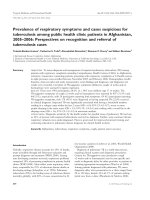
These effects are weIl known since, in 1929 von Neergard [6] described the
pressure-volume characteristics of the liquid-filled and air-filled lungs (Fig. 1):
liquid filling eliminates all air interfaces between cell walls and their lumina, so
that interfacial tensions are negligible, and only the resistance of tissue forces
remain. For many years knowledge about surface tension in situ was derived
from studies based on the difference between air-filled and liquid-filled lungs.
In 1977 Hoppin and Hildebrandt [7] presented a number of arguments, including those that relate to possible differences between tissue contribution in air-
-- -
Air·filled lung
Saline·filled lung
Pressure
Fig. 1. Volume-pressure diagrams of isolated lungs inflated from minimallung volume
with air or saline. In saline-filled lung the interfacial tension of the lung lining layer is
though to be largely eliminated when air is replaced by saline. The saline curve is typically
displaced to the left, and has a lower hysteresis than the air-filled curve. A "knee" in the
inflation arm of the air-filled loop is characteristically seen
122
P.Y. Romero
filled and liquid-filled status, which indicated clearly that the use of pressurevolume (PV) diagrams for calculation of y is unreliable. Between 1976 and 1989
Shürch et al. [8,9] developed a method of continuously measuring surface tensions in vivo, by monitoring the deformation of test droplets of fluids with different y deposited on the alveolar surface by means of a micropipette. Surface
tension-Iung volume and surface tension-recoil pressure relationships have
been since then measured in different species. The most important biomechanical features related to surface tension per se can be summarized as follows.
1. The surface tension-Iung volume relationship in static conditions is similar
for different species, particularly along the deflation limb: surface tension
decreases quasilinearlY with lung volume from totallung capacity (TLC) to
functional residual capacity (FRC) level [4]
2. Static recoil pressure is linearly related to y, but this relationship differs
between species. This difference has been related to the interspecies variability of the alveolar surface to volume ratio and the different participation
of lung tissue (tissue component of the recoil pressure, Pt). According to the
model proposed by Wilson and Bachofen [10], the component of recoil pressure due to surface tension (Pr) is directly proportional to y/Vl/3, where V is
the alveolar volume: Py=K.yV1I3
3. There is a prominent hysteresis in the y- V relationships with values of y
ranging from near zero at low lung volumes during deflation to transiently
high tensions near 40 dyn/ern during dynamic inflation. The amplitude of
the hysteresis and shapes of y- V relationships differ between quasistatic and
dyamic states and with volume history, and are therefore dependent on the
surface film kinetic behavior [11].
Biomechanics of lung tissue
Biomechanical structure oflung tissue
The major constituents of tissue matrix are elastic and collagen fibers, proteoglycans, fibronectin, and the constituents of the basement membran es of
endothelium and epithelium. The fiber strands (mainly elastin and collagen)
form the scaffold of alveolar walls, and allow the plastic deformation of the
lungs during respiration.
Collagen is a basic structural element for soft and hard tissues in animals. It
gives mechanical integrity and strength to our bodies. It is present in a variety
of structural forms in different tissues and organs. In the lung, collagen represents 15%-20% of dry weight, the major collagen types being land III. The primary building unit of collagen is the tropocollagen molecule, which is composed of polypeptide chains. In each tropocollagen molecule there are three
amino acid chains coiled into a left-handed helix. The molecule itself consist of
a right handed superhelix formed by these three chains. Basically a collection of
tropocollagen moleeules forms a collagen fibril. Under electron microscopy, the
collagen fibrils appear to be cross-striated with a periodicity of 64oA. This
Alveolar micromechanics
123
cross-banded staining pattern is a consequence of the parallel arrangement of
molecules in the fibril: molecules on adjacent axes are staggered by approximately one-quarter the length of an individual molecule. Bundles of fibrils
form fibers. Collagen fibers have great tensile strength due to an extensive system of cross-links between a-chains. The collagen fibers in lung tissue at deflation are loosely arranged and are wavy, so they do not become tight until the
parenchyma is distended.
Elastin is a protein found in vertebrates. It is present as thin strands in areolar connective tissue. It forms quite a large proportion of the material in the
walls or arteries, and in lung tissue. The function of elastin in lung parenchyma
is to provide elasticity to the tissue, especially at lower stress levels. Elastic
fibers are composed of an amorphous elastin component and a highly structured microfibrilar component. The microfibrils are found at the periphery of
the fiber, but in larger fibers they also occur as fine bundles in the interior of
the amorphous core. It is believed that the amorphous core represents the actual elastin, and thus has the elastic properties typical of elastic fibers, namely a
relatively high extensibility and a low tensile strength when compared with collagen fibers. In fact, elastin is the most linearly elastic biosolid material known:
its loading curve is almost a straight line. Loading and unloading do lead, however, to two different stress-strain curves (hysteresis), showing the existence of
an energy dissipation mechanism in the material.
Biomechanics
The first information about lung tissue mechanical properties was derived from
the liquid pressure-volume diagram (Fig. 1), established by von Neergard [4]:
liquid filling eliminates all air interfaces between cell walls and their lumina, so
that interfacial tensions are negligible and only the res ist an ce of tissue forces
remain. The early model of Setnikar and Meschia [12] explained the liquid PV
diagram as representing the resistance of elastin to stretch over most of the volurne range, while collagen, which is poorly extensible, would establish resistance to stretch at the highest lung volumes. Since then, many studies have analyzed the stress-strain relationships of small pieces of lung parenchyma,
assumed to be a model for the tissue network of the alveolar wall. Although
reservations have to be acknowledged, the comparison of the tissue stressstrain behavior with PV diagrams from liquid-lung was fairly good, and the
hypothesis first proposed by Setnikar and Meschia (SM) was straightened.
Karlinsky et al. in 1976 [13] found that in liquid-filled excised lungs destruction
of elastin by the enzyme elastase raised the compliance in the low and middle
volume ranges but affected neither volume nor compliance at high transpulmonary pressures. Destruction of collagen by collagen ase increased compliance
at high lung volumes but left the behavior at low lung volumes the same.
Similar results have been observed by Moretto et al. [14] in alveolar wall preparations. These results agreed with the SM model. Morphologic studies have
shown that in relaxed state the elastic fibers form a network of more or less
straight fibers, whereas the collagen fibers appear to be wavy. Elastin and colla-
124
P.V. Romero
gen were considered to be structured as complete and independent networks.
According to the SM model the system will function as folIows: if the tissue is
stretched, the elastic fibers elongate until the collagen fibers are straight. Then,
the low extensibility of collagen would prevent further stretching of the tissue.
This model would predict a biphasic length-tension relationship with an abrupt
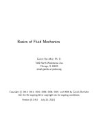
decrease in compliance near maximum lung volumes. However, the stressstrain loop of lung tissue is smoothly curved over its entire range (Fig. 2), and
uniaxial deformation of lung strips does not allow us to distinguish two different elastic behaviors [15]. Recent structural observations have stated that to
accomplish its dual structural function of scaffolding and stress-bearing, the
extracellular fiber matrix has to integrate its separate components into a functional whole, the so-called integral fiber strand [16] . Instead of independent
networks, collagen and elastic fibers form a macrostructure of interwoven
fibers that provide the characteristic network (nylon stocking) extensibility:
stretching in one direction leads to a temporary rearrangement of the fibers.
Elastic fibers will res tore the original arrangement upon relaxation. When this
30,0
25 ,0
--c..
tI:I
20,0
:5
'"
~
Vl
15.0
10.0
5.0 " r - - - -- - , - -- - -- . - - - -- - - , - -- --,0.0
0.5
1.0
1.5
2,0
train (UL
0)
Fig. 2. Stress-strain curves obtained in a subpleural sampie of rat lung (rat lung strip),
submitted to uniaxial oscillatory deformation of increasing amplitudes around the operative length (obtained at 14 hPa basal stress). The smooth curvature over the entire range
of deformation can be observed
Alveolar micromechanics
125
system is submitted to a radial stress, it will convert the extern al traction into
interior tension and transmit it throughout the lung via the elastic parenchymal network as in the ideallung of Mead [2]. Under the action of a distorting
force, structural intermolecular links in the proteins oppose deformation. No
structural change can be performed without a remaking of interactions at the
molecular level in the net. In these molecular re arrangements reside the biomechanical properties of lung parenchyma.
Nonlinearity and lung tissue structure
In vivo, alveoli are subject to finite deformation. Like many biological materials,
lung tissue exhibits prominent time-dependent and frequency-dependent phenomena. Even if hysteresis and time-dependent phenomena are disregarded, the
relationship between stress and strain is nonlinear over the range of physiological deformations. Many studies provide evidence that the nonlinear features of
lung dynamics arise largely from elastic nonlinearities in lung tissue. Hildebrandt
[17] studied the dynamic properties of excised cat lungs in a liquid plethysmograph. Lung elastic modulus and viscosity rose markedly with lung volume.
Moreover, the magnitude of the unit step response fell with increasing step size
and rose with lung volume. By measuring alveolar pressure to study parenchymal
mechanics in mechanically ventilated rabbits, Romero et al. [18] observed an
increase in both tissue elasticity and viscosity with transpulmonary pressure. As
with the mechanical tissue behavior of whole lungs, several authors [19,20,21]
have recently addressed the quest ion of the marked dependence of the elastic
modulus on the mean distending stress in isolated strips of lung parenchyma.
Therefore, the elastic recoil of the lung at normal breathing is dominated by the
nonlinear stress-strain characteristics of lung tissue (Fig. 2). The origin of the
curvilinear stress-strain behavior is generally thought to be one of recruitment.
Maksym and Bates [22] have developed a model of lung tissue based on the collagen fiber recruitment concept, by representing the collagen and elastic fibers as a
series of spring-string pairs. In this model, collagen is the recruited element
(string), while elastin (spring) is responsible for load-bearing at low strains when
much of the collagen is "wavy" and, therefore not contributing to the tension. As
strain increases, the collagen fibers become straight and so progressively take up
more load, thereby stiffening the tissue. This model explains the curvilinear quasistatic stress-strain characteristics of lung tissue, but does not account for
dynamic nonlinearities observed in alveolar wall preparations.
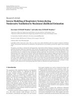
The model developed by Romero et al. [21] is represented in Fig. 3. In this
model, molecular interactions presenting a linear viscoelastic behaviour are
progressively recruited. Elastin and collagen interact in a more active way, and
the lung behaves as a complex polymer that can be modelled as a material with
two components. One is the set of alliung constituents which participate in the
mechanical response in a continuous, uninterrupted way during any mechanical test. This element is known as continuum or matrix. The second component
is formed by those elements whose participation in the mechanical response of
126
P.V. Romero
EIM
2M
CI
CT
EIM
E2M
C2
R2M
Fig. 3. Spring-dashpot scheme of the recruitment-based model of lung tissue behaviour,
ordered according to Takayagashi's block diagram. The basic element is a Kelvin's element
composed of elastic res ist an ces in parallel and a viscous resistance in series with one of
the elastic resistances. This Kelvin diagram represents the behaviour of the continuous
part of the system or "matrix" (M) either in series or in parallel with the recruiting portion of the system or "filler" (R). This model assumes that the nonlinearity between stress
and strain is due to the fact that the number of fibers with identical mechanical properties
participating in the lung's mechanical response is not constant, but depends, due to
recruitment, on the strain to wh ich sampie is subjected. (From [21])
the lung is dependent on the deformation to which the sample is subjected.
Consequently, a given element is incorporated into the lung's mechanical
response after a threshold value has been reached. This phenomenon is known
as recruitment. If the sample is shortened to adeformation value lower than
that at which recruitment of the element occurs, then this element will not participate in the mechanical response. This second component is assumed to be
embedded in the continuum, forming a discontinuous phase and which, by
analogy with compound materials is called filler. The mechanical behaviour of
the continum+filler as a whole can be studied using the block model of
Takayanagi [23] for complex polymers. This model assumes that the behaviour of
the material as a whole corresponds to the behaviour of the filler material
ordered in parallel with a fraction of the continuum (paraBel matrix), and this set
was then ordered in series with the rest of the matrix (serial matrix). Standard
viscoelastic Kelvin's model has been used to represent viscoelastic behavior,
both for the matrix and for each of the fiber elements composing the fiBer. This
model assumes that the nonlinearity between stress and strain is due to the fact
that the number of fibers with identical mechanical properties participating in
the lung's mechanical response is not constant, but depends, due to recruitment,
on the strain to which the tissue is subjected. It accounts fairly weB for both static
Alveolar micromechanics
127
(stress relaxation, stress recovery) and dynamic (oscillatory) properties of lung
tissue [21].
Alveolar stability
As a result of the interaction between surface and tissue forces in normal lungs,
alveoli remain permanently opened at FRC. Only if volume is forcibly reduced to
near RV (residual volume) are peripheral airways seen to elose. The older, wellknown arguments about the potential effects of surface forces on lung stability,
based on a picture of the lung microstructure as a collection of independent bubble-like airspaces, have now been replaced by arguments that treat lung stability as
a structural phenomenon. Morphological data indicate that surface tension (y)
a
"
0 .7
"
E
0 .6
=<"
......
:0
zo
ZON E C
0 .8
ZONE A
'E 8
I
0
..
0 .5
~
0.4
>
ö"
>
<:
0.3
0
2
4
6
8
10
12
14
Pr ess ure c m H 2 0
b
V
o~~--============~
_________
p
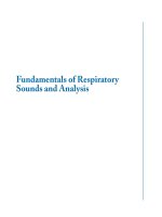
Fig. 4. (a) Experimental data of alveolar volume (derived from alveolar pressure) plotted
against driving pressure in an unstable region of a rabbit lung. Rabbit was pretreated with
ethchlorovynol to induce lung permeability edema; (b) pressure-volume (PV) diagram
describing a uniform expansion of parenchyma with constant surface tension, according to
the theoretical analysis from Stamenovic and Smith [24). This analysis predicts that particular nonuniformities of lung expansion, representing parenchymal instability, would occur
over the region where the PV curve has a negative slope. (From [26) with permission)
128
P.V. Romero
distorts alveolar geometry, and new models for the microstructural mechanics
consider that the outward pull of y exerted on the alveolar ducts is in equilibrium
with the tissue forces of the duct structure [10]. According to Eq. I, if surface tension were constant and high enough, the lung would be unstable at low lung volurne [24,25]. This conclusion is based on the fact that the contribution of surface
tension to the transpulmonary pressure is proportional to the product of surface
tension and interfacial surface-to-volume ratio and that the surface-to-volume
ratio increases as the volume decreases. Therefore, if surface tension is constant
and large enough, recoil pressure increases with decreasing volume and the lung
is unstable. According to Stamenovic and Smith [24], alveolar pressure-volume
curves from areas with constant surface tension would pass through a region of
instability (Fig. 4), in agreement with the experimental observations made in
rabbits after induced permeability edema [26].
Parenchymal constriction
Many studies have shown that bronchoconstrictor agents induce a substantial
increase in tissue resistance (Rti) and dynamic elastance (Edyn) in several
species. Several mechanisms have been invoked to induce changes in Rti and
Edyn after constrictor challenge: parallel heterogeneities, lung tissue constriction, and airways-to-tissue interaction are the most relevant. Recently Romero et
al. [27] have shown that pharmacologically induced changes in tissue resistance
and tissue hysteresivity precede to changes in alveolar heterogeneity (Fig. 5) and
are out of phase with airway resistance, whereas dynamic elastance changes are
in phase with changes in the airways. Hysteresivity being an intrinsic property of
the tissue dissipative behaviour at structurallevel [19], the authors concluded
that changes in tissue resistance and tissue hysteresivity reflect the active constriction of contractile cells and smooth muscle in the parenchyma. The conclusion that parenchymal tissue is affected by bronchoconstricting agents is significant because it implies that asthma may be a dis order of lung parenchyma, not
just of airways. But it has other important physiological implications in the regulation of the tensile equilibrium at the level of the acinus. At this respect, quantitative differencies between the changes in mechanical properties of lung strips
submitted to pharmacological agents in vitro and the pharmacological response
of the whole lung in vivo have been observed. The elast an ce and resistance of
parenchymal strips exposed to bronchoconstrictor agents increase by less than
50%, whereas apparent lung elastance and resistance increase manifold [18, 19].
Because of this disparity between the magnitude of changes in both preparations, some authors have concluded that most of the increased impedance of the
constricted lung is caused by large nonuniform airway resistance, mainly at the
level of terminal bronchioles [28]. Indeed, alveolar capsule technique has allowed
detection of important parallel heterogeneities once the constriction is fully
established. However, the lag between the increase in tissue resistance and hysteresivity (immediate after i.v. injection of methacholine), and the increase in
parallel airways inhomogeneity (Fig. 5) suggest that there is a real, not artifac-
Alveolar micromechanics
..c
c;J
2.0
~'
Cl
..l
~
~
a
/ :.,'"
:;
•
1.5
,
'
1'
I' "
,
1.0
2.5
.c1=
<:>
129
b
2.0
1.5
1.0
.-..
c
15
'-'
;>
U
10
5
0
... '. '-'_./~'; ..,:....
0
50
100
150
Time ( )
Fig. 5a-c. Time course of changes immediately after an Lv. injection of methacholine. (a)
Dynamic elastance (EL); (b) lung parenchyma hysteresivity (11); (c) coefficient of variation
of alveolar pressure at end expiration (CVe) and end inspiration (Cvi). A clear phase lag is
observed between alveolar heterogeneity and hysteresivity changes on one hand, and
between parenchyma hysteresivity and elastance on the other. (From (27) with permission)
tual increase in Rti, reflecting the activation of the contractile machinery at the
level of the parenchyma. An alternative explanation of the disparity of the
mechanical response to constrictor agents in the alveolar wall preparation and
in the whole lung resides in the structural behavior of the acinus, and particularly in the tissue forces-surface forces interaction. Smooth musde is distributed in the acinus in dose relation with the fiber rings at the alveolar mouths.
Contractile fibers have been described in the interstitial spaces in dose contact
with the fiberous network that forms the connective scaffold of the acinus.
According to the model of interaction proposed by Wilson and Bachofen [10 I, if
tissue tensions increase at a given alveolar volume, the interfacial press ure has
to increase to keep alveolar stability. Consequently, tissue constriction would
act as a regulatory mechanism of alveolar micromechanics.
130
P. V. Romero
References
1.
2.
3.
4.
5.
6.
7.
8.
9.
10.
11.
12.
13.
14.
15.
16.
17.
18.
19.
20.
21.
22.
Wilson TA (1981) The relations among recoil pressure, surface area and surface tension in the lung. J Appl Physiol Respirat Environ Exercise Physiol 50:921-926
Mead J (1961) Mechanical properties oflungs. Physiol Rev 41:281-330
Schürch S, Bachofen H, Weibel ER (1985) Alveolar surface tensions in exeised rabbit
lungs: effects of temperature. Respir PhysioI62:31-45
Bachofen H, Wilson TA (1991) Micromechanics of the acinus and the alveolar wall.
In: Crystal RG, West JB et al (eds) The Lung: seientific foundations. Vol. 1. Raven
Press, New York, pp 809-819
Pattle RE (1955) Properties, function and origin of the alveolar lining layer. Nature
175:1125-1127
Von Neergard K (1929) Neue Auffassungen über einen Grundbegriff der Atemmechanik:
Die Retraktionskraft der Lunge, Abhangig von der Oberflächensprannung in den
Alveolen. Z Gesamte Exp Med 66:373-394
Hoppin FG, Hildebrandt J (1977) Mechanical properties of the lung. In: West JB (ed)
Bioengineering aspects of the lung. Marcel Dekker, New York, pp 83-157
Schürch S, Goerke J, Clements JA (1976) Direct determination of surface tension in
the lung. Proc Natl Acad Sei 73:4698-4702
Schürch S, Bachofen H, Goerke J, Possmayer F (1989) A captive bubble method reproduces the in situ behavior oflung surfactant monolayers. J Appl PhysioI67:2389-2396
Wilson TA, Bachofen H (1982) A model of mechanical structure of alveolar duct. J
Appl PhysioI53:1512-1520
Smith JC, Stamenovic D (1986) Surface forces in the lungs. I Alveolar surface tensionlung volume relationships. J Appl PhysioI60:1341-1350
Setnikar I, Meschia G (1952) Propieta elastiche deI polmone e di modelli meccaniche.
Arch Fisiol 52:288-302
Karlinsky JB, Snyder GL, Franzlau C, Stone PJ, Hoppin FG Jr (1960) In vitro effects of
elastase and collagenase on mechanical properties of hamster lungs. Am Rev Respir
Dis 82:186-194
Moretto A, Dallaire M, Romero P, Ludwig M (1994) Effect of elastase on oscillation
mechanics oflung parenchymal strips. J Appl Physiol77:1623-1629
Romero PV, Caiiete C, Lopez-Aguilar J, Romero FJ (1998) Elastieity, viscosity and
plastieity in lung parenchyma. In: Milic-Emili J (ed) Applied physiology in respiratory mechanics. Springer-Verlag, Berlin Heidelberg New York, pp 57-72
Weibel ER, Crystal RG (1991) Structural organization of the pulmonary interstitium.
In: Crystal RG, West JB et al (eds) The lung: seientific foundations. VolL Raven Press,
NewYork,pp 369-380
Hildebrandt J (1969) Dynamic properties of air-filled excised cat lungs determined
by liquid pletismograph. J Appl PhysioI27:246-250
Romero PV, Robatto FM, Simard S, Ludwig MS (1992) Lung tissue behavior during
methacholine challenge in rabbits in vivo. J Appl PhysioI73:207-212
Fredberg JJ, Bunk D, Ingenito E, Shore SA (1993) Tissue resistance and the contractile
state oflung parenchyma. JAppl PhysioI74:1387-1397
Navajas D, Maksym GN, Bates JHT (1995) Dynamic viscoelastic nonlinearity of lung
parenchymal tissue. J Appl Physiol 79:348-356
Romero FJ, Pastor A, Lopez, J, Romero PV (1998) A recruitment-based rheological
model for mechanical behavior of soft tissues. Biorheology 35:17-35
Maksym GN, Bates JHT (1997) A distributed nonlinear model of lung tissue elastieity. JAppl PhysioI82:32-41
Alveolar micromechanics
23.
131
Takayanagi M (1963) Viscoelastic properties of crystalline polymers. Mem Fac Eng
Kyushu Univ 33(1):41-96
24. Stamenovic D, Smith Je (1986) Surface forces in lungs II. Microstructural mechanics
and lung stability. J Appl Physiol 60: 1351-1357
25. Stamenovic D, Wilson TA (1992) Parenchymal stability. J Appl PhysioI73:596-602
26. Romero PV; Lopez Aguilar J, Blanch L (1998) Pulmonary mechanics beyond peripheral airways. In: Milic-Emili J (ed) Applied physiology in respiratory mechanics.
Springer-Verlag, Berlin Heidelberg New York, pp 199-210
27. Romero PV; Rodriguez B, Lopez-Aguilar J, Manresa F (1998) Parallel airways inhomogeneity and lung tissue mechanics in transition to constricted state in rabbits. J
Appl PhysioI84:1040-1047
28. Hubmayr RD, Hill M, Wilson TA (1996) Nonuniform expansion of constricted dog
lungs. JAppl PhysioI80:522-530
Chapter 11
Partitioning of lung responses into airway
and tissue components
M.S.LuDWIG
This chapter deals with the role of the lung parenchyma in contributing to the
contractile response of the overalliung during induced constriction. Addressing
the contribution of the parenchyma has been made easier in recent years because
of the development of the alveolar capsule technique which permits direct measurement of alveolar pressure [1]. Resistive losses across the lung can, thereby, be
partitioned into a component due to airway resistance (Raw) and a component
due to tissue resistance (Rti). Similarly, resistance changes during induced constriction can be apportioned into the component related to changes in airway
calibre and the component related to alterations in tissue mechanical behaviour.
Recent studies in a number of different animal species have shown that much of
the resistive pressure drop across the lung under baseline conditions is due to
the resistive pressure drop at the level of the lung tissues [2-6]. Furthermore,
numerous animal studies have now shown that increases in lung resistance (RL)
during exogenous or endogenous constriction are due, in large part, to changes
in tissue resistance [2,5-10]. Traditionally, changes in lung resistance with
induced constriction were thought to be due to changes in airway calibre.
However, if increases in tissue resistance account for a large part of the increase
in lung resistance, then the pathophysiology of diseases such as asthma needs to
be reconsidered.
Background
The lung parenchyma was first described as a viscoelastic material by Bayliss
and Robertson in 1939 [11]. Hildebrandt and colleagues [12-14] in aseries of
elegant studies described the hysteretic properties of the lung parenchyma in a
number of different species and with lungs in the air-filled or fluid-filled state.
However, the relative importance of tissue resistance in determining the overall
resistive losses of the lung during cyclic ventilation has been a matter of some
controversy. Contribution of tissue resistance to lung resistance has been
reported to range from 15%-85% of total RL [11,15,16]. Some of the confusion
arises because many of these measurements were made using different regimes
of ventilation, i.e. different frequencies and tidal volumes or at different lung
volumes; both tissue and airway resistance are sensitive to changes in these
variables. Furthermore, alveolar pressure was measured indirectly in all these
134
M.S. Ludwig
studies. It was only with the introduction and application of the alveolar capsule technique that direct measurement of alveolar pressure became possible.
Alveolar capsule technique
The first use of an alveolar capsule to measure alveolar pressure (PA) was
reported by Takashima et al. in 1971 [17]; Fredberg et al. [1, 18] further refined
this approach. Basieally, a hollow capsule is glued to the pleural surface of the
lung and punctures are made in the underlying pleura to bring the capsule
chamber into communieation with the underlying alveoli. Pressure is then measured in the chamber with a miniature transducer. Once measurement of alveolar pressure can be obtained, lung resistance can be partitioned into airway and
tissue components by measuring pressure at the airway opening (Pao), PA, and
flow. While alveolar pressure is measured directly with this method, regional
flow is not. Rather, flow is measured at the airway opening and it is assumed
that flow is homogeneously distributed throughout the lung, an assumption
that is reasonable under baseline conditions [19] but can become somewhat
more problematic after induced constriction [20]. The pressure drop between
Pao and PA in phase with flow represents airway resistance while the pressure
drop between PA and the pleural space represents tissue resistance.
Animal studies: tissue resistance at baseline
Tissue resistance is dependent on the frequency and tidal volume of oscillation
as well as the lung volume at whieh the measurement is made [2,3,13]. My colleagues and I [2,6,21] and others [22] have shown in several different species
that tissue resistance increases as the transpulmonary pressure is increased.
Hence the contribution of tissue resistance to overalliung resistance will vary
as the regime of ventilation varies.
In studies conducted in my laboratory, measurements were made of tissue
and airway resistance at "physiologie" breathing frequencies, tidal volumes and
lung volumes. Results in dogs, rabbits, guinea pigs and rats are shown in Table
1. Under baseline conditions, tissue resistance accounts for a substantial proportion of overalliung resistance.
Animal studies: tissue resistance after induced constriction
Alveolar capsules were applied to canine lungs, and airway and tissue resistances
were measured before and after inhalations of histamine and prostagiandin Fz a ,
and after vagal stimulation [2]. Increases in tissue resistance accounted for
roughly half of the increase in RL after vagal stimulation and for most of the
increase after histamine and PGFz a inhalation. In subsequent experiments, concentration-response curves of airway and tissue resistance were examined after
Partitioning of lung response into airway and tissue eomponents
135
Table 1. Values of RL, raw and rti under baseline eonditions (mean ± standard error)
Dogs
(ern H.O s 1'1)
Rabbits
(ern H.O s mt1)
Rats
(ern H.O s ml' l )
Guineapigs
(ern H.O s mI' I )
RL
O.9S±O-24
O.O24±O.OO6
O.OS2±O.OO5
O.105±O.OOS
Raw
O.lS±O.02
O.OlO±O.OO5
O.O50±O.OO5
O.O79±O.OO5
Rti
O.SO±O.23
O.O14±O.OO3
O.O32±O.OO2
O.O26±O.OO5
RL, lung resistanee; raw, airway resistanee; Rti, tissue resistanee
inhalations of histamine or methacholine in dogs, rabbits, rats and guinea pigs
[6,21,23, and unpublished data]. Although there was some interspecies variation, much of the increase in RL was attributable to the increase in Rti (Fig. 1).
Several other investigators have reported similar results using alveolar capsules
to partition the response to different smooth muscle agonists delivered exogenously to both mature and immature animals. Sly and Lanteri [7] showed that
increases in tissue resistance accounted for most of the increase in lung resistance after methacholine nebulization in 8-10 week old mongrel puppies. Sakae
et al. [24] showed that alveolar pressures increased to a greater degree than airway pressures after inhalation of methacholine in rats. Shardonofsky and collegues [22] reported increases in tissue resistance in rabbits after intravenous
route can effeet changes in tissue behaviour.
6.0
liI 5.0
~
"-
~ 4.0
:c
E
3.0
w
u
~ 2.0
~
Vl
Vi 1.0
a::::
w
HISTAMINE (mg/mi)
Fig. 1. Resistanee values for lung resistance (RL), airway resistanee (Raw) and tissue viseanee or resistanee (Vti) du ring histamine ehallenge in dogs (n=6). Values are me an ±
standard error. Cant, eontrol; 5a1, saline. (From [23] with permission)
136
M.S. Ludwig
My eolleagus and I have also studied the role of the lung tissues in the allergie
response in the Brown Norway rat model of intrinsie asthma [25,26]. After
inhalation of aerosols of ovalbumin in previously sensitized rats, airway and tissue resistanee inereased during both the early and the late response [9]. Rti
aecounted for roughly half of the inerease in RL during the early response and
60% of the inerease in RL during the late response. As expeeted, studies of lung
morphology during the late response showed signifieant airway eonstrietion
(Fig. 2). In addition, the alveolar arehiteeture was also substantially altered (Fig.
3). Ihere was widespread tissue distortion with areas of hyperinflation adjaeent
to areas of ateleetasis. Ihis ateleetasis was not to airway closure as none of the
more than 200 airways sampled after ovalbumin exposure showed histologie evidence of airway closure.
A seeond model investigated is that of hyperpnea-induced constriction (HIC)
in the guinea pig [10] . Ihis model shares several common features with exereiseindueed asthma, including the time course of the onset of constrietion, the spontaneity of resolution, and the relationship between the amount of hyperpnea and
the degree of response elicited [27]. During HIC, approximately two-thirds of the
inerease in RL was aeeounted for by the inerease in Rti. Morphologie and morphometrie studies of the lung tissues during the HIC response again showed substantial tissue distortion, with areas of atelectasis and relative hyperinflation.
Fig. 2a,b. Photomicrographs
of airway from (a) a previously sensitized, ovalbumin-challenged Brown Norway rat
during the late asthmatic
response (basement membrane=1.508 mm), and (b) a
time-matched saline control
(basement membrane= 1.416
mm). Lungs fIxed at 3 cm H20
transpulmonary pressure.
Hemaetoxylin-eosin stain.
MagnifIcation. 100. (From [9)
with permission)
Partitioning of lung response into airway and tissue components
137
Fig. 3a,b. Photomicrographs of
lung tissues from (a) a previousIy sensitized, ovalbumin-challenged Brown Norway rat during the late asthmatic response,
and (b) a time-matched saline
control. Lungs fIxed at 3 cm H20
transpu monary press ure.
Hemaetoxylin-eosin stain.
Magnification • 63. (From [9)
with permission)
Human studies
Measurements of tissue resistance in humans have been more difficult to
obtain because of the invasiveness of the alveolar capsule technique. Verbeken
et al. [28,29] made measurements in autopsy specimens, oscillating the lungs
with pseudorandom noise. In normal autopsy lungs, at 4 Hz, Rti accounted for
36% of total resistance at distending pressures of 6 cm H20, and 74% of total
resistance at distending press ures of 20 cm H20. In lungs from patients with
emphysema, the proportion of RL attributable to Rti decreasedj in patients
with fibrosis the proportion remained the same. More recently investigators
made measurements of complex impedence to partition resistance into airway
and tissue components. Kaczka and colleagues [30] used the optimal ventilator
waveform technique, whereby a complex signal was simultaneously delivered
to a subject along with tidal volume ventilation. Data were fit to a model wh ich
included an airway resistance component and a tissue damping or resistance
component. Their data showed that, at typical breathing frequencies, Rti
accounted for roughly 60% of intrathoracic RL. After induced constriction,
however, most of the increase in RL was due to a change in the airway component. Similarly, Peslin and Duvivier [31] made measurements of airway and tissue impedence during pressure oscillations in normal subjects seated in a body
138
M.S. Ludwig
plethysmograph. They showed that Raw and Rti were of a similar magnitude
under baseline conditions. Induced constriction caused a change primarily in
the airway component. Whether similar responses would be seen in patients
with asthma is not known. Arecent study in chronic stable asthmatics showed
that much of the inflammation present in the lung occurs at the level of the
alveolar tissue [32]. To the extent that inftammation would alter the viscoelastic
properties of the alveolar tissues, one might expect a change in the tissue resistance.
Mechanisms contributing to increased tissue resistance during
induced constriction site of response
Contractile element
Kapanci et al. first described "contractile interstitial cells" wh ich bound antiactin antibodies in the alveolar wall [33]. Subsequently other investigators
described myoepithelial cells which contain molecules of actin and myosin [34]
(Fig. 4). It is possible that constriction of the contractile elements in these cells
leads to the increase in Rti seen during exogenous and endogenous constriction. The contractile element responding may be at the level of the alveolar
duct. Lai et a1. [35], in a preliminary study of parenchymal strips in an organ
bath, used confocal microscopy to show changes in alveolar duct geometry in
response to histamine. Alternately, the responding element could be at the level
of the terminal or respiratory bronchiole or even reftect a response in more
proximal airways [36]. Because of the mechanical interdependence between airways and surrounding parenchyma, airway smooth muscIe constriction could
cause changes in the stress on the tethered parenchymal attachments and
thereby affect local parenchymal mechanics [37].
Alterations in alveolar geometry and the air-liquid interface
The co11agen-elastin-proteoglycan matrix may be responsible for the hysteretic
or resistive pressure losses at the level of the lung parenchyma. Individual co11a-
Fig. 4. Detail of alveolar sep-
tum from adult rabbit fIxed
at 004 total lung capacity.
Interstitial cell (IC) with
contractile element (CE). A,
alveolar air space; C, capillary; S, small pool of alveolar
lining layer; EN, endothelium; EP, epithelium. (From
(34) with permission)
Partitioning of Iung response into airway and tissue components
139
gen and elastic fibres demonstrate little hysteresis; however, when fibres are
organized into a network, the behaviour of the network may be different from
that of the individual constituents [38]. Proteoglycans, moleeules which constitute the ground substance of the matrix, are highly hydrophilie and can alter
the tissue turgor and thereby, its viscoelastic properties. Constriction of contractile elements can cause distortion of alveolar geometry which would result
in changes in the hysteretic or resistive behaviour of the lung of the tissues.
Furthermore, microvascular leak caused by the agonists employed or by release
of mediators during allergen or hyperpnea challenge [39] could alter the water
content of the tissues. The surface film (surfactant) is highly hysteretic [40].
Changes in the surface layer could also occur as a consequence of microvascular leak. Finally, the interactions between the matrix and the surface film could
be altered once the lung is constricted.
Regional heterogeneities and microatelectasis
In addition to the mechanisms described above, regional heterogeneities can
cause alterations in the dynamic mechanical behaviour of the lung tissues.
Frequency dependence of compliance, i.e. changes in compliance due to heterogeneous distribution of airflow, has been well described, but heterogeneous distribution of airflow could also affect tissue resistance. For example, if the tidal
volume is distributed primarily to regions of the lung where the alveoli are relatively hyperinflated, then Rti will increase because it is related to lung volume
[2]. If the tidal volume is distributed to areas of the lung where atelectasis is
present, then Rti will increase on the basis of the energy required to recruit and
derecruit atelectatic airspaces [41]. Finally, constriction-induced airway heterogeneties can contribute to the measured increase in tissue resistance [42].
Conclusions
This chapter describes the important role of tissue resistance in determining
the overall resistance of the lung in both animals and humans. Tissue resistance
increases during induced constriction and in different animal models of asthma. While preliminary data suggest that the parenchymal tissues in normal
humans respond modestly to inhaled constrictors, studies in human asthmatics
or in tissue from asthmatics are necessary to define the role of the tissue
response in asthmatic disease. The mechanism of the tissue resistance response
is unclear at the present time, but may involve a response of contractile myoepithelial cells, constriction-induced changes in alveolar geometry and in the airliquid interface, or alterations in dynamic mechanical behaviour because of
prominent tissue distortion and mechanical heterogeneities. Understanding the
mechanisms giving rise to the tissue resistance response may have important
implications for understanding the underlying pathophysiology of obstructive
lung diseases.
140
M.S. Ludwig
References
Fredberg JJ, Keefe DH, Glass GM, Castile RG, Frantz III ID (1984) Alveolar pressure
nonhomogeneity during small-amplitude high-frequency oscillation. J Appl
Physiol 57:788-800
2. Ludwig MS, Dreshaj I, Solway J, Munoz A, Ingram Jr RH (1987) Partitioning of pulmonary resistance during constriction in the dog: effects of volume history. J Appl
PhysioI62:807-815
3. Brusasco V, Warner DO, Beck KC, Rodarte JR, Rehder K (1989) Partitioning of pulmonary resistance in dogs: effect of tidal volume and frequency. J Appl Physiol
66:1190-1196
4. Warner DO, Vetter mann J, Brusasco V, Rehder K (1989) Pulmonary resistance during
halothane anesthesia is not determined only by airway caliber. Anesthesiology
70:453-460
5. Romero PV, Ludwig MS (1991) Maximal methacholine-induced constriction in rabbit
lung: interactions between airways and tissue? J Appl PhysioI70:1044-1050
6. Nagase T, Ito T, Yanai M, Martin JG, Ludwig MS (1993) Responsiveness of and interactions between airways and tissue in guinea pigs during induced constriction. J Appl
PhysioI74:2848-2854
7. Sly PD, Lanteri CI (1991) Partitioning of pulmonary responses to inhaled methacholine in puppies. J Appl Physiol71:886-891
8. Martins MA, Dolhkinoff M, Zin WA, Saldiva PHN (1993) Airway and pulmonary tissue responses to capsaiein in guinea pigs assessed with the alveolar capsule technique. Am Rev Respir Dis 147:466-470
9. Nagase T, Moretto A, Dallaire MJ, EideIman DH, Martin JG, Ludwig MS (1994) Airway
and tissue responses to antigen challenge in sensitized Brown Norway rats. Am J
Respir Crit Care Med 150:218-226
10. Nagase T, Dallaire MJ, Ludwig MS (1994) Airway and tissue responses during hypnea-induced constriction in guinea pigs. Am J Respir Crit Care Med 149:1342-1347
11. Bayliss LE, Robertson GW (1939) The viscoelastic properties of the lungs. Q J Exp
PhysioI29:27-47
12. Hildebrandt J (1969) Dyamic properties of air-filled exeised cat lung determined by
liquid plethysmograph. J Appl PhysioI27:246-250
13. Bachofen H, Hidebrandt J (1971) Area analysis of pressure-volume hysteresis in
mammalian lung. J Appl Physiol 30:493-497
14. Bachofen H, Hildebrandt J, Bachofen M (1970) Pressure-volume curves of air- and
liquid-filled excised lungs - Surface tension in situ. J Appl PhysioI29:422-431
15. Marshall R, Dubois AB (1956) The measurement of the viscous resistance of the lung
tissues in normal man. Clin Sei 15:161-170
16. Loring SH, Drazen JM, Smith JC, Hoppin Jr FG (1981) Vagal stimulation and aerosol
histamine increase hysteresis of lung recoil. J Appl Physiol 51:477-484
17. Takashima T, Ishikawa T, Sasaki T, Nakamura T (1971) Measurement of collateral
flow at quasialveolar levels in excised dog lung. Tohuku J Exp Med 105:405-406
18. Fredberg JJ, Ingram Jr RH, Castile RG, Glass GM, Drazen JM (1985) Nonhomogeneity
of lung response to inhaled histamine assessed with alveolar capsules. J Appl Physiol
58: 1914-1922
19. Bates JHT, Ludwig MS, Sly PD, Brown K, Martin JG, Fredberg JJ (1988) Interrupter
resistance elucidated by alveolar pressure measurement in open-chested normal
dogs. J Appl PhysioI65:408-414
1.
· Partitioning of lung response into airway and tissue components
20.
21.
22.
23.
24.
25.
26.
27.
28.
29.
30.
31.
32.
33.
34.
35.
36.
37.
38.
39.
40.
141
Lauzon AM, Dechman G, Bates JHT (1995) On the use of alveolar capsule technique
to study bronchoconstriction. Respir Physiol 99: 139-146
Romero PV, Robatto FM, Simard S, Ludwig MS (1992) Lung tissue behaviour during
methachoIine challenge in rabbits in vivo. J Appl PhysioI73:207-212
Shardonofsky FR, McDonough JM, Grunstein MM (1993) Effects of positive endexpiratory pressure on lung tissue mechanics in rabbits. J Appl Physiol 75:25062513
Ludwig MS, Romero PV, Bates JHT (1989) A comparison of the dose-response behaviour of canine airways and parenchyma. J Appl PysioI67:1220-1225
Sakae RS, Martins MA, Criado PMP, Zin WA, Saldiva PHN (1992) In vivo evaluation
of airway and pulmonary tissue response to inhaled methacholine in the rat. J Appl
Toxic 12:235-238
Eidelman DH, Bellofiore S, Martin JG (1988) Late airway response to antigen challenge in sensitized inbred rats. Am Rev Respir Dis 137:1033-1037
Sapienza S, Du T, Eideiman DH, Wang NS, Martin JG (1991) Structural changes in the
airways of sensitized Brown Norway rats after antigen challenge. Am Rev Respir Dis
144:423-427
Ray DW, Hernandez C, Munoz N, Leff AR, Solway J (1988) Bronchoconstriction elicitated by isocapnic hyperpnea in guinea pigs. J Appl PhysioI65:934-939
Verbeken EK, Cauberghs M, Mertens I, Lauweryns JM, Van de Woestijne KP (1992)
Tissue and airway impedence of excised normal, senile, and emphysematous lungs. J
Appl Physiol 72:2343-2353
Verbeken EK, Cauberghs M, Lauweryns JM, Van de Woestijne KP (1994) Structure
and function in fibrosing alveolitis. J Appl PhysioI76:731-742
Kaczka D, Ingenito EP, Suki B, Lutchen KR (1997) Partitioning airway and lung tissue
resistance in humans: effects ofbronchoconstriction. J Appl PhysioI82:1531-1541
Peslin R, Duvivier C (1998) Partitioning of airway and respiratory tissue mechanical
impedences by body plethysmography. J Appl PhysioI84:553-561
Kraft M, Djukanovic R, Wilson S, Holgate ST, Martin RJ (1996) Alveolar tissue inflammation in asthma. Am J Respir Crit Care Med 154:1505-1510
Kapanci Y, Assimacopoulos A, Irle C, Zwahlen A, Gabbiani G (1974) "Contractile
interstitial cells" in pulmonary alveolar septa: a possible regulator of ventilation/perfusion ratio. J Cell BioI60:375-392
Gil J, Bachofen JGH, Gehr P, Weibel ER (1979) Alveolar volume-surface area relation
in air- and saline-filled lungs flXed by vascular perfusion. J Appl Physiol 47:9901001
Lai J, Rogers RA, Ekstein BA, Fredberg JJ (1994) Dynamic changes in alveolar duct
geometry in response to 10-3 M histamine. Am J Respir Crit Care Med 149:A539
Mitzner W, Blosser S, Yager S, Wagner E (1992) Effect of bronchial smooth muscle
contraction on compIiance. J Appl Physiol72:158-167
Mead J, Takishima T, Leith D (1970) Stress distribution in lungs: a model of pulmonary elasticity. J Appl Physiol 28:596-608
Bull HB (1957) Protein structure and elasticity. In: Remington JW (ed) Tissue elasticity. Waverly Press, Washington, pp 33-42
Erjefalt I, Greiff L, Alkner U, Persson CGA (1993) Allergen-induced biphasic plasma
exudation responses in guinea pig large airways. Am Rev Respir Dis 148:695-701
Schurch S, Bachofen H, Goerke J, Green F (1992) Surface properties of rat pulmonary
surfactant studied with the captive bubble method: adsorption, hysteresis, stability.
Biochim BiophysActa 1103:127-136
142
M.S. Ludwig
41. Smaldone Ge, Mitzner W, !toh H (1983) Role of alveolar recruitment in lung inflammation: influence on pressure-volume hysteresis. J Appl Physiol 55:1321-1332
42. Lutchen KR, Hantos Z, Petak F, Adamicza A, Suki B (1996) Airway inhomogeneities
contribute to apparent lung tissue mechanics during constriction. J Appl Physiol
80:1841-1849
THE WORK OF THE RESPIRATORY SYSTEM
Chapter 12
How the diaphragm works in normal subjects
N.B. PRIDE
About 25 years ago, it was proposed that the diaphragm was the only inspiratory muscle active in quiet breathing, but subsequent work has shown that this is
not the case. Indeed most recent developments have been in understanding the
inter relations between the actions of the diaphragm and the muscles acting on
the rib cage and abdominal muscles. Thus, while the diaphragm plays the major
role in sustaining ventilation, it is not absolutely essential for life; other muscles
can sustain ventilation - albeit with little reserve capa city for use on exercise when there is undoubted bilateral diaphragm paralysis [1].
Resting breathing
Contraction of the diaphragm (Fig. 1) enlarges the lungs by two actions: caudal
movement of the dome, and elevation and expansion of the lower rib cage.
Enlargement of the lungs by diaphragm contraction usually leads to outward
movement of the anterior abdominal wall on inspiration. On inspiration pres-
I
)
Zone 01
Apposition
Fig. 1. Mechanisms of lung inflation
by contraction of the diaphragm.
Contraction of the diaphragm muscle
fibres leads to shortening of the zones
of apposition (ZOA) and results in:
(1) descent of the dome (piston-like
action); (2) elevation and lateral
expansion of the lower rib cage
(insertional action); (3) lateral expansion of the lower rib cage by increase
in abdominal pressure (appositional
action). These actions reduce pleural
surface and hence alveolar pressure
leading to inspiratory flow. The
reduction in pleural pressure potentiaHy can reduce lateral dimensions
of the pulmonary-apposed rib cage:
in practice this does not occur
because of co-activation of muscles
acting on the upper rib cage