Ebook Lippincott’s illustrated review of neuroscience: Part 2
Bạn đang xem bản rút gọn của tài liệu. Xem và tải ngay bản đầy đủ của tài liệu tại đây (38.66 MB, 237 trang )
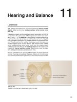
Hearing and Balance
11
I. OVERVIEW
Both hearing and balance are sensations carried by special somatic
afferent fibers that form the vestibulocochlear nerve (cranial nerve
[CN] VIII).
The sensory organs and the peripheral ganglia associated with CN VIII
are located in the petrous part of the temporal bone in the base of the
skull (Figure 11.1). The labyrinth is specialized to translate motion of the
head into information about balance, and the afferents from the labyrinth
that carry balance information are bundled together as the vestibular division. The afferents from the cochlea, which carry sound information, are
bundled together as the cochlear division. Both divisions come together
as the vestibulocochlear nerve, which travels from the receptor organs
in the temporal bone through the auditory canal into the cranial cavity
through the internal auditory meatus. Afferents then enter the brainstem at the pontomedullary junction (Figure 11.2).
Hearing and balance are two very different types of senses. Both the
cochlear (hearing) and vestibular (balance) divisions of CN VIII receive
stimuli from specialized end organs that contain mechanoreceptors called
Cochlea
Semicircular
canals
Anterior
Lateral
Posterior
Petrous part of temporal bone
Internal acoustic meatus
Vestibule
Figure 11.1
Position of the inner ear in the temporal bone of the skull.
199
Krebs_Chap11.indd 199
5/9/2011 7:10:10 PM
200
11. Hearing and Balance
“hair cells” because of their appearance. Although similar in appearance, hair cells respond to different stimuli. They respond to sound in the
cochlear division, and position and head movement in relation to gravity
in the vestibular division.
II. HEARING
Vestibulocochlear
nerve (CN VIII)
Pontomedullary
junction
For hearing, sound waves are interpreted in terms of their pitch, loudness, and their location of origin. The human ear has the remarkable
capability to distinguish a large range of sounds that can be either very
close together in pitch (maybe just a quarter note apart) or far apart in
pitch (ranging from the low rumblings of a pipe organ to the highest notes
of a piccolo flute).
Hearing is an integral component of communication. The sounds of
speech are perceived and then relayed to higher centers where they are
reassembled to make sense as words and phrases.
Figure 11.2
The vestibulocochlear nerve at the pontomedullary junction of the brainstem.
CN = cranial nerve.
A. Structures involved in hearing
The structures involved in hearing are specialized to bundle, amplify,
and fine-tune the sounds that surround us so that we can make sense
of them.
The outer ear is shaped to collect sound waves and focus them onto
the tympanic membrane, which separates the outer ear from the middle ear. The middle ear is an air-filled space, which contains three
small bones that amplify the sound energy from the tympanic membrane to the fluid-filled inner ear. The inner ear contains the cochlea,
which contains the sensory organ of hearing, the organ of Corti.
1. Outer ear: The outer ear is the visible part of the ear on the side of
the head. It is composed of the auricle and the external auditory
meatus, or outer ear canal. These structures gather sound energy
and focus this energy on the tympanic membrane, also referred to
as the eardrum, at the medial end of the outer ear canal (Figure 11.3).
Interestingly, the external ear also reflects sound, causing it to
reach the tympanic membrane in a time-delayed manner. This
plays a role in sound localization, as is discussed below.
The external auditory meatus also plays a role in how sound waves
are transmitted to the middle ear. Sound pressure at frequencies
around 3 kHz (the frequency to which the human ear is most sensitive) is boosted in the external auditory meatus through passive
resonance effects (echo).
2. Middle ear: The middle ear is located between the tympanic membrane and the inner ear. It is an air-filled chamber that contains
three small bones, or ossicles, that transfer the sound energy
from the tympanic membrane to the inner ear. The middle ear is
continuous with the nasopharynx through the pharyngotympanic
(Eustachian) tube (see Figure 11.3). This connection is important
to ensure that air pressure in the middle ear corresponds to the air
pressure around us. The pharyngotympanic tube opens to let air
into the middle ear and equilibrate the pressure (for example, during a plane landing when the ears “pop”).
Krebs_Chap11.indd 200
5/9/2011 7:10:11 PM
II. Hearing
201
Outer ear
Middle ear
Inner ear
Semicircular
canals
Cochlea
Incus
Malleus
Cochlear nerve
Concha
Auricle
Tympanic
membrane
(eardrum)
Stapes footplate
covering oval
window
External
auditory
meatus
Stapes
Tympanic Round
cavity
window
Pharyngotympanic
(Eustachian) tube
Figure 11.3
Overview of structures of the outer, middle, and inner ear.
a. Bones in the middle ear: The ossicles in the middle ear are
the malleus, the incus, and the stapes. The malleus is directly
attached to the tympanic membrane. The malleus articulates
with the incus, which is connected to the stapes. The stapes is
connected to the oval window of the inner ear (see Figure 11.3).
The function of these articulating ossicles is to boost the sound
energy from the tympanic membrane into the inner ear. This
boost is necessary so that the sound waves traveling through
the air can be transferred efficiently to the fluid-filled space of
the inner ear. Without a boost, the sound energy would be lost
through reflection once the sound waves hit fluid. The boost
is achieved through the lever action of the ossicles as well as
through compression of sound waves from the large-diameter
tympanic membrane to the small-diameter oval window.
b. Muscles in the middle ear: The middle ear also contains two
muscles: the tensor tympani, which attaches to the malleus
and is innervated by CN V, and the stapedius muscle, which
attaches to the stapes and is innervated by CN VII. Contraction
of the stapedius muscle can reduce the transmission of vibration into the inner ear, especially for low-frequency sounds, possibly to selectively filter out low-frequency background noises.
These two muscles also dampen movements of the ossicles in
response to loud sounds, which serves as a protective mechanism for the auditory nerve.
3. Inner ear: The inner ear contains the cochlea, the sensory organ
that mediates the transformation of the pressure waves of sound
into the electrical energy of a nerve impulse (Figure 11.4).
a. Cochlea: The cochlea sits in the petrous portion of the temporal bone, with its base facing medially and posteriorly. It is
Krebs_Chap11.indd 201
5/9/2011 7:10:11 PM
202
11. Hearing and Balance
COCHLEA CROSS SECTION
Vestibular
nerve
Auditory
nerve
Scala
vestibuli
Scala media
= cochlear duct
Tectorial
membrane
Oval
window
Spiral
ganglion
Round
window
Scala
tympani
Cochlea
Inner
hair cells
Basilar
membrane
Outer
hair cells
COCHLEA UNCOILED
Incus
Oval window
Scala vestibuli
Scala media = cochlear duct
Helicotrema
Stapes
Round window
Scala tympani
Endolymph
Perilymph
Figure 11.4
Structures of the inner ear: the cochlea.
a bony tube that coils through two and three-quarter turns in
the shape of a snail’s shell (cochlea is Latin for “snail”), from a
relatively broad base to a narrow apex.
b. Three chambers: A membranous tube or membranous labyrinth, also called the cochlear duct, is suspended within the
bony labyrinth.
Viewed in cross section, the bony labyrinth and cochlear duct
together form three chambers (or scalae) along most of their
length (see Figure 11.4). The cochlear duct, anchored to the
bony labyrinth, has a triangular shape in cross section. It forms
the middle chamber, or scala media (cochlear duct). The chamber above the cochlear duct is the scala vestibuli and is continuous with the vestibule (see below). The chamber below the
cochlear duct is called the scala tympani because it ends at the
round window or secondary tympanic membrane. Both the bony
labyrinth and the membranous labyrinth are filled with fluid. The
fluid in the bony labyrinth (scalae vestibuli and tympani) is called
perilymph, which is similar in composition to cerebrospinal fluid
(and also to extracellular fluid). Perilymph is low in K+ and high in
Krebs_Chap11.indd 202
5/9/2011 7:10:13 PM
II. Hearing
203
Na+. The cochlear duct (or scala media) is filled with endolymph,
which is similar in composition to intracellular fluid, and is high
in K+ and low in Na+. Endolymph is produced by the stria vascularis, a layer of cells on the lateral surface of the scala media
(Figure 11.5). The high concentration of K+ in the endolymph
plays a critical role in signal transduction, as discussed below.
The scalae tympani and vestibuli are joined at the apex of
the cochlea by a small opening called the helicotrema (see
Figure 11.4), where perilymph can pass from one chamber to
the other. The scala media is separated from the scala vestibuli
Scala vestibuli
is a perilymph-filled space
continuous with scala tympani
at the apex of the vestibule.
Scala media (cochlear duct)
is an endolymph-filled tube, continuous
with membranous labyrinth.
Spiral ganglion
contains cell bodies
of cochlear afferents.
Scala tympani
perilymph-filled space,
continuous with scala vestibuli.
CN VIII—cochlear division
gives afferents from the inner hair cells
and efferents to the outer hair cells.
Reissner membrane
separates the scala media
from the scala vestibuli.
Stria vascularis
produces the K+-rich
endolymph.
Tectorial membrane
is where the stereocilia of the
outer hair cells are embedded.
CN VIII—cochlear division
gives afferents from the inner
hair cells and efferents to the
outer hair cells.
Outer hair cells
amplify and fine-tune the
sound information.
Inner hair cells
transmit sound information
to cochlear nerve fibers.
Basilar membrane
is displaced in a frequencydependent manner by sound
waves.
Figure 11.5
Histology of the cochlea. CN = cranial nerve.
Krebs_Chap11.indd 203
5/9/2011 7:10:15 PM
204
11. Hearing and Balance
3
4
Sound energy is amplified
through the articulation of
the ossicles in the middle ear.
Incus
Malleus
Stapes transmits the sound
energy to the oval window,
into the fluid-filled scala vestibuli.
Oval
Stapes window
2
The tympanic
membrane
deflects.
Tympanic
membrane
“ear drum”
Cochlear
nerve
Spiral
ganglion
Scala
vestibuli
Scala media
contains organ
of Corti
External
auditory
meatus
Middle Ear
air-filled
1
5
Pharyngotympanic
tube
Sound waves travel
through the external
auditory meatus to the
tympanic membrane.
Round
window
6
The round window bulges
out as the sound wave travels
through the scala tympani.
Scala
tympani
The sound wave causes
frequency-specific
displacement of the basilar
membrane, which causes
activation of the hair cells
in the organ of Corti.
Figure 11.6
Sound transduction in the ear.
by the Reissner (or vestibular) membrane and from the scala
tympani by the flexible basilar membrane.
Sound energy is transmitted onto the oval window, which displaces the fluid in the scala vestibuli. Vibrations are then transmitted along the cochlea to the end, where it joins the scala
tympani and ultimately causes the round window at the end
of the scala tympani to bulge. The sound energy or vibrations
also cause the basilar membrane, which separates the scala
tympani from the scala media, to vibrate (Figure 11.6).
Scala
vestibuli
Tectorial
membrane
Supporting
cell
Inner
hair
cell
Outer
hair cells
Basilar
membrane
Spiral
ganglion
Scala tympani
Figure 11.7
Organ of Corti.
Krebs_Chap11.indd 204
Scala media
(cochlear duct)
filled with endolymph
c. Organ of Corti: The auditory sensory organ, or the organ of
Corti, is located within the scala media and sits on the flexible
basilar membrane. One row of inner hair cells and three rows
of outer hair cells, along with supporting cells, comprise the
organ of Corti. The hair cells are the signal-transducing cells.
Their name comes from the hair-like microvilli, known as stereocilia, that are arranged symmetrically and in graded height
(with the tallest toward one side of the hair cell) in a V shape
on the apex of the cells. The tectorial membrane, a gelatinous
extracellular structure, extends over the hair cells.
Both the inner and the outer hair cells are anchored to the basilar membrane. Importantly, the outer hair cells are also directly
embedded in or coupled to the tectorial membrane via their stereocilia. The inner hair cells do not have direct contact with the
tectorial membrane but respond to fluid movement in the scala
media (Figure 11.7).
5/9/2011 7:10:19 PM
II. Hearing
Frequency (Hz) = number of repeats of
the wave within a set interval
Amplitude (dB) = height
of the sound wave
The spiral ganglion, which contains the nerve cell bodies of the
primary auditory afferents, sits within the turns of the cochlea,
close to the organ of Corti (see Figure 11.5). Peripheral processes
travel to the scala media where they receive input from the receptor cells.
205
B. Physiology of sound perception in the inner ear
Sound is a pressure wave that travels through the air. It is then amplified in the outer and middle ear before it reaches the fluid-filled inner
ear, where the sensory organ of Corti sits. The organ of Corti transduces this pressure to a neuronal signal. Sound waves have different shapes and sizes. The amplitude of a sound wave determines
its loudness and is measured in decibels (dB). The frequency of
a sound wave determines the pitch and is measured in Hertz (Hz)
(Figure 11.8). The human ear can hear frequencies between 20 and
20,000 Hz. The lowest note on a large pipe organ is at 20 Hz, and the
highest note on a piano is at 4,200 Hz (Figure 11.9). The human voice
ranges between 300 and 3,000 Hz.
1. Basilar membrane: When a sound wave reaches the inner ear,
it sets off a wave in the basilar membrane at the same frequency
as the sound. This wave propagates from the base to the apex until
it reaches a point of maximal displacement of the basilar membrane. This point is reached because of the geometry and flexibility of the basilar membrane. The base of the basilar membrane
is narrow and stiff and is where the propagation of each sound
wave begins. High-frequency sounds produce their maximal displacement at the base. The apex of the basilar membrane, on the
other hand, is wider and more flexible and is where low-frequency
sounds are perceived (Figure 11.10). These mechanical properties result in the tonotopy of the inner ear, with distinct locations
interpreting discrete frequencies. Tonotopy is then carried forward
throughout the auditory pathway.
Figure 11.8
The physics of frequency and amplitude.
Lowest note on large pipe organ: 20 Hz
Most of the sounds we hear are a combination of different frequencies. As the sound waves travel into our inner ear, they are broken
up into their component parts. Each component will individually
reach its point of maximal displacement on the basilar membrane.
2. Inner and outer hair cells: Basilar membrane vibrations create
a shearing force against the stationary tectorial membrane, causing the stereocilia of the outer hair cells to be displaced in that
plane (Figure 11.11). The inner hair cells are not in direct contact
with the tectorial membrane and are activated through fluid movement in the scala media. Stereocilia are arranged symmetrically by
height. Displacement toward the tallest stereocilium causes depolarization of the cell, whereas displacement toward the shortest
stereocilium causes hyperpolarization of the cell (Figure 11.12).
Depolarization of the cell occurs when cation channels open at the
apex of the stereocilia. Stereocilia are connected to each other via
tip links that transmit force to an elastic gating spring, which, in
turn, opens the cation channel (see Figure 11.12). These cation
channels are examples of mechanotransduction channels, which
have the advantage of conferring immediate effects. In fact, hair
cells can respond to a stimulus within 50 μs. Such a rapid response
Krebs_Chap11.indd 205
Human voice: 300–3000 Hz
Highest note on a piano: 4200 Hz
Figure 11.9
Examples of different frequencies.
5/9/2011 7:10:21 PM
206
11. Hearing and Balance
Basilar membrane
Stapes
The basal end of the basilar
membrane is narrow and
stiff. It is “tuned” for high
frequencies.
Cochlear
base
Scala
vestibuli
The apical end of the
basilar membrane is
wide and flexible. It is
“tuned” for low
frequencies.
“Uncoiled”
cochlea
Helicotrema
Helicotrema
10,000 Hz
4000 Hz
Stapes on
oval window
Round
window
Traveling
wave
A specific frequency reaches
its point of maximal displacement
at a specific point along
the basilar membrane.
Scala
tympani
Basilar
membrane
(in scala
media)
Cochlear
apex
200 Hz
Different frequencies reach their point
of maximal displacement along the
basilar membrane.
Figure 11.10
Basilar membrane tuning.
A
would not be possible with a slow chemical signal transduction
process. Another advantage of mechanotransduction channels is
that they do not require receptor potentials, thereby increasing the
sensitivity of the response (see Chapter 1, “Introduction to the
Nervous System and Basic Neurophysiology”). The sensitivity of
the ion channel opening is remarkable: even small vibrations of 0.3
nm (the size of an atom) will cause channel opening.
Resting position
Tectorial
membrane
Inner
hair cell
B
Outer
hair cells
Basilar
membrane
Sound-induced vibration
Shear
force
Upward
phase
Shear
force
Downward
phase
Figure 11.11
Because the stereocilia are bathed in the K+-rich endolymph of the
scala media, the opening of the cation channels will cause a rapid
influx of K+ to the cell (the driving force for K+ uptake is about 150
mV). The hair cells then depolarize, which causes Ca2+ channels
at the base of the cells to open. Calcium influx causes neurotransmitter-filled vesicles to fuse with the basal membrane and release
the neurotransmitter glutamate into the synaptic cleft. The afferent cochlear neurons are stimulated and transmit this signal to the
central nervous system (CNS).
The inner hair cells are responsible for hearing. About 90% of cochlear
nerve fibers come from the inner hair cells. The outer hair cells
amplify the signals that are then processed by the inner hair cells.
3. Frequency selectivity: The frequency selectivity, or tuning,
of the basilar membrane is due to and limited by its mechanical
properties. The sound wave traveling along the basilar membrane
and its associated point of maximal displacement cannot be as
selective in frequency tuning as our hearing is, which suggests
The organ of Corti during displacement by sound waves.
Krebs_Chap11.indd 206
5/9/2011 7:10:23 PM
II. Hearing
207
A Hyperpolarization
B Depolarization
Hyperpolarization
Depolarization
Tip links
Movement away from the
kinocilium, or toward the
shortest steriocilium, prevents
opening of the mechanicallygated K+ channels.
Tip links
K+
K+
K+
Kinocilium
Movement toward the kinocilium, or
toward the longest stereocilium,
causes the opening of the mechanically
gated K+ channels. K+ from the K+-rich
endolymph enters the cell.
K+
Kinocilium
The increase in K+ leads to
depolarization of the cell.
Depolarization
Ca2+
Ca2+ channel
Vesicles
Ca2+
Transmitter
Afferent
nerve
To brain
Vesicles
Transmitter
Afferent
nerve
The depolarization of the cell
leads to opening of voltage-gated
Ca2+ channels.
To brain
The increase in intracellular Ca2+
causes neurotransmitter-filled
vesicles to fuse with the membrane
in the synaptic cleft, leading to
excitation of the afferents to the CNS.
Figure 11.12
Hyperpolarization and depolarization of hair cells in the inner ear. CNS = central nervous system.
involvement of an additional mechanism of sound amplification and
tuning. This additional mechanism is from movement of the outer
hair cells in response to specific frequencies. When the outer hair
cells are depolarized, their cell bodies actively contract. When they
are hyperpolarized, their cell bodies actively lengthen. High frequencies cause contraction of the outer hair cells at the base, and
low frequencies cause contraction at the apex. This mechanism
influences the movement of the basilar membrane in that particular
segment, increasing the fluid displacement around the inner hair
cells. This amplifies the magnitude of the K+ influx into the inner
hair cells, increasing the signal to the cochlear nerve. Because of
this fine-tuning and amplification of the sound wave through the
outer hair cells, we can both discriminate tones of neighboring frequencies with astounding accuracy and detect low-level sounds. In
addition, the outer hair cells are innervated by efferents originating from the auditory pathway (Figure 11.13). These inputs hyperpolarize, or inhibit, the outer hair cells, reducing their response to
displacement of the basilar membrane through sound and allowing
the central auditory pathway to influence sound amplification in the
inner ear. A possible function of this mechanism is to help focus the
inner ear on relevant sounds while filtering out background noises.
4. Otoacoustic emissions: Because the motility of the outer hair
cells can cause the basilar membrane to move, it is conceivable
Krebs_Chap11.indd 207
5/9/2011 7:10:26 PM
208
11. Hearing and Balance
that this movement could be retrograde, or backward, toward the
oval window and through the middle ear via the ossicles to cause
displacement of the tympanic membrane. This process would
result in the ear itself producing a sound and is, indeed, what actually happens. These sounds can be measured in the external auditory meatus as otoacoustic emissions. Such measurements are
routinely done in infants to assess the function of the inner and
middle ears.
CLINICAL APPLICATION 11.1
Cochlear Implants
Hearing loss can have several underlying causes and is divided into two main categories: conductive hearing loss and sensorineural hearing loss.
Conductive hearing loss is from obstruction in the conduction of sound energy from the outer ear to the
inner ear. The causes can be either in the outer ear (such as earwax or rupture of the tympanic membrane) or
in the middle ear (for example, fluid or arthritis of the ossicles). A hearing aid, which amplifies sound energy,
can significantly ameliorate conductive hearing loss.
Sensorineural hearing loss is from a problem in the inner ear, either with hair cells or with the cochlear
nerve itself. Hair cells are very susceptible to damage and do not regenerate in humans. Common causes for
2
Receiver under the skin receives
signal and transmits it to the
electrode array in the cochlea.
1
Microphone and speech
processor convert sounds
into a digital signal.
3
Electrode array in the cochlea
stimulates the cochlear nerve
tonotopically along the basilar
membrane.
4
Sound information is relayed
via the cochlear nerve to
the brainstem.
The cochlear implant.
Krebs_Chap11.indd 208
5/9/2011 7:10:28 PM
II. Hearing
209
congenital hearing loss include genetic causes and prenatal infection with TORCH organisms (toxoplasmosis,
other [syphilis], rubella, cytomegalovirus, and herpes), which lead to dysfunctional hair cells with an intact
cranial nerve (CN) VIII.
For the latter patients, a cochlear implant can improve sound perception, or hearing.
A cochlear implant consists of an external and an internal component. The external component includes
a microphone, a speech processor, and a transmission system. Sound information is broken down into its
component parts and converted into electrical signals, which are then relayed to the internal component. The
internal component includes a receiver and an electrode array. The receiver decodes the signal and delivers
the electrical signals to the electrode array.
The electrode array is inserted into the cochlea through the oval window where it sits in the cochlear duct
along the afferents from CN VIII. Electrical signals anywhere along the electrode array will stimulate a particular cochlear nerve afferent along the basilar membrane. The electrode array mimics the tonotopy of the basilar
membrane and stimulates nerves at discrete frequencies. This information is then relayed centrally, resulting
in the perception of sound.
The interpretation of sound heard is a central process that requires new neuronal connections to be made,
and the understanding of speech must be learned.
For patients with damage to CN VIII, devices that will directly relay sound information to brainstem nuclei are
currently being developed.
C. Central auditory pathways
The central auditory pathways carry the signal from the cochlea to the
CNS. The auditory system analyzes different aspects of sound including the frequency (pitch), the amplitude (volume), and the location
of the sound in space.
Outer hair cells
Inner
hair cell
The pitch and volume of the sound travel centrally in a relatively
straightforward pathway. The localization of sound, however, is more
complicated, and the pathways differ depending on whether high-frequency or low-frequency sounds are being analyzed.
1. Central pathway for pitch and volume: The frequencies of a
sound are broken down in the cochlea and then relayed to the
cochlear nerve fibers innervating the hair cells at different locations along the basilar membrane. Each cochlear nerve fiber
only transmits information of a specific frequency spectrum. The
nerve cell bodies of these cochlear afferents are located in the
spiral ganglion. The central processes of the first-order neurons synapse in the cochlear nuclei. These are columns of cells
adjacent to the inferior cerebellar peduncle and can be divided
into a posterior and anterior nucleus. Most fibers then cross
the midline and travel in the contralateral lateral lemniscus to
the inferior colliculus in the caudal midbrain, a major integration center in the auditory pathway. From there, fibers travel to
the medial geniculate nucleus (MGN) of the thalamus via the
inferior brachium. From the thalamus, fibers travel through the
internal capsule to the primary auditory cortex, which is located
Krebs_Chap11.indd 209
The majority of afferents in the
cochlear nerve come from the
inner hair cells.
Spiral
ganglion
cells
Cochlear
nerve
Inhibitory efferents to the outer hair cells
reduce their response to the displacement
of the basilar membrane. The auditory
pathway can inhibit the amplification of
sound in the cochlea.
Figure 11.13
Afferents to and efferents from the hair
cells in the cochlea.
5/9/2011 7:10:30 PM
210
11. Hearing and Balance
Transverse temporal gyri
CORTEX
Transverse temporal gyri
Insula
View into the lateral fissure. The superior surface
of the temporal lobe with the transverse temporal
gyri is visible.
Medial
geniculate
nucleus
Inferior
brachium
Medial geniculate
body
Inferior
colliculus
Inferior brachium
Inferior colliculus
Lateral
lemniscus
Lateral
lemniscus
CN VIII
Cochlear
nuclei
Posterior and
anterior cochlear
nuclei
Inferior cerebellar
peduncle
Midline
Figure 11.14
Central pathway for pitch and volume. CN = cranial nerve.
Path 1
direct
sound
Auricle
2. Central pathways for sound localization: We live in threedimensional space, and sounds are perceived as coming from
within this space. The auditory system can, in fact, map sounds
even though space is not directly represented in the auditory
system. We can map sounds on a vertical plane (whether
sounds come from above or below), and we can also map sound
in a horizontal plane. Vertical analysis can be done with only
one ear, whereas horizontal analysis relies on the input from
both ears.
Path 1
reflected
sound
Path 2
direct
sound
Path 2
reflected
sound
Auditory
canal
Path 3
reflected
sound
Path 3
direct
sound
Figure 11.15
Vertical sound mapping through reflection of sound in the outer ear.
Krebs_Chap11.indd 210
on the superior surface of the superior temporal gyrus in the temporal lobe (Figure 11.14).
Vertical sound mapping occurs in the external ear. Sounds reach
the tympanic membrane both directly and through reflection in
the external ear. The brain can localize sounds in the vertical plane
through analysis of the differences in the direct and reflected sound
inputs. The neuronal mechanisms of this are not fully understood
(Figure 11.15).
In order to achieve horizontal spatial mapping, the input to both
ears is compared in brainstem nuclei. For low-frequency sounds,
5/9/2011 7:10:31 PM
II. Hearing
211
the sound waves will reach the ear farther away from the sound
after they reach the ear closer to the sound, and a time difference
is detected and analyzed. For high-frequency sounds, the sound
waves are closer to each other, and the head forms a barrier for
these waves as they travel to the ear farther away from the sound
stimulus. The far ear will hear the sound at a lesser intensity than
the near ear due to the “sound shadow” created by the head.
a. Time difference detection at low frequencies: For lowfrequency sounds (below 3 kHz), the ear closer to the sound
source will perceive the sound waves before the ear farther away
from the source. These low-frequency sounds from both ears
project to the medial superior olivary nucleus (MSO) where the
time delay of the sound perception is analyzed (Figure 11.16). The
axons projecting to the MSO will vary in length. The longest axons
from the left will converge on the same neuron in the MSO as the
shortest axons from the right. Because axon diameter and the
degree of myelination are the same for all neurons coming from
the cochlear nuclei, the speed of action potential propagation is
the same. Only the length of the axon will determine how long it
takes for the signal to get to the MSO.
For example, when a sound reaches the left ear first, the neurons in the cochlear nucleus on the left will start sending action
potentials before the neurons on the right. The cochlear nucleus
neuron with the longest axon on the left will converge on the
same neuron in the MSO as the one with the shortest axon
from the right. The action potentials will arrive at that particular
neuron at the same time. The neurons in the MSO act as coincidence detectors. The temporal summation of signals from
the left and right resulting from the time delay and the different
axon lengths allow the localization of sound. Each neuron in the
MSO is sensitive to a sound originating from a particular area,
resulting in a sound map for low-frequency signals.
b. Intensity difference detection at high frequencies: At frequencies above 3 kHz, the head forms a barrier for sound transmission. A sound originating on the left side will be more intense
on the left than on the right because of the acoustical shadow of
the head. The intensity of the stimulation on the left side will be
higher than the intensity on the right side (Figure 11.17).
The intensity of the stimulation is transmitted to the cochlear
nuclei and from there to the lateral superior olivary nucleus
(LSO). At the same time, a signal encoded at the same intensity is sent to the contralateral medial nucleus of the trapezoid body, which will inhibit the LSO on that side. The LSO
then compares the amount of intensity-dependent excitation
from the ipsilateral side with the intensity-dependent inhibition from the contralateral side. Only when the excitation
outweighs the inhibition is the sound information relayed to
higher centers.
Each LSO can only relay information from the ipsilateral side
of the soundscape. In order to get a full appreciation of the
sound-filled space, both lateral superior olivary nuclei must
function.
Krebs_Chap11.indd 211
5/9/2011 7:10:36 PM
212
11. Hearing and Balance
Sound waves reach the ear
close to the source first and
the ear farther away from the
source with a time delay.
Sound waves
Sound source
200 Hz
To Higher Centers
Cochlear
nucleus
Input from both ears
converges in the medial
superior olivary nucleus,
where the delay in signal
is analyzed.
Cochlea
Auditory
nerve
Medial superior
olivary (MSO) nuclei
2
1
Sound arrives at
the ear closer to
the source first.
The action potential travels
to cochlear nucleus and
from there to the contra3
lateral MSO.
Sound arrives at
the far ear later.
Auditory
stimulus
Right ear
Left ear
Cochlea
Cochlea
Cochlear
nucleus
Cochlear
nucleus
MSO
4
1 2 3 4
The action potential travels
to cochlear nucleus and from
there to the ipsilateral MSO.
A B C D
4 3 2 1
5
6
The neuron on which both pathways
converge, neuron “D” in this diagram,
acts as a coincidence detector.
The pathway from the contralateral side, where the
sound was heard first, is longer. The pathway from
the ipsilateral side, where the sound was heard
later, is shorter. Both signals then arrive at the
same time, and they converge in the MSO.
Figure 11.16
Time difference detection at low frequencies.
Krebs_Chap11.indd 212
5/9/2011 7:10:36 PM
II. Hearing
213
Sound intensity is higher on the side
of the source. The sound shadow of
the head lessens the intensity on the
side away from the source.
Sound waves
Sound shadow
Sound source
2/10
0
Cochlear
nucleus
Cochlear
nucleus 10/10
8/10
+
10/10
+
Only when excitation
outweighs inhibition is
the sound information
relayed to higher centers.
LSO
10 – 8 = 2
LSO compares the intensitydependent excitation from
the ipsilateral side with the
intensity-dependent inhibition
from the contralateral side.
LSO
8 – 10 = –2
10/10
–
8/10
–
MNTB
MNTB
MNTB receives input
from contralateral LSO
and inhibits ipsilateral
LSO.
+
8/10
+
10/10
Figure 11.17
Intensity difference detection of sound at high frequencies. LSO = lateral superior olivary nucleus; MNTB = medial nucleus
of the trapezoid body.
c. Convergence of pathways: Both the intensity level and time
difference–encoded sound localization pathways converge in
the inferior colliculus. Similar to the visual map in the superior
colliculus, the inferior colliculus contains an auditory space map.
Here, both the vertical and the horizontal analyses of sound are
integrated, resulting in a precise sound localization. The inferior
colliculus also analyzes the temporal patterns of sound. From
Krebs_Chap11.indd 213
5/9/2011 7:10:38 PM
214
11. Hearing and Balance
CORTEX
Medial
geniculate
nucleus
Inferior
brachium
Analysis of spatial sound map and
analysis of pitch and volume.
Info is relayed to cortex via the
MGN of thalamus.
Inferior
colliculus
Lateral
lemniscus
MSO
Cochlear
nuclei
LSO
High-frequency sound
localization through
intensity difference.
Low-frequency sound
localization through
time delay.
Information on
pitch and volume.
Midline
Figure 11.18
Convergence of pathways in the auditory system. MGN = medial geniculate nucleus; MSO = medial superior olivary
nucleus; LSO = lateral superior olivary nucleus.
the inferior colliculus, the information is relayed to the MGN
of the thalamus. There the frequency-analyzed component and
the temporal component of sound converge in a tonotopically
mapped pathway (Figure 11.18).
The signal is then relayed to the primary auditory cortex,
which is also organized tonotopically and can interpret sounds
and spatial distribution patterns. The secondary auditory or
auditory association areas of the cortex are localized around
the primary area and process complex sounds necessary
for communication. In the human brain, the cortical area for
speech comprehension (Wernicke area) is directly adjacent to
the primary and association auditory areas. See Chapter 13,
“The Cerebral Cortex,” for more information.
III. BALANCE
Although we have no conscious appreciation of balance, it is a key sense
that interacts with many systems to ensure stable posture and coordinated movements.
Krebs_Chap11.indd 214
5/9/2011 7:10:39 PM
III. Balance
215
Carried in the vestibular component of the vestibulocochlear nerve
(CN VIII), the sense of balance allows perception of the body in
motion, head position, and the orientation of the head in relation to
gravity.
A. Structures involved in balance
The vestibular organ is embryologically and structurally related to
the cochlea. The part of the bony labyrinth related to balance
is adjacent to and continuous with the cochlea in the temporal
bone. It consists of three semicircular canals, which are attached
to the central vestibule and roughly orthogonal (at 90°) to each
other. Like the cochlea, these bony structures contain a membranous labyrinth, which is continuous with the cochlear duct (scala
media) of the cochlea. The membranous labyrinth contains K+-rich
endolymph, whereas the space between the membranous and the
bony labyrinth is filled with perilymph, with low K+ concentrations
(Figure 11.19).
1. Semicircular canals: The semicircular canals contain the membranous semicircular ducts. At the base of each duct is a bulging
ampulla. Each ampulla contains the receptor cells, which are hair
cells that sit on a crista (crista ampullaris) and, analogous to the
Horizontal semicircular canal and duct
Ampulla
Anterior semicircular canal and duct
Posterior semicircular canal and duct
Endolymphatic sac and duct
Dura mater
Vestibule contains utricle and saccule
Saccule
Utricle
Helicotrema
Stapes in oval window
Utricosaccular duct
Round window
Scala vestibuli
Cochlear duct
Scala tympani
Location of hair cells
Endolymph-filled space
Figure 11.19
The membranous labyrinth and the location of hair cells in the inner ear.
Krebs_Chap11.indd 215
5/9/2011 7:10:39 PM
216
11. Hearing and Balance
hair cells of the cochlear duct, are embedded in a gelatinous mass
in this location called the cupula (Figure 11.20).
2. Vestibule: The vestibule contains two endolymph-filled sacs or
enlargements of the membranous labyrinth, the utricle and the
saccule. The three semicircular ducts and their ampullae are continuous with the utricle, which is connected to the saccule via
the utricosaccular duct. The utricle and saccule comprise the otolithic organ. Each contains a macula (equivalent to the organ
of Corti), where the receptor hair cells are located. The utricular
macula is on the floor of the utricle, in a horizontal plane. The
saccular macula is on the medial wall of the saccule, in a vertical plane. Like the hair cells of the organ of Corti and the semicircular canals, the hair cells of the maculae are embedded in a
gelatinous mass. In this case, the gelatinous mass has an outer
layer covered in calcium carbonate crystals (otoconia, or otoliths). This gives this structure its name, the otolithic membrane
(Figure 11.21).
Semicircular
canals
Ampullae
Vestibular
nerve
Crista
ampullaris
and cupula
Vestibular
nerve
branch
Crista
ampullaris
B. Physiology of balance
Cupula
Hair
cell
Nerve
fibers
to vestibular
nerve branch
Figure 11.20
The semicircular canals with the ampullae containing crista and cupula.
We can move our bodies and our heads along all three axes of our
three-dimensional space. We can move in a linear way, along any one
of these axes (linear acceleration), or we can rotate around any one
of these axes (rotational acceleration). Our movements are often a
combination of linear and rotational acceleration, and the labyrinth of
the inner ear detects the different components of our movements and
faithfully relays them to central nuclei.
The system is best equipped to detect changes in movement. Vestibular afferents will fire most at the beginning and end of an acceleration.
1. Rotational acceleration: Rotational acceleration is detected in
the three semicircular canals, where identically oriented hair
cells sit atop cristae. Each hair cell has a long kinocilium and
A
B
Kinocilium
Otoliths in gelatinous mass
Microvilli (stereocilia)
Utricular
macula
Saccular
macula
Hair
cell
Utricle
Saccule
Hair
cell
Support
cells
Nerve
fibers of
vestibular
nerve
Part of macula
Figure 11.21
The otolithic organs: utricle and saccule.
Krebs_Chap11.indd 216
5/9/2011 7:10:41 PM
III. Balance
217
several microvilli, called stereocilia, in graded height as in the
cochlea.
During rotational acceleration, the endolymph is set into motion.
The movement of endolymph causes a deformation of the cupula
(Figure 11.22). This deformation causes deflection of the stereocilia of the hair cells. Movement toward the kinocilium will cause the
mechanically gated ion channels to open, resulting in depolarization
of the cell and increased signal transduction to the vestibular nerve
(Figure 11.23). Movement away from the kinocilium causes the
cation channels to close and, thereby, hyperpolarizes the cell with
a decrease of signal transduction in the vestibular nerve. All hair
cells in the ampulla have the same orientation and will respond
similarly to deformation of the cupula.
The semicircular canals on one side of the head are a mirror
image of the semicircular canals on the other side of the head.
The two horizontal canals are in the same plane, the left posterior
and right anterior canals are paired and in the same plane, and
the left anterior and right posterior canals are paired and in the
same plane (Figure 11.24). Rotational acceleration is detected on
both sides and sets endolymph in motion in the same direction on
both sides, but with different effects. Movement of endolymph will
cause hyperpolarization of hair cells on one side and depolarization on the other side, depending on whether the stereocilia are
deflected away from or toward the kinocilium, respectively (see
Figure 11.23).
LEFT
Horizontal
canal
on the right
Horizontal
canal
on the left
Steady state
Steady state
Depolarization
Head rotation
to the right
Hyperpolarization
Head rotation
Hyperpolarization to the left
Depolarization
Figure 11.22
Rotational acceleration and
deflection of the cupula in the
horizontal semicircular canal.
RIGHT
Turning the head to the left
causes movement of endolymph in the left horizontal
canal to the right.
Turning the head to the left
causes movement of endolymph in the right horizontal
canal to the right.
The acceleration or movement
of endolymph to the right
(clockwise) pushes onto the
cupula, where the hair cells
are located.
The acceleration or movement
of endolymph to the right
(clockwise) pushes onto the
cupula, where the hair cells
are located.
Hair cells are deflected toward
the kinocilium; this causes the
opening of the mechanically
gated K+ channels.
The hair cells depolarize and
action potentials are sent to the
CNS for the duration of the
horizontal acceleration.
K+
K+K+
Hair cells are deflected away
from the kinocilium; this causes
the closing of the mechanically
gated K+ channels.
The hair cells hyperpolarize,
and fewer action potentials
are generated.
Figure 11.23
How rotational acceleration leads to signal transduction. CNS = central nervous system.
Krebs_Chap11.indd 217
5/9/2011 7:10:43 PM
218
11. Hearing and Balance
For example, if the head is rotated to the left (or counterclockwise), endolymph in both horizontal canals will rotate to the right
(or clockwise). This will lead to displacement of the cupula on both
sides. On the left side, hair cells are displaced toward their kinocilia, which causes the opening of cation channels and increased
signal transduction. On the right side, hair cells are displaced away
from their kinocilia, which causes the closing of cation channels
and decreased signal transduction.
A
This system of hyperpolarization in one canal and depolarization
in the paired canal works in all pairs of canals. Increased signal
transduction will always occur in the canal toward which the head
is rotating.
B
Anterior
semicircular
canal
Posterior
semicircular
canal
Figure 11.24
The pairing of semicircular
canals and their orientation in the
head relative to each other (the
size of the inner ear is exaggerated for diagrammatic purposes).
Otolithic organ
Otoconia
Cupula
Steady
state
Macula
Head tilted
forward
Head moving
forward in one
plane
2. Linear acceleration: Linear acceleration is detected by the
otolithic organs. The otoconia make the otolithic membrane
heavier than the surrounding endolymph. During tilting head
movements, gravity pulls on the otolithic membrane causing
it to shift relative to the underlying macula and the hair cells
anchored there. This shift causes displacement of the hair cells
and, with that, the opening or closing of the cation channels,
depending on the direction of the shift. Similarly, when the head
moves forward without a tilt (pure linear acceleration), the same
shearing motion occurs between the otolithic membrane and the
macula. Because of the weight of the otoconia, the inertia of
the otolithic membrane is greater than that of the macula, and
the otolithic membrane will lag behind the movement of the macula (Figure 11.25).
The location of the macula and the orientation of the hair cells
determine which type of linear acceleration can be detected.
The saccular and utricular maculae on one side of the head are
mirror images to those on the other side of the head. This results
in opposing effects on corresponding hair cells of the two maculae, similar to that of the hair cells of two paired semicircular
canals.
a. Utricle: In the utricle, the macula is located at the bottom of the
sac. The hair cells can be divided into two groups with different
orientations, separated by the striola, a depression in the otolithic membrane. In the utricle, kinocilia are oriented toward the
striola. This enables the utricle to detect linear movement in a
horizontal plane in two directions, such as head tilts to the right
or left or rapid lateral displacements (Figure 11.26).
b. Saccule: In the saccule, the macula is located in the medial
wall of the sac. Again, the striola divides the hair cells into two
groups with different orientations. In the saccule, kinocilia are
oriented away from the striola. The saccule detects head movement in a vertical plane, such as up and down movements or
forward and backward tilts (see Figure 11.26).
C. Central vestibular pathways
Figure 11.25
Linear acceleration and deflection
of the macula in the saccule and
utricle.
Krebs_Chap11.indd 218
The afferents from the labyrinth have their cell bodies in the vestibular (or Scarpa) ganglion, located close to the spiral ganglion. The
central processes enter the brainstem as the vestibular portion of the
vestibulocochlear nerve at the pontomedullary junction and project
5/9/2011 7:10:45 PM
III. Balance
to the vestibular nuclear complex. The vestibular nuclei are located in
the posterior portion of the tegmentum, at the junction between the
pons and the medulla, adjacent to the inferior cerebellar peduncle
and the cochlear nuclei. The vestibular nuclei can be subdivided into
two functionally distinct groups: lateral vestibular nuclei and medial
vestibular nuclei (Figure 11.27).
The vestibular nuclei participate in three major reflex pathways. The
vestibuloocular reflex adjusts eye movements to head movements
and stabilizes images on the retina (see Chapter 9, “Control of Eye
Movements”). The vestibulocervical reflex is important for postural
adjustments of the head, and the vestibulospinal reflex is important
for the postural stability of the body.
The vestibular nuclei are integration centers that receive afferents not
only from the inner ear but also feedback loops from the cerebellum
as well as visual and somatosensory input. As a result, outflow from
the vestibular nuclei incorporates more than just raw input from the
inner ear.
219
rior
Supe
Macula of
the saccule
Striola
An
ter
ior
Striola
al
ter
La
Macula of
the utricle
Figure 11.26
Orientation of the saccule and utricle in
the inner ear and orientation of hair cells
on the maculae.
1. The vestibulocervical reflex: Postural adjustments of the head
are most relevant in response to rotational movements, which
are detected in the semicircular canals.
Afferents from the semicircular canals project to the medial vestibular nuclei. From there, fibers travel in the descending medial
longitudinal fasciculus or medial vestibulospinal tract to the
upper cervical levels of the spinal cord. Here, they cause postural
adjustments of the head and neck muscles in response to head
movements (Figure 11.28).
2. The vestibulospinal reflex: Postural adjustments of the body
occur in response to both linear and rotational acceleration. Linear
acceleration is detected by the otolithic organs, and afferents project mainly to the lateral vestibular nuclei.
Projections from both the medial and the lateral vestibular nuclei
travel through the descending medial longitudinal fasciculus
(or medial vestibulospinal tract) and lateral vestibulospinal
tract, respectively, to the spinal cord. In the anterior horn of the
spinal cord, the lateral vestibulospinal tract provides excitatory
input to the extensor muscles of the legs, which are key muscles in
mediating balance and postural stability in upright gait. They also
influence proximal trunk musculature, particularly in response to
rotational accelerations.
Lateral
vestibular
nucleus
Medial
vestibular
nucleus
Figure 11.27
The lateral and medial vestibular nuclei
in the rostral medulla.
The vestibulospinal reflex is a direct modulator of lower motor neuron function to allow for rapid postural adjustments in response to
a change in balance (see Figure 11.28).
3. Cortical projections: Although there is no conscious appreciation
of balance, there are projections from the vestibular nuclei to the
cortex via the thalamus. The cortical targets are in the primary and
secondary somatosensory areas, which receive additional visual
and proprioceptive inputs. These cortical areas are thought to be
important for the conscious appreciation of the position of our bodies in space as well as for the perception of extrapersonal space.
Krebs_Chap11.indd 219
5/9/2011 7:10:46 PM
220
11. Hearing and Balance
CLINICAL APPLICATION 11.2
Benign Paroxysmal Positional Vertigo and the Epley Maneuver
Benign paroxysmal positional vertigo (BPPV) is the most common peripheral vestibular disorder. Patients
report brief spells of vertigo directly related to movements of the head.
Pathophysiologically, BPPV is caused by sensitivity to gravity in the posterior semicircular canal because of
the presence of floating otoliths in that canal. These otoliths are thought to have dislodged from the utricle in
the vestibule from which they floated into the posterior semicircular canal. Here, they “bump into” the cupula
in the ampulla and stimulate the hair cells in response to certain head movements. This isolated stimulation of
the posterior semicircular canal on one side results in vertigo.
To assess the side from which the BPPV originates (or, in which inner ear the otoliths are floating in the semicircular canal), the patient is in a supine position, and the head is rotated to one side and then to the other.
During this procedure, the eyes are carefully observed. On the affected side, otoliths will stimulate the cupula
in the semicircular canal in response to the head rotation, resulting in nystagmus as well as vertigo.
The treatment of BPPV aims to remove the debris from the semicircular canal back into the vestibule through
a sequence of head positioning maneuvers. This sequence is referred to as the Epley maneuver and is
summarized in the figure.
1
The patient is sitting upright. The
otoliths are in the posterior canal
on the right side, where they can
stimulate the cupula and cause
vertigo.
2
Anterior canal
Lateral canal
Vestibule
3
In order to further move the otolith
debris down the semicircular canal,
the patient’s head is now rotated to
reposition in a 45°-angle to the left.
The patient is lying on his back
with his head off the edge of the
bed. The patient’s head is rotated
45° to the right. This moves the
floating otoliths down the posterior
semicircular canal.
Posterior
canal
4
The patient is asked to lie on
his side with his head facing
down to the floor. This helps
the otolith debris to enter to
the vestibule.
5
As the patient sits up, the otolith
debris falls into the vestibule.
The Epley maneuver.
Krebs_Chap11.indd 220
5/9/2011 7:10:49 PM
III. Balance
221
Medial vestibular nuclei
Process information from
semicircular canals relating
to rotational acceleration.
Lateral vestibular nuclei
Process information from
otolithic organs relating to
linear acceleration.
Descending medial
longitudinal fasciculus
= medial vestibulospinal tract
Lateral vestibulospinal tract
Cervical spinal cord
Postural adjustments of
head and neck muscles.
Spinal cord
Excitation to extensor muscles of the lower limb
(lateral vestibular nuclei). Innervation of proximal
musculature to stabilize body in response to rotational
acceleration (medial vestibular nuclei).
Figure 11.28
Overview of the central vestibular pathways.
Chapter Summary
• The inner ear contains the organs of hearing (cochlea) and balance (semicircular canals and vestibule).
The organs are connected through the membranous labyrinth, the endolymph-filled space in the inner
ear. Each organ uses the same type of receptor cell, the hair cell. The hair cells are mechanoreceptors that
open ion channels in response to movement of the endolymph.
• In the cochlea, movement of endolymph occurs as a consequence of sound waves displacing the basilar
membrane in the cochlea. The basilar membrane is organized in a tonotopic manner, and specific frequencies cause the basilar membrane to be displaced in discrete areas. This is further fine-tuned by the
movement of outer hair cells, which also amplify the signal. The localization of sound requires input from
both ears. For low frequencies, the time difference of sound waves reaching the two ears is analyzed.
For high frequencies, the head forms a “sound shadow,” and the intensity of sound between the two ears
is analyzed. The brainstem nuclei analyze pitch, volume, and temporal patterns of sound, and the cortical
regions assign meaning to sounds such as language, music, traffic noise, etc.
• Balance is analyzed according to head movements. These movements can be rotational or linear.
Rotational movements are detected through deflection of the cupula in the semicircular canals. Each
semicircular canal is coupled with a canal in the same plane on the other side of the head. The information
from both sides is relayed to the vestibular nuclei. Linear acceleration is detected in the otolithic organs,
which are sensitive to gravity because of the otoconia (“ear rocks”) that sit on the sensory organ. Gravity
will pull on the otoconia and cause a movement of the otolithic membrane, which, in turn, displaces the hair
cells and either depolarizes them or hyperpolarizes them depending on their orientation on the macula.
There is no conscious appreciation of balance. Rather, the vestibular nuclei interact with motor systems to
ensure stable posture and adjustments of movement.
Krebs_Chap11.indd 221
5/9/2011 7:10:50 PM
222
11. Hearing and Balance
Study Questions
Choose the ONE best answer.
11.1 A patient comes to the office with symptoms of vertigo
and difficulty hearing. She also reports having a dull
headache that gets worse throughout the day. During the
neurological examination, the clinician notices a weakness in the muscles of facial expression on the entire
right side of the face. A computed tomography scan
shows a tumor, which is pushing onto cranial nerves VII
and VIII. Where is this tumor most likely to be localized?
A.
B.
C.
D.
E.
Midpons.
Midbrain.
Caudal medulla.
Pontomedullary junction.
Rostral medulla.
11.2 A child is brought to the office with otitis media, an
infection involving the middle ear. Which statement
about the middle ear is correct?
A. The middle ear is a fluid-filled cavity.
B. The middle ear contains three ossicles: the malleus, incus, and stapes.
C. The middle ear acts to dampen sound from the
external ear.
D. The middle ear is connected to the nasopharynx.
E. The middle ear lies in the frontal bone.
11.3 A young man loses his hearing in one ear and now
needs to learn how to localize sound effectively. Which
statement about vertical sound mapping is correct?
A. Vertical sound mapping relies on input from both
ears.
B. Vertical sound mapping occurs in the internal ear.
C. Vertical sound mapping measures whether sounds
come from below or above.
D. Vertical sound mapping analyzes directional sound.
E. Vertical sound mapping depends on the differences
between high- and low-frequency sounds.
11.4 Benign paroxysmal vertigo is due to otoliths floating
in the vestibular organ, causing stimulation of the vestibular system without head movements. Which of the
following comprise(s) the otolithic organ?
A.
B.
C.
D.
E.
Cupula.
Utricle and saccule.
Otoliths.
Ampulla.
Semicircular ducts.
Krebs_Chap11.indd 222
Correct answer is D. Both the facial (cranial
nerve [CN] VII) and the vestibulocochlear (CN
VIII) nerves emerge from the brainstem at the
pontomedullary junction. A lesion of the nerves
themselves rather than of the nuclei associated
with the nerves is the most likely location of the
tumor because the brainstem nuclei associated
with these nerves are located throughout the
brainstem. A large lesion involving all of these
nuclei would also have other symptoms (motor
and/or sensory deficits). The headache is due to
increased intracranial pressure and irritation of
the dura mater from the tumor growth.
Correct answer is B. The three bones of the
middle ear are the malleus, incus, and stapes.
The middle ear cavity is filled with air. Sound
energy is increased in the middle ear cavity,
largely through the lever action of the ossicles.
The middle ear connects to the oropharynx by
the auditory (eustachian) tube. The middle ear
cavity lies within the petrous part of the temporal bone. An infection in the middle ear usually
involves fluid. This is painful and reduces the
sound energy transferred, which makes hearing
more difficult with that ear.
Correct answer is C. The brain can localize
sounds in the vertical plane through analysis
of the differences in the direct and reflected
sound inputs. Vertical sound mapping relies on
input from one ear, not both ears. Vertical sound
mapping occurs only in the external ear. Directional sound is measured by horizontal sound
mapping. Differences between high- and lowfrequency sounds are detected by horizontal
sound mapping.
Correct answer is B. The utricle and saccule
comprise the otolithic organ. The cupula is a
gelatinous mass in which hair cells are embedded. Otoliths are calcium carbonate crystals
covering the gelatinous mass containing the
hair cells. The ampulla is the enlargement or
bulge at the base of each semicircular canal
that contains the receptor cells. The semicircular ducts are part of the membranous labyrinth.
5/9/2011 7:10:50 PM
Brainstem Systems
and Review
12
I. OVERVIEW
In the previous chapters, we looked at the ascending and descending
pathways that travel through the brainstem as well as the blood supply
to the brainstem and the cranial nerve (CN) nuclei and their connections
within the brainstem.
In this chapter, we discuss the intrinsic systems of the brainstem, which
are interconnected with virtually all parts of the central nervous system
(CNS). The most important of these intrinsic systems is the reticular
formation (Figure 12.1). The reticular formation consists of a diffuse
Ventral tegmental area:
dopaminergic
Rostral Medulla
Nucleus and
tractus solitarius
Locus coeruleus:
noradrenergic
Vestibular
nuclei
Inferior
cerebellar
peduncle
Spinal
nucleus
and tract
of V
Nucleus
ambiguus
Inferior
olivary
nucleus
Medial
lemniscus
Lateral zone:
afferent and sensory
Hypoglossal
nucleus
Reticular
formation
Medial zone:
efferent and motor
Raphe nuclei:
serotonergic
Figure 12.1
Overview of the reticular formation in the brainstem.
223
Krebs_Chap12.indd 223
5/9/2011 8:45:49 PM
![Lippincott''s Illustrated Q&A Review of Rubin''s Pathology, 2nd Edition[Ussama Maqbool]](https://media.store123doc.com/images/document/2018_11/01/medium_Gnipn7UYn2.jpg)