Ebook Harper’s illustrated biochemistry (31/E): Part 2
Bạn đang xem bản rút gọn của tài liệu. Xem và tải ngay bản đầy đủ của tài liệu tại đây (6.72 MB, 904 trang )
SECTION
VIII
Biochemistry of
Extracellular &
Intracellular
Communication
CHAPTER
40
Membranes: Structure & Function
P. Anthony Weil, PhD
OBJECTIVES
After studying this chapter, you should be able to:
Know that biologic membranes are mainly composed of a lipid
bilayer and associated proteins and glycoproteins. The major lipids
are phospholipids, cholesterol, and glycosphingolipids.
Appreciate that membranes are asymmetric, dynamic structures
containing a mixture of integral and peripheral proteins.
Describe the widely accepted fluid mosaic model of membrane
1120
structure.
Understand the concepts of passive diffusion, facilitated diffusion,
active transport, endocytosis, and exocytosis.
Recognize that transporters, ion channels, the Na+ − K+-ATPase,
receptors, and gap junctions are important participants in
membrane function.
Be aware that a variety of disorders result from abnormalities of
membrane structure and function, including familial
hypercholesterolemia, cystic fibrosis, hereditary spherocytosis,
among others.
BIOMEDICAL IMPORTANCE
Membranes are dynamic, highly fluid structures consisting of a lipid
bilayer and associated proteins. Plasma membranes form closed
compartments around the cytoplasm to define cell boundaries. The plasma
membrane has selective permeabilities and acts as a barrier, thereby
maintaining differences in composition between the inside and outside of
the cell. Selective membrane molecular permeability is generated through
the action of specific transporters and ion channels. The plasma
membrane also exchanges material with the extracellular environment by
exocytosis and endocytosis, and there are special areas of membrane
structure—gap junctions—through which adjacent cells may
communicate by exchanging material. In addition, the plasma membrane
plays key roles in cell–cell interactions and in transmembrane signaling.
Membranes also form specialized compartments within the cell. Such
intracellular membranes help shape many of the morphologically
distinguishable structures (organelles), for example, mitochondria,
endoplasmic reticulum (ER), Golgi, secretory granules, lysosomes, and the
nucleus. Membranes localize enzymes, function as integral elements in
excitation-response coupling, and provide sites of energy transduction,
such as in photosynthesis in plants (chloroplasts) and oxidative
phosphorylation (mitochondria).
Changes in membrane components can affect water balance and ion
flux, and therefore many processes within the cell. Specific deficiencies or
alterations of certain membrane components (eg, caused by mutations in
genes encoding membrane proteins) lead to a variety of diseases (see
Table 40–7). In short, normal cellular function critically depends on
normal membranes.
1121
MAINTENANCE OF A NORMAL INTRA- &
EXTRACELLULAR ENVIRONMENT IS
FUNDAMENTAL TO LIFE
Life originated in an aqueous environment; enzyme reactions, cellular and
subcellular processes have therefore evolved to work in this milieu,
encapsulated within a cell.
The Body’s Internal Water Is Compartmentalized
Water makes up about 60% of the lean body mass of the human body and
is distributed in two large compartments.
Intracellular Fluid (ICF)
This compartment constitutes two-thirds of total body water and provides
a specialized environment for the cell to (1) make, store, and utilize
energy; (2) to repair itself; (3) to replicate; and (4) to perform cell-specific
functions.
Extracellular Fluid (ECF)
This compartment contains about one-third of total body water and is
distributed between the plasma and interstitial compartments. The
extracellular fluid is a delivery system. It brings to the cells nutrients (eg,
glucose, fatty acids, and amino acids), oxygen, various ions and trace
minerals, and a variety of regulatory molecules (hormones) that coordinate
the functions of widely separated cells. Extracellular fluid removes CO2,
waste products, and toxic or detoxified materials from the immediate
cellular environment.
The Ionic Compositions of Intracellular &
Extracellular Fluids Differ Greatly
As illustrated in Table 40–1, the internal environment is rich in K+ and
Mg2+, and phosphate is its major inorganic anion. The cytosol of cells
contains a high concentration of protein that acts as a major intracellular
buffer. Extracellular fluid is characterized by high Na+ and Ca2+ content,
and Cl− is the major anion. These ionic differences are maintained due to
various membranes found in cells. These membranes have unique lipid
1122
and protein compositions. A fraction of the protein constituents of
membrane proteins are specialized to generate and maintain the
differential ionic compositions of the extra- and intracellular
compartments.
TABLE 40–1 Comparison of the Mean Concentrations of Various
Molecules Outside and Inside a Mammalian Cell
MEMBRANES ARE COMPLEX STRUCTURES
COMPOSED OF LIPIDS, PROTEINS, &
CARBOHYDRATE-CONTAINING MOLECULES
We shall mainly discuss the membranes present in eukaryotic cells,
although many of the principles described also apply to the membranes of
prokaryotes. The various cellular membranes have different lipid and
protein compositions. The ratio of protein to lipid in different membranes
is presented in Figure 40–1, and is responsible for the many divergent
functions of cellular organelles. Membranes are sheet-like enclosed
structures consisting of an asymmetric lipid bilayer with distinct inner and
outer surfaces or leaflets. These structures and surfaces are proteinstudded, sheet-like, noncovalent assemblies that form spontaneously in
aqueous environments due to the amphipathic nature of lipids and the
proteins contained within the membrane.
1123
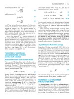
FIGURE 40–1 Membrane protein content is highly variable. The
amount of proteins equals or exceeds the quantity of lipid in nearly all
membranes. The outstanding exception is myelin, an electrical insulator
found on many nerve fibers.
The Major Lipids in Mammalian Membranes Are
Phospholipids, Glycosphingolipids & Cholesterol
Phospholipids
Of the two major phospholipid classes present in membranes,
phosphoglycerides are the more common and consist of a glycerolphosphate backbone to which are attached two fatty acids in ester linkages
and an alcohol (Figure 40–2). The fatty acid constituents are usually
1124
even-numbered carbon molecules, most commonly containing 16 or 18
carbons. They are unbranched and can be saturated or unsaturated with one
or more double bonds. The simplest phosphoglyceride is phosphatidic
acid, a 1,2-diacylglycerol 3-phosphate, a key intermediate in the formation
of other phosphoglycerides (see Chapter 24). In most phosphoglycerides
present in membranes, the 3-phosphate is esterified to an alcohol such as
choline, ethanolamine, glycerol, inositol, or serine (see Chapter 21).
Phosphatidylcholine is generally the major phosphoglyceride by mass in
the membranes of human cells.
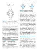
FIGURE 40–2 A phosphoglyceride showing the fatty acids (R1 and
R2), glycerol, and a phosphorylated alcohol component. Saturated fatty
acids are usually attached to carbon 1 of glycerol, and unsaturated fatty
acids to carbon 2. In phosphatidic acid, R3 is hydrogen.
The second major class of phospholipids comprises sphingomyelin
(see Figure 21–11), a phospholipid that contains a sphingosine rather than
a glycerol backbone. A fatty acid is attached by an amide linkage to the
amino group of sphingosine, forming ceramide. When the primary
hydroxyl group of sphingosine is esterified to phosphorylcholine,
sphingomyelin is formed. As the name suggests, sphingomyelin is
prominent in myelin sheaths.
Glycosphingolipids
The glycosphingolipids (GSLs) are sugar-containing lipids built on a
backbone of ceramide. GSLs include galactosyl- and glucosyl-ceramides
(cerebrosides) and the gangliosides (see structures in Chapter 21), and are
mainly located in the plasma membranes of cells, displaying their sugar
components to the exterior of the cell.
1125
Sterols
The most common sterol in the membranes of animal cells is cholesterol
(see Chapter 21). The majority of cholesterol resides within plasma
membranes, but smaller amounts are found within mitochondrial, Golgi
complex, and nuclear membranes. Cholesterol intercalates among the
phospholipids of the membrane, with its hydrophilic hydroxyl group at the
aqueous interface and the remainder of the molecule buried within the
lipid bilayer leaflet. From a nutritional viewpoint, it is important to know
that cholesterol is not present in plants.
Lipids can be separated from one another and quantified by techniques
such as column, thin-layer, and gas-liquid chromatography and their
structures established by mass spectrometry and other techniques (see
Chapter 4).
Membrane Lipids Are Amphipathic
All major lipids in membranes contain both hydrophobic and hydrophilic
regions and are therefore termed amphipathic. If the hydrophobic region
were separated from the rest of the molecule, it would be insoluble in
water but soluble in organic solvents. Conversely, if the hydrophilic region
were separated from the rest of the molecule, it would be insoluble in
organic solvents but soluble in water. The amphipathic nature of a
phospholipid is represented in Figure 40–3 and also Figure 21–24. Thus,
the polar head groups of the phospholipids and the hydroxyl group of
cholesterol interface with the aqueous environment; a similar situation
applies to the sugar moieties of the GSLs (see below).
1126
FIGURE 40–3 Diagrammatic representation of a phospholipid or
other membrane lipid. The polar head group is hydrophilic, and the
hydrocarbon tails are hydrophobic or lipophilic. The fatty acids in the tails
are saturated (S) or unsaturated (U); the former is usually attached to
carbon 1 of glycerol and the latter to carbon 2 (see Figure 40–2). Note the
kink in the tail of the unsaturated fatty acid (U), which is important in
conferring increased membrane fluidity.
The S-U phospholipid on the left contains the C16 saturated lipid
palmitic acid, and the monounsaturated C18 lipid cis-oleic acid; both are
esterified to glycerol (see Figure 40-2). The S-S phospholipid schematized
on the right contains the C16 saturated lipid palmitic acid and the saturated
C18 lipid, stearic acid.
Saturated fatty acids form relatively straight tails, whereas unsaturated
fatty acids, which generally exist in the cis form in membranes, form
“kinked” tails (Figure 40–3; see also Figures 21–1, 21–6). As the number
of double bonds within the lipid side chains increase, the number of kinks
in the tails increases. As a consequence, the membrane lipids become less
tightly packed and the membrane more fluid. The problem caused by the
presence of trans fatty acids in membrane lipids is described in Chapter
21.
1127
Detergents are amphipathic molecules that are important in
biochemistry as well as in the household. The molecular structure of a
detergent is not unlike that of a phospholipid. Certain detergents are
widely used to solubilize and purify membrane proteins. The hydrophobic
end of the detergent binds to hydrophobic regions of the proteins,
displacing most of their bound lipids. The polar end of the detergent is
free, bringing the proteins into solution as detergent-protein complexes,
usually also containing some residual lipids.
Membrane Lipids Form Bilayers
The amphipathic character of phospholipids suggests that the two regions
of the molecule have incompatible solubilities. However, in a solvent such
as water, phospholipids spontaneously organize themselves into micelles
(Figure 40–4 and Figure 21–24), an assembly that thermodynamically
satisfies the solubility requirements of the two chemically distinct regions
of these molecules. Within the micelle the hydrophobic regions of the
amphipathic phospholipids are shielded from water, while the hydrophilic
polar groups are immersed in the aqueous environment. Micelles are
usually relatively small in size (eg, ~200 nm) and consequently are limited
in their potential to form membranes. Detergents commonly form micelles.
FIGURE 40–4 Diagrammatic cross-section of a micelle. The polar head
groups are bathed in water, whereas the hydrophobic hydrocarbon tails are
1128
surrounded by other hydrocarbons and thereby protected from water.
Micelles are relatively small (compared with lipid bilayers) spherical
structures.
Phospholipids and similar amphipathic molecules can form another
structure, the bimolecular lipid bilayer, which also satisfies the
thermodynamic requirements of amphipathic molecules in an aqueous
environment. Bilayers are the key structures in biologic membranes.
Bilayers exist as sheets wherein the hydrophobic regions of the
phospholipids are sequestered from the aqueous environment, while the
hydrophilic, charged portions are exposed to water (Figure 40–5 and
Figure 21–24). The ends or edges of the bilayer sheet can be eliminated by
folding the sheet back on itself to form an enclosed vesicle with no edges.
The closed bilayer provides one of the most essential properties of
membranes. The lipid bilayer is impermeable to most water-soluble
molecules since such charged molecules would be insoluble in the
hydrophobic core of the bilayer. The self-assembly of lipid bilayers is
driven by the hydrophobic effect, which describes the tendency of
nonpolar molecules to self-associate in an aqueous environment, while in
the process excluding H2O. When lipid molecules come together in a
bilayer, the entropy of the surrounding solvent molecules increases due to
the release of immobilized water.
FIGURE 40–5 Diagram of a section of a bilayer membrane formed
from phospholipids. The unsaturated fatty acid tails are kinked and lead
to more spacing between the polar head groups, and hence to more room
for movement. This in turn results in increased membrane fluidity.
Two questions arise from consideration of the information described
above. First, how many biologically important molecules are lipid-soluble
1129
and can therefore readily enter the cell? Gases such as oxygen, CO2, and
nitrogen—small molecules with little interaction with solvents—readily
diffuse through the hydrophobic regions of the membrane. The
permeability coefficients of several ions and a number of other molecules
in a lipid bilayer are shown in Figure 40–6. The electrolytes Na+, K+, and
Cl− cross the bilayer much more slowly than water. In general, the
permeability coefficients of small molecules in a lipid bilayer correlate
with their solubilities in nonpolar solvents. For instance, steroids more
readily traverse the lipid bilayer compared with electrolytes. The high
permeability coefficient of water itself is surprising, but is partly
explained by its small size and relative lack of charge. Many drugs are
hydrophobic and can readily cross membranes and enter cells.
FIGURE 40–6 Permeability coefficients of water, some ions, and
other small molecules in lipid bilayer membranes. The permeability
coefficient is a measure of the ability of a molecule to diffuse across a
permeability barrier. Molecules that move rapidly through a given
membrane are said to have a high permeability coefficient.
The second question concerns non–lipid-soluble molecules. How are
the transmembrane concentration gradients for these molecules
maintained? The answer is that membranes contain proteins, many of
which span the lipid bilayer. These proteins either form channels for the
movement of ions and small molecules or serve as transporters for
molecules that otherwise could not readily traverse the lipid bilayer
(membrane). The nature, properties, and structures of membrane channels
and transporters are described below.
Membrane Proteins Are Associated With the Lipid
1130
Bilayer
Membrane phospholipids act as a solvent for membrane proteins, creating
an environment in which the latter can function. As described in Chapter 5,
the α-helical structure of proteins minimizes the hydrophilic character of
the peptide bonds themselves. Thus, proteins can be amphipathic and form
an integral part of the membrane by having hydrophilic regions protruding
at the inside and outside faces of the membrane but connected by a
hydrophobic region traversing the hydrophobic core of the bilayer. In fact,
those portions of membrane proteins that traverse membranes do contain
substantial numbers of hydrophobic amino acids and almost invariably
have a high α-helical content. For most membranes, a stretch of ~20 amino
acids in an α-helical configuration will span the lipid bilayer (see Figure 52).
It is possible to calculate whether a particular sequence of amino acids
present in a protein is consistent with a transmembrane location. This
can be done by consulting a table that lists the hydrophobicities of each of
the 20 common amino acids and the free energy values for their transfer
from the interior of a membrane to water. Hydrophobic amino acids have
positive values; polar amino acids have negative values. The total free
energy values for transferring successive sequences of 20 amino acids in
the protein are plotted, yielding a so-called hydropathy plot. Values of
over 20 kcal mol−1 are consistent with—but do not prove—the
interpretation that the hydrophobic sequence is a transmembrane segment.
Another aspect of the interaction of lipids and proteins is that some
proteins are anchored to one leaflet of the bilayer by covalent linkages to
certain lipids; this process is termed protein lipidation. Lipidation can
occur at protein termini (N- or C-) or internally. Common protein
lipidation events are C-terminal protein isoprenylation, cholesterylation,
and glycophosphatidylinositol (GPI; see Figure 46-1); N-terminal protein
myristoylation and internal cysteine S-prenylation and S-acylation. Such
lipidation only occurs on a specific subset of proteins and typically plays
key roles in their biology.
Different Membranes Have Different Protein
Compositions
The number of different proteins in a membrane varies from less than a
dozen very abundant proteins in the sarcoplasmic reticulum of muscle cells
to hundreds in plasma membranes. Proteins are the major functional
1131
molecules of membranes and consist of enzymes, pumps and
transporters, channels, structural components, antigens (eg, for
histocompatibility), and receptors for various molecules. Because every
type of membrane possesses a different complement of proteins, there is
no such thing as a typical membrane structure. The enzymes associated
with several different membranes are shown in Table 40–2.
TABLE 40–2 Enzymatic Markers of Different Membranesa
Membranes Are Dynamic Structures
Membranes and their components are dynamic structures. Membrane
lipids and proteins undergo turnover, just as they do in other compartments
of the cell. Different lipids have different turnover rates, and the turnover
rates of individual species of membrane proteins may vary widely. In some
instances, the membrane itself can turn over even more rapidly than any of
its constituents. This is discussed in more detail in the section on
endocytosis.
Another indicator of the dynamic nature of membranes is that a variety
of studies have shown that lipids and certain proteins exhibit lateral
diffusion in the plane of their membranes. Many nonmobile proteins do
not exhibit lateral diffusion because they are anchored to the underlying
1132
actin cytoskeleton. By contrast, the transverse movement of lipids across
the membrane (flip-flop) is extremely slow (see below), and does not
appear to occur at an appreciable rate in the case of membrane proteins.
Membranes Are Asymmetric Structures
Proteins have unique orientations in membranes, making the outside
surfaces different from the inside surfaces. An inside-outside
asymmetry is also provided by the external location of the carbohydrates
attached to membrane proteins. In addition, specific proteins are located
exclusively on the outsides or insides of membranes.
There are also regional heterogeneities in membranes. Some, such as
occur at the villous borders of mucosal cells, are almost macroscopically
visible. Others, such as those at gap junctions, tight junctions, and
synapses, occupy much smaller regions of the membrane and generate
correspondingly smaller local asymmetries.
There is also inside-outside asymmetry of the phospholipids. The
choline-containing phospholipids (phosphatidylcholine and
sphingomyelin) are located mainly in the outer leaflet; the
aminophospholipids (phosphatidylserine and phosphatidylethanolamine)
are preferentially located in the inner leaflet. Obviously, if this lipid
asymmetry is to exist at all, there must be limited transverse mobility, or
‘flip-flop’ the membrane phospholipids. In fact, phospholipids in synthetic
bilayers exhibit an extraordinarily slow rate of flip-flop; the half-life of the
asymmetry in these synthetic bilayers is on the order of several weeks.
The mechanisms involved in the lipid asymmetry are not well
understood. The enzymes involved in the synthesis of phospholipids are
located on the cytoplasmic side of microsomal membrane vesicles.
Translocases (flippases) exist that transfer certain phospholipids (eg,
phosphatidylcholine) from the inner to the outer leaflet. Specific proteins
that preferentially bind individual phospholipids also appear to be present
in the two leaflets; thus, lipid binding also contributes to the asymmetric
distribution of specific lipid molecules. In addition, phospholipid
exchange proteins recognize certain phospholipids and transfer them from
one membrane (eg, the ER) to others (eg, mitochondrial and peroxisomal).
A related issue is how lipids enter membranes. This has not been studied
as intensively as the topic of how proteins enter membranes (see Chapter
49) and knowledge is still relatively meager. Many membrane lipids are
synthesized in the ER. At least three pathways have been recognized: (1)
transport from the ER in vesicles, which then transfer the contained lipids
1133
to the recipient membrane; (2) entry via direct contact of one membrane
(eg, the ER) with another, facilitated by specific proteins; and (3) transport
via the phospholipid exchange proteins (also known as lipid transfer
proteins) mentioned above, which only exchanges lipids, but does not
cause net transfer.
There is further asymmetry with regard to glycosphingolipids and
glycoproteins; the sugar moieties of these molecules all protrude outward
from the plasma membrane and are absent from its inner face.
Membranes Contain Integral & Peripheral Proteins
It is useful to classify membrane proteins into two types: integral and
peripheral (Figure 40–7). Most membrane proteins fall into the integral
class, meaning that they interact extensively with the phospholipids and
require the use of detergents for their solubilization. Also, they generally
span the bilayer as a bundle of α-helical transmembrane segments. Integral
proteins are usually globular and are themselves amphipathic. They consist
of two hydrophilic ends separated by an intervening hydrophobic region
that traverses the hydrophobic core of the bilayer. As the structures of
integral membrane proteins were being elucidated, it became apparent that
certain ones (eg, transporter molecules, ion channels, various receptors,
and G proteins) span the bilayer many times, whereas other simple
membrane proteins (eg, glycophorin A) span the membrane only once (see
Figures 42–4 and 52–5). Integral proteins are asymmetrically distributed
across the membrane bilayer. This asymmetric orientation is conferred at
the time of their insertion in the lipid bilayer during biosynthesis in the ER.
The molecular mechanisms involved in insertion of proteins into
membranes and the topic of membrane assembly are discussed in Chapter
49.
1134
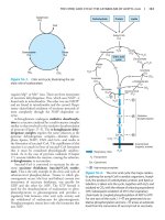
FIGURE 40–7 The fluid mosaic model of membrane structure. The
membrane consists of a bimolecular lipid layer with proteins inserted in it
or bound to either surface. Integral membrane proteins are firmly
embedded in the lipid layers. Some of these proteins completely span the
bilayer and are called transmembrane proteins, while others are embedded
in either the outer or inner leaflet of the lipid bilayer. Loosely bound to the
outer or inner surface of the membrane are the peripheral proteins. Many
of the proteins and all the glycolipids have externally exposed
oligosaccharide carbohydrate chains. (Reproduced, with permission, from
Junqueira LC, Carneiro J: Basic Histology: Text & Atlas, 10th ed.
McGraw-Hill, 2003.)
Peripheral proteins do not interact directly with the hydrophobic cores
of the phospholipids in the bilayer and thus do not require use of
detergents for their release. They are bound to the hydrophilic regions of
specific integral proteins and head groups of phospholipids and can be
released from them by treatment with salt solutions of high ionic strength.
For example, ankyrin, a peripheral protein, is bound to the inner aspect of
the integral protein “band 3” of the erythrocyte membrane. Spectrin, a
cytoskeletal structure within the erythrocyte, is in turn bound to ankyrin
and thereby plays an important role in maintenance of the biconcave shape
1135
of the erythrocyte.
ARTIFICIAL MEMBRANES MODEL
MEMBRANE FUNCTION
Artificial membrane systems can be prepared by appropriate techniques.
These systems generally consist of mixtures of one or more phospholipids
of natural or synthetic origin that have been treated by using mild
sonication to induce the formation of spherical vesicles in which the lipids
form a bilayer. Such vesicles, surrounded by a lipid bilayer with an
aqueous interior, are termed liposomes (see Figure 21–24).
The advantages and uses of artificial membrane systems for the
biochemical study of membrane function are as follows:
1. The lipid content of the membranes can be varied, allowing
systematic examination of the effects of varying lipid composition on
certain functions.
2. Purified membrane proteins or enzymes can be incorporated into
these vesicles in order to assess what factors (eg, specific lipids or
ancillary proteins) the proteins require to reconstitute their function.
3. The environment of these systems can be rigidly controlled and
systematically varied (eg, ion concentrations and ligands).
4. When liposomes are formed, they can be made to entrap certain
compounds within the vesicle such as drugs and isolated genes. There
is interest in using liposomes to distribute drugs to certain tissues, and
if components (eg, antibodies to certain cell surface molecules) could
be incorporated into liposomes so that they would be targeted to
specific tissues or tumors, the therapeutic impact would be
considerable. DNA entrapped inside liposomes appears to be less
sensitive to attack by nucleases; this approach may prove useful in
attempts at gene therapy.
THE FLUID MOSAIC MODEL OF MEMBRANE
STRUCTURE IS WIDELY ACCEPTED
The fluid mosaic model of membrane structure proposed in 1972 by
Singer and Nicolson (Figure 40–7) is now widely accepted. The model is
often likened to integral membrane protein “icebergs” floating in a sea of
(predominantly) fluid phospholipid molecules. Early evidence for the
1136
model was the finding that well characterized, fluorescently labeled
integral membrane proteins could be seen microscopically to rapidly and
randomly redistribute within the plasma membrane of a hybrid cell formed
by the artificial fusion of two different (mouse and human) parent cells
(one labeled the other not). It has subsequently been demonstrated that
phospholipids undergo even more rapid lateral diffusion with subsequent
redistribution within the plane of the membrane. Measurements indicate
that within the plane of the membrane, one molecule of phospholipid can
move several micrometers per second.
The phase changes—and thus the fluidity of membranes—are largely
dependent on the lipid composition of the membrane. In a lipid bilayer, the
hydrophobic chains of the fatty acids can be highly aligned or ordered to
provide a rather stiff structure. As the temperature increases, the
hydrophobic side chains undergo a transition from the ordered state
(more gel-like or crystalline phase) to a disordered one, taking on a more
liquid-like or fluid arrangement. The temperature at which membrane
structure undergoes the transition from ordered to disordered (ie, melts) is
called the “transition temperature” (Tm). Longer and more saturated
fatty acid chains interact more strongly with each other via their extended
hydrocarbon chains and thus cause higher values of Tm—that is, higher
temperatures are required to increase the fluidity of the bilayer. On the
other hand, unsaturated bonds that exist in the cis configuration tend to
increase the fluidity of a bilayer by decreasing the compactness of the side
chain packing without diminishing hydrophobicity (Figures 40–3 and 40–
5). The phospholipids of cellular membranes generally contain at least one
unsaturated fatty acid with at least one cis double bond.
Cholesterol acts as a buffer to modify the fluidity of membranes. At
temperatures below the Tm, it interferes with the interaction of the
hydrocarbon tails of fatty acids and thus increases fluidity. At temperatures
above the Tm, it limits disorder because it is more rigid than the
hydrocarbon tails of the fatty acids and cannot move in the membrane to
the same extent, thus limiting, or “buffering” membrane fluidity.
The fluidity of a membrane significantly affects its functions. As
membrane fluidity increases, so does its permeability to water and other
small hydrophilic molecules. The lateral mobility of integral proteins
increases as the fluidity of the membrane increases. If the active site of an
integral protein involved in a given function is exclusively in its
hydrophilic regions, changing lipid fluidity will probably have little effect
on the activity of the protein; however, if the protein is involved in a
1137
transport function in which transport components span the membrane,
lipid-phase effects may significantly alter the transport rate. The insulin
receptor (see Figure 42–8) is an excellent example of altered function with
changes in fluidity. As the concentration of unsaturated fatty acids in the
membrane is increased (by growing cultured cells in a medium rich in such
molecules), fluidity increases. Increased fluidity alters the receptor such
that it binds insulin more effectively. At normal body temperature (37°C),
the lipid bilayer is in a fluid state. Underscoring the importance of
membrane fluidity, it has been shown that bacteria can modify the
composition of their membrane lipids to adapt to changes in temperature.
Lipid Rafts, Caveolae, & Tight Junctions Are
Specialized Features of Plasma Membranes
Plasma membranes contain certain specialized structures whose
biochemical natures have been investigated in some detail.
Lipid rafts are specialized areas of the exoplasmic (outer) leaflet of
the lipid bilayer enriched in cholesterol, sphingolipids, and certain proteins
(Figure 40–8). They have been hypothesized to be involved in signal
transduction and other processes. It is thought that clustering certain
components of signaling systems closely together may increase the
efficiency of their function.
FIGURE 40–8 Schematic diagram of a lipid raft. Shown in schematic
form are multiple lipid rafts (red membrane shading) that represent
localized microdomains rich in the indicated lipids and signaling proteins
1138
(blue, green, yellow). Lipid rafts are stabilized through interactions (direct
and indirect) with the actin cytoskeleton (red bihelical chains; see Figure
51–3). (Figure modified from: The lipid raft hypothesis revisited—new
insights on raft composition and function from super-resolution
fluorescence microscopy. Bioessays 2012;34:739-747. Wiley Periodical,
Inc. Copyright © 2012.)
Caveolae may derive from lipid rafts. Many, if not all, contain the
protein caveolin-1, which may be involved in their formation from rafts.
Caveolae are observable by electron microscopy as flask, or tube-shaped
indentations of the cell membrane into the cytosol (Figure 40–9). Proteins
detected in caveolae include various components of the signal transduction
system (eg, the insulin receptor and some G proteins; see Chapter 42), the
folate receptor, and endothelial nitric oxide synthase (eNOS). Caveolae
and lipid rafts are active areas of research, and ideas concerning them and
their roles in various biologic processes are rapidly evolving.
FIGURE 40–9 Schematic diagram of a caveola. A caveola is an
invagination in the plasma membrane. The protein caveolin appears to
play an important role in the formation of caveolae and occurs as a dimer.
Each caveolin monomer is anchored to the inner leaflet of the plasma
membrane by three palmitoyl molecules (not shown).
Tight junctions are other structures found in surface membranes. They
are often located below the apical surfaces of epithelial cells and prevent
the diffusion of macromolecules between cells. They are composed of
various proteins, including occludin, various claudins, and junctional
1139
adhesion molecules.
Yet other specialized structures found in surface membranes include
desmosomes, adherens junctions, and microvilli; their chemical natures
and functions are not discussed here. The nature of gap junctions is
described below.
MEMBRANE SELECTIVITY ALLOWS
ADJUSTMENTS OF CELL COMPOSITION &
FUNCTION
If the plasma membrane is relatively impermeable, how do most molecules
enter a cell? How is selectivity of this movement established? Answers to
such questions are important in understanding how cells adjust to a
constantly changing extracellular environment. Metazoan organisms also
must have means of communicating between adjacent and distant cells, so
that complex biologic processes can be coordinated. These signals must
arrive at and be transmitted by the membrane, or they must be generated as
a consequence of some interaction with the membrane. Some of the major
mechanisms used to accomplish these different objectives are listed in
Table 40–3.
TABLE 40–3 Transfer of Material and Information Across
Membranes
1140
Passive Diffusion Involving Transporters & Ion
Channels Moves Many Small Molecules Across
Membranes
Molecules can passively traverse the bilayer down electrochemical
gradients by simple diffusion or by facilitated diffusion. This
spontaneous movement toward equilibrium contrasts with active
transport, which requires energy because it constitutes movement
against an electrochemical gradient. Figure 40–10 provides a schematic
1141
representation of these mechanisms.
FIGURE 40–10 Many small, uncharged molecules pass freely
through the lipid bilayer by simple diffusion. Larger uncharged
molecules, and some small uncharged molecules, are transferred by
specific carrier proteins (transporters) or through channels or pores.
Passive transport is always down an electrochemical gradient (shown
schematically, right), toward equilibrium. Active transport is against an
electrochemical gradient and requires an input of energy, whereas passive
transport does not. (Redrawn and reproduced, with permission, from
Alberts B, et al: Molecular Biology of the Cell. Garland, 1983.)
Simple diffusion is the passive flow of a solute from a higher to a
lower concentration due to random thermal movement. By contrast,
facilitated diffusion is passive transport of a solute from a higher to a
lower concentration mediated by a specific protein transporter. Active
transport is vectorial movement of a solute across a membrane against a
concentration gradient, and thus requires energy (frequently derived from
1142
the hydrolysis of ATP); a specific transporter (pump) is involved.
As mentioned earlier in this chapter, some solutes such as gases can
enter the cell by diffusing down an electrochemical gradient across the
membrane and do not require metabolic energy. Simple diffusion of a
solute across the membrane is limited by three factors: (1) the thermal
agitation of that specific molecule; (2) the concentration gradient across
the membrane; and (3) the solubility of that solute (the permeability
coefficient, Figure 40–6) in the hydrophobic core of the membrane bilayer.
Solubility is inversely proportional to the number of hydrogen bonds that
must be broken in order for a solute in the external aqueous phase to
become incorporated in the hydrophobic bilayer. Electrolytes, poorly
soluble in lipid, do not form hydrogen bonds with water, but they do
acquire a shell of water from hydration by electrostatic interaction. The
size of the shell is directly proportional to the charge density of the
electrolyte. Electrolytes with a large charge density have a larger shell of
hydration and thus a slower diffusion rate. Na+, for example, has a higher
charge density than K+. Hydrated Na+ is therefore larger than hydrated
K+; hence, the latter tends to move more easily through the membrane.
The following affect net diffusion of a substance: (1) concentration
gradient across the membrane—solutes move from high to low
concentration; (2) electrical potential across the membrane: solutes move
toward the solution that has the opposite charge. The inside of the cell
usually has a net negative charge; (3) permeability coefficient of the
substance for the membrane; (4) hydrostatic pressure gradient across the
membrane: increased pressure will increase the rate and force of the
collision between the molecules and the membrane; and (5) temperature,
since increased temperature will increase particle motion and thus increase
the frequency of collisions between external particles and the membrane.
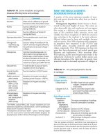
Facilitated diffusion involves either certain transporters or ion
channels (Figure 40–11). Active transport is mediated by other
transporters most of which are ATP-driven. A multitude of transporters
and channels exist in biologic membranes that route the entry of ions into
and out of cells. Table 40–4 summarizes some important differences
between transporters and ion channels.
1143
FIGURE 40–11 A schematic diagram of the two types of membrane
transport of small molecules.
TABLE 40–4 Comparison of Transporters and Ion Channels
Transporters Are Specific Proteins Involved in
Facilitated Diffusion & Also Active Transport
Transport systems can be described in a functional sense according to the
number of molecules moved and the direction of movement (Figure 40–
12) or according to whether movement is toward or away from
equilibrium. The following classification depends primarily on the former.
A uniport system moves one type of molecule bidirectionally. In
cotransport systems, the transfer of one solute depends on the
stoichiometric simultaneous or sequential transfer of another solute. A
symport moves two solutes in the same direction. Examples are the
proton-sugar transporter in bacteria and the Na+-sugar transporters (for
1144