Ebook Manual of practical medicine (4/E): Part 2
Bạn đang xem bản rút gọn của tài liệu. Xem và tải ngay bản đầy đủ của tài liệu tại đây (23.99 MB, 412 trang )
Chapter
8
Nervous System
428
Manual of Practical Medicine
Abnormal flexor response
(decorticate posture)
Extensor response (decerebrate posture)
No response
Higher Functions
Definitions
Consciousness
It is defined as the state of awareness of self and the
environment.
Confusion
It is lack of clarity and coherence of thought, perception,
understanding or action. It is often the first feature of
cognitive impairment.
3
2
1
GCS is useful in assessing level of consciousness in a
patient with head injury.
Best total score is
15
Mild injury
13 to 15
Moderate injury
9 to 12
Severe injury
8
This gives an indication of the patient’s state of consciousness and is not a substitute for neurological
examination.
Coma
It is a state of unconsciousness in which the patient does
not respond to any type of external stimuli or inner
need.
Abbreviated Coma Scale (AVPU)
Stupor (or) Semiconsciousness
A
V
P
U
It is a state of disturbed consciousness from which only
vigorous external stimuli can produce arousal.
Coma Vigil (Vegetative State)
Glasgow Coma Scale (GCS)
Eye Opening
Spontaneous
To speech
To pain
No response
4
3
2
1
Best Verbal Response
Fully oriented
Mild confusion
Moderate confusion (inappropriate)
Severe confusion (incomprehensible)
No response
Best Motor Response
Obeys commands
Localises pain
Withdrawal to pain
5
4
3
2
1
6
5
4
alert
responds to vocal stimuli
responds to pain
unresponsive.
Patient is comatose, but the eyelids are open giving the
appearance of being awake. Patient may perform random limb and head movements, but there is complete
inability to respond to command or to communicate.
Akinetic Mutism
This refers to a partial or fully awake patient who is
immobile and silent. This state may be seen in
hydrocephalus, mass in the region of third ventricle or
large bilateral hemispherical lesions.
Abulia
This is a mild form of akinetic mutism, in which patient
is hypokinetic, but is able to communicate. This is seen
in lesions in the periaqueductal region or lower
diencephalon.
Unresponsive States in Neurology
Disorder
Awareness
Sleep cycle
Motor function
Experiences
suffering
Respiratory
function
EEG
Persistent
vegetative state
Absent
Intact
No purposeful
movement
No
Normal
Polymorphic
delta and theta
Brain death
Absent
Absent
None or only reflex
spinal movements
No
Absent
Silent
Locked-insyndrome
Present
Intact
Quadriplegia
Preserved vertical eye
movements
Yes
Normal
Normal
Akinetic mutism
Present
Intact
Paucity of movements
Yes
Normal
Non-specific slowing
Nervous System
Locked-in Syndrome (Pseudo Coma)
Patients are awake, alert and selectively de-efferented.
They are non communicable with intact lid movements,
eye movements in the vertical plane and quadriplegia
with involvement of lower cranial nerves. The site of
lesion is either ventral pons or bilateral medulla with
intact tegmentum (which contains fibres of Reticular
Activating System). Infarction of ventral pons transects
all descending corticospinal and corticobulbar tracts,
but spares ARAS, which maintains arousal.
Causes
1.
2.
3.
4.
Demyelination (central pontine myelinolysis)
Ventral pontine infarction (basilar artery occlusion)
Bilateral infarction of lateral 2/3 of cerebral peduncle
Peripheral disorders associated with locked in
syndrome
a. Severe polyneuropathy
b. Myasthenia gravis
c. Neuromuscular blocking agents.
Catatonia
Patient appears awake and blink spontaneously. There
is a waxy flexibility (limbs maintain the posture implemented by the examiner). This is seen in schizophrenia.
Delirium
This is synonymous with acute confusional state characterised by periods of agitation, heightened mental
activity, increased wakefulness, hallucinations, motor
hyperactivity and autonomic stimulation. There is an
associated impairment of attention.
Causes of Delirium
Head injury
CVA
Cerebral infections
Epilepsy
Hypoglycaemia, DKA
Hypoxia
Renal or hepatic failure
Electrolyte or acid-base imbalance
Wernicke’s encephalopathy
Septicaemia, malaria, SBE, pneumonia
Heat stroke, hypothermia
Toxins
Alcoholic intoxication
*Alcohol and drug (Barbiturates and narcotics) withdrawal
429
Psychiatric disorders
Acute mania
Extreme anxiety
Schizophrenia (auditory hallucinations)
Hysteria.
Note: *Alcohol withdrawal causes delirium tremens
which is characterised by delirium, tremors and visual
hallucinations.
Dementia
It is a syndrome of acquired global or multifocal
impairment of cognitive function involving decline in
intellect, memory or personality in the presence of
normal consciousness.
Causes of Dementia
1. Primary dementias
a. Alzheimer’s disease (diffuse cortical atrophy)
b. Pick’s disease (circumscribed cortical atrophy,
early frontal and temporal)
c. Frontal lobe degeneration.
2. Secondary dementias
a. Degenerative disorders
Parkinson’s disease
Hereditary ataxias
Progressive supranuclear palsy (SteeleRichardson syndrome)
Motor neuron disease
Huntington’s chorea
Multiple sclerosis.
b. Conditions with raised intracranial tension
• Primary and secondary tumours
• Hydrocephalus
• Chronic subdural hematoma
• Carcinomatous meningitis.
c. Vascular dementia
• Multiinfarct dementia
— Lacunar infarct
— Thalamic infarct
— Diffuse atherosclerosis
• Vasculitis: SLE, polyarteritis nodosa, Behcet’s
disease.
d. Chronic infections
• Syphilis, GPI
• Tuberculosis
• Fungal, protozoal infections
Slow viral diseases:
* Subacute sclerosing panencephalitis
* Creutzfeldt-Jacob disease
* Papova virus
* HIV.
430
Manual of Practical Medicine
3. Dementia due to diffuse brain damage
Anoxia
Encephalitis
Acute head injury
Pugilistic dementia (boxers).
4. Endocrine disorders
• Chronic hypoglycaemia
• Hypothyroidism
• Hypo and hyperparathyroidism
• Adrenal insufficiency
• Cushing’s syndrome.
5. Vitamin deficiencies
• Vitamin B12 deficiency
• Thiamine deficiency
• Niacin deficiency.
6. Toxins
• Alcohol
• Drug and narcotic poisoning
• Heavy metal intoxication
• Dialysis dementia.
7. Dementia in adolescents and young adults
• Wilson’s disease
Progressive myoclonic epilepsy
Tuberous sclerosis
Leukodystrophies
Storage diseases.
Note: • Treatable causes
Presenile Dementia
It occurs before 65 years of age
(Pick’s disease, Alzheimer’s
disease)
Senile Dementia
It occurs after 65 years of age
Cortical Dementia
It occurs in Pick’s disease and
Alzheimer’s disease
Subcortical Dementia It occurs in Huntington’s disease,
multiple sclerosis and HIV.
Differences between Alzheimer’s Disease and
Pick’s Disease
Features
Alzheimer’s disease
Pick’s disease
1.
Portion of
brain
affected
2.
Pathology
3.
4.
Age of onset
Clinical
features
Diffuse cortical involve- Confined to
ment (esp. hippocampus frontal and
and temporal lobes)
temporal lobes
(lobar sclerosis)
Neurofibrillary tangles, Pick’s bodies
senile plaques seen
seen
Presenile or senile
Presenile
Features of diffuse
Prominent
cortical involvement
frontoseen: Frontotemporal
temporal
features less prominent features seen
Amnesia
It is a disorder of memory characterised by inability to
remember past events and to learn new information
despite normal consciousness and attention.
As a result of head injury, memory disturbance
occurs for events before (retrograde amnesia) and after
the time of injury (post-traumatic amnesia).
Anterograde Amnesia
Impairment in learning new material which accompanies post-traumatic amnesia.
Duration of post-traumatic amnesia indicates the
severity of head injury; the ability to learn new material
often being the last cognitive deficit to recover.
Transient Global Amnesia
It is a syndrome in which a previously normal person
suddenly becomes confused and amnesic. It is usually
of spontaneous origin but also may be due to immersion
in cold or hot water, emotional stimuli, exertion,
intercourse or travel in motor vehicles.
There is severe impairment of recall of recent and
sometimes most distant events. Immediate memory is
intact. There is no other neurological sign. It is usually
benign. Rarely it may be due to temporal lobe tumour,
migraine or temporal lobe epilepsy.
Examination of Higher Mental
Functions
Consciousness
Find out the level of consciousness of the patient
(whether the patient is comatose, stuporose or delirious).
Causes of Coma
Trauma
Cerebral contusion, concussion and laceration
Subdural haematoma
Extradural haematoma.
Cerebrovascular Disease
Subarachnoid haemorrhage
lntracerebral haemorrhage
Massive cerebral infarction
Brainstem infarction or haemorrhage
Cerebellar infarction or haemorrhage
Cerebral venous sinus thrombosis.
Nervous System
Infections
Meningitis
Encephalitis
Cerebral abscess
Cerebral malaria.
Seizure Disorders and Raised ICT
Epilepsy
Space occupying lesions.
Endocrine and Metabolic Disturbances
a. Diabetes mellitus: Hypoglycaemia, ketoacidosis,
hyperosmolar coma
b. Myxoedema
c. Hypocalcaemia
d. Hypercalcaemia
e. Hypoadrenalism
f. Hypopituitarism
g. Hepatic failure
h. Respiratory failure
i. Cardiac failure
j. Uraemia
k. Metabolic acidosis
1. Metabolic alkalosis
m. Electrolyte disturbances (hypo and hypernatraemia).
Cardiovascular Disorders
Congestive cardiac failure
Hypertensive encephalopathy
Shock
Arrhythmias.
Physical Agents
Hyperpyrexia
Hypothermia
Electric shock
Lightning.
Toxins and Others
Acute poisoning
Alcohol
Thiamine deficiency.
Tropical Coma
Cerebral malaria
Typhoid fever
Trypanosomiasis
Rabies.
431
Metabolic Coma
Aaetiology
Neurologic signs
Diagnostic workup
Hypoxia
Myoclonus, flaccid
muscle tone
Cardiorespiratory
disorder, polytrauma,
Diabetic
ketoacidosis
Clouding of
conciousness/coma
Blood sugar > 400 mg
with ketonuria
Hyperosmolar
coma
Coma, seizure,
focal signs
Blood sugar > 800 mg
High serum osmolarity
Hypoglycaemic
coma
Coma, seizure,
focal signs
Blood sugar < 50 mg%
Hepatic coma
Asterixis, jaundice
Elevated ammonia level
Uraemia
Myoclonus, asterixis,
oliguria
Raised renal
parameters
Disequilibrium
syndrome
Muscle cramps,
seizure
Postdialysis syndrome
Hyponatraemia
Coma and seizure
Serum sodium
< 126 mmol
Hypernatraemia
Muscle weakness,
coma
Serum sodium
> 156 mmol
Hypercalcaemia
Muscle weakness,
headache
Calcium, phosphate,
and parathormone
Hypocalcaemia
Tetany, seizure, coma
Calcium, phosphate
and parathormone
Approach to Coma
A comatose patient has to be approached systematically
to derive maximum information. The aim of physical
examination is to arrive at following conclusions.
1. Localisation of coma
2. Aetiology of coma (structural vs metabolic)
Approach to the Patient
I. History and general examination.
A meticulous history and detailed general examination will give clue regarding the aetiology of coma.
II. Neurological examination.
The neurological examination of a comatose patient
serves 3 purposes.
a. To aid in determining the cause of coma
b. To help determine the prognosis of coma
c. To provide a base line.
For localisation of structural lesion and to assess the
prognosis, the following examinations are the most
helpful
1. State of consciousness
2. Respiratory pattern
3. Pupillary size and reactivity
432
Manual of Practical Medicine
4. Ocular motility
5. Skeletal muscle motor response.
1. State of Consciousness
Auditory, visual and noxious stimuli of progressively
increasing intensity should be applied to the patient.
The maximal state of arousal, intensity of stimuli
required for that and the response of the patient has to
be noted. Any asymmetry in the response to stimuli
points towards structural lesion.
All patients in coma should be asked to open their
eyes and look up and down. Because in locked in
syndrome only these voluntary movements are spared.
Patient will be alert and aware, but quadriplegic with
lower cranial nerve paralysis, thus mimicking coma.
2. Respiration
Respiratory patterns that are helpful in localising level
of involvement are the following (Fig. 8.1):
A. Cheyne-Stokes breathing.
i. Rate of respiration will be around 30 per minute
ii. There is waxing and waning of respiration
iii. Waning of respiration is followed by apnoea for
about 15 seconds.
Causes
i. Bilateral hemispheric damage
ii. Diencephalic insults
iii. Bilateral damage anywhere between forebrain and
upper pons
iv. Prolonged circulation time as in cardiac failure.
ii. Emergence of Cheyne-Stokes breathing in a patient
with unilateral mass lesion may be a sign of
herniation
iii. Change in pattern from Cheyne-Stokes to other
patterns described is ominous.
B. Central neurogenic hyperventilation.
i. Refers to rapid breathing (40-70 per minute)
ii. Lesions of low midbrain ventral to aqueduct of
Sylvius and of upper pons ventral to fourth
ventricle.
iii. Hyperpnoea cannot be ascribed to CNS lesion if
PaO2 is < 70-80 mm Hg and PCO2 is greater than
40 mmHg
C. Apneustic breathing.
Apneustic breathing is a prolonged inspiratory gasp
with a pause at full inspiration. It is caused by lesions
of the dorsolateral lower half of pons.
D. Cluster breathing.
Cluster breathing results from high medullary
damage, involves periodic respirations that are
irregular in frequency and amplitude, with variable
pauses between clusters of breaths.
E. Ataxic breathing.
This is irregular in rate and rhythm and is usually
due to medullary lesions. Ataxic breathing and
bilateral VI nerve lesion may be a warning sign of
brainstem compression from an expanding lesion in
posterior fossa.
3. Pupil Size and Reactivity (Fig. 8.2)
a. Thalamic lesions cause small, reactive pupils, which
are often referred to as diencephalic pupils. Similar
Prognosis
i. Stable pattern of Cheyne-Stokes respiration is a
good prognostic sign
Fig. 8.1: Respiratory patterns in coma
Fig. 8.2: Pupillary defects in coma
Nervous System
b.
c.
d.
e.
pupillary findings are noted in many toxic-metabolic
conditions resulting in coma.
Hypothalamic lesions or lesions elsewhere along the
sympathetic pathway result in Horner’s syndrome.
Midbrain lesions produce three types of pupillary
abnormality, depending on where the lesion occurs.
i. Dorsal tectal lesions interrupt the pupillary light
reflex, resulting in midposition eyes, which are
fixed to light but react to near, although the
reaction is impossible to test in the comatose
patient. Spontaneous fluctuations in size occur,
and the ciliospinal reflex is preserved.
ii. Nuclear midbrain lesions usually affect both
sympathetic and parasympathetic pathways,
resulting in fixed, irregular midposition pupils,
which may be unequal.
iii. Lesions of the third nerve in the brainstem, or after
the nerve exits the brainstem parenchyma, cause
wide pupillary dilation unresponsive to light.
Pontine lesions interrupt sympathetic pathways to
cause small pupils (pinpoint pupils), which remain
reactive, although magnification may be needed to
observe this.
Lesions above the thalamus and below the pons
should leave pupillary function intact, except for
Horner’s syndrome in medullary or cervical spinal
cord lesions.
4. Ocular Motility
Preservation of normal ocular motility implies that a
large portion of brainstem is intact, from the oculomotor
nucleus in the midbrain to the vestibular nuclei at the
pontomedullary junction.
Evaluation of ocular movement consists of three main
elements.
i. Abnormalities of resting position including eye
deviation.
ii. Spontaneous eye movements.
a. Purposeful appearing eye movements occur in
locked in syndrome, catatonia, pseudocoma,
and persistent vegetative state.
b. Rowing eye movements indicates brainstem is
relatively intact and coma is due to metabolic
or toxic cause or bilateral lesions above the
brainstem.
c. Nystagmus occurring in comatose patients
suggests an irritative or epileptogenic supratentorial focus.
d. Spontaneous conjugate vertical eye movements like ocular bobbing which is characterised by rapid downward jerk of both eyes
followed by a slow return to the midposition.
The centre of lesion is at Pons.
433
e. Oculopalatal nystagmus occurs due to damage
to the lower brainstem involving the GuillainMollaret triangle, which extends between the
cerebellar dentate nucleus, red nucleus and
inferior olive.
iii. Reflex ocular movements
This constitutes:
a. Oculocephalic reflex (Dolls eye movement)
b. Vestibulo-oculogyric reflex (Cold caloric
testing).
a. Dolls eye phenomenon. This is tested by sudden passive
rotation of head in both directions laterally and flexion
and extension of the neck while observing the motion
of the eyes.
b. Cold caloric testing. Clinical caloric testing is
commonly done by applying cold water to the tympanic
membrane with the head tilted back 60 degrees from
the horizontal. The head tilt allows maximal stimulation
of the lateral semicircular canal, which is most
responsible for reflex lateral eye movements. After
checking to make certain that the ear canal is patent
and the tympanic membrane is free of defect, 10 ml of
ice-cold water is slowly injected into one ear canal. Cold
water applied to the tympanic membrane causes
currents to be set up in the endolymph of the
semicircular canal. This results in a change in the
baseline firing of the vestibular nerve and slow (tonic)
conjugate deviation of the eyes toward the stimulated
ear. In an awake person, the eye deviation is minimal
and is corrected with a nystagmus fast phase towards
the opposite side. Warm water irrigation produces
reversal of flow of the endolymph, which causes a slow
phase away from the stimulated ear and a normal
corrective phase towards the ear. By tradition, the
nystagmus is named by the direction of the fast phase.
The mnemonic COWS (cold opposite, warm same) refers
to the fast phases. Simultaneous bilateral cold water
application results in slow phase down and fast phase
up, whereas the reverse occurs with bilateral warm
water application.
Interpretation
i. Normal response indicate intact brainstem
ii. Absent response indicate brainstem involvement
iii. Abnormal dysconjugate responses occur with
cranial nerve palsies, internuclear ophthalmoplegia,
or restrictive eye disease.
5. Motor System
Resting posture and adventitious movements are
analysed.
434
Manual of Practical Medicine
Adventitious Movements
i. Tonic-clonic or other stereotyped movements signal
seizure as the probable cause of decreased alertness.
ii. Myoclonic jerking, nonrhythmic jerking movements in single or multiple muscle groups, is seen
with anoxic encephalopathy or other metabolic
comas, such as hepatic encephalopathy.
iii. Rhythmic myoclonus, which must be differentiated
from epileptic movements, is usually a sign of
brainstem injury.
iv. Tetany occurs with hypocalcaemia.
v. Cerebellar fits, resulting from intermittent tonsillar
herniation, are characterised by a deterioration of
level of arousal, opisthotonos, respiratory rate
slowing and irregularity, and pupillary dilation.
Differentiating Features between Structural and
Metabolic Coma
Fig. 8.3: Postures
Postures (Fig. 8.3)
i. Head and eye deviation to one side and
contralateral hemiparesis indicate supratentorial
lesion, while ipsilateral hemiparesis indicates
brainstem lesion.
ii. Decerebrate posturing is bilateral extensor posture,
with extension of the lower extremities and
adduction and internal rotation of the shoulders
and extension at the elbows and wrist. Bilateral
midbrain or pontine lesions are usually responsible
for decerebrate posturing. Less commonly, deep
metabolic encephalopathies or bilateral supratentorial lesions involving the motor pathways
may produce a similar pattern.
iii. Decorticate posturing is bilateral flexion at the
elbows and wrists, with shoulder adduction and
extension of the lower extremities. It is a much
poorer localising posture because it may result
from lesions in many locations but usually above
the brainstem. Decorticate posture is not as
ominous a sign as decerebrate posture because the
former occurs with many relatively reversible
lesions.
iv. Unilateral decerebrate or decorticate postures are
generally less ominous than bilateral posturing.
Lesions causing unilateral posturing may be
anywhere in the motor system from cortex to
brainstem. Unilateral extensor posturing is
common immediately after an acute hemispheric
event, followed in time by a flexor response.
1. State of consciousness: Patients with metabolic
problems often have mild alterations in arousal and
tend to have waxing and waning of the behavioural
state. Patients with acute structural lesions tend to
stay at the same level of arousal or progressively
deteriorate. Toxins may also cause progressive
decline in level of arousal.
2. Respiration: Deep, frequent respiration is most
commonly due to metabolic abnormalities, but
rarely it is caused by pontine lesions or by
neurogenic pulmonary oedema secondary to acute
structural lesions.
3. Funduscopic examination: Subhyaloid haemorrhage or papilloedema is almost pathognomonic
of structural lesions. Papilloedema due to increased
ICP may indicate an intracranial mass lesion or
hypertensive encephalopathy. Papilloedema does
not occur in metabolic diseases, except hypoparathyroidism, lead intoxication, and malignant
hypertension.
4. Pupil size: The pupils are usually symmetrical in
coma from toxic-metabolic causes. Patients with
metabolic or toxic encephalopathies often have
small pupils with preserved reactivity. Exceptions
occur with methyl alcohol poisoning, which may
produce dilated and unreactive pupils, or late in
the course of toxic or metabolic coma if hypoxia or
other permanent brain damage has occurred. In
terminal asphyxia, the pupils dilate initially and
then become fixed at midposition within 30
minutes. The initial dilation is attributed to massive
sympathetic discharge.
5. Pupil reactivity: Assessment of the pupillary reflex
is one of the most useful means of differentiating
Nervous System
6.
7.
8.
9.
10.
metabolic from structural causes of coma. Pupillary
reactivity is relatively resistant to metabolic insult
and is usually spared in coma from drug intoxication or metabolic causes, even when other
brainstem reflexes are absent. Hypothermia may
fix pupils, as does severe barbiturate intoxication;
neuromuscular blocking agents produce midposition or small pupils, and glutethimide and
atropine dilate them.
Ocular motility: Dysconjugate eye movements are
typically a feature of structural lesions.
Spontaneous eye movements: Roving eye movements with full excursion most often suggest
metabolic or toxic abnormalities.
Reflex eye movements: Reflex eye movements are
normally intact in toxic-metabolic coma, except
rarely in phenobarbital or phenytoin intoxication
or deep metabolic coma from other causes.
Adventitious movement: Coma punctuated by
periods of motor restlessness, tremors, or spasm is
often due to toxins or drugs, such as chlorpromazine or lithium. Brainstem herniation or
intermittent CNS ischaemia may also produce
unusual posturing movements. Myoclonic jerking
is generally metabolic and often anoxic in origin.
Muscle tone: Muscle tone is usually symmetrical
and normal or decreased in metabolic coma.
Structural lesions cause asymmetrical muscle tone.
Tone may be increased, normal, or decreased by
structural lesions.
Appearance and Behaviour
This can be assessed as the patient walks into the
consulting room. A note is made of the way the patient
carries himself including the way he has attired himself
and his personal hygiene. Note also from his behaviour
whether he is disturbed, apathetic, agitated or confused.
Emotional State
Assess whether the patient is elated, euphoric, excited
or depressed.
Mood: It is the prevailing emotional state.
Affect: It is an emotional experience evoked by a particular stimulus.
Mood is characterised by a feeling of cheerfulness
and happiness, a state of exceptional mental well-being
or a feeling of depression.
Depression: This is a mood of dejection and gloom for no
reason. Depression may be of two types:
435
1. Major depressive disorder (single or recurrent episodes)
2. Dysthymic disorder (chronic, less intense form of
depression lasting for atleast two years).
Emotional instability: Inappropriate elation and depression for no reason; It is seen in pseudobulbar palsy.
Mania: It is a distinct period of abnormal and persistently
elevated or irritable mood.
Anxiety: It is an anticipatory reaction. It may present
with somatic symptoms related to autonomic nervous
system or psychic symptoms or both.
Orientation
Questions are put to the patient to test his orientation to
time, place and person as follows:
Time
Ask the patient to tell the year, season, date,
day and month.
Place
Ask for the state, country, town, hospital
and floor in which he is admitted.
Person Ask for the identity of his nearby relatives or
neighbours.
Self
Ask the patient’s name, age, address and
qualifications.
Rule out confabulation, which is a filling in of forgotten
memories by inappropriately recalled material from
previous experience, e.g. Korsakoff’s psychosis.
Handedness
It is the preference to use the hand of a particular side
(right or left) for complicated, fine and skillful motor
acts.
Dominant hand is the hand used for combing the hair
or buttoning the shirt or picking up a coin. It can also be
tested indirectly by asking the patient to kick a ball or to
use his or her eye to see through a small hole. The leg or
the eye used preferentially, gives a clue to the side of
cerebral dominance.
On asking the patient to fold his arms across the
chest, the dominant arm is placed anteriorly. Similarly,
while asking the patient to stand at ease, the dominant
hand comes posteriorly.
There is an anatomic difference between the sizes of
dominant and nondominant cerebral hemispheres.
‘Planum temporale’, which is adjacent to the auditory
centre of Heschl’s transverse gyrus, is larger in the left
hemisphere in the right handed individuals. Left
handedness may be hereditary or may result from
disease of the left hemisphere in early life.
436
Manual of Practical Medicine
Left hemisphere dominance for language occurs in
95% of right handed people. Even in 50% of left handed
individuals, left hemisphere is dominant.
General Intelligence
It is necessary to ascertain the patient’s general
intellectual ability as evidenced by his educational standard and work records before assessing his intelligence.
Intelligence is assessed by testing the following:
a. Abstract thinking: It is tested by asking the patient to
explain the meaning of a common proverb.
b. Reasoning: This is tested by asking him to compare
objects or by asking him to differentiate between a
lie and a mistake. Test his power to appreciate
similarities and dissimilarities between two objects,
animals, etc.
c. Judgement: It is tested by asking the patient various
questions, like what he would do on seeing a house
on fire or what he would do when he finds a stamped
envelope on the road.
d. Attention: It is tested by asking the patient to do
sequential subtraction of 7 from 100 down to zero
and by forward and reverse digit spans.
e. Calculation: It is tested by asking the patient to solve
simple numerical problems.
drome. Immediate memory requires sustained attention also.
b. Recent memory: It means recall of information
presented within minutes, hours and days. It is tested
by asking the patient to recall certain important
recent events or current affairs and by asking him to
remember three unrelated common objects or a
simple address told to him, a few minutes ago.
It is impaired in dementia, acute confusional
syndromes and amnesia. This is tested after two
minutes and five minutes depending on the degree
of amnesia.
c. Long-term or Secondary or Remote memory: It means
memory for past events. It can be tested by asking
the dates and salient facts of some well known but
distant public events or names of political figures or
locations of major cities. It incorporates the meaning
of information rather than exact words or pictures.
Perceptions
Delusions: These are false beliefs which continue to be
held despite evidence to the contrary.
Hallucinations: These are false impressions referred to
the organs of special senses in the absence of a stimulus,
e.g., temporal lobe epilepsy, alcohol withdrawal, schizophrenia.
Memory
Illusions: These are misinterpretations of stimuli.
It is the power to retain and recall past experiences.
Obsessions: These are recurrent and persistent thoughts,
which intrudes into the patient’s mind despite best effort
to get rid of them.
Components of Memory
Reception
Registration
Retention
Recall.
Types
a. Immediate or short-term memory: It is the memory for
events of a few seconds duration. This holds
information close to consciousness for a few seconds
only.
This is tested by asking the patient to reproduce
a string of numerals.
Example:
• Digit span, 7 forwards, 5 backwards
• Spell ‘World’ backwards.
Immediate memory is impaired in acute confusional syndrome, Wernicke-Korsakoff syndrome
and mostly retained in dementia and amnesic syn-
Visuospatial Functions
Ask the patient to copy a drawing of a five pointed star
or three dimensional box. Constructional apraxia or
visuospatial agnosia results in difficulty in drawing the
lines required in the correct spatial orientation or
position. ‘Perseveration’ or visual neglect is revealed by
this test.
Apraxia
It is a defect in the ability to carry out known acts in the
absence of motor weakness, sensory loss or ataxia.
Consequently, the apraxic patient is unable to make use
of objects, though their use can be recognised and
described. It results from damage to the left parietal
cortex or to parietal white matter of the left or of both
hemispheres, or from disease of the connections between
the two hemispheres through the corpus callosum.
Nervous System
It is tested by asking the patient to use objects
(lighting a cigar, copying a cube, star, clock) or to carry
out or imitate certain movements.
Types of Apraxia
1. Limb kinetic apraxia.
This involves a specific motor disability of one limb,
usually an arm, in the absence of gross weakness or
ataxia
2. Ideomotor apraxia.
This refers to the condition in which patient is unable
to carry out the motor command, despite adequate
comprehension of the command and adequate motor
and sensory functions to perform the commands.
This is the most frequent type of apraxia. Here the
concept is normal, but execution is defective.
3. Ideational apraxia.
This refers to the condition in which patients are
apraxic because they have lost the ideas (concepts)
behind the skilled movements. Here the patient will
name and define an object. But not know how to
manipulate the object when it is placed in the hand.
4. Buccofacial apraxia.
This refers to the condition in which the patient
cannot perform learned skilled movements of the
mouth, lips, cheeks, tongue and throat in the absence
of motor paralysis of concerned muscles.
Agnosia
Agnosia is defined as failure to recognise known objects
in the presence of intact sensory, visual and auditory
pathway.
437
A. Tactile Agnosia
Patient is not able to recognise known objects in the
presence of intact sensory system and he/she should
have sufficient motor function and coordination to
explore the object.
B. Visual Agnosia
It is the inability to recognize what is seen with the eyes
in the presence of intact visual pathway. At the same
time, they can describe the colour, size, and shape of
the object without recognising it.
C. Prosapagnosia
It is the inability to identify a familiar face which occurs
in parieto-occipital lesion.
D. Anosognosia
In right parietal lobe lesion, there is lack of awareness
to recognize the paralysed limb.
Sleep
Sleep is an elemental phenomenon of life and an
indispensable phase of human existence. Sleep represents one of basic 24-hour circadian rhythms.
Most adults sleep for 7 to 8 hours/day.
Age
Duration of sleep
Newborn
Child
10 years
Adolescence and adults
Late adult life
16–20 hours
10–12 hours
9–10 hours
7–8 hours
About 6.5 hours
Types of Agnosia
Modality
Subtypes
Neuroanatomical correlates
Vision
Visual object agnosia
Bilateral occipitotemporal
Left occipitotemporal
Bilateral occipitotemporal
Right occipitotemporal and
occipitoparietal
Associative prosopagnosia
Apperceptive prosopagnosia
Audition
Environmental sound
agnosia
Phonagnosia
Amusia
Bilateral posterior superior
Temporal
Right inferior parietal
Right posterior temporal and
inferior parietal
Somatosensory
Tactile object agnosia
(complete)
Tactile object agnosia
(nonmanipulable stimuli)
Right and left parietal
operculum, posterior insula
Right superior mesial parietal
Perception
of disease
Anosognosia
Right parietal and bilateral
ventromedial frontal
438
Manual of Practical Medicine
States and Stages of Sleep
It comprises of 2 distinct physiological states namely
REM and Non-REM sleep.
1. REM (Rapid eye movement sleep/dreaming/desynchronised/D-sleep)
2. Non-REM Sleep (orthodox/synchronised/S-sleep).
Non-REM sleep has 4 stages, two of which are known
as ‘slow-wave’ or deep sleep because they are associated
with low frequency synchronised waves on EEG.
Stage 1: Transition from wakefulness is characterised
by disappearance of regular α pattern and emergence
of a low amplitude mixed frequency pattern the theta
range (2–7 Hz). It is associated with slow rolling eye
movements.
Stage 2: There is occurrence of K complexes and sleep
spindles superimposed upon a background activity
similar to that of stage 1 (low amplitude).
Stage 3: There is predominance of delta EEG activity in
20 to 50% of the record (increased amplitude and
decreased frequency).
Stage 4: More than 50% of the record is dominated by
delta EEG activity.
Types of waves in EEG
α
β
θ
δ
Rate
7–13/sec
> 13/sec
4–6/sec
< 4/sec
REM Sleep
This comprises of low amplitude, mixed frequency
waves.
REM sleep develops after progression through
various stages of NREM sleep, usually within 90
minutes. It is the stage in which most dreaming occurs.
During a night’s sleep, there is a cycle of Non-REM and
REM sleep with episodes of REM becoming relatively
longer.
Tonic muscle activity is minimal during REM sleep.
Eye movements are rapid and conjugate in all directions.
Gross body movements occur every 15 minutes or so in
all stages of sleep, but are maximal in the transition
between REM and NREM sleep.
REM sleep has phasic and tonic components. During
the phasic period in addition to eye movements, the
pupils dilate and constrict alternately and BP, pulse,
and respiration increase and may become irregular.
During the nonphasic period there is flaccidity, atonia
of upper airways, intercostal muscles and abdomen
which may pose a threat to life in infants with excessive
respiratory difficulty and in patients with kyphoscoliosis, muscular dystrophy, and paralytic
poliomyelitis.
About 20 to 25% of total sleep time in young adults
is spent in REM sleep, 3 to 5% in stage 1, 50 to 60% in
stage 2 and 10 to 20% in stage 3 and 4 combined. Stage
3 and stage 4 sleep decreases with age and in elderly
over 70 years, there is no stage 4 sleep virtually.
Most adults sleep 7–8 hours/night usually. At the
extremes of age, infants and the elderly have frequent
interruptions of sleep.
Adults with habitual sleep durations of less than 4
hours or greater than 9 hours have increased mortality
rates as compared to those who sleep for 7–8 hr/night.
Rapid onset of REM sleep in adults may suggest:
Endogenous depression
Narcolepsy
Circadian rhythm disorders
Drug withdrawal.
During sleep there is:
a. Fall in body temperature, mainly during NREM
period
b. During REM sleep, glucose metabolism is increased
in comparison with the waking state
c. In urine, absolute sodium and potassium excretion
decreases
d. Secretion of cortisol and TSH are decreased at the
onset of sleep. Cortisol secretion increases at
awakening
e. Melatonin (from pineal gland) is secreted at night
and ceases upon retinal stimulation by sunlight
f. During stages 3 and 4, growth hormone is secreted
till middle and late adult life
g. Prolactin secretion increases at night in both men
and women
h. Sleep associated secretion of LH occurs in pubertal
boys and girls.
Physiologic mechanism governing NREM and REM
sleep lie in the pontine reticular formation.
Neuroanatomy of Sleep
(Sleep Centre)
Generation of sleep is from medullary reticular formation, the thalamus and basal forebrain. Generation
of wakefulness or EEG arousal is maintained by
brainstem reticular formation, the midbrain, the
subthalamus, the thalamus and the basal forebrain.
Current hypothesis suggests that the capacity for sleep
and wake generation is distributed along an axial ‘core’
Nervous System
of neurons extending from the brainstem rostrally to
the basal forebrain. There is no specific sleep centre.
Function of Sleep
Sleep is thought to be useful for body restitution,
facilitation of motor function and for consolidation of
learning and memory.
Effect of Sleep Deprivation
Deprivation of sleep (REM, NREM) for about 60–200
hours causes increased sleepiness, fatigue, irritability
and difficulty to concentrate. Performance of skilled
motor activity decreases. Self care is neglected. Later,
stages of microsleep occurs leading to all types of errors
and accidents. Illusions, hallucinations (visual and
tactile) may occur.
Patient may have nystagmus, impairment of saccades, loss of accommodation, slight tremor of hands,
ptosis, expressionless face, thickness of speech,
mispronunciation, etc. Seizure threshold is reduced.
Concentration of 17-OH corticosteriods increases and
catecholamine output rises. Rarely, psychotic episodes
of screaming and sobbing may occur. During recovery,
patients go straight into stage IV NREM at the expense
of stage II and REM sleep. The next day, REM sleep
occurs with a longer duration.
International Classification of
Sleep Disorders
Dyssomnias
Intrinsic Sleep Disorders
1. Psychophysiologic insomnia
2. Idiopathic insomnia
3. Narcolepsy
4. Recurrent or idiopathic hypersomnia
5. Post-traumatic hypersomnia
6. Sleep apnoea syndromes
7. Periodic limb movement disorder
8. Restless leg syndrome.
Extrinsic Sleep Disorders
1.
2.
3.
4.
5.
6.
7.
8.
Inadequate sleep hygiene
Environment sleep disorders
Altitude insomnia
Adjustment sleep disorders
Sleep onset association disorders
Food allergy insomnia
Nocturnal eating/drinking syndrome
Drug/alcohol dependent sleep disorders.
439
Circadian Rhythm Sleep Disorders
1. Time-Zone changes (jet lag) syndrome
2. Shift work sleep disorder
3. Delayed sleep phase syndrome (patient goes to bed
late (2–3 am) and gets up late (11 am)
4. Advanced sleep phase syndrome (patient goes to
bed early (8–9 pm) and gets up early (4–5 am)
5. Non-24 hours sleep wake disorders.
Parasomnias
Arousal Disorders
1. Confusional arousal
2. Sleep walking
3. Sleep terrors.
Sleep Wake Transition Disorders
1. Rhythmic movement disorders
2. Sleep talking
3. Nocturnal leg cramps.
Parasomnias Associated with REM Sleep
1. Nightmares
2. Sleep paralysis
3. Impaired sleep related penile erection
4. Sleep related painful erection
5. REM sleep related arrhythmias
6. REM sleep behaviour disorders.
Others
1. Sleep bruxism
2. Sleep enuresis
3. Nocturnal paroxysmal dystonia.
Sleep Disorders Associated with Medical or
Psychiatric Disorders
Associated with Mental Disorders
Schizophrenia, anxiety, affective illness, obsessivecompulsive neurosis, chronic alcoholism, depression.
Associated with Neurological Disorders
a. Cerebral degenerative disorders
b. Parkinsonism
c. Fatal familial insomnia
d. Sleep related epilepsy
e. Sleep related headaches.
Associated with Other Medical Disorders
1. Sleeping sickness
2. Nocturnal cardiac ischaemia
440
Manual of Practical Medicine
3. COPD, cystic fibrosis
4. Sleep related asthma
5. Sleep associated gastro-oesophageal reflux, peptic
ulcer disease
6. Chronic renal failure, liver failure
7. Hyperthyroidism
8. Drugs (theophylline, adrenergic agonists, glucocorticoids can disrupt sleep)
9. Chronic pain.
Insomnia
It is a complaint of inadequate sleep. It can be
a. Sleep onset insomnia—difficulty in falling asleep.
b. Sleep maintenance insomnia (frequent or sustained
awakenings).
c. Non-restorative sleep—persistent sleepiness despite
sleep of adequate duration.
Sleep Apnoea Syndromes
There is respiratory dysfunction during sleep. Cough
reflex is depressed. There is falling back of tongue or
epiglottis. The cessation of breathing may be due to
either occlusion of the airway (obstructive sleep apnoea)
absence of respiratory effort (central sleep apnoea) or a
combination of these (mixed sleep apnoea). These are
common in obese men and elderly, often associated with
hypertension.
Parasomnias
These are behavioural disorders occuring during sleep
that are associated with brief or partial arousals but not
without marked sleep interruption. There is no
impairment of daytime alertness.
The two most important parasomnias are sleep
walking and night terror both of which occur in slow
wave sleep.
Somnambulism (Automatic Motor Activities
during Sleep)
Patients may walk, urinate inappropriately or exit from
the house while remaining unconscious or uncommunicative. Arousal is difficult. It occurs in stages 3 and 4 of
NREM sleep. It is common in children and adolescents.
Diazepam can be tried in severe cases.
Nightmares occur during REM sleep and cause full
arousal with memory for the dream associated
unpleasant episode. It occurs following withdrawal of
alcohol or sedatives or may be due to barbiturate
intoxication. Autonomic changes are less frequent. As
an isolated event they can occur following fever,
indigestion, reading blood curdling stories or exposure
to terrifying movies.
REM Sleep Behaviour Disorders
It is common in men of middle or old age. There is
previous history of GBS, degenerative disorders, dementia, subarachnoid haemorrhage or stroke. Commonly
there is injury to the bystander. Upon waking patient
reports vivid dreams. It has to be differentiated from
nocturnal seizures after a polysomnogram.
One-third of patients will go onto develop
Parkinsonism.
Narcolepsy and Cataplexy
There is excessive daytime sleepiness with involuntary
daytime sleep episodes, disturbed nocturnal sleep and
cataplexy (sudden weakness or loss of muscle tone often
elicited by emotion). It consists of a clinical tetrad of
a. Excessive daytime somnolence
b. Cataplexy
c. Hypnogogic hallucinations
d. Sleep paralysis.
Associated symptoms are automatic behaviour during
wakefulness, amnesia lasting for a few seconds to hours,
sudden burst of words without meaning or relevance
terminating the attack.
The cause of this disorder is unknown. Rarely it may
follow cerebral trauma, multiple sclerosis, craniopharyngioma, tumours of third ventricle or brainstem
and diabetes insipidus.
Treatment
Sleep Terrors (Pavor Nocturnus)
1. Strategically placed 15–20 minute naps
2. Use of stimulant drugs (dextroamphetamine sulphate, methylphenidate, pemoline)
3. Tricyclic antidepressant for the control of cataplexy
4. Modafinil 200-400 mg/day single dose is a novel
weight promoting agent for the treatment of
excessive daytime somnolence in narcolepsy.
It occurs in young children during first several hours of
sleep (stage 3 or 4 of NREM). Child screams with
autonomic arousal (sweating, tachycardia, hyperventilation) and usually does not remember the episode.
They should be warned of the danger of sleep attacks
and analogous lapses of consciousness while driving or
during engagement in other activities that require
constant alertness.
Nervous System
Sleep Bruxism
This is a involuntary, forceful grinding of teeth during
sleep that affects 10–20%. The typical age of onset is 17–
20 years. Spontaneous remission may occur by the age
of 40 years.
Malocclusion of teeth and central neural mechanisms
may be involved in the pathophysiology. Severe cases
are treated with rubber tooth guard and stress
management should be given.
Sleep Enuresis (Bed Wetting)
This occurs during slow wave sleep in the young. It is
normal before 5 or 6 years. The condition improves at
puberty and is rare in adulthood.
Primary enuresis: Failure to attain continence since birth.
Secondary enuresis: Patient fully continent for 6 to 12
months and then becomes incontinent. It may be due to
a. Emotional disturbances
b. UTI
c. Cauda equina lesions
d. Epilepsy
e. Sleep apnoea
f. Urinary tract malformations.
Treatment
1. Bladder training exercises
2. Behavioural therapy
3. Stress management
a. Oxybutynin chloride
b. Imipramine
c. Intranasal desmopressin.
Sleep Disorders with Neurologic Disorders
This may be due to
1. Pain (cervical spondylosis)
2. Dementia (nocturnal wandering, exacerbation of
symptoms at night)
3. Epilepsy may present during sleep.
Nocturnal epilepsy occurs soon after the onset of
sleep or during the lst hour after awakening, mainly
at stage 4 NREM or REM sleep. Deprivation of sleep
on prior days may be conducive to a seizure. Sleeping epileptics attract attention to their seizures by
cry, violent motor activity or laboured breathing.
They fall into a state from which they cannot be
aroused. Sometimes, disheveled bed clothes or a few
drops of blood on the pillow, urinary incontinence,
bitten tongue or sore muscles indicate seizures. Rarely
they may die during an attack or due to arrhythmias.
441
4. Movement disorders (Parkinson’s disease, hemiballismus, Huntington’s chorea, Gilles de La Tourette
syndrome—patients have extrapyradimal symptoms
and coprolalia) are associated with disturbed
sleep.
5. Headache syndromes may show sleep associated
exacerbations (migraine, cluster headache).
6. Fatal familial insomnia: It is a hereditary disorder. There
is bilateral degeneration of anterior and dorsomedial
nuclei of the thalamus. Later autonomic dysfunction,
dysarthria, myoclonus, coma and death may occur.
Circadian Rhythm Sleep Disorders
These are disorders of sleep timing rather than sleep
generation.
It can be organic if the defect is in the circadian
pacemaker or it can be environmental.
Jet-Lag Syndrome
It is associated with excessive daytime sleepiness, sleep
onset insomnias, frequent arousals or GI discomfort; it
occurs up to 2–14 days depending on the number of time
zones crossed, the direction of travel, age and phase
shifting capacity of the traveller. Those who spend a lot
of time outdoors can adapt quickly. East bound travellers
fall asleep late and face an early sunrise. West bound
travellers face late sunset, a long night sleep and adapt
early.
Shift-Work Sleep Disorders
Sleep deprivation and misalignment of circadian phase
produce decreased alertness and performance and cause
increased safety hazards among night shift workers.
There is improvement if the following criteria are
followed.
i. Work schedule should favour a clockwise rotation
of shift.
ii. Minimise the frequency of shift rotation (Alteration
in shift timings should be done every 2–3 weeks).
iii. Consecutive night work days should be restricted
to 4–5 days only per week.
Speech and Language
Definitions
Aphasia: Disturbance in the comprehension or production of language in written or spoken forms.
Dysphasia: It means difficulty in speech. The disorder is
usually incomplete.
442
Manual of Practical Medicine
Language: This refers to the selection and serial ordering
of words according to learned rules by which a person
can use spoken or written modalities to communicate
with others and to express cerebral activities involved
with thinking and learning.
Anarthria: Total loss of articulation.
Dysarthria: Difficulty in articulation usually related to
poor pronunciation of consonants.
Agraphia: Inability to write.
Dysgraphia: Faulty writing skills due to disturbances of
motor skills in writing.
Alexia: Inability to read.
Dyslexia: Difficulty in reading.
Word deafness: It means difficulty in understanding the
meaning of words heard.
Word blindness: It means difficulty in understanding the
meaning of words seen.
Paraphasia: Simple syllabic or word elements are missing
and are replaced by substitutions so that desired
response is only approximated.
Paraphasia may be
a. Literal
incorrect letters (Grass is greel)
b. Verbal
incorrect words (Grass is blue)
c. Neologisms nonsense words (Grass is grumps).
Aphonia: Total loss of production of voice.
Dysphonia: This means difficulty in phonation (voice). It
is due to disease of larynx or its innervation causing
inability to produce basic vowel sounds, often with
reduced voice volume.
Bradylalia: Slowness of speech, e.g. depression, hypothyroidism, parkinsonism.
Echolalia: This means repetition of examiner’s words by
the patient, due to cortical or temporoparietal lesions or
schizophrenia.
Palilalia: This means repetition of terminal words of own
speech, e.g. parkinsonism, diffuse cortical lesions.
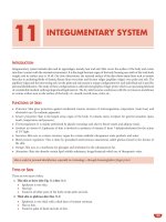
Speech Areas (Fig. 8.4)
Broca’s area (motor speech area/area 44): It is the posterior
most portion of inferior third frontal convolution of the
dominant hemisphere. It is important for fluency,
rhythm of speech and for the maintenance of grammar
and syntax.
Broca’s aphasia (expressive aphasia or motor aphasia):
Damage to motor area results in poorly articulated and
Figs 8.4A and B: Functional areas of cerebral cortex
non-fluent speech, with reduced number of words, with
errors of grammar and syntax.
Wernicke’s area (sensory speech area/posterior part of area 22
and parietotemporal junction): It is the dominant tempero
parieto-occipital region and is important in the
comprehension of received speech and in the selection
of words to express ideas.
Wernicke’s aphasia (receptive aphasia or sensory aphasia): With
damage to sensory area, the output of spontaneous speech
may be normal or increased, the speech is fluent and the
articulation of phonemes is usually intact. Speech may
contain paraphasias, neologisms, jargons. When lesion is
restricted to temporal region, there is disturbance in
words heard. When lesion is restricted to parieto-occipital
region, there is disturbance in words seen.
Conduction aphasia: The defect is inability of the patient
to repeat phrases or words spoken by the examiner
(impaired repetition). The lesion lies in the perisylvian
area with damage to the fibres of arcuate fasciculus.
Transcortical aphasia
Motor
Anterosuperior to Broca’s area
Sensory Posteroinferior to Wernicke’s area
The speech disturbance in these two conditions will be
of Broca’s and Wernicke’s type aphasias respectively
with normal repetition.
Nervous System
Differentiating Features of Various Speech Syndromes
Syndromes
Clinical Features
Site of Lesion
Causes
Associated Features
Global aphasia
Minimal speech;
nonfluent; comprehension for spoken and
written language—poor
Infarction, trauma,
tumour
Contralateral hemiplegia,
hemisensory loss, hemianopsia
Broca’s aphasia
Nonfluent; agrammatic;
dysprosodic; may
be mute
Internal carotid and
middle cerebral
arteries (dominant
frontal, parietal,
superior temporal lobe)
Superior frontal
branch of MCA
Haemorrhage, tumour,
infarction.
Wernicke’s
aphasia
Fluent speech; incomprehension; no repetition; alexia; agraphia;
paraphasias
Paraphasia; difficulty
in repetition and in
reading aloud. Comprehension normal
Impaired auditory
comprehension;
inability to repeat words
or write a dictation
Impaired visual
comprehension; cannot read or write
Normal spoken
language and writing;
inability to read
Lower division of
MCA
Haemorrhage, tumour,
herpes simplex
encephalitis
Contralateral hemiplegia,
minimal sensory loss, oral
dyspraxia, cortical
dysarthria, impairment
in writing
Parietal lobe sensory
deficit, hemianopsia; no
motor disturbance
Posterior branch of
MCA (upper bank of
sylvian fissure, inferior
parietal lobule)
Superior temporal
gyrus
Embolism
Parieto- occipital
region
Infarction, tumour,
lobar haemorrhage
Hemianopsia
Posterior cerebral
artery (left occipito
striate cortex, adjacent
association cortex,
posterior corpus
callosum)
Watershed zones
between ACA, MCA
and PCA territories
PCA territory (deep
temporal lobe, parahippocampal or
hippocampal gyrus)
Infarction, tumour,
lobar haemorrhage
Hemianopsia
Hypotension,
hypoxia, cardiac
arrest
Tumour, Alzheimer’s
disease, infarction
of PCA, herpes simplex
encephalitis
Decreased alertness &
responsiveness; bilateral
leg weakness
Apraxia; dementia; no
motor or sensory loss;
upper quadrantic field
defects
Conduction
aphasia
Pure word
deafness
(mainly auditory)
Dyslexia with
dysgraphia
(mainly visual)
Pure word
blindness
Isolation of
speech areas
Parrot like speech
(echolalia)
Amnesicdysnomic aphasia
Inability in recalling
names of objects or
parts of objects;
difficulty in recent
memory
Infarction, abscess,
tumour
Contralateral hemihypesthesia, homonymous
hemianopsia, optokinetic
nystagmus
Rarely deafness
Global aphasia: In this condition, there are marked
elements of both anterior (Broca) and posterior
(Wernicke) aphasias. This is due to large lesions in the
middle cerebral artery territory or left internal carotid
artery or a large haemorrhage or a major tumour or
trauma (Fig. 8.5).
Dysarthria
Cerebellar dysarthria: Patient speaks slowly and deliberately, syllable by syllable as if scanning a line of
poetry and the normal prosodic rhythm is lost (scanning speech). When the speech has explosive character and slurring of consonants it is called staccato
speech.
Fig. 8.5: Algorithm for approach to aphasia
443
444
Manual of Practical Medicine
Pseudo-bulbar (spastic) dysarthria: Individual syllables are
slurred and the precision of consonant pronuciation is
lost. It is due to lesions in corticospinal fibres supplying
muscles of face, larynx, tongue and respiration (multiple
lacunar infarcts, motor neuron disease and atherosclerosis), e.g. British constitution becomes Brizh
conshishusion.
Bulbar dysarthria: Lower motor neuron bulbar palsy
affect the muscles of articulation. There is non-specific
slurring of speech. Other features like dysphagia and
nasal regurgitation are present, e.g. motor neuron
disease.
Rigid dysarthria: This is due to extrapyramidal involvement, e.g. low volume, monotonous speech of
parkinsonism.
Cortical dysarthria: There is irregular hesitancy in word
production associated with difficulties in abstract,
volitional movements of the lips and tongue (Orofacial
apraxia). It is usually associated with aphasia. The lesion
is in the left frontal and temporal regions. It never occurs
as an isolated defect.
Peripheral Disorders
This can be due to the involvement of the following:
1. Neuromuscular—myasthenia gravis
2. Muscular—myopathy (oculopharyngeal)
3. Structural—cleft lip, cleft palate, dentures.
Examination of Speech and Language
1. Spontaneous speech
Articulation
Fluency
Paraphasias
Grammar
Syntax
2. Naming objects, concepts
3. Comprehension of spoken commands
4. Repetition of spoken phrases
5. Reading aloud
6. Handwriting.
Lobar Functions
Functions of Left Hemisphere
a.
b.
c.
d.
e.
f.
Verbal
Linguistic description
Mathematical
Sequential
Analytical
Direct link to consciousness.
Functions of Right Hemisphere
a.
b.
c.
d.
e.
f.
Almost non-verbal
Musical
Geometrical
Spatial comprehension
Temporal synthesis
Doubtful link to consciousness.
Frontal Lobe Functions
Personality
Emotional response
Social behaviour.
Frontal Lobe Lesions
Effects of Unilateral Frontal Lobe
Disease Either Left or Right
a. Contralateral spastic hemiplegia.
b. In prefrontal lesions, no hemiplegia; grasp and suck
reflexes may be released. There is slight elevation of
mood, increased talkativeness and tendency to joke
(disinhibition), loss of initiative, lack of tact.
c. Anosmia with involvement of orbital parts.
d. Impaired memory.
Effects of Nondominant Right Frontal Lobe Disease
Same features as mentioned above.
Effects of Dominant Left Frontal Lobe Disease
In addition to the above features,
a. Motor speech disorder with agraphia with or without
apraxia of the lips and tongue
b. Loss of verbal associative fluency; perseveration
c. Sympathetic apraxia of left hand.
Effects of Bilateral Frontal Lobe Disease
a. Bilateral hemiplegia
b. Pseudo-bulbar palsy
c. In prefrontal lesions, abulia or akinetic mutism, lack
of ability to solve problems, lack of attention, rigidity
of thinking, bland affect, labile mood, and varying
combination of grasping, sucking, decomposition of
gait, and sphincteric incontinence.
Functions of Parietal Lobe
Dominant side
Calculation
Language
Planned movement
Appreciation of size, shape, weight and texture.
Nervous System
445
Nondominant side
Spatial orientation
Constructional skills.
b. Dreamy states with uncinate seizures
c. Homonymous superior quadrantanopia
d. Emotional and behavioural changes.
Parietal Lobe Lesions
Effects of Dominant Left Temporal Lobe Disease
Effects of Unilateral Parietal Lobe Disease
Either Left or Right
a. Cortical sensory loss and sensory extinction
b. Mild hemiparesis, unilateral muscular atrophy in
children
c. Homonymous inferior quadrantanopia (incongruent), visual inattention, anosognosia, neglect of
one half of body
d. Abolition of optokinetic nystagmus to one side.
In addition to the above features—
a. Wernicke’s aphasia
b. Amusia
c. Impairment in tests of verbal material presented
through the auditory sense
d. Dysnomia or amnesic aphasia.
Effects of Dominant Left Parietal Lobe Disease
In addition to the above features, patients have
a. Disorders of language (alexia)
b. Gerstmann syndrome (defect in calculation, writing,
finger naming and right to left orientation)
c. Tactile agnosia
d. Bilateral ideomotor and ideational apraxia.
Effects of Nondominant Right Parietal Lobe Disease
a.
b.
c.
d.
Visuospatial disorders
Topographic memory loss
Anosognosia and dressing apraxia
Construction apraxia.
Effects of Bilateral Parietal Lobe Disease
a. Visuospatial imperception
b. Optic ataxia
c. Spatial disorientation
d. Severe forms of construction apraxia.
Functions of Temporal Lobe
Dominant
Auditory perception, speech, language, verbal
memory and olfaction.
Nondominant
Auditory perception
Music tone sequences
Non-verbal memory (faces, shapes, music)
Olfaction.
Lesions of Temporal Lobe
Effects of Unilateral Temporal Lobe Disease
Either Left or Right
a. Auditory, visual, olfactory and gustatory hallucinations
Effects of Nondominant Right Temporal Lobe Disease
In addition to the above features—
a. Inability to judge spatial relationships in some cases
b. Impairment in tests of visually presented non-verbal
material (non-verbal memory)
c. Agnosia for sounds and some qualities of music.
Effects of Bilateral Temporal Lobe Disease
a. Korsakoff’s amnesic effect
b. Apathy and placidity
c. Increased sexual activity
d. Sham rage
b, c and d—Kluver-Bucy syndrome.
Functions of Occipital Lobe
Analysis of vision.
Lesions of Occipital Lobe
Effects of Unilateral Disease, Either Right or Left
a. Contralateral (congruent) homonymous hemianopia,
which may be central (splitting the macula) or
peripheral; also homonymous hemiachromatopsia
b. Irritative lesions—elementary hallucinations.
Effects of Left Occipital Lobe Disease
In addition to the above features—
a. Splenium of corpus callosum—alexia, colour anomia
b. Object agnosia.
Effects of Right Occipital Lobe Disease
In addition to the above features—
a. In extensive lesions, visual illusions, hallucinations
b. Loss of topographic memory and visual orientation.
Effects of Bilateral Occipital Lobe Disease
a. Cortical blindness (pupils reactive)
b. Anton’s syndrome (denial of blindness in patients
who cannot see)
446
Manual of Practical Medicine
c. Loss of perception of colour
d. Prosopagnosia (inability to identify a familiar face),
simultanagnosia (a cognitive defect in the synthesis
of visual impressions leading to lack of ability to read
all but, the shortest words spelled out letter by letter;
there is a quantitative defect in the capacity for
perceptual analysis and form synthesis, resulting in
decrease in the span of visual form apprehension
e. Balint’s syndrome (inability to look voluntarily into
and to scan the peripheral field despite full eye
movements; failure to precisely grasp or touch an
object under vision; visual inattention mainly
affecting the peripheral field).
Cognitive Scales
Total score is 30. Maximum score of 30 is normal.
Scores between 15 and 22 suggest mild to moderate
dementia.
Scores lower than 21 are associated with severe cognitive
impairment.
Cognitive function is assessed by
1. Mini mental state examination
Orientation: 1 point for each correct answer
What is the: (orientation to time)
time
date
day
month
year
3 points if three-stage commands correctly obeyed
‘Take this piece of paper in your right hand, fold it
in half, and place it on the floor’.
3 points
1 point for correct response to a written command
such as ‘close your eyes’.
1 point
Have the patient write a sentence. Award 1 point if
the sentence is meaningful, has a verb and a subject.
1 point
Test the patient’s ability to copy a complex diagram
of two intersected pentagons.
1 point
5 points
What is the name of this: (orientation to place)
ward
hospital
district
town
country
5 points
Registration
Name three objects
Score 1, 2, 3 points according to how many are
repeated
Re-submit the list until the patient is word perfect in
order to use this for a later test of recall
Score only for first attempt
3 points
Attention and calculation
Have the patient subtract 7 from 100 and then from
the result a total of five times.
Score 1 point for each correct subtraction 5 points
Recall
Ask for three objects used in the registration test,
one point being awarded for each correct answer
3 points
Language
1 point each for two objects correctly named (pencil
and watch)
2 points
1 point for correct repetition (No ifs and buts)
1 point
2. Mental status questionnaire (MSQ)
1. What is the name of this place (where are we now)?
2. What is the address of this place?
3. What is the date?
4. What month is it?
5. What year is it?
6. How old are you?
7. When is your birthday?
8. What year were you born?
9. Who is the Prime Minister?
10. Who was the previous Prime Minister?
Total score 10 (1 for correct response, 0 for incorrect
response)
Normal subjects score 9 or 10; scores less than 8 imply a
degree of mental confusion.
Examination of the Cranial
Nerves (Fig. 8.6)
First Cranial Nerve (Olfactory Nerve)
Olfactory nerve subserves the sense of smell.
Anatomical Peculiarity
This is the only sensory pathway having no thalamic
connection (Fig. 8.7).
Testing Sense of Smell
1. It is tested separately in each nostril, after confirming
the patency of the nostrils, and with the eyes closed.
2. Substances with familiar odours like coffee,
peppermint oil, clove oil, soap, etc. can be used.
Irritating and pungent substances like ammonia are
avoided as trigeminal nerve is also stimulated.
Nervous System
447
2. Head injuries may result in shearing strain and tear
of olfactory filaments. This is the most common
neurological cause of anosmia
3. Tumours of the anterior cranial fossa from frontal
lobe
4. Chronic basal meningitis (tuberculous, syphilitic
or neoplasm)
5. Kallman’s syndrome (anosmia, obesity, hypogonadism, midline defects)
6. Tabes dorsalis
7. Internal hydrocephalus
8. Ageing
9. Alzheimer’s disease
10. Parkinson’s disease
11. Huntington’s chorea
12. Down syndrome.
Unilateral Anosmia is a useful sign of anteriorly situated
space occupying lesion like sub-frontal meningioma or frontal
lobe tumour.
Fig. 8.6: Cranial nerve nuclei
Increased Olfactory Acuity (Hyperosmia)
1. It is occasionally a feature of premonitory phase of
migraine
2. Addison’s disease
3. Hyperemesis gravidarum
4. Mucoviscidosis
5. Strychnine poisoning.
Perversion of Smell (Parosmia)
1. It is seen during partial recovery, from traumatic
anosmia
2. Severe nasal infection
3. Ingestion of drugs (phenytoin)
4. Psychological.
Fig. 8.7: Anatomy of the olfactory nerve
Foul Smell (Cacosmia)
This is said to be present when the patient perceives
unpleasant odours in the absence of a stimuli. It is seen
in severe upper respiratory tract infections and in
atrophic rhinitis.
3. There is a strong relationship between the sense of
smell and taste, which combine to give a perception
of flavour. Normal olfaction is therefore necessary
to appreciate taste.
Olfactory Hallucinations
Interpretation
Second Cranial Nerve (Optic Nerve)
Loss of Sense of Smell (Anosmia)
This nerve subserves the sense of vision.
Examination of this nerve consists of testing of
1. Visual acuity
1. This most commonly occurs due to nasal diseases
like catarrh, sinusitis, hay fever
They are usually of unpleasant nature and characteristic
of epilepsy arising in the uncinate gyrus of temporal
lobe. They may also occur in psychosis.
448
Manual of Practical Medicine
Some Common Features of Olfaction and
Taste Sensations
1.
2.
3.
4.
5.
2.
3.
4.
5.
Olfaction
Taste
The olfactory fibres do not
relay in the thalamus
Bacterial and viral
infections (URI) can
cause loss of olfaction
Toxins (toxic chemicals),
drugs that affect cell
turnover and irradiation
all affect olfaction
Abnormalities of mucous
secretion in which the
olfactory cilia are bathed
can result in decreased
olfaction
Zinc and vitamin therapy
may improve olfaction
Only a part of the taste
fibres relay in the thalamus
Bacterial colonisation of the
taste pores leads to loss of
taste sensation
Toxins (heavy metals),
drugs that affect cell turnover and irradiation all
affect taste sensation
Abnormalities of the
salivary milieu in which
the taste receptors are
bathed can lead to loss of
taste sensation
Zinc and vitamin therapy
may improve taste
sensation
Visual fields
Colour vision
Pupillary responses
Inspection of optic nerve head and fundus by
ophthalmoscopy.
Testing of Visual Acuity
It is a measurement of the efficacy of the macular or
central vision and depends on the intactness of this part
of the retina and its nervous connection. Peripheral
retinal lesions do not significantly affect visual acuity.
This is done for both near and distant vision. Standard Snellen’s type chart is used for testing distant vision.
Each eye is tested separately at a distance of six metres.
Acuity is recorded as a fraction. Normal visual acuity is
6/6. Corrected visual acuity of 6/60 bilaterally
constitutes legal blindness. If visual acuity is severely
depressed, finger counting, hand movement and perception of light should be tested (Fig. 8.8).
Jaegar type card held at a distance of one foot
from patient’s eye is used for testing near vision of each
eye.
Causes of Decreased Visual Acuity
1. Papillitis
2. Retrobulbar neuritis
3. Refractive errors
a. Myopia
b. Presbyopia
c. Astigmatism
4. Primary ocular disorders
Fig. 8.8: Snellen’s chart
a.
b.
c.
d.
e.
f.
Iridocyclitis
Corneal opacities
Cataracts
Vitreous opacities
Retinal detachment
Glaucoma.
Loss of visual acuity is commonly due to refractive errors
of the eye, cataracts, vitreous and corneal opacities.
Pin hole test: It is useful in detecting whether poor vision
is due to refractory error or disease of the eyeball or
visual pathway. If patient is able to see better through a
pin hole then patient most probably has refractory error.
Visual acuity is not affected in lesions posterior to optic
chiasma except in cortical blindness.
Testing of Visual Fields
The full extent of vision observed while visualising an
object is known as the visual field. The field of vision is
limited by the area of the retina and by the margins of
the orbit, nose and cheek.
Normal visual field using 5 mm white object is
approximately 100o temporally, 60o nasally, 60o superiorly and 75o inferiorly.
Concentric contraction of the visual fields binocularly to less than 10o constitutes legal blindness.
Nervous System
449
Automated perimetry: Automated perimetry utilises
computer to programme visual field sequences.
They provide exact repeatable tests through a
selection of visual field testing procedure. They are more
sensitive than manual perimetry and always
reproducible.
Changes in the Field of Vision (Fig. 8.10)
1. Central scotoma
It is the loss of vision confined to central field of vision.
Unilateral central scotoma is commonly due to
demyelination of the optic nerve (multiple sclerosis) and
diseases of the choroid or retina and bilateral scotoma
is due to toxic causes like alcoholism, vitamin B 12
deficiency.
Fig. 8.9: Confrontation method
Confrontation method: This method is useful for testing
peripheral field of vision (Fig. 8.9).
The examiner must have a normal visual field, as
field defects present in the patient is detected by
comparing his field of vision with that of the examiner.
The examiner is seated at a distance of one metre from
the patient. Both eyes are tested simultaneously first
and then each eye separately. The test is carried out by
asking the patient to fix his gaze on the examiner’s eye
(patient’s right eye is fixed on the examiner’s left eye
and vice versa). The eye not being tested, is covered.
The patient is instructed not to move his head or shift
his gaze. The examiner then moves his finger, kept
midway between him and patient from the periphery
to the centre in the temporal, nasal, superior and inferior
directions.
By this method, approximate defects in the visual
fields can be made out.
Red pin test: This outlines the central field by using a red
hat-pin. The test is carried out as in the confrontation
method. Central area of impaired vision (central
scotoma) can be detected by this method. This test also
determines the size of the physiological blind spot.
A red hat-pin is used for testing the central field of
vision as the macula, which is the area for perceiving
the central field of vision, contains a large number of
cones, which in turn perceive coloured objects (especially red) better than white.
Perimetry: This surveys the monocular field of vision,
e.g. Goldmann perimeter used to chart patient’s visual
field on Bjerrum’s screen. It is the most accurate method
for testing the field of vision.
2. Hemianopia
It is the loss of sight in one half of the visual field.
a. Homonymous hemianopia: It is the loss of nasal field
of vision in one eye and temporal field of vision in the
other eye.
b. Heteronymous hemianopia: It is the loss of either the
nasal or the temporal field of vision in both eyes.
c. Incongruous hemianopia: The outline of visual field
loss in both eyes are different, e.g. lesions of optic tract,
or chiasma (pregeniculate lesions).
Lesions of lateral geniculate body have been found
to produce incongruous wedge-shaped homonymous
field defects but when the aaetiology is ischaemic, the
defect is usually congruous.
d. Congruous hemianopia: The outline of visual field loss
in both eyes are similar, e.g. lesions of optic radiation
(postgeniculate lesions).
Lesion of the optic radiation close to the calcarine
cortex in the occipital lobe produces congruous hemianopia, as the fibres in the optic radiation are closely
packed together.
e. Bitemporal hemianopia: This is produced by lesions of
the optic chiasma caused by tumour of pituitary gland
or sella turcica or by an inflammatory or traumatic lesion
of optic chiasma. This may occur in 80% of people in
whom the nasal fibres at sella turcica are affected. In
10%, the decussation may be pre-fixed, when a lesion at
the sella turcica may cause a lesion of the optic tract. In
the other 10% in whom the decussation may be postfixed, the above lesion may involve the optic nerve.
Field defects of chiasmal lesions are produced by
the following conditions:
1. Pituitary tumours
2. Craniopharyngioma
450
Manual of Practical Medicine
Fig. 8.10: Visual field defects
3. Meningioma
4. Chiasmal glioma
5. Distension of the third ventricle (hydrocephalus)
6. Internal carotid artery aneurysm
7. Mucocele of the sphenoid sinus
8. Granulomatous meningitis (TB, syphilis, sarcoidosis)
9. Head injury
10. Ischaemia.
Compression of the optic chiasma in the midline
produces bitemporal hemianopia, along with progressive loss of visual acuity.
Compression of the optic chiasma in the lateral aspect
on both sides produces binasal hemianopia (example:
compression by atherosclerotic internal carotid or
anterior cerebral arteries).
Pressure upon the optic chiasma from below
produces bilateral upper temporal quadrantanopia
(example: in the early stages of pituitary tumour).
Pressure upon the optic chiasma from above
produces bilateral lower temporal quadrantanopia
(example: distension of the third ventricle as occurs in
hydrocephalus in the early stage).
f. Inattention hemianopia: It is an example of perceptual
rivalry. It occurs in patients having a lesion in the parietal
lobe, where patients fail to perceive an object in one
half of visual field when presented simultaneously and
bilaterally.
g. Quadrantic hemianopia: Superior and inferior quadrantic hemianopia means loss of upper and lower
quadrants of the visual field respectively.
In temporal lobe lesions, affection of the optic radiation causes superior quadrantic hemianopia.
In parietal lobe lesions, affection of the optic radiation causes inferior quadrantic hemianopia.
Hemianopia with macular sparing is seen in
i) Lesion of calcarine cortex.
h. Altitudinal hemianopia: It is due to partial lesion of
the blood supply of the optic nerve as in vascular
accidents or trauma (Fig. 8.11).
3. Concentric constriction of visual field
It occurs in long standing papilloedema, bilateral
lesion of visual cortex, retinitis pigmentosa, and in
hysteria.
Nervous System
451
Fig. 8.11: Altitudinal hemianopia
Monocular Visual Loss
• Optic neuritis
Viral (childhood)
Multiple sclerosis
Epstein-Barr virus
Post-infectious
Sphenoid sinusitis
Ischaemia-Giant cell arteritis
• Orbital tumour
• Vascular occlusion—Arterial/Venous
Binocular Visual Loss
•
•
•
•
Chiasmal compression
Elevated intracranial pressure
Toxic- Methyl alcohol
Infiltrative disorders
Malignancy
Lymphoma
Leukaemia
• Leber’s optic atrophy
Fig. 8.12: Ishihara chart
Inspection of Optic Nerve Head and
Fundus by Ophthalmoscopy (Fig. 8.13)
Colour Vision
The primary colours are red, green and blue. Blue colour
has the maximum field of vision. Colour vision is tested
by use of pseudo-isochromatic plates (Ishihara chart) (Fig.
8.12). Most common anomaly of colour vision are the
various types of red-green deficiency inherited as sex
linked recessive condition. Acquired defects of colour
vision occur in macular and optic nerve diseases, and due
to certain drugs, e.g. ethambutol, chloroquine.
Swinging Light Test for Afferent (Optic
Nerve) Pupillary Abnormality
This test is done to detect a lesion in the afferent
pathway, i.e. optic nerve. In this test, a bright light is
swung from one eye to the other alternatively. The eye
with optic nerve lesion will show a positive consensual
light reflex, but will not show a positive direct light
reflex. So, the affected pupil starts to dilate when direct
light is thrown into that eye (Marcus Gunn’s Pupil).
Fig. 8.13: Ophthalmoscopy
A. Papilloedema
It is the oedema of the optic disc > 3 dioptres (Fig. 8.14).