Ebook Dental management of the medically compromised patient (9/E): Part 2
Bạn đang xem bản rút gọn của tài liệu. Xem và tải ngay bản đầy đủ của tài liệu tại đây (14.44 MB, 371 trang )
PA R T
VII
Immunologic Disease
308
18
AIDS, HIV Infection, and Related Conditions
DEFINITION
On June 5, 1981, when the Centers for Disease Control
and Prevention (CDC) reported five cases of Pneumocystis
carinii (now jiroveci) pneumonia in young homosexual
men in Los Angeles, few suspected that it heralded a
pandemic of acquired immunodeficiency syndrome (AIDS).
In 1983, a retrovirus (later named the human immunodeficiency virus [HIV]) was isolated from a patient with
AIDS. Since that first report, more than 70 million persons
have been infected with HIV, and more than 30 million
have died of AIDS.1 The total number of deaths has
exceeded those caused by the Black Death of 14th-century
Europe and the influenza pandemic of 1918 and 1919.
About 95% of HIV-infected persons live in low- to middleincome regions and countries and in sub-Saharan Africa.
More than 40% of new infections (excluding those in
infants) occur in young people 15 to 24 years of age.1,2
AIDS is an infectious disease caused by HIV, which is
transmitted predominantly through intimate sexual contact
and by parenteral means. In view of the nature of this
bloodborne pathogen, HIV infection and AIDS have
important implications for dental practitioners. Although
HIV has rarely been transmitted from patients to health
care workers, this may occur, and patients with HIV
infection or AIDS may be medically compromised and
may need special dental management considerations. On
the basis of current statistics, the average dental practice
is predicted to encounter at least two patients infected
with HIV per year.
The definition of AIDS provided by the CDC has
been revised several times over the years, and in 2008,
it was revised to be laboratory-confirmed evidence of
HIV infection in a person who has stage 3 HIV infection
(i.e., a CD4+ lymphocyte count <200 cells/µL).3,4 This
definition also includes HIV-infected persons whose CD4+
count may be above 200/µL but have an AIDS-defining
condition, as shown in Box 18.1. Of note, because of
the provision of antiretroviral drug regimens, not all
patients progress to AIDS or develop life-threatening
opportunistic infections.3,4
CRITICAL COMPLICATIONS: Patients with HIV/AIDS undergoing dental treatment may not be diagnosed and may be at
risk for either transmitting the infection or sustaining complications such as infection, bleeding, drug interactions, and side
effects. These events could prove serious. Dentists must be
able to identify these patients, assess risk based on history
and clinical findings, and work closely with the managing
physician to develop a dental management plan that will be
effective and safe for the patient as well as others.
INCIDENCE AND PREVALENCE
An estimated 2.7 million people across the globe are
newly infected with HIV annually.1
Since the onset of the worldwide pandemic, more than
70,000,000 people have been infected with HIV, of whom
approximately 35,000,000 have died as a consequence
of AIDS3,4 (Tables 18.1 and 18.2). HIV prevention efforts
have been “front and center” since the virus was discovered
as the cause of AIDS, and behavioral interventions focused
on HIV-negative persons have likely played a role in the
falling population level incidence in some countries
reported by the United Nations Program in HIV/AIDS
(UNAIDS) in its 2012 report.3
A majority of those infected are between 25 and 29
years of age, male, and disproportionately black. Recent
estimates for cases of HIV infection diagnosed in the
United States by age, race, and transmission category are
shown in Table 18.1.
From 2010 through 2014, the rate for persons aged
25 to 29 years increased. The rates for children (aged
younger than 13 years) and persons aged 13 to 14, 15
to 19, 35 to 39, 40 to 44, 45 to 49, 50 to 54, 55 to 59,
and 60 to 64 years decreased. The rates for persons aged
20 to 24, 30 to 34, and 65 years and older remained
stable. In 2014, the highest rate was for persons aged 25
to 29 years (35.8 in 100,000), followed by the rate for
persons aged 20 to 24 years (34.3 in 100,000).1,2
Overall in the United States, the estimated rate of HIV
infection in 2014 was 13.8 in 100,000.
Race and Ethnicity: From 2010 through 2014, the
rates for American Indians and Alaska Natives and Asians
increased. The rates for blacks and African Americans,
Native Hawaiians and other Pacific Islanders, and persons
of multiple races decreased. The rates for Hispanics and
Latinos and whites remained stable. In 2014, the rates
were 49.4 in 100,000 for blacks and African Americans,
18.4 in 100,000 for Hispanics and Latinos, 15.4 in
100,000 for persons of multiple races, 10.6 in 100,000
for Native Hawaiians and other Pacific Islanders, 9.5 in
100,000 for American Indians and Alaska Natives, 6.2
in 100,000 for Asians, and 6.1 in 100,000 for whites.1,2
309
310
CHAPTER 18 AIDS, HIV Infection, and Related Conditions
BOX 18.1 AIDS-Defining Conditions
•
•
•
•
•
•
•
•
•
•
•
•
•
•
•
•
•
•
•
•
•
•
•
•
•
•
•
Bacterial infections, multiple or recurrent*
Candidiasis of bronchi, trachea, or lungs
Candidiasis of esophagus†
Cervical cancer, invasive‡
Coccidioidomycosis, disseminated or extrapulmonary
Cryptococcosis, extrapulmonary
Cryptosporidiosis, chronic intestinal (>1 month in duration)
Cytomegalovirus disease (other than liver, spleen, or nodes),
onset at age >1 month
Cytomegalovirus retinitis (with loss of vision)†
Encephalopathy, HIV related
Herpes simplex: chronic ulcers (>1 month’s duration) or
bronchitis, pneumonitis, or esophagitis (onset at age >1 month)
Histoplasmosis, disseminated or extrapulmonary
Isosporiasis, chronic intestinal (>1 month’s duration)
Kaposi sarcoma†
Lymphoid interstitial pneumonia or pulmonary lymphoid
hyperplasia complex*†
Lymphoma, Burkitt (or equivalent term)
Lymphoma, immunoblastic (or equivalent term)
Lymphoma, primary, of brain
Mycobacterium avium complex or Mycobacterium kansasii
infection, disseminated or extrapulmonary†
Mycobacterium tuberculosis infection of any site, pulmonary,†‡
disseminated,† or extrapulmonary†
Mycobacterium infection, other species or unidentified species,
disseminated† or extrapulmonary†
Pneumocystis jiroveci pneumonia†
Pneumonia, recurrent†‡
Progressive multifocal leukoencephalopathy
Salmonella septicemia, recurrent
Toxoplasmosis of brain, onset at age >1 month†
Wasting syndrome attributed to HIV
*Only among children younger than 13 years of age. (Data from
Centers for Disease Control and Prevention: 1994 revised
classification system for human immunodeficiency virus infection
in children less than 13 years of age, MMWR Recomm Rep 43:1,
1994 and Centers for Disease Control and Prevention: 2008
revised surveillance case definitions for HIV infection among
adults, adolescents, and children aged <18 months and for HIV
infection and AIDS among children aged 18 months to <13
years—United States, MMWR Recomm Rep 57:9, 2008.)
†
Condition that might be diagnosed presumptively.
‡
Only among adults and adolescents 13 years of age and older.
(Data from Centers for Disease Control and Prevention: 1993
Revised classification system for HIV infection and expanded
surveillance case definition for AIDS among adolescents and
adults, MMWR Recomm Rep 41:1, 1993.)
Sex: From 2010 through 2014, the rate for female
adults and adolescents decreased; the rate for males
remained stable. In 2014, males accounted for 81% of
all diagnoses of HIV infection among adults and adolescents. The rate for male adults and adolescents was 27.4
in 100,000, and the rate for females was 6.1 in 100,000.1,2
Transmission Category: From 2010 through 2014,
among male adults and adolescents, the annual number
of diagnosed HIV infections attributed to male-to-male
sexual contact increased. The number of infections
attributed to injection drug use, male-to-male sexual
contact and injection drug use, or heterosexual contact
decreased. Among female adults and adolescents, the
number of infections attributed to injection drug use or
heterosexual contact decreased. In 2014, among male
and female adults and adolescents, the diagnosed infections
attributed to male-to-male sexual contact (70%, including
male-to-male sexual contact and injection drug use), and
those attributed to heterosexual contact (24%) accounted
for approximately 94% of diagnosed HIV infections in
the United States.1,2
The estimated number of AIDS diagnoses in the United
States for 2014 was approximately 15,6001,2 (see Table
18.2). Adult and adolescent AIDS accounted for about
99% of the cases, 75% of which occurred in males and
25% in females. The cumulative estimated number of
AIDS diagnoses through 2014 in the United States was
approximately 1.2 million.1,2
Since the introduction of protease inhibitors in 1996
and the advent of highly active antiretroviral therapy
(HAART), the epidemic of AIDS in the United States has
slowed and stabilized.3,4 As of the end of 2014, approximately 566,000 deaths have been reported in the United
States from AIDS. In the United States, AIDS is the leading
cause of death in men 25 to 44 years of age. Worldwide,
there are 2 million deaths per year, and more than 30
million persons have died of AIDS3-5 (see Table 18.2).
With improved survival of persons with HIV infection,
fortunately, there are more people living with HIV infection
and therefore more people who may seek dental treatment.
Consequently, dentists will be treating more people living
with HIV infection. At present, there is no effective vaccine
to prevent HIV infection, although large research efforts
have and continue to be made in this arena. Also, a
nonpandemic relating strain of HIV, known as HIV-2,
occurs less commonly throughout the world.6 Most cases
of HIV-2 infection have occurred in West Africa, with a
limited number of cases occurring in Canada and the
United States. Most persons infected with HIV-2 are
long-term nonprogressors because viral loads generally
are low, and the immunosuppression is not as severe.4-6
ETIOLOGY
AIDS is caused by HIV, a nontransforming retrovirus of
the lentivirus family. There are two HIV subtypes, HIV-1
and HIV-2, and many strains of each. HIV-1 was first
identified in 1983 by Francoise Barre-Sinoussi in the
laboratory of Luc Montaignier of the Pasteur Institute.
They first called it lymphadenopathy-associated virus.7
Within 1 year of this discovery, a team led by Robert
Gallo from the National Institutes of Health (NIH) isolated
a retrovirus identified as the human T lymphotropic virus
III (HTLV-III) and labeled it as the etiologic agent for
AIDS.8 In 1984, Jay Levy’s team in San Francisco also
isolated a retrovirus, AIDS-related virus (ARV), and
designated it as the causative agent for AIDS.9 All three
311
CHAPTER 18 AIDS, HIV Infection, and Related Conditions
TABLE 18.1 Select Patient Characteristics in HIV Infection
2010
2011
2012
2013
2014
Estimated*
Estimated*
Estimated*
Estimated*
Estimated*
No.
No.
Rate
No.
No.
232
42
2071
7079
6366
5504
5046
5230
4851
3510
2077
1064
790
238
43
2118
7245
6520
5639
5171
5361
4972
3602
2132
1091
810
0.4
0.5
9.6
33.4
30.8
28.1
25.8
25.6
22.0
16.1
10.8
6.4
2.0
192
43
2002
7069
6346
5272
4463
4800
4595
3364
1999
1072
816
198
45
2068
7311
6563
5455
4622
4971
4758
3487
2072
1111
848
American Indian
174
or Alaska
Native
Asian
708
Black or African
20,461
American
Hispanic or
9072
Latino†
Native Hawaiian
57
or other Pacific
Islander
White
11,864
Multiple races
1526
177
7.8
159
163
4.9
774
55.2 19,345
802
20,064
Rate
No.
No.
Rate
No.
No.
Rate
No.
No. Rate
0.5
0.6
9.2
33.1
31.7
27.4
22.4
22.1
20.8
14.9
9.7
6.2
2.0
180
40
1689
7035
6711
5235
4031
3997
3995
3024
2047
1098
867
191
43
1792
7483
7151
5574
4288
4257
4268
3235
2184
1170
930
0.4
0.5
8.5
32.7
33.1
26.2
21.8
20.4
20.1
14.3
10.3
6.5
2.1
159
33
1664
7144
7114
5449
4212
3799
3647
2928
1949
960
819
174
35
1828
7868
7870
6026
4662
4196
4021
3242
2166
1069
914
0.3
0.4
8.7
34.3
35.8
28.0
23.4
20.4
19.3
14.4
10.1
5.8
2.0
8.4
178
186
8.0
208
222
9.5
5.4
809
859
50.5 17,993 19,252
5.3
941
49.2 17,592
1046
19,540
6.2
49.4
AGE AT DIAGNOSIS (YR)
<13
13–14
15–19
20–24
25–29
30–34
35–39
40–44
45–49
50–54
55–59
60–64
≥65
0.4
0.5
9.5
33.0
30.8
26.6
23.6
23.6
21.5
15.4
10.2
6.2
2.0
240
44
1875
7157
6470
5472
4176
4433
4317
3219
1923
1052
819
250
46
1964
7489
6777
5729
4374
4646
4527
3377
2019
1106
861
RACE/ETHNICITY
727
20,987
7.1
187
193
5.3
809
52.3 18,632
848
19,581
9291
18.3
8919
9230
17.8
8954
9372
17.7
8829
9386
17.3
9227
10,201
18.4
58
11.7
56
58
11.4
58
60
11.6
54
56
10.6
53
58
10.6
6.1 11,376
27.7
1404
11,738
1456
5.9 11,259
25.0
1298
11,752
1358
5.9 10,914 11,581
22.6
1172
1246
5.9 10,967
20.1
889
12,025
982
6.1
15.4
—
21,566
29,418
—
12,135
1565
TRANSMISSION CATEGORY
Male Adult or Adolescent
Male-to-male
21,834
sexual contact
Injection drug use
1307
Male-to-male
1238
sexual contact
and injection
drug use
Heterosexual
2891
contact‡
Other§
6771
Subtotal
34,041
27,034
—
21,828
27,001
—
21,758
27,588
—
2115
1562
—
—
1080
1100
1819
1393
—
—
916
1036
1642
1342
—
—
874
924
1575
1216
—
—
809
870
1590
1217
—
—
4111
—
2739
3683
—
2500
3617
—
2398
3545
—
2049
3285
—
—
6245
27.9 32,992
50
34,146
—
6491
27.0 32,701
69
34,259
—
6163
57
26.9 31,953 34,034
—
6891
26.4 32,185
60
35,571
52
34,871
21,594 27,642
—
27.4
Female Adult or Adolescent
Injection drug use
Heterosexual
contact‡
Other§
Subtotal
803
4740
1455
8340
—
—
672
4307
1284
7833
—
—
617
3905
1178
7439
—
—
559
3782
1073
7213
—
—
495
3282
1045
7242
—
—
4046
9589
36
9831
—
7.5
3870
8849
49
9166
—
6.9
3734
8256
39
8656
—
6.5
3475
7816
55
8340
—
6.2
3756
7533
41
8328
—
6.1
—
—
0.4
142
50
192
147
51
198
—
—
0.4
168
72
240
175
75
250
—
—
0.5
119
61
180
127
64
191
—
—
0.4
115
44
159
127
48
174
—
—
0.3
Child (<13 yr at Diagnosis)
Perinatal
Other∥
Subtotal
181
51
232
185
53
238
Continued
312
CHAPTER 18 AIDS, HIV Infection, and Related Conditions
TABLE 18.1 Select Patient Characteristics in HIV Infection—cont’d
2010
2011
2012
2013
2014
Estimated*
Estimated*
Estimated*
Estimated*
Estimated*
No.
No.
8381
5554
21,997
7930
43,862
8597
5664
22,550
8129
44,940
Rate
No.
No.
15.5
7800
8.5
5424
19.6 21,316
11.3
7493
14.5 42,033
8087
5580
22,079
7764
43,510
Rate
No.
No.
Rate
No.
No.
Rate
No.
No. Rate
REGION OF RESIDENCE
Northeast
Midwest
South
West
Total¶
14.5
7646
8.3
5507
19.0 20,469
10.7
7575
14.0 41,197
8039
5717
21,480
7929
43,165
14.4
7236
7750
8.5
5376
5654
18.3 20,131 21,508
10.8
7206
7654
13.7 39,949 42,566
13.8
7137
8.4
5099
18.1 20,065
10.3
7576
13.4 39,877
7953
5529
22,196
8395
44,073
14.2
8.2
18.5
11.2
13.8
Note. Data include persons with a diagnosis of HIV infection regardless of stage of disease at diagnosis.
*Estimated numbers resulted from statistical adjustment that accounted for reporting delays and missing transmission category but not for incomplete
reporting. Rates are per 100,000 population. Rates are not calculated by transmission category because of the lack of denominator data.
†
Hispanics and Latinos can be of any race.
‡
Heterosexual contact with a person known to have or to be at high risk for HIV infection.
§
Includes hemophilia, blood transfusion, perinatal exposure, and risk factor not reported or not identified.
∥
Includes hemophilia, blood transfusion, and risk factor not reported or not identified.
¶
Because column totals for estimated numbers were calculated independently of the values for the subpopulations, the values in each column may not sum
to the column total.
This is from (pages 18 and 19).
TABLE 18.2 Select Patient Characteristics in AIDS
2010
2011
2012
2013
2014
Estimated*
Estimated*
Estimated*
Estimated*
Estimated*
No.
No. Rate
No.
No. Rate
No.
No. Rate
No.
No. Rate
Cumulative†
No. Rate
No.
No.
Est. No.*
8
30
423
2112
2974
3099
3010
3270
3534
2963
1893
1087
811
0.0
0.4
2.0
9.2
13.8
14.6
15.3
15.7
16.6
13.1
8.9
6.0
1.8
94
20
207
1339
2303
2368
2330
2424
2611
2253
1548
835
713
104
22
227
1467
2531
2598
2556
2659
2866
2468
1702
913
784
0.2
0.3
1.1
6.4
11.5
12.1
12.8
12.9
13.7
10.9
7.9
4.9
1.7
9561
1462
8909
51,371
141,002
224,940
243,597
204,493
139,973
83,736
46,497
24,762
20,882
9588
1477
9027
51,986
142,069
226,254
245,042
205,990
141,345
84,738
47,105
25,078
21,137
American Indian
116
118 5.2
103
105 4.6
94
96 4.2
91
94 4.0
or Alaska
Native
Asian§
363
372 2.5
362
375 2.5
354
369 2.3
358
379 2.3
Black or African 13,419 13,745 36.2 12,363 12,816 33.4 12,030 12,574 32.4 11,548 12,219 31.2
American
Hispanic or
5617 5745 11.3
5237 5415 10.4
5015 5222 9.8 4868 5132 9.5
Latino∥
Native Hawaiian
39
40 8.0
31
32 6.3
26
27 5.2
27
28 5.2
or other
Pacific
Islander
White
6953 7102 3.6
6483 6693 3.4
6111 6354 3.2 6005 6328 3.2
Multiple races
1134 1165 20.6
1064 1108 19.0
992 1044 17.4
971 1034 16.7
90
95
4.0
3498
3523
323
352 2.1
9119 10,045 25.4
9689
499,734
9815
504,354
3924
4279
7.7
217,650
219,578
18
19
3.5
842
850
5303 2.7
803 12.6
436,952
32,820
439,455
33,260
AGE AT DIAGNOSIS (YR)
<13c
13–14
15–19
20–24
25–29
30–34
35–39
40–44
45–49
50–54
55–59
60–64
≥65
21
49
451
1989
2886
3305
3717
4324
4292
3168
1761
936
742
21
50
461
2038
2956
3382
3804
4425
4392
3240
1801
957
759
0.0
0.6
2.1
9.4
14.0
16.9
18.9
21.2
19.4
14.5
9.1
5.6
1.9
15
40
426
1970
2776
3133
3205
3776
3965
2908
1802
906
721
15
42
440
2042
2877
3242
3318
3909
4107
3009
1863
936
744
0.0
0.5
2.0
9.2
13.5
15.8
16.9
18.6
18.5
13.3
9.2
5.3
1.8
10
30
349
1895
2730
3236
2919
3431
3650
2888
1771
991
722
10
31
363
1981
2853
3377
3047
3577
3805
3010
1848
1033
751
0.0
0.4
1.7
8.8
13.3
16.1
15.6
17.0
17.5
13.3
8.9
5.8
1.7
8
28
401
2001
2809
2931
2848
3095
3344
2807
1796
1031
769
RACE/ETHNICITY
4850
721
CHAPTER 18 AIDS, HIV Infection, and Related Conditions
313
TABLE 18.2 Select Patient Characteristics in AIDS—cont’d
2010
2011
2012
2013
2014
Estimated*
Estimated*
Estimated*
Estimated*
Estimated*
No.
No. Rate
No.
No. Rate
No.
No. Rate
No.
No. Rate
No.
No. Rate
Cumulative†
No.
Est. No.*
TRANSMISSION CATEGORY
Male Adult or Adolescent
Male-to-male
11,695 14,448 — 11,238 13,863 — 10,826 13,664 — 10,781 13,785 —
8,180 11,277 —
sexual
contact
Injection drug
1485 2072 —
1240 1773 —
1116 1649 —
957 1477 —
812 1268 —
use
Male-to-male
1177 1431 —
1018 1242 —
929 1166 —
774 1005 —
555
782 —
sexual
contact and
injection drug
use
Heterosexual
2255 3092 —
2112 2896 —
1946 2728 —
1891 2730 —
1453 2211 —
contact¶
Other**
4063
118 —
3613
127 —
3699
117 —
3696
133 —
3,243
85 —
Subtotal
20,675 21,161 16.9 19,221 19,901 15.8 18,516 19,324 15.2 18,099 19,130 14.9 14,243 15,624 12.0
529,245
586,385
165,386
187,372
78,824
85,290
66,501
84,200
107,624
947,580
11,627
954,875
Female Adult or Adolescent
Injection drug
use
Heterosexual
contact¶
Other**
Subtotal
981
1469 —
864
1302
—
781
1205
—
671
1089
—
523
913
—
75,745
90,413
3603
5502 —
3316
5195
—
3113
5039
—
2899
4839
—
2203
4150
—
114,383
149,980
2361
6945
133 —
7105 5.4
2227
6407
131
6628
—
5.0
2202
6096
109
6353
—
4.8
2191
5761
147
6075
—
4.5
1982
4708
106
5168
—
3.8
53,916
244,044
5979
246,372
12
3
15
12
3
15
—
—
0.0
8
2
10
8
2
10
—
—
0.0
8
0
8
8
0
8
—
—
0.0
83
11
94
92
12
104
—
—
0.2
8690
871
9561
8715
874
9588
Child (<13 Yr at Diagnosis)
Perinatal‡
Other††
Subtotal
15
6
21
15 —
6 —
21 0.0
REGION OF RESIDENCE‡‡
Northeast
Midwest
South
West
Total
5631 5782 10.4
5153 5356 9.6
4765 4992 8.9 4360 4636 8.3
3409 3793
3365 3431 5.1
3248 3341 5.0
3106 3215 4.8 2952 3087 4.6
2431 2627
13,747 14,059 12.2 12,949 13,402 11.5 12,608 13,155 11.2 12,667 13,376 11.3 10,003 10,965
4898 5016 7.0
4293 4445 6.1
4143 4325 5.9 3889 4115 5.5
3202 3511
27,641 28,287 9.1 25,643 26,545 8.5 24,622 25,687 8.2 23,868 25,214 8.0 19,045 20,896
6.8
351,863
354,392
3.9
127,677
128,670
9.2
483,173
487,539
4.7
238,472
240,235
6.6 1,201,185 1,210,835
Note. Reported numbers less than 12, as well as estimated numbers (and accompanying rates and trends) based on these numbers, should be interpreted
with caution because the numbers have underlying relative standard errors greater than 30% and are considered unreliable.
*Estimated numbers resulted from statistical adjustment that accounted for reporting delays and missing transmission category but not the incomplete
reporting. Rates are per 100,000 population. Rates are not calculated by transmission category because of the lack of denominator data.
†
From the beginning of the epidemic through 2014.
‡
The criteria for stage 3 (AIDS) classification among pediatric cases were expanded in 2014.
§
Includes Asian Pacific Islander legacy cases (see Technical Notes).
∥
Hispanics and Latinos can be of any race.
¶
Heterosexual contact with a person known to have or to be at high risk for HIV infection.
**Includes hemophilia, blood transfusion, perinatal exposure, and risk factor not reported or not identified.
††
Includes hemophilia, blood transfusion, and risk factor not reported or not identified.
‡‡
Because column totals for estimated numbers were calculated independently of the values for the subpopulations, the value in each column may not sum
to the column total.
This is from (pages 22 and 23).
314
CHAPTER 18 AIDS, HIV Infection, and Related Conditions
viruses were similar retroviruses, but minor differences
were observed in their amino acid sequences. Variation
in disease patterns are attributed to the slight sequence
differences among HIV strains, which also makes difficult
the production of a vaccine. The three groups essentially
were describing the same retrovirus, which can change
its antigenicity. Until 1986, most workers in the field
referred to the virus as HTLV-III and considered it to be
the causative agent for AIDS. In 1986, the World Health
Organization (WHO) recommended that the AIDS virus
be called the human immunodeficiency virus7-9 (Fig. 18.1).
Subsequent analysis of frozen tissue and serum samples
from select patients who died of uncertain causes in the
1950s and 1960s demonstrated that HIV had infected
these patients, indicating its presence in humans for more
than 60 years.10
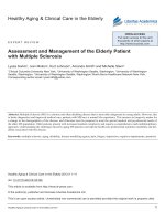
HIV is an enveloped RNA retrovirus about 100 nM
in diameter. Glycoproteins (gp41 and gp120) stud the
surface of the envelope and serve to bind to human cells
(Fig. 18.2). Internal to the envelope is a protein capsid
(p24) that surrounds essential viral enzymes (protease,
integrase, reverse transcriptase) and an RNA inner core.
It infects most human cells. However, the cells most
commonly infected are those with CD4+ receptors, including T helper lymphocytes (CD4+ cells) and macrophages.
Accordingly, these cells are most deeply involved in HIV
infection. Additional coreceptors that allow HIV to infect
human cells include CCR5, CXCR4 (fusin), and CCR2.4,5,11
HIV-1 infection is divided into stages: entry, reverse
transcription of RNA to DNA, export of the viral DNA
from the cytoplasm to the nucleus and integration into
the host chromosome, transcription, translation and
cleavage of the polyproteins produced, assembly of virions,
and budding of virions. The process is largely regulated
by the proteins tat, rev, and nef, which are necessary for
viral replication. Virulence has been mapped to the
carboxyl-terminal half of the gp120, which has been
referred to as the V3 loop.4,5,11
p17 matrix
p24 capsid
Lipid
bilayer
Protease
Integrase
RNA
gp41
gp120
Pathophysiology and Complications
Reverse
transcriptase
Transmission of HIV is by exchange of infected bodily
fluids from sexual contact and through blood and blood
products. The most common method of sexual transmission in the United States is anal intercourse in men who
have sex with men (MSM), in whom the risk of HIV
infection is 40 times higher than in other men and in
women.4,5,11 Heterosexual transmission (male to female
100 nm
FIG 18.1 The structure of human immunodeficiency virus,
showing the p24 capsid protein surrounding two strands of
viral RNA. (From Copstead LC, Banasik JL: Pathophysiology,
ed 4, St. Louis, 2010, Saunders.)
HIV
virion
Viral
assembly
and budding
HIV
virion
Injects viral RNA
Fuses with cell
Cell
Reverse
transcriptase
transforms
into DNA
Migrates
to nucleus
Provirus
integrated
into host
cell’s DNA
Cell
nucleus
Activates
cell
HIV
RNA
Protease
cleaves
protein
Viral
proteins
Messenger
RNA
FIG 18.2 Life Cycle of the Human Immunodeficiency Virus. (From Copstead LC, Banasik JL:
Pathophysiology, ed 4, St. Louis, 2010, Saunders.)
CHAPTER 18 AIDS, HIV Infection, and Related Conditions
or female to male) is the second most common form of
transmission in the United States but accounts for 80%
of the world’s HIV infections. Heterosexual transmission
can occur through sexual contact of carriers who are
heterosexual injection drug users, bisexual men, or blood
recipients of either gender. Transmission from sharing
needles is the third largest group affected in the United
States.4,5,11
HIV is found in blood, seminal fluid, vaginal secretions,
tears, breast milk, cerebrospinal fluid, amniotic fluid, and
urine. Blood, semen, breast milk, and vaginal secretions
are the main fluids that have been shown to be associated
with transmission of the virus.4,5,11
Vertical transmission, from mother to infant, can occur
during pregnancy, at birth, during breastfeeding, or from
providing premasticated food from HIV-infected parents
to infants.12 Casual contact has not been demonstrated
as a means of transmission. Inflammation and breaks in
the skin or mucosa (e.g., presence of other sexually
transmitted diseases) and high concentrations of HIV in
bodily fluids increase the risk of transmission.13,14 The
risk of transmission from a blood transfusion is estimated
to be less than 1 in 1 million because of current screening
measures. Occupational exposure is also a source of
transmission, and transmission from health care provider
to patient has occurred (see later under “Dental
Management”).
After HIV has gained access to the bloodstream, the
virus selectively seeks out T lymphocytes (specifically T4
or T helper lymphocytes) (see Fig. 18.2).4,5,11 The virus
binds to the CD4+ lymphocyte cell surface specifically
through the highly glycosylated outer surface envelope
(gp120) proteins. Upon infection, reverse transcriptase
catalyzes the synthesis of a haploid, double-stranded DNA
provirus, which becomes incorporated into the chromosomal DNA of the host cell. After integration, the provirus
genetic material may remain latent in an unexpressed
form until events occur that activate it. Activation leads
to DNA transcription and the production of new
virions.4,5,11 The virus is lymphotropic; hence, the cells it
selects for replication are soon destroyed. When the virus
takes hold, the infection causes progressive loss in the
total number of T helper cells and a marked shift in the
ratio of CD4+ to CD8+ lymphocytes. The normal ratio
of T helper to T suppressor lymphocytes is about two to
one (60% T helper, 30% T suppressor). In AIDS, the
T4-to-T8 ratio is reversed.4,5,11 This marked reduction in
T helper lymphocytes, to a great degree, explains the lack
of an effective immune response seen in patients with
AIDS and contributes to the increase in malignant disease
that has been found to be associated with AIDS, including
Kaposi sarcoma, lymphoma, carcinoma of the cervix,
and carcinoma of the rectum.5,11
Table 18.3 presents the clinical stages of HIV infection
through frank AIDS. More than 50% of persons exposed
to HIV develop an acute and brief viremia (seroconversion
sickness) within 2 to 6 weeks of exposure and then develop
315
antibodies (anti-gag, anti-gp120, anti-p24) between weeks
6 and 12. A few may take 6 months or longer to achieve
seroconversion. A concomitant, transient fall in CD4+
cells occurs (lymphopenia, along with high titers of plasma
HIV), but patients do not develop evidence of immunosuppression. Various flulike symptoms occur during this acute
infection, which usually lasts about 2 to 4 weeks. Only
an estimated 20% of affected persons seek medical attention. During the early phase of HIV disease, the virus
disseminates throughout lymphoid tissue, incubates,
replicates, and alters many physiologic processes, resulting
in hyperimmune activation, persistent inflammation, and
impaired gut function and flora.4,11
As time progresses, a steady-state viremia develops,
and several thousand copies of HIV are present in the
blood (Fig. 18.3).4,11 This clinical latency period is characterized by evolution of the virus within its host to
generate closely related yet distinct mutant viruses that
serve to evade the surveying immune response and circulating antibodies. Although the infection is clinically latent,
there is a progressive decline in immune function evident
as progressive depletion of CD4+ lymphocytes with
ultimate pancytopenia, impaired lymphocyte proliferation,
and cytokine responses to mitogens and antigens; impaired
cytotoxic lymphocyte function and natural killer cell
activity; anergy to skin testing; and diminished antibody
responses to new antigens.4,11
In untreated persons and in persons in whom therapy
is ineffective, the CD4+ count continues to decline while
HIV proliferates. As the CD4+ count drops and approaches
200 cells/µL, persons can exhibit weight loss, diarrhea,
and night sweats (see Fig. 18.3).4,11 When the CD4+ count
drops to below 200 cells/µL, the person has AIDS and
is susceptible to opportunistic infections, including
Pneumocystis pneumonia, toxoplasmosis, cryptococcosis,
influenza, histoplasmosis, tuberculosis, and cytomegalovirus
(CMV) infection; mucocutaneous diseases such as candidiasis; and neoplasms previously discussed. Neurologic
disease is common and includes secondary opportunistic
infections as well as primary HIV infection of macrophages, neurons, and microglial cells in the CNS that
leads to rapidly progressive dementia. HIV infection also
leads to immune activation and dysregulated lipid metabolism, resulting in hyperlipidemia, hypertension, cardiovascular events, diabetes, and premature aging.4,5,11
Evidence suggests that persons most susceptible to
developing AIDS are those with repeated exposure to the
virus who also have an immune system that has been
challenged by repeated exposure to various antigens
(semen, hepatitis B, or blood products).4,5,11 The median
time from primary infection to the development of AIDS
in untreated patients is about 10, and notably, there are
gender-based differences in HIV-pathogenesis with progression to AIDS being faster in infected women than infected
men.5,11 About 30% of patients with AIDS can be expected
to live approximately 2 to 3 years; most others live 10
years or longer. Long-term survival with HIV infection
316
CHAPTER 18 AIDS, HIV Infection, and Related Conditions
TABLE 18.3 Features of HIV Infection and Disease Progression
Status
Signs/Symptoms
Laboratory Findings
Comments
Recent infection
No signs or symptoms
Stage 1: acute
seroconversion
syndrome
Symptoms occur within about 1–3 wk
after infection in ≈70% of infected
patients: fever, weakness, diarrhea,
nausea, vomiting, myalgia, headache,
weight loss, pharyngitis, skin rashes
(roseola-like or urticarial),
lymphadenopathy; symptoms clear in
about 1–2 wk
Patient is unaware of his or her HIV
infection.
Can transmit the infection by blood
or sexual activity
The severity of the acute syndrome
varies among infected persons.
The period for seroconversion of
30% of patients without acute
symptoms varies and can be
1–6 mo or longer.
Stage 2: latent period
(asymptomatic
stage)
Median time from initial infection to
onset of clinical symptoms: 8–10
years
≈50%–70% of patients develop PGL
Stage 2: early
symptomatic stage
Without treatment, lasts for 1–3 yr; any
of the following: PGL, fungal
infections, vaginal yeast and
trichomonal infections, oral hairy
leukoplakia, herpes zoster, herpes
simplex, HIV retinopathy
Constitutional symptoms: fever, night
sweats, fatigue, diarrhea, weight
loss, weakness
Stage 3: AIDS
Opportunistic infection(s): Pneumocystis
jiroveci pneumonia, cryptococcosis,
tuberculosis, toxoplasmosis,
histoplasmosis, others
Malignancies: Kaposi sarcoma, Burkitt
lymphoma, non-Hodgkin lymphoma,
primary CNS lymphoma, invasive
cervical cancer, carcinoma of
rectum, slim (wasting) disease
HIV nucleic acid: positive p24
antigen; positive DNA PCR assay;
ELISA and Western blot may or
may not be positive
HIV antibody–negative at start of
syndrome
Seroconversion occurs near end of
the syndrome
CD4+ and CD8+ lymphocytes
reduced in numbers, but >500
cells/µL
After acute symptoms, they tend to
return toward normal levels.
ELISA and Western blot are positive.
ELISA and Western blot are positive.
A slow but usually steady increase
in viral load
Usually, a steady decline in CD4+
cell count; CD4+/CD8+ ratio
begins to approach 1
ELISA and Western blot are positive.
HIV antigen, RNA, and DNA tests
are positive.
Signs and symptoms increase as
CD4+ cell count declines and
approaches 200/µL; often
between 200 and 300/µL
Viral load continues to increase
Platelet count may decrease in
about 10% of patients.
High viral load; CD4+ cell count
<200/µL
CD4+ cell count <50/µL at high risk
for lymphoma and death
Platelet count may be low.
Neutrophil count may be low.
ELISA and Western blot are positive.
HIV antigen, RNA, and DNA tests
are positive.
Viral replication is ongoing and
progressive.
A steady decline in CD4+ cell counts
occurs, except in the fewer than
1% who are nonprogressors (also
have low viral load).
The spectrum of disease changes as
CD4+ cell count declines.
Death usually occurs because of
wasting, opportunistic infection,
or malignancies.
The use of combination antiretroviral
agents has slowed the death rate,
but long-term outlook must
depend on vaccines for
prevention and treatment because
the virus promotes resistance to
these agents.
AIDS, Acquired immunodeficiency syndrome; CNS, central nervous system; ELISA, enzyme-linked immunosorbent assay; HIV, human immunodeficiency virus;
PCR, polymerase chain reaction; PGL, persistent generalized lymphadenopathy.
(beyond 15 years) occurs and is associated with less virulent HIV strains, lower level viremia, HAART, and robust
immune responses.5,11
CLINICAL PRESENTATION
Signs and Symptoms
During the first 2 to 6 weeks after initial infection with
HIV, more than 50% of patients develop an acute flulike
syndrome marked by viremia that may last 10 to 14 days.
Others may not manifest this symptom complex. Symptomatic persons often develop lymphadenopathy, fever,
pharyngitis, and a skin rash but generally do not display
circulation antibodies until the sixth week to sixth month.
The severity of the initial acute infection with HIV (i.e.,
level of viremia) is predictive of the course the infection
will follow.4,5,11 In one study, 78% of persons with a
long-lasting acute illness developed AIDS within 3 years;
by contrast, only 10% of those patients with no acute
illness at seroconversion developed AIDS within 3 years.15
The CDC defines three stages of HIV infection.1 Box
18.2 illustrates the definitions for each stage. Briefly, stage
1 generally begins immediately after HIV exposure and
may last for years. Affected persons are HIV antibody
positive but are asymptomatic and show no other laboratory abnormalities. Stage 2 is characterized by progressive
CHAPTER 18 AIDS, HIV Infection, and Related Conditions
317
FIG 18.3 The natural history of human immunodeficiency virus infection. (From Brookmeyer R,
Gail MH: AIDS epidemiology: a quantitative approach, New York, 1994, Oxford University Press.)
BOX 18.2 Centers for Disease Control
and Prevention Staging of
HIV Infection in Adults
and Adolescents
Stage 1: Laboratory confirmation of HIV infection, no AIDS-defining
conditions and CD4+ T lymphocyte count of ≥500 cells/µL or CD4+
T lymphocyte percentage of total lymphocytes of ≥29.
Stage 2: Laboratory confirmation of HIV infection, no AIDS-defining
condition, and laboratory confirmation of HIV infection and CD4+ T
lymphocyte count of 200–499 cells/µL or CD4+ T lymphocyte percentage of total lymphocytes of 14–28.
Stage 3 (AIDS): Laboratory confirmation of HIV infection and CD4+
T lymphocyte count is <200 cells/µL or CD4+ T lymphocyte percentage
of total lymphocytes is <14 or documentation of an AIDS-defining
condition (see Box 18.1). Documentation of an AIDS-defining condition
supersedes a CD4+ T lymphocyte count of ≥200 cells/µL and a CD4+
T lymphocyte percentage of total lymphocytes of ≥14.
immunosuppression and symptomatic disease. Patients
who demonstrate various laboratory changes (i.e., lymphopenia: ratio of T helper to T suppressor usually <1)
in addition to HIV antibody positivity also may show
clinical signs or symptoms, such as enlarged lymph nodes,
night sweats, weight loss, oral candidiasis, fever, malaise,
and diarrhea. Persons in stage 3 have AIDS and can
demonstrate a variety of immunosuppression-related
diseases.4,5,11 Opportunistic infections predominate as the
CD4+ T count approximates 200 cells/µL; then malignancies, wasting syndrome, and a progressive form of dementia
can develop. Patients may become confused and disoriented
or may experience short-term memory deficits. Others
develop severe depression or paranoia and show suicidal
tendencies. Fig. 18.3 depicts the natural history of HIV,
and Table 18.2 lists the diseases associated with the
progression of HIV infection through frank AIDS.4,11
Laboratory and Diagnostic Findings
Most patients exposed to the virus, with or without clinical
evidence of disease, show antibodies to the virus by the
sixth month of infection. Patients with advanced HIV
infection or AIDS have an altered ratio of CD4+/CD8+
lymphocytes, a decrease in total number of lymphocytes,
thrombocytopenia, anemia, a slight alteration in the
humoral antibody system, and a decreased ability to show
delayed allergic reactions to skin testing (cutaneous
anergy).4,11 CD4+ and CD8+ cell counts should be performed
at the time of HIV diagnosis and then every 3 to 4 months.4,11
The enzyme-linked immunosorbent assay (ELISA) is
the screening test for identification of antibodies to HIV.
It is 90% sensitive but has a high rate of false-positive
results. Current practice is to screen first with ELISA. If
the results are positive, a second ELISA is performed. All
positive results are then confirmed with Western blot
analysis. This combination of tests is accurate more than
99% of the time. Positive ELISA and Western blot test
results indicate only that the individual has been exposed
to the AIDS virus.4,11 If results of the Western blot are
indeterminate, HIV infection is rarely, if ever, present.
These tests, however, do not indicate the status of the
HIV infection or whether AIDS is present. However,
patients with positive results on the ELISA and Western
318
CHAPTER 18 AIDS, HIV Infection, and Related Conditions
blot test are considered potentially infectious. ELISA testing
for HIV in saliva is an alternative approach that is 98%
sensitive in detecting antibodies to HIV.4,16 Abbott has
developed a combination assay, the ARCHITECT HIV
Ag/Ab Combo assay (Abbott Laboratories, Abbott Park,
IL), that can simultaneously detect the combined presence
of HIV antigens (the p24 antigen produced by HIV) and
antibodies to HIV. This test is important for diagnosing
HIV infection in the acute phase of the disease when
antibodies are not yet present and for ongoing monitoring
of patients.16
Nucleic acid amplification using polymerase chain
reaction (PCR)–based assays of the viral RNA is performed
to determine the viral load in the blood (i.e., degree of
viremia) and monitor response to therapy. Detection ranges
are from 40 copies/mL to more than 750,000 copies/mL.
The greatest viral load is found during the first 3 months
after initial infection and during late stages of the disease.
Direct detection of HIV by PCR assay is superior to
testing for HIV antigen in serum but more expensive.17
Antiviral resistance testing is recommended when treatment
is failing.18
MEDICAL MANAGEMENT
Medical management of the HIV-infected patient has four
main treatment goals: (1) to reduce HIV-associated
morbidity and prolong the duration and quality of survival,
(2) to restore and preserve immunologic function, (3) to
maximally and durably suppress plasma HIV viral load,
and (4) to prevent HIV transmission.4,18 Physicians managing these patients should be experts in infectious disease
and in the use of antiretroviral drugs. Antiretroviral
therapy (ART) should be used in a manner that will
achieve viral suppression and immune reconstitution while
at the same time preventing emergence of resistance and
limiting drug toxicity. Long-term goals are to delay disease
progression, prolong life, and improve quality of life.
Treatment often is organized into three major areas: (1)
ART, (2) prophylaxis for opportunistic infections, and
(3) treatment of HIV-related complications. Monitoring
response to therapy is a long-term requirement because
more than 70% of HIV-infected persons survive beyond
10 years from the time of diagnosis in the United States,
especially if treatment is not delayed.4,19-22
ART and HAART
Over the past decade, much progress has been made in
the treatment of AIDS because of ART. Both ART and
HAART involve use of combinations of antiretroviral
drugs; however, strictly speaking, HAART is defined as
the use of at least three active antiretroviral medications.
The benefits of ART are now well known. ART
increases survival, reduces systemic complications, and
improves the quality of life in patients infected with
HIV.4,19-22 The major goal of ART is to inhibit HIV replication completely such that the viral load is below the
detection limit of the assay at 4 to 6 months. However,
there are no conclusive studies that show when therapy
should be initiated. Experts recommend starting treatment
in all patients with symptoms ascribed to HIV infection,
all pregnant mothers infected with HIV, and all HIVinfected infants. ART currently is recommended when
the CD4+ count is less than 350 cells/µL and in those
with plasma HIV RNA levels greater than 55,000 copies/
mL.4,19-22 Treatment is generally initiated for asymptomatic
patients who have a rapid drop in CD4+ T cell count or
high viral loads. Asymptomatic patients with stable CD4+
T cell counts and low viral loads are generally followed
without treatment. ART is strongly recommended for
patients with CD4+ T cell counts lower than 200/µL and
for those with AIDS.4,19-22
Antiretroviral drugs are used to restore immune dysfunction by inhibiting viral replication. More than 20 antiretroviral drugs are currently available for the management
of HIV infection/AIDS (Table 18.4). The antiretroviral
agents available are classified into five categories: protease
inhibitors (PIs), nucleoside reverse transcriptase inhibitors
(NRTIs), non-nucleoside reverse transcriptase inhibitors
(NNRTIs), nucleotides, and entry inhibitors. These agents
usually are used in combinations known as ART or
HAART and should be given long term.4,19-22
The development of effective ART for HIV infection
is one of the most notable achievements in modern
medicine.4 Triple-drug therapy was first introduced in the
mid-1990s and resulted in a two-thirds decrease in HIVrelated deaths within 2 years in developed countries. Today,
a total of 29 antiretroviral drugs are approved by the
Food and Drug Administration, and three-drug combination regimens are the standard of care.4,19-22 The benefits
of ART were extended to developing countries, and an
estimated more than 16 million people currently are taking
ART worldwide. The life expectancy of an HIV-infected
individual appropriately treated with ART is now estimated
to be nearly that of the general population, both in
developed and developing countries, although it also is
estimated to be about 1.7-fold higher than in healthy
people with no comorbid conditions.4,19-22
Current guidelines from around the world now recommend starting ART in all HIV-infected patients, regardless
of CD4 cell count because of both clinical benefits to the
patient and reduction in HIV transmission to others (Box
18.3).4,19-22 This recommendation is supported by the fact
that current ART regimens are potent, convenient, and
generally well tolerated by randomized controlled clinical
trials data and by supportive clinical cohort data.4,19-22
The drug regimen that is initiated should be individualized to be potent enough to suppress the viral load to
below the level of assay detection for a prolonged period
while reducing the virus mutation rates that can lead to
drug resistance. Currently, preferred regimens for an
ART-naive patient consist of either efavirenz + tenofovir
+ emtricitabine or ritonavir-boosted atazanavir–darunavir
plus tenofovir–emtricitabine, or raltegravir + tenofovir +
TABLE 18.4 Antiretroviral Drugs Used to Treat HIV Infection
Drug
Toxicity
Interactions
Amprenavir
Atazanavir
Darunavir
Fosamprenavir
Indinavir
Lopinavir*
Nelfinavir
Ritonavir*
Saquinavir
Tipranavir
Nausea, vomiting
Nausea, vomiting, liver, tingling arms or legs
Nausea, diarrhea, lipodystrophy
Nausea, vomiting
Diarrhea
Abdominal discomfort
Paresthesias
Fatigue
Anemia, leukopenia
Thrombocytopenia, altered
taste, hypercholesterolemia,
hypertriglyceridemia, xerostomia
Nausea, vomiting, diarrhea, liver damage
Abacavir†
Emtricitabine
Didanosine
Lamivudine†
Stavudine
Zalcitabine
Zidovudine†
Headache
Avoid mixing zidovudine and
Insomnia
stavudine, ribavirin, or
Fatigue
doxorubicin.
Anemia, neutropenia
Ganciclovir and interferon-α
Nausea
must be avoided.
Diarrhea
Neuropathy, pancreatitis, myopathy, xerostomia
Comments
PROTEASE INHIBITORS (PIS)
Amiodarone
PIs act at the end of the virus
Midazolam, triazolam
replication cycle, blocking the
Midazolam, triazolam, quinidine
catalytic center of the protease
Midazolam, triazolam
enzyme, resulting in viral particles
Quinidine
that are ineffective and immature.
Rifampin
Ergotamine
St. John’s wort
Midazolam
Triazolam
Midazolam, triazolam, quinidine
NUCLEOSIDE REVERSE TRANSCRIPTASE INHIBITORS (NRTIS)
Drug adverse effects often are dose
related and can be minimized with
lower doses. Use of zalcitabine is
restricted because of the small
therapeutic window. Stavudine is
the most frequently used drug in the
group.
NON-NUCLEOSIDE REVERSE TRANSCRIPTASE INHIBITORS (NNRTIS)
Delavirdine
Efavirenz
Etravirine
Nevirapine
Dizziness, insomnia, dyslipidemia
Confusion, agitation
Rash, nausea
Hallucinations, depression, mania
Midazolam
Triazolam
Clarithromycin
Clarithromycin (rash,
Sertraline (
Warfarin (>drug effect)
Ketoconazole (
Skin rashes, nausea, vomiting
Diarrhea
Stevens-Johnson syndrome, xerostomia,
taste alteration
The most important negative adverse
effects are neuropsychiatric events,
skin reactions, GI alterations, and
liver alterations.
NUCLEOTIDES
Adefovir
Tenofovir
Dizziness
Nausea, diarrhea, weakness, depression,
anxiety, skin rash—allergy, neuropathy,
liver, kidney failure, lactic acidosis (rapid
breathing, drowsiness, muscle aches)
NSAIDs, acyclovir, and
ganciclovir affect the
metabolism of tenofovir.
Vancomycin, NSAIDs, and
cyclosporine increase the
risk for kidney disease.
Adefovir is not used often because of
GI and renal toxicity. Tenofovir is
used in patients on multiple-drug
therapy who are not responding.
Tenofovir usually is well tolerated.
ENTRY INHIBITORS
Enfuvirtide
Maraviroc
Bacterial pneumonia, rash, fever, nausea,
No significant drug interactions Inhibits fusion of HIV-1 and
vomiting, glomerulonephritis, Guillain-Barré
CD4+ T cells.
syndrome, taste disturbance, hyperglycemia,
Only one fusion inhibitor has been
myalgia, xerostomia, anorexia
approved (enfuvirtide), and it
Liver
None
has to be injected.
Three other entry inhibitors are
available.
IMMUNE-BASED THERAPIES‡
Chloroquine,
Stomach upset, muscle weakness,
hydroxychloroquine
retinopathy
Interleukin-2
Fever, chills, nausea, vomiting
Interleukin-7
Transient elevations of liver function tests
Gold salts
Pain medications, steroids
None yet reported.
These drugs reduce cellular activation,
thus reducing HIV replication, and
boost the immune response. Several
others are in testing.
*Available in combination as Kaletra.
†
Available in combination as Combivir, Epzicom, Trizivir, and Truvada.
‡
Although not antiretroviral therapy (ART) drugs, immune-based therapies also are being used in the management of human immunodeficiency virus (HIV)
infection. ART is associated with many drug interactions; only a few are listed. For more detailed recommendations, see guidelines at />guidelines.
GI, Gastrointestinal; NSAID, nonsteroidal antiinflammatory drug.
320
CHAPTER 18 AIDS, HIV Infection, and Related Conditions
BOX 18.3 Typical Antiretroviral Drug
Regimens
Number of Antiretroviral Drugs
• A two-drug regimen is effective, but three drugs are preferred;
28 days of treatment is recommended.
Preferred Antiretroviral Regimen
•TDF + 3TC (or FTC) as the first two drugs
• LPV/r or ATV/r is the preferred third drug, but RAL, DRV/r, or
EFV are alternatives.
ATV, Atazanir; DRV, darunavir; EFV, efavirenz; FTC, emtricitabine;
HIV, human immunodeficiency virus; LPV, lopinavir; /r, boosted
with ritonavir; RAL, raltegravir; TDF, tenofovir; 3TC, lamivudine.
emtricitabine.4,18-22 Several alternative drug regimens also
appear in recent Department of Health and Human
Services guidelines; however, no regimen has proved
superior to efavirenz-based regimens with respect to
virologic responses.4,18-22 Patients who respond to therapy
generally show an increase in CD4+ count in the range
of 50 to 150 cells/µL per year and viral loads of less than
75 copies/mL.4,18-22 Virologic suppression is defined as
less than 48 copies/mL, and virologic failure is defined
as a confirmed viral load of greater than 200 copies/mL
in the presence of ART.4,18-22
Patients who are taking ART medications must be
closely monitored for drug effectiveness (which often
wanes over time), development of antiviral resistance,
drug toxicity, and drug interactions. Some important
toxicities include hyperlactemia, mitochondrial dysfunction,
peripheral neuropathy, hepatotoxicity, and lipodystrophy.
Compliance also is a major challenge for patients in
view of recognized drug toxicities, costs, and inconvenience.4,18-22 To this end, several drugs are now formulated
as combination agents to simplify and improve treatment
of the disease. Atripla, Epzicom, and Trizivir are combinations of three antiretrovirals, and Combivir, Epzicom,
Trizivir, and Truvada are combinations of two nucleoside–
nucleotide reverse transcriptase inhibitors. Only a decade
ago, when cocktails of AIDS drugs began to be used,
patients sometimes had to take two dozen or more pills
a day. Currently, immune modulators and stem cell
therapies also are being tested in conjunction with ART.23
In about 25% of patients, particularly those with very
low CD4+ T cell counts, weeks after initiation of ART,
an exacerbation of preexisting opportunistic infections
occurs.20 This condition, known as immune reconstitution
inflammatory syndrome (IRIS), probably results from
elicitation of an inflammatory response in association
with the antiviral drugs, leading to focal lymphadenitis
and reactivation of a viral disease (e.g., shingles) or
granulomatous infection.20,24
Chemoprophylaxis
Chemoprophylaxis regimens are recommended when CD4+
lymphocyte counts drop to specific levels to prevent initial
episode of a disease or to suppress a developing opportunistic infection. These regimens exist for the prevention
of Pneumocystis pneumonia, tuberculosis, toxoplasmosis,
and other opportunistic diseases.4,25-27 Also, select vaccines
are recommended for HIV-infected adults before the CD4+
T cell count drops to below 200/µL. Standard resources
such as the NIH’s AIDS information website (http://
www.aids.info.nih.gov) are available for more information
on this topic.
Hope exists for improving outcomes with HIV infection.
Vaccine development is ongoing, and stem cell transplantation with CCR5-deficient cells has led to reduction of the
HIV viral reservoir in one patient and may prove effective
in eradicating HIV in the clinical setting.4,25-27
DENTAL MANAGEMENT
Health history, head and neck examination, intraoral
soft tissue examination, and complete periodontal and
dental examinations should be performed on all new
patients. History and clinical findings may indicate that
the patient has HIV infection or AIDS. Of note, however,
is that patients who know they are seropositive and
those at high risk for these conditions may not answer
questions honestly on account of the stigma or concern
for privacy. Accordingly, the patient history should be
obtained whenever possible with this understanding;
verbal communication in a quiet, private location; and
the sharing of knowledge and facts in an atmosphere
of honesty and openness.28,29
Patients who, on the basis of history or clinical findings,
are found to be at high risk for AIDS or related conditions
should be referred for HIV testing and medical evaluation.
The dentist can undertake diagnostic laboratory screening
using saliva (OraQuick Advance; OraSure Technologies,
Bethlehem, PA), or serum testing can be done with a
referral to a medical facility. Discussions with the patient
should emphasize importance of testing and should
ascertain risk factors, including sexual habits, intravenous
drug use, and so forth. Patients with high-risk factors
should be strongly encouraged to seek diagnostic
testing.25-29
Patients at high risk for AIDS and those in whom
AIDS or HIV has been diagnosed should be treated in a
manner identical to that for any other patient—that is,
with standard precautions. Several guidelines have emerged
regarding the rights of dentists and patients with AIDS,
including the following:
• Dental treatment may not be withheld if the patient
refuses to undergo testing for HIV exposure. The dentist
may then assume that the patient is a potential carrier
of HIV and should treat the person using standard
precautions, just as for any other patient.
• A patient with AIDS who needs emergency dental
treatment may not be refused care simply because the
dentist does not want to treat patients with AIDS.
CHAPTER 18 AIDS, HIV Infection, and Related Conditions
• No medical or scientific reason exists to justify why
patients with AIDS who seek routine dental care may
be declined treatment by the dentist, regardless of the
practitioner’s personal reason. However, if the dentist
and the patient agree, the dentist may refer the patient
to another provider who is more willing or better suited
(in keeping with the patient’s oral health status) to
provide treatment.
• A patient who has been under the care of a dentist
and then develops AIDS or a related condition must
be treated by that dentist or receive a referral that is
satisfactory for and agreed to by the patient.
• The CDC and the American Dental Association recommend that infected dentists inform their patients of
their HIV serostatus and should receive consent or
refrain from performing invasive procedures.30
Treatment Planning Considerations
A major consideration in dental treatment of the patient
with HIV infection/AIDS involves determining the current
CD4+ lymphocyte count and level of immunosuppression
of the patient.4 Another point of emphasis in dental
treatment planning is the level of viral load, which may
be related to susceptibility to opportunistic infections and
rate of progression of AIDS.31 The dentist should be
knowledgeable about the presence and status of opportunistic infections and the medications that the patient
may be taking for therapy or prophylaxis for such conditions. Patients who have been exposed to the AIDS virus
and are HIV seropositive but asymptomatic may receive
all indicated dental treatment. Generally, this is true for
patients with a CD4+ cell count of more than 350/µL.
Patients who are symptomatic for the early stages of
AIDS (i.e., CD4+ cell count <200/µL) have increased
susceptibility to opportunistic infections and may be
medicated with prophylactic drugs.18,31
Patients with AIDS can receive almost any dental care
needed and desired after the possibility of significant
immunosuppression, neutropenia, or thrombocytopenia
has been ruled out. Complex treatment plans should not
be undertaken before an honest and open discussion about
the long-term prognosis of the patient’s medical condition
has occurred.
Dental treatment of HIV-infected patients without
symptoms is no different from that provided for any
other patient in the practice.31 Standard precautions must
be used for all patients. Any oral lesions found should
be diagnosed and then managed by appropriate local and
systemic treatment or referred for diagnosis and treatment.
Patients with lesions suggestive of HIV infection must be
evaluated for possible HIV.32
In planning invasive dental procedures, attention must
be paid to the prevention of infection and excessive
bleeding in patients with severe immunosuppression,
neutropenia, and thrombocytopenia. This may involve
the use of prophylactic antibiotics in patients with CD4+
cell counts below 200/µL or severe neutropenia (neutrophil
321
count <500/µL).32 White blood cell (WBC) and differential
counts, as well as a platelet count, should be ordered
before any surgical procedure is undertaken. Patients with
severe thrombocytopenia may require special measures
(platelet replacement) before surgical procedures (including
scaling and curettage) are performed. Medical consultation
should precede any dental treatment for patients with
these abnormalities.32
Patients may be medicated with drugs that are prophylactic for Pneumocystis pneumonia, candidiasis, herpes
simplex virus (HSV) or CMV infection, or other opportunistic disease, and these medications must be carefully
considered in dental treatment planning. Care in prescribing other medications must be exercised with these, or
any, medications after which the patient may experience
adverse drug effects, including allergic reactions, toxic
drug reactions, hepatotoxicity, immunosuppression,
anemia, serious drug interactions, and other potential
problems. Most often, consultation with the patient’s
physician is beneficial.32 For example, acetaminophen
should be used with caution in patients treated with
zidovudine (Retrovir) because studies have suggested that
granulocytopenia and anemia, associated with zidovudine,
may be intensified; also, aspirin should not be given to
patients with thrombocytopenia. Meperidine should be
avoided in patients taking ritonavir because ritonavir
increases the metabolism of meperidine to normeperidine,
which is associated with adverse effects such as lethargy,
agitation, and seizures. Propoxyphene levels may be
increased by ritonavir, which may potentially lead to toxic
effects such as drowsiness, slurred speech, or incoordination. Antacids, phenytoin, cimetidine, and rifampin should
not be given to patients who are being treated with
ketoconazole because of the possibility of altered absorption and metabolism. Also, midazolam and triazolam
should be avoided in patients taking select protease
inhibitors because benzodiazepine metabolism may be
inhibited, leading to excessive sedation or respiratory
depression.32
Medical consultation is necessary for symptomatic
HIV-infected patients before surgical procedures are
performed. The patient’s current platelet count and WBC
count should be available. Patients with abnormal test
results may require special management. All of these
matters must be discussed in detail with the patient’s
physician. Any source of oral or dental infection should
be eliminated in HIV-infected patients, who often require
more frequent recall appointments for maintenance of
periodontal health. Daily use of chlorhexidine mouth rinse
may be helpful.
In patients with periodontal disease whose general
health status is not clear, periodontal scaling for several
teeth can be provided to allow assessment of tissue
response and bleeding. If no problems are noted, the rest
of the mouth can be treated. Adjunctive antibacterial
measures may be required if the patient’s CD4+ cell count
is below 200/µL or if tissues remain unresponsive to
322
CHAPTER 18 AIDS, HIV Infection, and Related Conditions
routine therapy. Root canal therapy has good success in
patients with HIV infection, and no modifications are
required. Infection can be treated through local and
systemic measures.32
Occupational Exposure to HIV
The risk of HIV transmission from infected patients to
health care workers is very low, reportedly about 3 of
every 1000 cases (0.3%) in which a needlestick or other
sharp instrument transmitted blood from a patient to a
health care worker.34 In comparison, the risk of infection
from a needlestick is 3% for hepatitis C and is 30% for
hepatitis B.
After a needlestick, the rate of transmission of HIV
can be reduced by postexposure prophylaxis (PEP).33 The
CDC recommends PEP as soon as possible after exposure
to HIV-infected blood.29 The number of PEP drugs recommended is based on the severity of the exposure as well
as the HIV status of the source patient.33 A less severe
exposure (solid needle or superficial injury) from a source
patient who is asymptomatic or has a low viral load
(<1500 viral copies/mL) has a two-drug PEP. Use of at
least a three-drug PEP regimen is recommended for more
severe exposure (large-bore hollow needle, deep puncture,
visible blood on device or needle used in patient’s artery
or vein) or when the patient is symptomatic, has AIDS,
or a high viral load. The recommended basic regimen for
HIV PEP is tenofovir plus emtricitabine or zidovudine
plus lamivudine.33 The expanded regimen includes a
standard two-drug regimen plus a protease inhibitor such
as ritonavir-boosted (/r) lopinavir, darunavir/r, atazanavir/r,
or raltegravir. PEP should be continued for 4 weeks, during
which time the exposed clinician should be provided expert
consultation and follow-up monitoring for compliance,
adverse events, and possible seroconversion. Tests for
seroconversion should be performed at 3, 6, and 12
months. To date, there have been six reports of occupational HIV seroconversion despite combination PEP.33
If the exposed dental health care worker is pregnant,
the risk of infection versus unknown yet possible risks
of PEP to the fetus should be discussed.
manifestations include candidiasis (erythematous or
pseudomembranous) of the oral mucosa (Figs. 18.4 to
18.7), bluish purple or red lesion(s) that on biopsy are
identified as Kaposi sarcoma (Figs. 18.8 to 18.11), and
hairy leukoplakia of the lateral borders of the tongue
(Fig. 18.12).34-39 Other oral conditions that occur in
association with HIV infection are HSV, CMV, EpsteinBarr virus (EBV), herpes zoster, deep tissue infections
(e.g., cryptococcus, histoplasmosis), recurrent aphthous
ulcerations, linear gingival erythema (Fig. 18.13), necrotizing ulcerative periodontitis (Fig. 18.14), necrotizing
stomatitis, tuberculosis, syphilis, oral warts (human
papillomavirus, condyloma acuminatum; Fig. 18.15), facial
palsy, trigeminal neuropathy, salivary gland enlargement,
xerostomia, and melanotic pigmentation.34-39 Candidiasis,
hairy leukoplakia, specific forms of periodontal disease
FIG 18.4 White lesions on the palate in a patient with AIDS.
The lesions could be scraped off with a tongue blade. The
underlying mucosa was erythematous. Clinical and cytologic
findings supported the diagnosis of pseudomembranous
candidiasis. (From Silverman S Jr: Color atlas of oral manifestations of AIDS, ed 2, St. Louis, 1996, Mosby.)
Risk of Transmission From Health Care Personnel
The risk of transmission in the dental setting is minimized
by adherence to standard infection control procedures.32,33
Oral Complications and Manifestations
Oral lesions can be one of the early signs of HIV infection
and risk for progression to AIDS and occur commonly
(30%–80%) in infected patients.35,36 Currently, patients
with HIV/AIDS that is being treated can live comfortable
lives with few complications for many years.34-38 For these
reasons, the clinician should be cognizant of the oral
manifestations of HIV infection and AIDS. The overall
prevalence of oral manifestations has changed significantly
since the advent of HAART (≈10%).34-38 The more serious
oral conditions have diminished.34-38 Common oral
FIG 18.5 Note the white lesions on the oral mucosa. The
diagnosis of pseudomembranous candidiasis was established.
(Courtesy of Eric Haus, Chicago, IL.)
CHAPTER 18 AIDS, HIV Infection, and Related Conditions
323
FIG 18.6 Erythematous palatal lesion in an HIV antibody–positive patient. Smears taken from the lesion showed hyphae
and spores consistent with Candida. The lesion healed after
a 2-week course of antifungal medications. A diagnosis of
erythematous candidiasis was made on the basis of clinical
laboratory findings. (Courtesy of Eric Haus, Chicago, IL.)
FIG 18.8 Multiple erythematous lesions on the face of a patient
with AIDS. With the use of biopsy, lesions were established
as Kaposi sarcoma. (Courtesy of Sol Silverman, San Francisco,
CA.)
FIG 18.7 Angular cheilitis in a patient with AIDS. The lesion
responded to antifungal medication. (Courtesy of Eric Haus,
Chicago, IL.)
(i.e., linear gingival erythema and necrotizing ulcerative
periodontitis), Kaposi sarcoma, and non-Hodgkin lymphoma are reported to be strongly associated with HIV
infection.34-39 Likewise as the condition progresses, these
conditions become more prevalent and more severe.34-39
Features and management of the oral manifestations of
HIV infection are discussed in Tables 18.5 and 18.6. In
addition, clinicians should be aware that oral lesions can
be a feature of the stage of the disease or a sign of treatment failure or disease progression.34-39
Worldwide, candidiasis is the most common oral
manifestation of HIV infection.35,36 Oral candidiasis
diagnosed in HIV-infected patients with persistent generalized lymphadenopathy may be of predictive value for the
subsequent development of AIDS. The appearance of
pseudomembranous candidiasis in HIV-infected persons
has been shown to be a strong indicator for progression
FIG 18.9 Multiple large, flat, erythematous lesions involving
the palatal mucosa. Biopsy revealed the lesions to be Kaposi
sarcoma, and the patient was eventually given a diagnosis of
AIDS. (Courtesy of Sol Silverman, San Francisco, CA.)
of infection to AIDS. The erythematous form of candidiasis
also indicates progression toward AIDS.34-39 This information might be helpful to dental clinicians in evaluating
patients for the initial diagnosis of HIV/AIDS or in
determining stage of infection and level of immunosuppression. However, the oral manifestations of candidiasis
Text continued on p. 328
324
CHAPTER 18 AIDS, HIV Infection, and Related Conditions
FIG 18.10 Palatal lesion in a patient with AIDS. Biopsy revealed
Kaposi sarcoma. (Courtesy of Sol Silverman, San Francisco,
CA.)
FIG 18.11 Kaposi sarcoma of the gingiva. (From Silverman S
Jr: Color atlas of oral manifestations of AIDS, ed 2, St. Louis,
1996, Mosby.)
FIG 18.12 Diffuse white lesion involving the tongue. Biopsy
supported the diagnosis of hairy leukoplakia. (From Silverman
S Jr: Color atlas of oral manifestations of AIDS, ed 2, St. Louis,
1996, Mosby.)
FIG 18.13 Band of linear gingival erythema involving the free
gingival margin of a human immunodeficiency virus–infected
patient. (From Neville B, Damm D, Allen C: Oral and maxillofacial
pathology, ed 3, St. Louis, 2009, Saunders.)
FIG 18.14 Necrotizing ulcerative periodontitis in a human
immunodeficiency virus–infected patient. The diagnosis was
established after the patient was referred for medical evaluation.
(Courtesy of Sol Silverman, San Francisco, CA.)
FIG 18.15 Multiple areas of condylomata acuminata on the
gingivae of an HIV-positive patient. (From Silverman S Jr:
Color atlas of oral manifestations of AIDS, ed 2, St. Louis,
1996, Mosby.)
CHAPTER 18 AIDS, HIV Infection, and Related Conditions
325
TABLE 18.5 Head, Neck, and Oral Lesions Commonly Associated With HIV Infection and AIDS
Oral Condition
Comments
Treatment
Persistent generalized
lymphadenopathy
An early sign of HIV infection found in about 70% of
infected patients during the latent stage of infection
Must be present >3 months and in two or more
extrainguinal locations
Anterior and posterior cervical, submandibular,
occipital, and axillary nodes are most frequently
involved.
Most common intraoral manifestation of HIV infection.
First found during the early symptomatic stage of
infection. This indicates that AIDS will develop
within 2 years in untreated patients.
≈90% of patients with AIDS will develop oral
candidiasis at some time during their disease
course.
Usually not treated directly; may need biopsy to
rule out lymphoma or other conditions
LGE does not respond to improved plaque control
procedures. Condition is associated with
candidiasis.
NUG relates to ulceration and necrosis of one or more
interdental papillae with no loss of periodontal
attachment.
NUP consists of gingival ulceration and necrosis with
attachment loss and does not respond to
conventional periodontal therapy.
May be seen as an extension of NUP or may involve
oral mucosa separate from the gingiva
Immunocompetent persons and HIV-infected patients
experience about the same rate of recurrent HSV
infection (10%–15%), but in HIV-infected patients,
the lesions are more widespread, occur in an
atypical pattern, and may persist for months.
Recurrent VZV infection is common in HIV-infected
patients, but the course is more severe. Intraoral
lesions are often severe and can lead to bone
involvement with loss of teeth.
White lesion most often found on the lateral border of
the tongue. OHL on rare occasions has been found
on the buccal mucosa, soft palate, and pharynx.
Associated with EBV infection
In an untreated patient with HIV symptomatic infection,
the finding of OHL indicates that AIDS will develop
in the near future.
HHV-8 is involved in KS development. ≈50% of patients
with KS have oral lesions, and the oral cavity is the
initial site of involvement in 20% to 25% of cases.
The most common sites are the hard palate,
gingival, and tongue. KS that occurs in an HIVinfected patient is diagnostic of AIDS.
LGE usually responds to plaque removal, improved
oral hygiene, and chlorhexidine rinses.
Persistent cases usually respond to local
measures plus systemic antifungal medications.
Therapy for NUG, NUP, and NS involves
debridement (removal of necrotic tissue and
povidone–iodine irrigation), chlorhexidine rinses,
metronidazole, follow-up care, and long-term
maintenance.
Oral candidiasis
Pseudomembranous
Erythematous
Hyperplastic
Angular cheilitis
HIV-associated
periodontal disease
Linear gingival
erythema (LGE)
Necrotizing ulcerative
gingivitis (NUG)16
Necrotizing ulcerative
periodontitis (NUP)
Necrotizing stomatitis
(NS)
Herpes simplex virus
(HSV) infection
Varicella-zoster virus
(VZV) infection
Oral hairy leukoplakia
(OHL)
Kaposi sarcoma (KS)
Nystatin often is ineffective. Topical clotrimazole is
effective but has high rate of recurrence.
Systemic fluconazole and itraconazole are
effective but have a number of drug interactions
and may result in drug-resistant candidiasis. If
azoles fail, then IV amphotericin B can be
administered.
Systemic acyclovir, valacyclovir, or famciclovir for
at least 5 days can be effective. Higher doses may
be needed during severe immunosuppression. An
elixir or syrup of diphenhydramine (Benadryl) of
12.5 mg/5 mL can be used for pain control.
Valacyclovir 1 g PO tid; famciclovir 500 mg PO tid;
acyclovir 800 mg PO 5 times per day. IV acyclovir
may be needed for severe herpes zoster in
patients with immunosuppression.
Treatment often is not needed. Acyclovir or
Desiclovir can result in rapid resolution, but
recurrence is likely. Retinoids or podophyllum
resin therapy can lead to temporary remission.
HIV therapy with ART can result in significant
regression.
Often regresses with HAART. Treatment involves
irradiation and local and systemic chemotherapy.
Focal symptomatic lesions can be excised or
injected with vinblastine or a sclerosing agent
(sodium tetradecyl sulfate). Other options for
dealing with these types of lesions are
cryotherapy, laser ablation, and electrosurgery,
but care must be taken to protect operating
personnel from aerosolization of viral particles
when the laser or electrosurgery unit is used.
AIDS, Acquired immunodeficiency syndrome; ART, antiretroviral therapy; EBV, Epstein-Barr virus; HAART, highly active antiretroviral therapy; HHV-8, human
herpes virus type; HIV, human immunodeficiency virus; IV, intravenous; PO, oral; tid, three times a day.
326
CHAPTER 18 AIDS, HIV Infection, and Related Conditions
TABLE 18.6 Less Common Oral Conditions Associated With HIV Infection
Oral Condition
Comments
Treatment
Aphthous stomatitis
Minor
Major
Herpetiform
≈66% of lesions are of the more uncommon
forms—major and herpetiform. With more
severe reduction of CD4+ cell count, major
lesions become more prevalent. Lesions that
are chronic or atypical or that do not
respond to treatment should be biopsied.
Human papillomavirus (HPV)
Verruca vulgaris (wart)
Oral squamous papilloma
The usual HPV types are found in oral lesions,
but some uncommon variants such as HPV-7
and HPV-32 also are found. Lesions usually
are multiple and may be found on any oral
mucosal site.
Histoplasmosis
Histoplasmosis is the most common endemic
respiratory fungal infection in the United
States and usually is subclinical and
self-limiting. Dissemination of infection
occurs in ≈5% of patients with AIDS who live
in areas in the United States where the
fungus is endemic.
Molluscum contagiosum is caused by a
poxvirus. The lesions are small papules with
a central depressed crater. In
immunocompetent persons, the lesions are
self-limiting and are found on the genitals
and trunk. In patients with AIDS, multiple
lesions (hundreds) are found that do not
regress (5%–10% of patients with lesions
have lesions of the facial skin).
Thrombocytopenia is found in ≈10% of
HIV-infected patients. It may occur during
any stage of the disease. Skin manifestations
are most common, but petechiae,
ecchymosis, and spontaneous gingival
bleeding can occur in the oral cavity.
Found in 5% of HIV-infected patients and can
occur any time during the infection. Bilateral
swelling of the parotid gland is most
common. In some patients, CD8+
lymphocytes infiltrate the gland and are
associated with lymphadenopathy.
Xerostomia may occur. Patients are at
increased risk for B-cell lymphoma.
Melanin pigmentation has been reported to
occur in HIV-infected patients. Several of the
medications (ketoconazole, clofazimine, and
zidovudine) used to treat these patients may
cause melanin pigmentation. Addison-like
pigmentation also may occur because of
destruction of the adrenal gland. HIV
infection itself may cause melanin
pigmentation.
Treatment of major lesions that persist involves
potent topical or intralesional corticosteroids.
Systemic steroids generally are avoided to
prevent further immunosuppression. Thalidomide
treatment has yielded good response but should
be used for only a short time because the drug
can enhance HIV replication. Granulocyte
colony-stimulating factor has produced significant
improvement in a limited number of patients.
Treatment of choice is surgical removal of the
lesion(s). Other treatment modalities include
topical podophyllin, interferon, and cryosurgery.
Laser ablation and electrocoagulation have been
used, but care must be taken because the plume
may contain infectious HPV.
The treatment of choice for disseminated
histoplasmosis is IV amphotericin B. Oral
itraconazole also has been found to be effective
and has fewer adverse effects, with better patient
compliance.
Molluscum contagiosum
Thrombocytopenia
HIV-associated salivary gland
disease
Hyperpigmentation
Curettage, cryosurgery, and cautery have been used
to treat these lesions, but they are painful, and
recurrences are common. Resolution of multiple
lesions has been reported with HAART.
Platelet counts <50,000/mm3 may result in significant
bleeding with minor surgical procedures. Platelet
replacement may be indicated for these patients.
Risk is increased for cysts of the parotid and
lymphoma. Treatment involves antiretroviral
therapy ± immune modulators. Associated
xerostomia can be managed with sialogogues and
saliva substitutes.
Usually no treatment is indicated. Single lesions may
have to be biopsied so that melanoma can be
ruled out. Patients with Addison disease may
require corticosteroids.
CHAPTER 18 AIDS, HIV Infection, and Related Conditions
327
TABLE 18.6 Less Common Oral Conditions Associated With HIV Infection—cont’d
Oral Condition
Comments
Treatment
Lymphoma
Found in ≈3% of patients with AIDS. Most are
found in extranodal locations. Most lesions
are non-Hodgkin B-cell lymphoma and are
related to EBV. The CNS is the most common
site, but oral lesions occur in the palate and
gingiva and in other locations.
Oral squamous cell carcinoma
(SCC)
Can be found in the oral cavity, pharynx, and
larynx in HIV-infected persons. The same
risk factors apply as for the general
population, but the cancer occurs at a
younger age (it appears that HIV infection
accelerates the onset of carcinoma).
Treatment usually involves a combination of
chemotherapy and radiation and is used for local
control of disease. The prognosis is very poor,
with death occurring within months of the
diagnosis. HAART has reduced the prevalence of
opportunistic infections and KS in HIV-infected
patients but has not affected the prevalence of
lymphoma.
Treatment of oral SCC is the same as for non–HIVinfected patients: surgery, irradiation,
chemotherapy, or combination therapy.
AIDS, Acquired immunodeficiency virus; CNS, central nervous system; EBV, Epstein-Barr virus; HAART, highly active antiretroviral therapy; HIV, human
immunodeficiency virus; IV, intravenous; KS, Kaposi sarcoma.
BOX 18.4
Dental Management Considerations in Patients With HIV Infection or AIDS
P
Patient Evaluation and Risk Assessment (see Box 1.1)
• Evaluate and determine whether HIV infection exists.
• Obtain medical consultation if poorly controlled or undiagnosed
problem or if uncertain.
C
Chair position
Cardiovascular
No issues
Confirm cardiovascular status. Some ART
drugs can increase risk of cardiovascular
disease.
D
Potential Issues and Factors of Concern
A
Analgesics
Antibiotics
Anesthesia
Anxiety
Allergy
Aspirin and other NSAID use can worsen
bleeding in a patient who has
thrombocytopenia. Avoid during
thrombocytopenic episodes. Check drug
interactions before use.
Prophylactic use not required unless severe
immune neutropenia (<500 cells/µL) is
present. Manage postoperative infections
with usual antibiotic use. Check for drug
interactions before use of antibiotics.
No issues
No issues
No issues
B
Bleeding
Breathing
Blood pressure
Excessive bleeding may occur in patients with
untreated or poorly controlled disease as a
result of thrombocytopenia, which fortunately
is not a common finding.
Ensure that patient does not have a pulmonary
infection. Delay treatment until pulmonary
infections are resolved.
No issues
ART, Antiretroviral therapy; NSAID, nonsteroidal antiinflammatory drug.
Devices
Drugs
No issues
There are many drug interactions and drug
toxicities associated with ART. Clinicians are
advised to check drug reference resources
before prescribing medications to patients on
ART to minimize drug interactions. Also, some
ART drugs can cause mucosal eruptions (see
Table 18.3).
E
Equipment
Emergencies/
urgencies
No issues
No issues
F
Follow-up
Routine and periodic follow-up evaluation is
advised for patients in stage 1. Patients in
stage 2 or 3 may require more frequent
follow-up or additional prophylactic agents
and may require hospital-like environment for
care. Inspect for oral lesions to monitor for
disease progression or ART treatment failure.
328
CHAPTER 18 AIDS, HIV Infection, and Related Conditions
that occurred more recently may be masked by earlier
use of prophylactic antifungal agents.34-39
Kaposi sarcoma is a malignant tumor of endothelial
cells caused by human herpesvirus type 8 (HHV-8). MSM
who are HIV-infected are more commonly affected.34-39
In these patients, Kaposi sarcoma most often is disseminated throughout the body and runs a fulminant clinical
course. Before 1996, the survival rate was 35% at 2
years. However, survival rates have improved to 81%
since the introduction of protease inhibitors into the ART
regimen.34-39
Hairy leukoplakia is an asymptomatic, corrugated white
lesion of the lateral borders of the tongue caused by
reactivation and replication of EBV.38 This lesion can
appear in any patient who is immunosuppressed, irregardless of HIV status. The diagnosis can be made on cell
scrapings or from a biopsy. Histologic features include
koilocytosis and hyperkeratotic, hairlike surface projections
from the lesion. Treatment is with antiviral agents.38
Lymphadenopathy at cervical and submandibular
locations often is an early finding in patients infected
with HIV. This condition is persistent and may be found
in the absence of any current infection or medications
known to cause lymph node enlargement. The nodes tend
to be larger than 1 cm in diameter, and multiple sites of
enlargement may be found.34-39
The overall general dental management of the patient
with AIDS is summarized in Box 18.4. Dentists should
perform head and neck and intraoral soft tissue examinations on all patients. White lesions in the mouth must be
identified and appropriate steps taken to establish a
diagnosis. This may involve cell study, culture, and biopsy
by the dentist or referral to an oral surgeon. If red or
purple lesions are found that cannot be explained by
history (e.g., trauma, burn, chemical, physical) or proved
by clinical observation (healing within 7–10 days), biopsy
is indicated. Persistent lymphadenopathy must be investigated by referral for medical evaluation, diagnosis, and
treatment.
REFERENCES
1. Centers for Disease Control and Prevention. https://www
.cdc.gov/hiv/statistics/overview/ataglance.html.
2. Centers for Disease Control and Prevention. HIV
Surveillance Report, 2015; vol. 26. />hiv/library/reports/surveillance.
3. AIDS epidemic update, Geneva, WHO/UNAIDS, 2009;
available at />contentassets/dataimport/pub/report/2009/jc1700_epi_
update_2009_en.pdf. Accessed on 31 March 2011.
4. Blankston JN, Siliciano RF. Immunopathogenesis of
human immunodeficiency virus infection. Chapter 386
in: Goldman L, Schafer AI, eds. Cecil Textbook of
Medicine. 25 ed. Elsevier; 2016:1580-1590, ISBN
978-1-4377-1604-7.
5. Del Rio C, Cohen MS. Prevention of human
immunodeficiency virus infection. Chapter 387 in:
Goldman L, Schafer AI, eds. Cecil Textbook of Medicine.
25 ed. Elsevier; 2016:1590-1612, ISBN 978-1-4377
-1604-7.
6. Campbell-Yesufu OT, Gandhi RT. Update on human
immunodeficiency virus (HIV)-2 infection. Clin Infect
Dis. 2011;52:780-787.
7. Barré-Sinoussi F, Chermann JC, Rey F, et al. Isolation of
a T-lymphotropic retrovirus from a patient at risk for
acquired immune deficiency syndrome (AIDS). Science.
1983;220:868-871.
8. Gallo RC, Salahuddin SZ, Popovic M, et al. Frequent
detection and isolation of cytopathic retroviruses
(HTLV-III) from patients with AIDS and at risk for
AIDS. Science. 1984;224:500-503.
9. Levy JA, Hoffman AD, Kramer SM, et al. Isolation of
lymphocytopathic retroviruses from San Francisco
patients with AIDS. Science. 1984;225:840-842.
10. Hillis DM. AIDS. Origins of HIV. Science.
2000;288:1757-1759.
11. Fauci AS, Lane HC. HIV disease: AIDS and related
disorders. In: Fauci AS, et al, eds. Harrison’s Principles
of Internal Medicine. ed 17. New York: McGraw-Hill;
2008:1772-1776.
12. Gaur AH, Dominguez KL, Kalish ML, et al. Practice of
feeding premasticated food to infants: a potential risk
factor for HIV transmission. Pediatrics.
2009;124:658-666.
13. Abu-Raddad LJ, Magaret AS, Celum C, et al. Genital
herpes has played a more important role than any other
sexually transmitted infection in driving HIV prevalence
in Africa. PLoS One. 2008;3:e2230.
14. Workowski KA, Berman S. Sexually transmitted diseases
treatment guidelines. MMWR Recomm Rep.
2010;59:1-110, 2010.
15. Pedersen C, Lindhardt BO, Jensen BL, et al. Clinical
course of primary HIV infection: consequences for
subsequent course of infection. BMJ. 1989;299:154-157.
16. Eshleman SH, Khaki L, Laeyendecker O, et al. Detection
of individuals with acute HIV-1 infection using the
ARCHITECT HIV Ag/Ab Combo assay. J Acquir
Immune Defic Syndr. 2009;52:121-124.
17. Stekler JD, Swenson PD, Coombs RW, et al. HIV testing
in a high-incidence population: is antibody testing alone
good enough? Clin Infect Dis. 2009;49:444-453.
18. Guidelines for the use of antiretroviral agents in HIV-1infected adults and adolescents, January 20, 2011.
Developed by the DHHS Panel on Antiretroviral
Guidelines for Adults and Adolescents—A Working
Group of the Office of AIDS Research Advisory Council
(OARAC) (publication online), sinfo
.nih.gov/ContentFiles/AdultandAdolescentGL.pdf.
Accessed on 31 March 2011.
19. Pérez-Molina JA, Suárez-Lozano I, Del Arco A, et al.
Gesida 5808 Study Group: Late initiation of HAART
among HIV-infected patients in Spain is frequent and
related to a higher rate of virological failure but not to
immigrant status. HIV Clin Trials. 2011;12:1-8.
20. Müller M, Wandel S, Colebunders R, et al. IeDEA
Southern and Central Africa: Immune reconstitution
inflammatory syndrome in patients starting antiretroviral
therapy for HIV infection: a systematic review and
meta-analysis. Lancet Infect Dis. 2010;10:251-261.
CHAPTER 18 AIDS, HIV Infection, and Related Conditions
21. Treatment. Early ART reduces transmission, non-AIDS
related health issues. AIDS Policy Law. 2015;30(10):1-4.
22. Lv R, Li G, Wu J, et al. Research on AIDS patients’
survival time after highly active antiretroviral therapy,
treatment effect and treatment modes. Saudi Pharm J.
2016;24(3):318-321.
23. Pernet O, Yadav SS, An DS. Stem cell-based therapies for
HIV/AIDS. Adv Drug Deliv Rev. 2016;103(1):187-201.
24. />2016.
25. Dale Sannisha K, Traeger L, O’Cleirigh C, et al. High
prevalence of metabolic syndrome and cardiovascular
disease risk among people with HIV on stable ART.
AIDS Patient Care STDS. 2016;30(5):215-220.
/>26. Muyanja D, Muzoora C, Muyingo A, et al. High
prevalence of metabolic syndrome and cardiovascular
disease risk among people with HIV on stable ART in
Southwestern Uganda. AIDS Patient Care STDS.
2016;30(1):4-10. doi:10.1089/apc.2015.0213.
27. Philbin MM, Parker CM, Parker RG, et al. The promise
of pre-exposure prophylaxis for black men who have sex
with men: an ecological approach to attitudes, beliefs,
and barriers. AIDS Patient Care STDS. 2016;30(6):
282-290. doi:10.1089/apc.2016.0037.
28. Campo J, Cano J, del Romero J, et al. Oral complication
risks after invasive and non-invasive dental procedures in
HIV-positive patients. Oral Dis. 2007;13:110-116.
29. Occupational postexposure prophylaxis for HIV: the
PEPline perspective. Top HIV Med. 2010;18:174-177.
30. Oral Health Topics: HIV (serial online), http://
www.ada.org/5166.aspx?currentTab=1. Accessed on 31
March 2011.
329
31. Patton LL, Shugars DC. Immunologic and viral markers
of HIV-1 disease progression: implications for dentistry. J
Am Dent Assoc. 1999;130:1313-1322.
32. Moswin AH, Epstein JB. Essential medical issues related
to HIV in dentistry. J Can Dent Assoc. 2007;73:945-948.
33. Panlilio AL, Cardo DM, Grohskopf LA, et al. U.S.
Public Health Service: Updated U.S. Public Health
Service guidelines for the management of occupational
exposures to HIV and recommendations for
postexposure prophylaxis. MMWR Recomm Rep.
2005;54:1-17.
34. Hodgson TA, Greenspan D, Greenspan JS. Oral lesions
of HIV disease and HAART in industrialized countries.
Adv Dent Res. 2006;19:57-62.
35. Hirata CHW. Oral manifestations of AIDS. Brazil J
Otorhinolaryngol. 2015;81(1):120-123.
36. Reznik D, O’Daniels C. Oral manifestations of AIDS in
the HAART era. />oralmanifestations/oralmanifestations.omhah0502.htm.
37. Greenspan D, Canchola AJ, MacPhail LA, et al. Effect of
highly active antiretroviral therapy on frequency of oral
warts. Lancet. 2001;357(9266):1411-1412.
38. Greenspan JS, Greenspan D, Webster-Cyriaque J. Hairy
leukoplakia; lessons learned: 30-plus years. Oral Dis.
2016;22 Suppl 1(1):120-127.
39. Lodi S, Guiguet M, Costagliola D, et al. CASCADE
Collaboration: Kaposi sarcoma incidence and survival
among HIV-infected homosexual men after HIV
seroconversion. J Natl Cancer Inst. 2010;102:784-792.
19
Allergy
Allergic diseases are a spectrum of clinical disorders that
result from immunologic reactions to a noninfectious
foreign substance (antigen) in a sensitized host. Allergic
reactions affect multiple organ systems by mobilizing cells
and chemical mediators within the immune system. Allergic
disorders are increasing in prevalence,1 and this contributes
significantly to increasing health care costs. An overview
of the significant principles of allergic disease, including
the various types of reactions that may be encountered
in the dental office, is presented.
EPIDEMIOLOGY
Allergy is an abnormal or hypersensitive response of the
immune system to a substance introduced into the body.
It is estimated that more than 25% of all Americans
demonstrate an allergy to some substance, including 10%
to 20% who have allergic rhinoconjunctivitis, 7% who
have a diagnosed food allergy, 7% who have asthma,
4% who are allergic to insect stings, and 5% who are
allergic to one or more drugs.2 Allergic reactions account
for about 6% to 10% of all adverse drug reactions. Of
these, 46% consist of erythema and rash, 23% urticaria,
10% fixed drug reactions, 5% erythema multiforme, and
1% anaphylaxis. About a 1% to 3% risk for an allergic
reaction is associated with administration of any drug.
Fatal drug reactions occur in about 0.01% of surgical
inpatients and 0.1% of medical inpatients.1,3,4
Drugs are the most common cause of urticarial reactions
in adults, and food and infection are the most common
causes of these reactions in children. Urticaria occurs in
15% to 20% of young adults. In approximately 70% of
patients with chronic urticaria, an etiologic agent cannot
be identified.1,3,4
Anaphylaxis in dental practice is estimated to occur
in 0.004 to 0.015 cases per dentist per year.5-7 One of
the more common triggers is penicillin. About 10% of
people who take penicillin develop an allergic reaction,8
and 0.04% to 0.2% of them experience anaphylaxis.
Death occurs in about 1% to 10% of those persons who
experience an anaphylactic reaction, and usually death
occurs within 15 minutes after administration of the drug.
Fifty percent of the time, the allergic reaction starts
immediately after drug administration. About 70% of
affected patients report that they have taken penicillin
previously.9 The most common causes of anaphylactic
330
death are penicillin, bee stings, and wasp stings10; people
with an atopic history are more susceptible to anaphylactic
death than are patients with no history of allergy. Significant causes of anaphylaxis in clinical practice are listed
in Box 19.1.1,3,4,11,12
In rare cases, antihistamines have been reported to
cause urticaria through an allergic response to the colored
coating material of the capsule. In addition, azo and
nonazo dyes used in toothpaste have been reported to
cause anaphylactic-like reactions. Aniline dyes used to
coat certain steroid tablets have caused serious allergic
reactions as well.1,3,4
Several drugs used in dentistry and medicine can cause
allergic reactions. For example, parabens (used as preservatives in local anesthetics) have caused anaphylactoid
reactions. Sulfites (sodium metabisulfite or acetone sodium
bisulfite) used in local anesthetic solutions to prevent
oxidation of the vasoconstrictors can cause serious allergic
reactions. The group most susceptible to allergic reactions
caused by sulfites includes the 25 million persons in the
United States in whom asthma has been diagnosed.1,3,4
Allergy to latex occurs in between 1% and 6% of the
general population and is much more common in persons
who have spina bifida as well as health care personnel
who wear latex gloves frequently.11,13,14 Also, use of
iodinated organic compounds as radiographic contrast
media results in laryngeal edema, seizure, or unconsciousness in about 3% of diagnostic procedures and between
one and five deaths per million procedures.15,16
ETIOLOGY
Allergic reactions classically involve contact with foreign
substances, called allergens or antigens, that trigger
hypersensitivity reactions, which involve elements of the
innate, humoral and cellular immune system and the
release of chemical mediators. The primary underlying
factor is aberrant regulatory activity of T lymphocytes.
The CD4+ T helper (Th) cells, specifically the Th2 lymphocytes, produce cytokines (interleukins-4, -13, and -5)
that stimulate B-lymphocyte synthesis of IgE antibody
and attract and activate eosinophils.17 The binding of IgE
to mast cells and basophils leads to degranulation and
release of additional vasoactive substances.18 Aspects of
the innate, humoral, and cellular branches of the immune
system and the four types of hypersensitivity reactions,
CHAPTER 19 Allergy
BOX 19.1 Causes of Human Anaphylactic
Reactions of Importance in
Health Care
Causative Agents
Antibiotics
• Penicillins, sulfonamides, vancomycin
• Amphotericin B, cephalosporins, nitrofurantoin
• Ciprofloxacin, tetracyclines, streptomycin, chloramphenicol
Miscellaneous Drugs and Therapeutic Agents
• Neuromuscular blocking agents (succinylcholine,
d-tubocurarine)
• Antitoxins, progesterone, thiopental
• Vaccines, protamine sulfate, mechlorethamine
• Acetylsalicylic acid, NSAIDs, opiates
Diagnostic Agents
• Sodium dehydrocholate, radiographic contrast media
• Sulfobromophthalein, benzylpenicilloyl polylysine (Pre-Pen)
Hormones
• Insulin, parathormone, corticotropin
• Synthetic ACTH
Enzymes
• Streptokinase, penicillinase, chymotrypsin
• Asparaginase, trypsin, chymopapain
Blood Products
• Whole blood, plasma, gamma globulin
• Cryoprecipitate, IgA
Latex
ACTH, Adrenocorticotropic hormone; IgA, immunoglobulin A;
NSAID, nonsteroidal antiinflammatory drug.
Data from Grammer LC, Greenberger PA, editors: Patterson’s
allergic diseases, ed 7, Philadelphia, 2009, Lippincott Williams &
Wilkins.
as originally described by Gell and Coombs,19 are shown
in Fig. 19.1.
PATHOPHYSIOLOGY AND COMPLICATIONS
Humoral Immune System
B lymphocytes recognize specific foreign chemical configurations via receptors on their cell membranes. For the
antigen to be recognized by specific B lymphocytes, it
must first be processed by T lymphocytes and macrophages.
Each clone (family) of B lymphocytes recognizes its own
specific chemical structure. Once recognition has taken
place, B lymphocytes differentiate and multiply, forming
plasma cells and memory B lymphocytes. Memory B
lymphocytes remain inactive until contact is made with
the same type of antigen. This contact transforms the
memory cell into a plasma cell that produces immunoglobulins (antibodies) specific for the antigen involved.
Box 19.2 lists the functions of the five classes of immunoglobulins. Note that immunoglobulin E is the key
331
BOX 19.2 Functions of Immunoglobulins
• Immunoglobulin (Ig) G
• Most abundant immunoglobulin
• Small size allows diffusion into tissue spaces
• Can cross the placenta
• Opsonizing antibody—facilitates phagocytosis of
microorganisms by neutrophils
• Four subclasses: IgG1, IgG2, IgG3, IgG4 (IgG can bind to mast
cells)
•IgA
• Two types
• Secretory (dimer, secretory components)—found in saliva,
tears, and nasal mucus; secretory component protects from
proteolysis
• Serum (monomer)
• Does not cross the placenta
• Last immunoglobulin to appear in childhood
•IgM
• Large molecule
• Confined to intravascular space
• First immunoglobulin produced
• Activates complement
• Good agglutinating antibody
•IgE
• Very low concentration in serum (0.004%)
• Increased in parasitic and atopic diseases
• Binds to mast cells and basophils
• Key antibody in pathogenesis of type I hypersensitivity
reactions
•IgD
• Low concentration in serum
• Minor importance
Adapted from Thomson NC, Kirkwood EM, Lever RS, editors:
Handbook of clinical allergy, Oxford, 1990, Blackwell Scientific, pp
1-36.
antibody involved in the pathogenesis of type I hypersensitivity reactions. Normal functions of the humoral
immune system are shown in Box 19.3.1,3,4,11
Type I, II, and III hypersensitivity reactions involve
elements of the humoral immune system.
Type I Hypersensitivity. Type I hypersensitivity commonly involves contact with common exposures such
as dust, mites, pollens, animal danders, food (e.g.,
shellfish, nuts, eggs, milk), drugs (e.g., antibiotics: sulfa
drugs, penicillins, cephalosporins), or insect bites (e.g.,
bee stings). This is an IgE-mediated reaction that leads
to the release of chemical mediators from mast cells
and basophils in various target tissues, which leads to
release of histamine, leukotrienes, and interleukins. These
mediators cause vascular dilation and endothelial leakage
and can induce smooth muscle contraction. In addition,
these molecules attract CD4+ T lymphocytes, eosinophils,
and basophils, which can extend the reaction time and
alter healing. Usually type I reactions occur soon after
second contact with an antigen; however, many people have
repeated contacts with a specific drug or material before
they become allergic to it (Fig. 19.2).1,3,4,11 Clinical
332
CHAPTER 19 Allergy
FIG 19.1 Four categories of hypersensitivity reactions.
FIG 19.2 This generalized urticarial reaction occurred after
injection of penicillin for treatment of an acute oral infection.
The patient had previously taken penicillin a number of times
without any problem.
manifestations include hay fever, asthma, urticarial,
angioedema, or anaphylaxis.
Anaphylaxis is an acute reaction involving the smooth
muscle of the bronchi in which antigen–IgE antibody
complexes form on the surface of mast cells, resulting in
sudden histamine release from these cells. The release of
histamine, as well as other vasoactive mediators, leads
to smooth muscle contraction and increased vascular
permeability. The potential end result is acute respiratory
compromise and cardiovascular collapse.
Atopy is a hypersensitivity state that is influenced by
hereditary factors. Hay fever, asthma, urticaria, and
angioedema are examples of atopic reactions. Lesions
most commonly associated with atopic reactions include
urticaria, which is a superficial lesion of the skin, and
angioedema, which is edema that occurs in the deeper
layers (i.e., dermis or subcutaneous tissues) and often
involves diffuse enlargement of the lips, infraorbital tissues,
larynx, or tongue. In true allergic reactions, these lesions
result from the effects of antigens and their antibodies
on mast cells in various locations in the body. As is typical
for type I hypersensitivity, the antigen–antibody complex
causes the release of mediators (histamine) from mast
cells. These mediators then produce an increase in the
permeability of adjacent vascular structures, resulting in
loss of intravascular fluid into surrounding tissue spaces—
this is seen clinically as urticaria, angioedema, and
secretions associated with hay fever.1,3,4,11