Ebook Pediatric radiology casebase (2nd edition): Part 2
Bạn đang xem bản rút gọn của tài liệu. Xem và tải ngay bản đầy đủ của tài liệu tại đây (23.67 MB, 220 trang )
V
Genitourinary
Section Editors
Joanna J. Seibert and Leah E. Braswell
Authors
Leann E. Linam and Nadir Khan
Case 83
■■
Clinical Presentation
A 13-year-old boy with acute right scrotal pain.
■■
Radiographic Studies
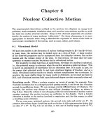
image (Fig. 83.1b) shows that there is no flow to this nodule.
Gray-scale ultrasound image of the same patient several days
later (Fig. 83.1c) shows interval increase in echogenicity of the
nodule (arrow) and increasing complexity of the hydrocele.
Longitudinal gray-scale ultrasound image of the right testis
(Fig. 83.1a) shows an enlarged, echogenic nodule located in
the groove between the epididymis and the testis (arrow).
There is a simple hydrocele (asterisk). Color flow ultrasound
a
b
c
185
Genitourinary
■■
Diagnosis
Torsion of the Appendix Testis
■■
Discussion and Differential Diagnosis
Torsion of the appendix testis is the most common cause of an
acute scrotum in children,1,2 with a prevalence of 30 to 60%.1,3
The appendix testis is a remnant of the müllerian duct and is
present in 80% of males.2–4 It is normally located in the groove
between the head of the epididymis and the superior pole of
the testis.3,4 The appendix epididymis is a wolffian duct remnant and is present in 25% of males.2–4 Patients typically pre
sent before puberty with acute onset of scrotal pain, usually
less than 12 hours in duration. On physical exam, tenderness
is usually localized to the superior pole of the testis, and the
testis should be normal to palpation.4 The “blue dot” sign is
due to the torsed appendix seen through the scrotal skin. This
is specific for a torsed appendix, but only seen in the minority
of patients.1 Torsion of a testicular or epididymal appendage is
self-limited; treatment is conservative, and symptoms gradually resolve in about a week.2,4
Sonographically, a normal appendix testis is an oval structure, isoechoic to the epididymis, located between the testis
and the head of the epididymis.3 The normal appendix epididymis is also isoechoic to the epididymis, and projects from the
head of the epididymis.3 A torsed appendix testis enlarges to
> 5 mm and can be echogenic or heterogeneous with punctate
areas of hyperechogenicity.1–5 Lack of flow within the appendix is not specific, as flow can be hard to detect in a normal
appendix.3,5 The adjacent epididymis, and occasionally the
testis, can also be enlarged and hyperemic. A reactive hydrocele is not uncommon.
The differential diagnosis for acute scrotum in the absence of
trauma in a child includes testicular torsion and epididymitis/
epididymo-orchitis. Testicular torsion can be easily differentiated sonographically due to decreased testicular blood flow.
Epididymitis/epididymo-orchitis presents more commonly
after puberty and generally has a longer duration of symptoms, ranging from 24 to 72 hours.4 On ultrasound, the epididymis is enlarged and heterogeneous, most markedly at the
head. There may be a reactive hydrocele and scrotal wall thickening. With color flow ultrasound imaging, the epididymis is
hyperemic. Epididymitis may have bacterial, viral, or postinflammatory etiology. In a young child, it can be associated
with congenital genitourinary anomalies. Ultrasound evaluation of the kidneys and bladder should be performed.1
Pearls
Pitfall
◆◆ An echogenic nodule ≥ 5 mm in the groove between the epi-
◆◆ Sonographically, torsion of the appendix testis is very difficult
didymal head and the testis is highly specific for a torsed appendix testis.5
◆◆ The “blue-dot” sign is a specific clinical finding, but only seen
20% of the time.1
to differentiate from epididymitis/epididymo-orchitis. Both have
enlargement and hyperemia of the epididymis and/or testis.
Patient age, duration of symptoms, and identification of the
enlarged appendix testis is key to diagnosing a torsed appendix
testis.
References
1. Baldisserotto M. Scrotal emergencies. Pediatr Radiol 2009;39:516–521
PubMed
2. Park SJ, Kim HL, Yi BH. Sonography of intrascrotal appendage torsion: varying
echogenicity of the torsed appendage according to the time from onset.
J Ultrasound Med 2011;30:1391–1396 PubMed
3. Baldisserotto M, de Souza JCK, Pertence AP, Dora MD. Color Doppler
sonography of normal and torsed testicular appendages in children. AJR Am
J Roentgenol 2005;184:1287–1292 PubMed
186
4. Munden MM, Trautwein LM. Scrotal pathology in pediatrics with sonographic
imaging. Curr Probl Diagn Radiol 2000;29:185–205 PubMed
5. Yang DM, Lim JW, Kim JE, Kim JH, Cho H. Torsed appendix testis: gray scale
and color Doppler sonographic findings compared with normal appendix
testis. J Ultrasound Med 2005;24:87–91 PubMed
Case 84
■■
Clinical Presentation
A term newborn with emesis, abdominal distention, and
anemia.
■■
Radiographic Studies
Longitudinal ultrasound image (Fig. 84.1a) shows a heterogeneous oval echogenic lesion superior to the right kidney
(arrowheads). A similar echogenic oval lesion is seen superior
to the left kidney (Fig. 84.1b, arrow) with no significant internal color flow vascularity. Serial ultrasound images of another
a
patient show decreasing size of a complex right suprarenal
cystic lesion (Fig. 84.1c). A similar left suprarenal cystic lesion
was noted in this patient; follow-up ultrasound at 6 weeks
showed resolution of these bilateral suprarenal lesions.
b
c
187
Genitourinary
■■
Diagnosis
Adrenal Hemorrhage
■■
Discussion and Differential Diagnosis
Adrenal hemorrhage can occur in the neonatal period and in
older age groups. Incidence of neonatal adrenal hemorrhage
ranges from 1.6 to 2.1 per 1,000 births and occurs more
commonly in term infants and in male neonates.1–3 Bilateral
hemorrhage occurs in 10 to 15% of cases.1,2 Neonatal adrenal
hemorrhage occurs in birth trauma, large-for-gestational-age
infants, infants of diabetic mothers, prolonged labor, perinatal
asphyxia, sepsis, hemorrhagic disorder, extracorporeal membrane oxygenation, and renal vein thrombosis.1 Neonates with
adrenal hemorrhage may be asymptomatic, with the lesion
detected incidentally at sonography.3,4 Alternatively, a neonate
may present acutely with fever, vomiting, jaundice, hypotension, anemia, and a palpable flank mass.3,4
Adrenal hemorrhage occurs more frequently in the right
adrenal gland, which is thought to be more vulnerable, as it
is compressed between the liver, kidney, and the spine. The
increased frequency may also be due to the fact that the right
venous drainage is directly into the inferior vena cava (IVC),
which when compressed causes a rise in the intra-adrenal
venous pressure.4 Adrenal hemorrhage may be secondary to
renal vein thrombosis, particularly on the left side.3 This association relates to the left adrenal vein anatomy, which drains
into the left renal vein.5
Traumatic adrenal hemorrhage is observed in older children
after blunt abdominal injury.3 Many mechanisms have been
postulated to explain adrenal hemorrhage in trauma, including direct trauma and compression of the gland between the
spine and liver, shearing of small vessels that perforate the adrenal capsule because of deceleration forces, and a short-term
rise in intra-adrenal venous pressure due to compression of
the IVC.6
Ultrasound is the preferred modality for initial detection
and follow-up of adrenal hemorrhage because it is portable,
rapid, sensitive, and lacks ionizing radiation. Initially the hemorrhage appears as a solid echogenic mass superior to the kidney; the lesion decreases in size and echogenicity over time.
The primary differential diagnosis of an echogenic adrenal mass
in a neonate is neuroblastoma, which will have branching
color flow vascularity within the mass on Doppler. Eventually
an adrenal hemorrhage will liquefy and become cystic and
multiloculated; the lesion completely resolves within 4 to 16
weeks.1 Peripheral calcifications can develop and may be seen
incidentally on plain abdominal radiographs and CT.1,3
Computed tomography shows an oval or triangular adrenal
hematoma that is of moderate to high attenuation on noncontrast CT and relatively lower in attenuation compared with
the enhancing liver and spleen on postcontrast CT.4 Associated
CT findings include periadrenal fat stranding and thickening
of the ipsilateral diaphragmatic crus.4 Adrenal hemorrhage is
reported in 3% of children following blunt abdominal trauma
and is associated with a high frequency of ipsilateral intra-
abdominal and intrathoracic injuries.6 Adrenal hemorrhage in
nonaccidental injury indicates the use of a severe force to injure the child and has been reported to occur in 10% of deaths.7
Magnetic resonance imaging is particularly useful to distinguish between adrenal hemorrhage and other causes of a cystic adrenal mass on ultrasound. Most importantly, MRI helps
differentiate adrenal hemorrhage from cystic neuroblastoma
in a neonate. Other differential diagnoses of adrenal hemorrhage include adrenal abscess, cortical renal cyst, obstructed
calyceal diverticulum, and an obstructed upper moiety collecting system in a duplicated kidney.1
Pearls
Pitfall
◆◆ Serial ultrasound is an acceptable method to differentiate
◆◆ Hemorrhage into a congenital neuroblastoma may make differ-
neuroblastoma from adrenal hemorrhage. Hemorrhage will
decrease in size, whereas neuroblastoma will remain stable or
increase in size.
◆◆ Adrenal hemorrhage occurs more commonly in the right adrenal gland.
◆◆ With right adrenal hemorrhage, always look carefully for a clot
in the adjacent IVC. With left adrenal hemorrhage, look carefully for left renal vein thrombosis.
entiation between adrenal hemorrhage and neuroblastoma
very difficult. Metaiodobenzylguanidine (MIBG) scintigraphy
and urinary catecholamines may help distinguish these two entities but may be equivocal in the newborn.
References
1. Mutlu M, Karagüzel G, Aslan Y, Cansu A, Okten A. Adrenal hemorrhage in
newborns: a retrospective study. World J Pediatr 2011;7:355–357 PubMed
2. Demirel N, Baş AY, Zenciroğlu A, Taşci-Yildiz Y. Adrenal bleeding in neonates:
report of 37 cases. Turk J Pediatr 2011;53:43–47 PubMed
3. Westra SJ, Zaninovic AC, Hall TR, Kangarloo H, Boechat MI. Imaging of the
adrenal gland in children. Radiographics 1994;14:1323–1340 PubMed
4. Paterson A. Adrenal pathology in childhood: a spectrum of disease. Eur Radiol
2002;12:2491–2508 PubMed
188
5. Orazi C, Fariello G, Malena S, Schingo P, Ferro F, Bagolan P. Renal vein
thrombosis and adrenal hemorrhage in the newborn: ultrasound evaluation
of 4 cases. J Clin Ultrasound 1993;21:163–169 PubMed
6. Sivit CJ, Ingram JD, Taylor GA, Bulas DI, Kushner DC, Eichelberger MR.
Posttraumatic adrenal hemorrhage in children: CT findings in 34 patients.
AJR Am J Roentgenol 1992;158:1299–1302 PubMed
7. deRoux SJ, Prendergast NC. Adrenal lacerations in child abuse: a marker of
severe trauma. Pediatr Surg Int 2000;16:121–123 PubMed
Case 85
■■
Clinical Presentation
A 2-year-old with abdominal mass.
■■
Radiographic Studies
Longitudinal and transverse ultrasound images (Fig. 85.1a,b)
show a large, heterogeneous, predominantly echogenic mass
infiltrating much of the kidney. Echogenic renal fat and normal-appearing kidney can be seen draped over the renal mass
(Fig. 85.1a, arrow). Postcontrast CT image (Fig. 85.1c) shows a
a
well-demarcated mass of renal origin, with functioning renal
parenchyma displaced medially and anteriorly (arrows, “claw
sign”). Postcontrast coronal reformatted CT image (Fig. 85.1d)
shows significant low density within the mass (asterisk) related to necrosis or hemorrhage.
b
c
d
189
Genitourinary
■■
Diagnosis
Wilms’ Tumor
■■
Discussion and Differential Diagnosis
Wilms’ tumor is the most common pediatric malignant renal
tumor and represents 8% of all childhood malignancies. The
most common presentation is an asymptomatic abdominal
mass. Peak incidence occurs at 3 to 4 years of age, and 4 to 13%
are bilateral. Associated anomalies include aniridia, hemi
hypertrophy, cryptorchidism, and hypospadias.1
Ultrasound is the screening modality of choice showing a
heterogeneous intrarenal echogenic mass displacing or splaying the collecting system. Doppler sonography is very useful in
demonstrating tumor extension into the renal vein and inferior vena cava. CT is useful for defining the organ of origin,
detecting nodal metastases, and identifying tumor thrombus.
Tumor thrombus may extend into the renal vein, the inferior
vena cava, and the right atrium, occasionally causing inferior
vena caval obstruction and pulmonary tumor emboli.2
Search for bilateral Wilms’ tumor and consideration of
nephroblastomatosis is imperative. Nephroblastomatosis in-
cludes rests of nephrogenic tissue or renal blastoma that
resemble Wilms’ tumor microscopically but lack mitosis.3
Nephroblastomatosis complex usually presents as a solid oval
lesion, generally in a subcapsular location. Follow-up imaging
is required because nephroblastomatosis is a precursor of
Wilms’ tumor.
Wilms’ tumor must be distinguished from neuroblastoma,
an extrarenal tumor, which involves adjacent lymph nodes,
encases vascular structures, and may extend across the midline.3 Mesoblastic nephroma, a neonatal renal mass, is distinguished by earlier clinical presentation but is indistinguishable
from Wilms’ tumor on ultrasound and CT. Renal cell carcinoma is a nonspecific solid renal mass indistinguishable on
ultrasound from Wilms’ tumor, but usually occurs in older
patients. Multilocular cystic nephroma, a complex cystic lesion, is difficult to distinguish from the atypical cystic Wilms’
except by pathological exam.
Pearls
Pitfall
◆◆ Familial Wilms’ tumor accounts for 1% of cases; genetic associ-
◆◆ The combination of renal mass with brain lesion favors rhab-
ation is seen in Wilms’ tumor occurring with syndromes such as
Beckwith-Wiedemann or the WAGR syndrome (Wilms’ tumor,
aniridia, genitourinary anomalies, and mental retardation).2
◆◆ Left renal vein tumor thrombus may obstruct the left gonadal
vein, causing a varicocele, a rare initial presentation of Wilms’
tumor.4
◆◆ If a right atrial tumor thrombus is identified, cardiopulmonary
bypass may be necessary at the time of tumor resection.4
doid tumor over Wilms’ tumor.
References
1. Green DM, D’Angio GJ, Beckwith JB, et al. Wilms tumor. CA Cancer J Clin
1996;46:46–63 PubMed
2. Lowe LH, Isuani BH, Heller RM, et al. Pediatric renal masses: Wilms tumor
and beyond. Radiographics 2000;20:1585–1603 PubMed
190
3. White KS, Grossman H. Wilms’ and associated renal tumors of childhood.
Pediatr Radiol 1991;21:81–88 PubMed
4. Swinson S, McHugh K. Urogenital tumours in childhood. Cancer Imaging
2011;11(Spec. No. A):S48–S64 PubMed
Case 86
■■
Clinical Presentation
A 5-month-old infant with vomiting, fever, and urinary tract
infection.
■■
Radiographic Studies
Longitudinal ultrasound image of the right kidney (Fig. 86.1a)
shows dilated pelvicaliceal systems separated by renal parenchyma into lower pole (arrowhead) and upper pole (asterisk)
moieties. Increased echogenicity of the upper pole moiety
renal parenchyma is noted. Longitudinal pelvic ultrasound
image (Fig. 86.1b) shows a dilated upper pole moiety ureter
(arrowheads) terminating in a cystic ureterocele in the bladder
a
(arrow). A dilated lower pole moiety ureter is also seen (asterisk). Nuclear medicine renogram in a different patient, a
1-year-old girl with left hydronephrosis (Fig.86.1c), shows
delayed excretion from the left upper pole moiety collecting
system (arrowhead) with subsequent delayed Lasix washout of
tracer (arrow) compatible with upper pole moiety obstruction.
b
c
191
Genitourinary
d
■■
e
f
Diagnosis
Collecting System Duplication/Ectopic Ureter/Ureterocele
■■
Discussion and Differential Diagnosis
A duplicated urinary collecting system is the most common
anomaly of the urinary tract.1 With incomplete duplication,
the two pelvicaliceal systems may join at the level of renal pelvis (bifid pelvis) or may drain into separate ureters that will
join at any level before draining into the urinary bladder.1,2
With complete duplication, the two pelvicaliceal systems drain
into separate ureters that do not join at any level; each enters
the urinary bladder separately.1,2 The lower pole moiety ureter joins the urinary bladder at the normal position on the
trigone.2 The ureter arising from the upper pole moiety drains
into an abnormal location more inferiorly and is referred to as
ectopic ureter.2 Sometimes this ectopic insertion is into the
prostatic urethra in males or vagina in females. These patients
present with urinary dribbling. Girls are affected about twice
as often as boys.1
Collecting system duplication may go undiagnosed into
adulthood. Clinical significance arises when it is complicated
by ureteral ectopia, vesicoureteral reflux (VUR), urinary tract
infection, or ureteropelvic junction (UPJ) obstruction. VUR is
the most commonly detected association, and it is more common with complete than incomplete duplication.2 The incidence of ectopic urterocele is 20%.1 Ureterocele is dilatation
of the intravesical component of the upper pole ureter. It is
usually associated with obstructive dilatation of the associated ureter/calices.
Ultrasound demonstrates a prominent segment of renal cortex between the two duplicated intrarenal collecting systems.2
The upper pole moiety and its ureter may be dilated and tor-
192
tuous on ultrasound secondary to obstruction at the insertion
into the bladder. The lower pole moiety may also be dilated
secondary to vesicoureteral reflux or UPJ obstruction.2,3 Ureterocele on ultrasound appears as a round intraluminal fluid-
filled structure.2 Ureterocele on voiding cystourethrogram
(VCUG) appears as a round filling defect in the expected location of the ureteral orifice (Fig. 86.1d, arrowheads). VCUG is
performed to document the presence and severity of reflux.
Higher grades of reflux correspond with higher rates of urinary tract infection.2 The classic VCUG appearance of reflux
is the inferior displacement of a contrast-filled lower pole
calyx by an obstructed and enlarged upper pole moiety, the
so-called drooping lily sign (Fig. 86.1e).
More recent advances have made CT urography and magnetic resonance (MR) urography useful in children.3 The excretory phase of a CT urogram protocol can be performed to
answer a specific question when a congenital anomaly is suspected.3 MR urography is superior to CT in that it provides
better tissue contrast resolution and does not utilize ionizing
radiation. This is especially favored in children, particularly
those who may need repeated examinations.3 Both anatomic
and functional information about the urinary tract can be
obtained in a single examination, potentially eliminating the
need for nuclear scintigraphy.3 Fig. 86.1f is an oblique MR
urography image in a 5-year-old girl with constant wetting
and shows duplicated collecting systems (arrowheads) and ectopic insertion of the upper pole moiety ureter (arrow) into
the vagina.
Genitourinary
Pearls
Pitfall
◆◆ Weigert-Meyer rule: In complete duplication, the upper pole
◆◆ Voiding cystourethrogram should be performed with small
moiety ureter almost always inserts in an ectopic location into
the bladder medially and inferiorly to the lower pole moiety
ureter (which inserts in the normal location in the trigone of
bladder).
◆◆ The ectopic upper pole moiety ureter is more prone to obstruction; the lower pole moiety ureter is more prone to vesicoureteral reflux.
amounts of dilute contrast to search for ureterocele early in the
filling phase of the exam. Dense contrast or a completely filled
bladder may completely obscure the ureterocele.
References
1. Siomou E, Papadopoulou F, Kollios KD, et al. Duplex collecting system
diagnosed during the first 6 years of life after a first urinary tract infection: a
study of 63 children. J Urol 2006;175:678–681, discussion 681–682 PubMed
2. Fernbach SK, Feinstein KA, Spencer K, Lindstrom CA. Ureteral duplication and
its complications. Radiographics 1997;17:109–127 PubMed
3. Silverman SG, Leyendecker JR, Amis ES Jr. What is the current role of CT
urography and MR urography in the evaluation of the urinary tract?
Radiology 2009;250:309–323 PubMed
193
Case 87
■■
Clinical Presentation
Male neonate with obstructive urinary symptoms.
■■
Radiographic Studies
Ultrasound image of the bladder (Fig. 87.1a) shows mild bladder wall thickening and a distended bladder. Dilated ureter is
seen posterior to the bladder (arrow). Longitudinal ultrasound
images of the kidneys (Fig. 87.1b,c) show mildly echogenic
kidneys bilaterally (right worse than left). There is grade 4 hydronephrosis on the right and grade 1 hydronephrosis on the
a
left. AP image from a VCUG (Fig. 87.1d) shows bilateral vesicoureteral reflux, right grade 4 and left grade 2. Oblique voiding
image from VCUG (Fig. 87.1e) shows a dilated posterior urethra (arrow) with a nondilated anterior urethra. Sagittal prenatal MRI in another patient (Fig. 87.1f) shows a thick-walled
distended bladder. The posterior urethra is dilated (arrow).
b
c
d
195
Genitourinary
e
■■
f
Diagnosis
Posterior Urethral Valves
■■
Discussion and Differential Diagnosis
Posterior urethral valves (PUVs) are the most common cause
of lower urinary tract obstruction in males. Despite the rarity,
PUVs are an important cause of end-stage renal disease, accounting for almost 17% of children with renal failure.1 The
most common clinical presentation of PUV is detection of hydronephrosis on routine prenatal sonography.1 Concurrent
ultrasound findings of dilated bladder, dilated posterior urethra, and oligohydramnios in a male fetus confirm the diagnosis. If not prenatally detected, a neonate with PUV may present
with abdominal mass (due to hydronephrosis or distended
bladder), urinary ascites, or respiratory distress. An older child
may present with urinary tract infections, failure to thrive, or
urinary insufficiency.1,2 PUVs cause urine outflow obstruction
with secondary bladder wall hypertrophy and trabeculation.
Massive hydronephrosis is common, secondary to either vesicoureteral reflux or obstruction. In either case, renal dysplasia
is common with chronic renal failure and end-stage renal disease. Even after ablation of the valves, hydronephrosis may
persist secondary to a hypertrophied noncompliant bladder.3
Persistent reflux after valve ablation can be due to ureteral dysplasia, peristaltic failure from ureteral fibrosis, or decreased
peristaltic force in a dilated tortuous ureter.4
Definitive postnatal diagnosis of PUVs is usually made with
a voiding cystourethrogram, which delineates the urethral
obstruction and searches for vesicoureteral reflux. Findings
196
suggesting good prognosis are unilateral high-grade reflux,
perinephric urinomas, and urinary ascites; these are believed
to represent a pressure “pop-off,” helping to preserve renal
function.2 Renal ultrasound may demonstrate hydronephrosis
and hydroureter from obstruction or reflux, a thickened bladder wall, and a dilated posterior urethra (keyhole appearance).
Rarely one can see a linear echogenic structure within it, representing the posterior urethral valve membrane.2,5 Transperineal ultrasound delineating the linear echogenic structure
representing the valves within the urethra helps differentiate
patients with PUVs from patients with urethral strictures.5
Other findings are echogenic kidneys and renal cysts, indicating renal dysplasia.2 Nuclear medicine studies may be useful
in assessing renal function and the degree of residual obstruction after valve ablation.
The differential diagnosis includes bilateral ureteropelvic
junction obstruction or bilateral ureterovesical obstruction;
these patients usually have a normal bladder. There are many
normal variants to the appearance of the male urethra, and
nonobstructed urethral ectasia or kinking should not be confused with PUVs.3 Prune-belly syndrome is indistinguishable
from PUVs in utero, but, at birth, the deficiency of abdominal
musculature confirms the diagnosis of prune-belly syndrome.
The urethra in prune-belly syndrome may be dilated but is not
elongated.
Genitourinary
Pearls
Pitfall
◆◆ Transient hydronephrosis of the newborn may be a normal
◆◆ Urethral strictures can be confused with PUVs, but strictures
variant seen on prenatal ultrasound and may resolve
spontaneously.3
◆◆ Of patients with PUVs, 50% have vesicoureteral reflux at presentation; 50% of these patients have bilateral reflux.
◆◆ A male newborn who presents with isolated ascites most likely
has PUVs.
■■
are usually irregular in outline and in a different location. Urethral strictures are inflammatory, congenital, traumatic, or iatrogenic in origin.3
Controversies
• In older children, primary valve ablation through a transurethral approach is usually the treatment of choice. If
hydronephrosis persists and renal function worsens, upper
tract diversion may be necessary.1 Many of these patients
go on to renal transplant.
• If the urethra is too small to accommodate the urethroscope/
cystoscope, temporary urinary diversion with vesicostomy
may be necessary until the urethra grows larger and the
patient is more stable.1
• Selected cases may undergo antenatal treatment to prevent pulmonary hypoplasia and preserve renal function.
Vesicoamniotic shunts and fetal cystoscopic valve ablation
have been reported.6
References
1. Hodges SJ, Patel B, McLorie G, Atala A. Posterior urethral valves. Scientific
WorldJournal 2009;9:1119–1126 PubMed
2. Perks AE, MacNeily AE, Blair GK. Posterior urethral valves. J Pediatr Surg
2002;37:1105–1107 PubMed
3. Macpherson RI, Leithiser RE, Gordon L, Turner WR. Posterior urethral valves:
an update and review. Radiographics 1986;6:753–791 PubMed
4. Lal R, Bhatnagar V, Mitra DK. Upper-tract changes after treatment of posterior
urethral valves. Pediatr Surg Int 1998;13:396–399 PubMed
5. Cohen HL, Zinn HL, Patel A, Zinn DL, Haller JO. Prenatal sonographic diagnosis
of posterior urethral valves: identification of valves and thickening of the
posterior urethral wall. J Clin Ultrasound 1998;26:366–370 PubMed
6. Chauvin NA, Epelman M, Victoria T, Johnson AM. Complex genitourinary
abnormalities on fetal MRI: imaging findings and approach to diagnosis. AJR
Am J Roentgenol 2012;199:W222-31 PubMed
197
Case 88
■■
Clinical Presentation
A 4-year-old boy with fever, abdominal pain, midline hypo
gastric mass, and urinary frequency.
■■
Radiographic Studies
Longitudinal pelvic ultrasound image (Fig. 88.1a) shows a
complex oval lesion (arrowheads) with mixed internal echogenic debris. The lesion abuts the bladder dome (asterisk). The
a
thickened cyst wall is hyperemic on color flow imaging (Fig.
88.1b). An associated tract extending to the umbilicus is seen
(Fig. 88.1c, arrowheads).
b
c
199
Genitourinary
■■
Diagnosis
Urachal Anomalies
■■
Discussion and Differential Diagnosis
The urachus is the embryonic remnant of the allantois and the
cloaca that lies in the midline between the transverse fascia
and the peritoneum.1,2 In the fetus, the urachus joins the bladder dome to the umbilicus, but the lumen obliterates at between 4 and 5 months of gestation as the bladder descends
into the pelvis.1–4 The fibrous remnant forms the median umbilical ligament.
Urachal anomalies are classified into four subgroups. A completely patent urachus is secondary to failure of obliteration of
the urachal channel in the fetus and represents 16% of urachal
anomalies.3 A patent urachus frequently presents in the newborn with urine leakage at the umbilicus.2 About one third of
these patients have an associated bladder outlet obstruction
such as posterior urethral valves or urethral atresia.2 Diagnosis
can be made with umbilical orifice contrast injection to show
communication to the bladder.1,2 Ultrasound often demonstrates a fluid-containing channel between the dome of the
bladder and the umbilicus. Voiding cystourethrography (VCUG)
shows contrast filling of the patent channel with drainage at
the umbilicus.
The second subgroup of urachal anomalies includes patients
with a blind-ending urachal sinus tract that opens into the
umbilicus and comprises 37% of urachal anomalies. Symptoms
may occur at any age and are usually secondary to infected
discharge from the sinus.2 The diagnosis is usually made by
contrast injection into the sinus orifice at the umbilicus.1 Ultrasound may show a thickened tubular structure below the
umbilicus in the midline.2
The third subgroup of urachal anomalies includes patients
with the rare urachal diverticulum, which is a cystic structure
Pearls
Pitfall
◆◆ A patent urachal anomaly should be suspected if there is a
◆◆ An ovarian cyst on a long stalk may mimic an urachal cyst, and
history of umbilical cord cyst, edematous umbilical cord, delayed sloughing of the umbilical cord, or umbilical soft tissue
protrusion.5
◆◆ Analysis of the umbilical drainage fluid for blood urea nitrogen
(BUN) and creatinine may confirm the diagnosis of a patent
urachal anomaly.
■■
final diagnosis may depend on surgical pathology.
Controversy
• Traditional thought has held that urachal remnants should
uniformly be surgically resected due to the risk of recurrent
infection and the small risk of urachal carcinoma. However,
recent studies have shown that asymptomatic urachal
remnants often spontaneously resolve in children younger
than 1 year of age.6 Infected urachal remnants may be
200
connecting with the anterior-superior aspect of the bladder.2
Diverticula are usually detected incidentally on ultrasound or
CT for unrelated reasons.2 They can be associated with or complicated by infection, stone formation, or increased risk of carcinoma in teenage.
The most common subgroup of urachal anomalies is the
urachal cyst, which accounts for 45% of reported urachal
anomalies.3,4 The cysts form within the isolated urachal channel, and the lumen enlarges over time secondary to des
quamated or degenerative tissue.2 Urachal cysts are usually
asymptomatic, but can become symptomatic when they enlarge or become infected. If direct communication with the
bladder occurs, infection is common. Complications include
rupture of the infected cysts into the preperitoneal tissues and
rupture into the intraperitoneal tissues with secondary peritonitis.2 On ultrasound or CT imaging, the urachal cyst is seen
as a fluid-filled structure in the midline below the umbilicus,
just deep to the rectus muscle. When infected, complicated
fluid, cyst wall thickening, and surrounding inflammatory
changes are seen.2
Urachal neoplasms, such as fibromas, fibroadenomas, fibromyomas, and hamartomas, are extremely rare in children and
are usually benign.2 Urachal carcinoma usually occurs in patients
40 to 70 years of age and is predominantly adenocarcinoma.2
The differential diagnosis of urachal cyst includes ovarian
cyst and mesenteric cyst. Differentiation can usually be made
based on location/configuration of the anomaly combined
with imaging. The differential diagnosis for symptomatic urachal anomalies includes appendicitis, omphalitis, and inflamed
umbilical stump.3
treated with drainage and antibiotics and followed with
clinical examination and serial ultrasound for resolution.4
If the urachal remnant does not resolve after 6 months of
age or if symptoms persist, then operative management
should be considered.4
Genitourinary
References
1. Cappele O, Sibert L, Descargues J, Delmas V, Grise P. A study of the anatomic
features of the duct of the urachus. Surg Radiol Anat 2001;23:229–235
PubMed
2. Yu J-S, Kim KW, Lee H-J, Lee Y-J, Yoon C-S, Kim M-J. Urachal remnant diseases:
spectrum of CT and US findings. Radiographics 2001;21:451–461 PubMed
3. Yiee JH, Garcia N, Baker LA, Barber R, Snodgrass WT, Wilcox DT. A diagnostic
algorithm for urachal anomalies. J Pediatr Urol 2007;3:500–504 PubMed
4. Galati V, Donovan B, Ramji F, Campbell J, Kropp BP, Frimberger D. Management of urachal remnants in early childhood. J Urol 2008;180(4, Suppl):
1824–1826, discussion 1827 PubMed
5. Frazier HA, Guerrieri JP, Thomas RL, Christenson PJ. The detection of a patent
urachus and allantoic cyst of the umbilical cord on prenatal ultrasonography.
J Ultrasound Med 1992;11:117–120 PubMed
6. Ueno T, Hashimoto H, Yokoyama H, Ito M, Kouda K, Kanamaru H. Urachal
anomalies: ultrasonography and management. J Pediatr Surg 2003;38:
1203–1207 PubMed
201
Case 89
■■
Clinical Presentation
A 16-year-old girl with crampy abdominal pain and delayed
onset of menses.
■■
Radiographic Studies
Longitudinal and transverse ultrasound images of the pelvis
(Fig. 89.1a,b) show fluid and echogenic debris within the
vagina (asterisk). Ultrasound image from a different patient,
a newborn (Fig. 89.1c), shows an oval cystic pelvic structure
(asterisk) with layering internal debris. Mass effect on the pos-
a
terior aspect of the bladder is noted. Fluoroscopic contrast
study (Fig. 89.1d) demonstrates communication between the
dilated vagina (asterisk) and bladder in the same patient as in
Fig. 89.1c. This newborn patient had cloacal malformation.
b
c
d
203
Genitourinary
■■
Diagnosis
Hydrocolpos
■■
Discussion and Differential Diagnosis
Hydrocolpos is the term for a dilated, fluid-filled vagina, typically resulting from a congenital vaginal obstruction.1 Hydrometrocolpos is present if there is dilatation of the uterus as
well as the vagina. In most cases, the obstruction is secondary
to an imperforate hymen, and simple lysis of the hymen provides resolution of the problem.2,3 In more complex forms,
vaginal atresia or stenosis is the cause of the obstruction.
These patients may have other complicated congenital anomalies, including anorectal malformation, cloacal anomalies, a
common urogenital sinus, duplicated/septated uterus or vagina, rectovaginal fistula, and renal anomalies with hydronephrosis.4–6 These conditions are obviously more complicated,
and correction of hydrocolpos or hydrometrocolpos must be
coordinated with correction of the other complex anomalies.
At the time of diagnosis, it is important to evaluate the upper
urinary tract for obstruction. Hydronephrosis and hydroureter
may be present due to compression of the distal ureters and
subsequent obstruction by the large pelvic mass.5
The typical patient is a female newborn with a palpable
elvic/abdominal mass. Diagnosis of more complex anomalies
p
is often made prenatally. With simple imperforate hymen,
diagnosis may be delayed, and the patient may present at puberty, when there is an increased accumulation of menstrual
blood and secretions causing uterine and vaginal distention.3
The differential diagnosis includes ovarian cysts, mesenteric
cysts, duplication cysts, anterior meningoceles, and sacrococcygeal teratomas.3,6 In patients with ovarian cysts and mesenteric cysts, the uterus and vagina are usually identified by
ultrasound but may be difficult to visualize because of severe
compression or displacement. Ultrasound of patients with anterior meningocele and sacrococcygeal teratomas usually shows
that the masses are related to the sacrum or distal neural
canal. In patients whose diagnosis is uncertain, or if further
delineation of adjacent structures is necessary, an MRI of the
pelvis may prove helpful.
Pearls
Pitfalls
◆◆ In isolated vaginal outlet obstruction, fluid in the vagina is
◆◆ If the bladder is completely empty, or markedly compressed
usually simple, but can contain small amounts of echogenic
debris due to mucus or debris.1,6 If there are associated complex anomalies, the fluid is more likely to be inhomogeneous,
with clumps of echogenic debris and septations. Multiple cystic
structures can be seen with cloacal anomalies.1
◆◆ In patients with associated congenital anomalies, the vagina
and uterus may be septated/duplicated.1
◆◆ A female with an imperforate anus with a single perineal orifice
has a cloacal anomaly. If there is an associated abdominal mass,
it is most likely hydrocolpos.7
against the anterior abdominal wall, it may not be identified on
ultrasound. In these patients, the dilated vagina/uterus may be
confused with dilated bladder.6
◆◆ Errors in diagnosis can result in unnecessary laparotomy or hysterectomy, which have reported mortality up to 50%.8
References
1. Dhombres F, Jouannic JM, Brodaty G, Bessiere B, Daffos F, Bénifla JL.
Contribution of prenatal imaging to the anatomical assessment of fetal
hydrocolpos. Ultrasound Obstet Gynecol 2007;30:101–104 PubMed
2. Elsayes KM, Narra VR, Dillman JR, et al. Vaginal masses: magnetic resonance
imaging features with pathologic correlation. Acta Radiol 2007;48:921–
933 PubMed
3. Yildirim G, Gungorduk K, Aslan H, Sudolmus S, Ark C, Saygin S. Prenatal
diagnosis of imperforate hymen with hydrometrocolpos. Arch Gynecol
Obstet 2008;278:483–485 PubMed
4. Subramanian S, Sharma R, Gamanagatti S, Agarwala S, Gupta P, Kumar S.
Antenatal MR diagnosis of urinary hydrometrocolpos due to urogenital sinus.
Pediatr Radiol 2006;36:1086–1089 PubMed
204
5. Breech L. Gynecologic concerns in patients with anorectal malformations.
Semin Pediatr Surg 2010;19:139–145 PubMed
6. Picone O, Laperelle J, Sonigo P, Levaillant JM, Frydman R, Senat MV. Fetal
magnetic resonance imaging in the antenatal diagnosis and management
of hydrocolpos. Ultrasound Obstet Gynecol 2007;30:105–109 PubMed
7. Levitt MA, Peña A. Anorectal malformations. Orphanet J Rare Dis 2007;
2:33 PubMed
8. Nazir Z, Rizvi RM, Qureshi RN, Khan ZS, Khan Z. Congenital vaginal
obstructions: varied presentation and outcome. Pediatr Surg Int
2006;22:749–753 PubMed
Case 90
■■
Clinical Presentation
A 15-year-old girl with right lower quadrant pain.
■■
Radiographic Studies
A CT exam was performed to evaluate the patient for appendicitis. Axial CT image (Fig. 90.1a) and coronal reformatted CT
image (Fig. 90.1b) show a large oval fluid attenuation lesion in
the abdomen and upper pelvis. Subsequent ultrasound images
(Fig. 90.1c,d) show a large oval cystic lesion with mass effect
on the bladder. The right ovary was not identified separate
from this cyst. Coronal T2-weighted prenatal MRI (Fig. 90.1e)
in another patient shows a large oval fluid signal lesion arising
from the pelvis.
a
b
c
d
e
205
Genitourinary
■■
Diagnosis
Ovarian Cyst
■■
Discussion and Differential Diagnosis
In the female neonate, ovarian cysts are the most common cystic abdominal mass.1 On ultrasound, the cysts are identified as
thin-walled cystic lesions, usually unilateral, and measure up
to 3 cm in diameter with prominent through-transmission
deep to the lesion.2 These small cysts are generally asymptomatic and usually resolve spontaneously in the weeks following
birth.3
In the fetus, follicular cysts develop under the influence of
maternal estrogen, placental human chorionic gonadotropin
(HCG), and fetal gonadotropins.1,4 There is an increased incidence of large ovarian cysts in infants of diabetic mothers, maternal toxemia, or maternal isoimmunization.1 Occasionally,
these cysts can become large; potential complications include
pulmonary hypoplasia, cyst rupture, visceral compression, and
polyhydramnios.1,2 These large cysts may be identified pre
natally and have a variable ultrasound appearance. Simple
cysts are generally follicular in origin, but more complex cysts
may be the result of torsion or hemorrhage.3 Shortly after
birth, the diagnosis of a large ovarian cyst is usually made
with ultrasound, which identifies a unilocular or septated cyst,
with or without internal echoes. The differential diagnosis of
large cystic abdominal masses includes hydronephrosis, gastrointestinal duplication cysts, mesenteric duplication cysts,
macrocystic lymphatic malformation, choledochal cysts, ante-
rior myelomeningocele, hydrocolpos, megacystis, and urachal
cysts.1,3,4 Diagnostic criteria for the sonographic diagnosis
of an ovarian cyst include female sex, a regular cystic structure off midline in location, identification of normal urinary
tract anatomy, and identification of normal gastrointestinal
structures.1
Ovarian cysts are less common in early childhood, once the
neonatal period has passed, due to low levels of gonadotropin
and estradiol.1,2 Small cysts usually result from failure of the
follicle to involute.1 Occasionally, cyst formation can be associated with precocious puberty, and larger cysts require an extensive endocrine evaluation.1,2
In the adolescent, ovarian cysts may be follicular in origin or
the result of persistence of the corpus luteum. Follicular cysts
are generally 2 to 3 cm in diameter and simple in appearance;
however, if ovulation does not occur, these cysts can continue
to grow and become very large.1 Corpus luteum cysts can also
grow up to 6 cm in size and are more likely to rupture, causing
hemorrhage.1 Resolution of both these cysts occurs spontaneously in over 90% of cases.1 The differential diagnosis for an
ovarian cyst in an adolescent includes a hemorrhagic/torsed
ovary, an ectopic pregnancy, pelvic inflammatory disease, or
endometriosis.1,2
Pearls
Pitfall
◆◆ Unilocular anechoic cysts are benign, regardless of the patient’s
◆◆ A complicated cyst can be difficult to distinguish by imaging
age or size.
◆◆ Most cysts smaller than 5 cm resolve spontaneously.
◆◆ Ovarian malignancies are extremely rare in the neonate.2
■■
alone from ovarian torsion; both entities have enlarged, echogenic ovaries.
Controversy
• Some authors advocate prenatal ovarian cyst aspiration for
large fetal ovarian cysts.1,2 Cyst aspiration is thought to
prevent complications such as pulmonary hypoplasia,
torsion, and rupture.2 However, cyst reaccumulation is
common, and there is a risk of hemorrhage into the cyst
or aspirating a cyst of nonovarian origin.1
References
1. Brandt ML, Helmrath MA. Ovarian cysts in infants and children. Semin Pediatr
Surg 2005;14:78–85 PubMed
2. Strickland JL. Ovarian cysts in neonates, children and adolescents. Curr Opin
Obstet Gynecol 2002;14:459–465 PubMed
206
3. Akın MA, Akın L, Özbek S, et al. Fetal-neonatal ovarian cysts—their monitoring and management: retrospective evaluation of 20 cases and review of the
literature. J Clin Res Pediatr Endocrinol 2010;2:28–33 PubMed
4. Helmrath MA, Shin CE, Warner BW. Ovarian cysts in the pediatric population.
Semin Pediatr Surg 1998;7:19–28 PubMed
Case 91
■■
Clinical Presentation
A 3-year-old boy with weight loss and irritability.
■■
Radiographic Studies
Longitudinal ultrasound image (Fig. 91.1a) shows a heterogeneous solid mass (arrowheads) between the kidney and the
liver. Postcontrast coronal reformatted CT image (Fig. 91.1b)
shows lateral displacement of the right kidney by the paraspinal mass; internal stippled calcifications are noted (arrow).
Postcontrast CT image (Fig. 91.1c) shows mild enhancement
of the mass, which invades the spinal canal (arrow). Axial
T2-weighted MRI (Fig. 91.1d) depicts spinal canal extent and
a
c
thecal sac displacement (arrow) more clearly. Postcontrast
CT image in another patient (Fig. 91.1e) shows a large heterogeneously enhancing retroperitoneal mass that encases
the vessels, crosses the midline, and displaces the aorta and
IVC from the spine. On whole-body nuclear medicine metaiodobenzylguanidine (MIBG) imaging (Fig. 91.1f), there is
increased uptake in the abdominal paraspinal mass and scattered throughout the skeleton.
b
d
207