Ebook Robbins and cotran review of pathology (4th edition): Part 2
Bạn đang xem bản rút gọn của tài liệu. Xem và tải ngay bản đầy đủ của tài liệu tại đây (14.61 MB, 241 trang )
CHAPTER
Head and Neck
16
PBD9 Chapter 16 and PBD8 Chapter 16: Head and Neck
BP9 Chapter 14 and BP8 Chapter 15: Oral Cavity and Gastrointestinal Tract
1 A 47-year-old man sees his dentist for a routine checkup.
He states that his gums bleed easily on brushing his teeth. On
examination, he is found to have marked gingival recession
with erythema, along with extensive plaque and calculus formation over tooth surfaces. Which of the following organisms
is most likely to be associated with development of his oral
lesions?
A
Actinobacillus
B
Candida
C
Epstein-Barr virus
D
Herpes simplex virus
E
Human papillomavirus
F
Mucor circinelloides
2 A 17-year-old girl notices a small, sensitive, gray-white
area forming along the lateral border of her tongue 2 days before the end of her final examinations. On examination by the
physician’s assistant, the girl is afebrile. There is a shallow, ulcerated, 0.3-cm lesion with an erythematous rim. No specific
therapy is given, and the lesion disappears within 2 weeks.
The history shows that the girl does not use tobacco or alcohol.
Which of the following is the most probable diagnosis?
A
Aphthous ulcer
B
Herpes simplex stomatitis
C
Leukoplakia
D
Oral thrush
E
Sialadenitis
3 A 55-year-old woman notes a nodule while rubbing her
tongue on the side of her mouth. On physical examination by
her dentist, there is a firm, nontender 0.6-cm nodule covered
by pink buccal mucosa at the bite line next to the first molar on
the lower right. The lesion is excised and does not recur. What
is the most likely diagnosis?
A
Candidiasis
B
Fibroma
C
Leukoplakia
D
Pyogenic granuloma
E
Sialadenitis
4 A 23-year-old primigravida has noticed a rapidly enlarging nodule next to a tooth for the past 16 days. On physical
examination there is a 1-cm, soft, reddish, pedunculated mass
above a left upper bicuspid. She is advised that the lesion will
likely regress. Which of the following pathologic findings is
most likely found in this lesion?
A
Granulation tissue
B
Lymphoid proliferation
C
Neutrophilic exudate
D
Rhabdomyosarcoma
E
Squamous hyperplasia
5 A 25-year-old man notices several 0.3-cm, clear vesicles
on his upper lip after a bout of influenza. The vesicles rupture,
leaving shallow, painful ulcers that heal over the course of 10
days. Three months later, after a skiing trip, similar vesicles
develop, with the same pattern of healing. Which of the following microscopic findings is most likely to be associated
with these lesions?
A
Budding cells with pseudohyphae
B
Mononuclear inflammatory infiltrates
C
Neutrophils within abscesses
D
Squamous epithelial hyperkeratosis
E
Intranuclear inclusions
6 A 35-year-old, HIV-positive man complains that he has
had a “bad” taste in his mouth and discoloration of his tongue
for the past 6 weeks. On physical examination, there are areas
of adherent, yellow-to-gray, circumscribed plaque on the lateral aspects of the tongue. This plaque can be scraped off as a
pseudomembrane to show an underlying granular, erythematous base. What is the most likely diagnosis?
A
Aphthous ulcer
B
Cheilosis
C
Hairy leukoplakia
D
Herpetic stomatitis
E
Leukoplakia
F
Oral thrush
253
254
U N I T I I Diseases of Organ Systems
7 A 42-year-old man has had a constant bad taste in his
mouth for the past month. On physical examination there are
white fluffy patches on the sides of his tongue. These cannot
be scraped off. A biopsy is taken and on microscopic examination shows squamous epithelial hyperkeratosis, parakeratosis,
and koilocytosis. Immunohistochemical staining for EpsteinBarr virus (EBV) is positive. Which of the following is the most
likely risk factor for his oral lesions?
A
Chronic alcohol abuse
B
Diabetes mellitus
C
HIV infection
D
Pernicious anemia
E
Sjögren syndrome
9 A 51-year-old man from Kolkata has an area of depression in his mouth that has enlarged over the past 7 months.
On oral examination, there is a 1.5 × 0.7 cm velvety, erythematous area with focal surface erosion on his left buccal mucosa.
The lesion is excised and on microscopic examination there is
dysplastic squamous epithelium. Which of the following is the
most likely risk factor for developing this lesion?
A
Candidiasis
B
Dental malocclusion
C
Epstein-Barr virus infection
D
Immunosuppression
E
Eating hot, spicy food
F
Tobacco chewing
A
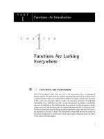
10 A 49-year-old man has used chewing tobacco and snuff
for many years. On physical examination the lesion shown in
the figure is seen on the hard palate. It cannot be removed by
scraping. A biopsy is performed, and microscopic examination
of the lesion shows a thickened squamous mucosa. Four years
later, a biopsy specimen of a similar lesion shows carcinoma in
situ. Which of the following is the most likely diagnosis?
A
Oral thrush
B
Lichen planus
C
Leukoplakia
D
Pyogenic granuloma
E
Xerostomia
B
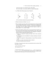
8 A 58-year-old man, a cigar smoker, visited his dentist for
a routine dental examination. The dentist noticed lesions with
the clinical (A) and histologic (B) appearance shown in the figure. The medical history showed no major medical problems.
Which of the following etiologic factors most likely contributed to the development of these lesions?
A
Chronic sialadenitis
B
Dental caries
C
Eating smoked foods
D
Herpes simplex virus type 1
E
Smoking tobacco
11 A 54-year-old man, a nonsmoker, has a nonhealing ulceration at the base of his tongue on the right side for
2 months. On examination this lesion is 1 cm in diameter
with irregular borders. Biopsy of the lesion is performed and
microscopic examination shows infiltrating squamous cell carcinoma. Which of the following infectious agents is most likely
to be associated with this lesion?
A
Candida albicans
B
Herpes simplex virus (HSV)
C
Human papillomavirus (HPV)
D
Prevotella intermedia
E
Group A streptococcus
C H A P T E R 1 6 Head and Neck
255
12 A 19-year-old woman has noted swelling in the back of
her mouth for 2 months. On dental examination, she has an
area of swelling in the location of the left third molar. Dental
radiographs show a radiolucent unilocular, well-circumscribed
cyst surrounding the crown of the unerupted third mandibular
molar. The lesion is excised, and on microscopic examination,
the cyst is lined by stratified squamous epithelium and surrounded by a chronic inflammatory infiltrate. What is the most
likely diagnosis?
16 On December 13, 1799, George Washington, recently
retired as first President of the United States, developed a
“cold” with mild hoarseness. By the next morning he had difficulty breathing and swallowing, with throat pain. He was
treated with the usual therapy of the time: bloodletting. Had
vital signs been recorded, they may have shown temperature
of 37.8° C, pulse 115/min, respiratory rate 24/min, and blood
pressure 90/60 mm Hg. Which of the following organisms
most likely caused his illness?
A
Ameloblastoma
B
Dentigerous cyst
C
Odontogenic keratocyst
D
Odontoma
E
Periapical cyst/granuloma
A
Coronavirus
B
Corynebacterium diphtheriae
C
Haemophilus influenzae
D
Parainfluenza virus
E
Prevotella intermedia
F
Group A streptococcus
13 A 19-year-old man noted progressive swelling on the
left side of his face over the past year. On physical examination, there is painless swelling in the region of the left posterior
mandible. Head CT scan shows a circumscribed multilocular
cyst of the left mandibular ramus. The lesion is surgically
excised with wide bone margins. On microscopic examination,
the lesion shows cysts lined by stratified squamous epithelium
with a prominent basal layer; no inflammation or granulation
tissue is seen. What is the most likely diagnosis?
A
Ameloblastoma
B
Dentigerous cyst
C
Odontogenic keratocyst
D
Odontoma
E
Periapical cyst/granuloma
14 A 26-year-old man has had difficulty breathing through
his nose for 3 years, but this problem has become progressively worse over the past 2 months. Physical examination shows
glistening, translucent, polypoid masses filling the nasal
cavities. Histologic examination of the excised masses shows
respiratory mucosa overlying an edematous stroma with scattered plasma cells and eosinophils. Which of the following
laboratory findings is most likely to be present in this patient?
A
Elevated serum hemoglobin A1c level
B
Increased serum IgE level
C
Nuclear staining for Epstein-Barr virus antigens
D
Positive ANA test result
E
Tissue culture positive for Staphylococcus aureus
15 A 39-year-old woman has been bothered by headache,
facial pressure, nasal obstruction with discharge, and diminished taste sensation for the past 6 months. On physical
examination there is discomfort on palpation over her left
maxillary sinus. No oral lesions are noted. Rhinoscopy shows
nasal
erythema, marked edema, and purulent discharge.
Which of the following complications is most likely to occur in
this patient?
A
Mucocele
B
Nasopharyngeal carcinoma
C
Osteomyelitis
D
Sinonasal papilloma
E
T-cell lymphoma
17 A 3-year-old child has had difficulty breathing for the
past 24 hours. On physical examination, the child is febrile
and has a harsh cough with prominent inspiratory stridor.
The lungs are clear on auscultation. An anterior-posterior
neck radiograph shows the steeple sign caused by edema producing loss of normal shoulders on the subglottic larynx. The
child’s oxygen saturation is normal with pulse oximetry. She
improves over the next 3 days while taking nebulized glucocorticoids. Which of the following organisms is the most likely
cause of the child’s condition?
A
Corynebacterium diphtheriae
B
Epstein-Barr virus
C
Haemophilus influenzae
D
Human papillomavirus
E
Parainfluenza virus
F
Streptococcus, group A
18 A 9-year-old girl has had a sore throat for the past
2 days. On physical examination there is pharyngeal erythema
with yellowish exudates over swollen palatine tonsils. A Gram
stain of the exudate shows gram-positive cocci in chains. She is
given penicillin therapy. What is the most likely complication
prevented by prompt treatment of this girl?
A
Carditis
B
Hepatitis
C
Meningitis
D
Otitis
E
Pneumonitis
256
U N I T I I Diseases of Organ Systems
19 A 48-year-old man from Hong Kong has had difficulty
breathing through his nose and has experienced dull facial
pain for the past 4 months. On physical examination, there is a
mass filling the right nasal cavity. CT scan of the head shows
a 5-cm mass in the nasopharynx on the right that erodes adjacent bone. The mass is excised, and microscopic examination
shows that it is composed of large epithelial cells with indistinct borders and prominent nuclei. Mature lymphocytes are
scattered throughout the undifferentiated neoplasm. Which of
the following etiologic factors most likely played the greatest
role in the development of this lesion?
A
Allergic rhinitis
B
ANCA-associated vasculitis
C
Epstein-Barr virus infection
D
Sjögren syndrome
E
Smoking tobacco
20 A 28-year-old man who is a singer/songwriter has been
experiencing hard times for the past 3 years. He has played at
a couple of clubs a night to earn enough to avoid homelessness. He comes to the free clinic because he has noticed that
his voice quality has become progressively hoarser over the
past year. On physical examination, he is afebrile. There are no
palpable masses in the head and neck area. He does not have
a cough or significant sputum production, but he has been advised on previous visits to give up smoking. Which of the following is most likely to produce these findings?
A
Croup
B
Epiglottitis
C
Reactive nodule
D
Squamous cell carcinoma
E
Squamous papillomatosis
21 A 6-year-old boy has had increased difficulty breathing, and the character of his voice has changed over the past
3 months. Endoscopic examination shows three soft, pink excrescences on the true vocal cords and in the subglottic region.
The masses are 0.6 to 1 cm in diameter. Microscopic examination of the excised masses shows fingerlike projections of
orderly squamous epithelium overlying fibrovascular cores.
Immunostaining for human papillomavirus 6 antigens is positive. Based on these findings, which of the following statements is the best advice to give the parents of this boy?
A
A total laryngectomy is necessary
B
Congenital heart disease may be present
C
The boy should not overuse his voice
D
The lesions are likely to recur
E
Therapy with acyclovir is indicated
22 A 58-year-old man bothered by increasing hoarseness
for almost 6 months now has an episode of hemoptysis. On
physical examination, no lesions are noted in the nasal or
oral cavity. There is a firm, nontender anterior cervical lymph
node. The lesion shown in the figure is identified by endoscopy. The patient undergoes biopsy, followed by laryngectomy
and neck dissection. Which of the following etiologic factors
most likely played the greatest role in the development of this
lesion?
A
Epstein-Barr virus infection
B
Human papillomavirus infection
C
Repeated bouts of aspiration
D
Smoking tobacco
E
Type I hypersensitivity
23 A 5-year-old boy has had repeated bouts of earache
for 3 years. Each time on examination, the bouts have been
accompanied by a red, bulging tympanic membrane, either
unilaterally or bilaterally, sometimes with a small amount of
yellowish exudate. Laboratory studies have included cultures
of Staphylococcus aureus, Pseudomonas aeruginosa, and Moraxella
catarrhalis. The most recent examination shows that the right
tympanic membrane has perforated. The boy responds to antibiotic therapy. Which of the following complications is most
likely to occur as a consequence of these events?
A
Cholesteatoma
B
Eosinophilic granuloma
C
Labyrinthitis
D
Otosclerosis
E
Squamous cell carcinoma
C H A P T E R 1 6 Head and Neck
24 A 64-year-old man has had progressive difficulty hearing, particularly with the left ear, over the past 10 years. Audiometric testing shows that he has a bone conduction type of
deafness. CT scan of the head shows no abnormal findings.
The patient’s brother and mother are similarly affected. What
is the most likely diagnosis?
A
Cholesteatoma
B
Chondrosarcoma
C
Otitis media
D
Otosclerosis
E
Schwannoma
25 A 25-year-old woman is concerned about a lump on the
left side of her neck that has remained the same size for the
past year. Physical examination shows a painless, movable,
3-cm nodule beneath the skin of the left lateral neck just above
the level of the thyroid cartilage. There are no other remarkable findings. Fine-needle aspiration of the mass is performed.
Her physician is less than impressed by the pathology report,
which notes, “Granular and keratinaceous cellular debris.”
Fortunately, she has saved her Robbins pathology textbook
from medical school. She consults the head and neck chapter
to arrive at a diagnosis, using the data from the report. Which
of the following terms best describes this nodule?
A
Branchial cyst
B
Metastatic thyroid carcinoma
C
Mucocele
D
Mucoepidermoid tumor
E
Paraganglioma
F
Thyroglossal duct cyst
26 A 17-year-old girl is concerned about a “bump” on her
neck that she has noticed for several months. It does not seem
to have increased in size during that time. On physical examination, there is a discrete, slightly movable nodule in the
midline of the neck just adjacent to the region of the hyoid.
The nodule is excised, and microscopic examination shows
a cystic mass lined by squamous and respiratory epithelium
surrounded by fibrous tissue with lymphoid nodules. Which
of the following additional histologic elements would most
likely be located adjacent to this cyst?
A
Malignant lymphoma
B
Noncaseating granulomas
C
Serous salivary glands
D
Squamous cell carcinoma
E
Thyroid follicles
27 A 56-year-old woman has noticed an enlarging lump on the
right side of her neck for the past 7 months. On physical examination, there is a 3-cm nodule in the right upper neck, medial to the
sternocleidomastoid muscle and lateral to the trachea at the angle
of the mandible. CT scan shows a circumscribed, solid mass adjacent to the carotid bifurcation. Microscopic examination of the
excised mass shows nests of round cells with pink, granular cytoplasm. Tests for immunohistochemical markers chromogranin
and S-100 are positive. Electron microscopy shows neurosecretory
granules in the tumor cell cytoplasm. The tumor recurs 1 year later and is again excised. What is the most likely diagnosis?
A
Metastatic squamous cell carcinoma
B
Metastatic thyroid medullary carcinoma
C
Mucoepidermoid carcinoma
D
Paraganglioma
E
Warthin tumor
257
28 A 67-year-old man with Parkinson disease has experienced an increasingly dry mouth for the past 3 months, and
this interferes with eating and swallowing. He has noted dry
eyes as well. On physical examination he has minimal tremor
at rest; there are no other abnormal findings. Laboratory studies show no detectable autoantibodies. Which of the following
is the most likely cause for his findings?
A
Alcohol ingestion
B
Anticholinergic drug use
C
Candidiasis
D
Sialadenitis with blockage of salivary duct
E
Sjögren syndrome
F
Tobacco use
29 A 69-year-old man has a major psychosis. He has been
bothered by pain on the left side of the face for 2 weeks. On
physical examination, there is a tender area of swelling 4 cm
in diameter beneath the skin, anterior to the left auricle above
the angle of the jaw. CT scan of the head shows cystic and
solid areas in the region of an enlarged left parotid gland.
After a course of antibiotic therapy, there is only minimal
improvement. A parotidectomy is performed. Microscopic
examination of the excised gland shows acute and chronic
inflammation, with fibrosis and abscess formation, duct lithiasis, and atrophy of acini. Which of the following infectious
agents is most likely to be found in this gland?
A
Epstein-Barr virus
B
Human papillomavirus
C
Prevotella intermedia
D
Rubeola virus
E
Staphylococcus aureus
30 A 95-year-old man has noted swelling of his lower lip
for the past month. On examination, there is a fluctuant, 1-cm
nodule with a blue, translucent hue just beneath the oral mucosa on the inside of his lip. The lesion is excised, and on microscopic examination shows granulation tissue. What is the
most likely etiology for this lesion?
A
Eating chili peppers
B
French kissing
C
HIV infection
D
Local trauma
E
Pipe smoking
31 A 65-year-old woman has noticed a slowly enlarging
nodule on her face for the past 3 years. On physical examination, a 3-cm, nontender, mobile, discrete mass is palpable on
the left side of the face, anterior to the ear and just superior to
the mandible. The mass is completely excised, and histologic
examination shows ductal epithelial cells in a myxoid stroma
containing islands of chondroidlike tissue and bone. This patient is most likely to have which of the following neoplasms?
A
Acinic cell tumor
B
Mucoepidermoid carcinoma
C
Pleomorphic adenoma
D
Primitive neuroectodermal tumor
E
Squamous cell carcinoma
F
Warthin tumor
258
U N I T I I Diseases of Organ Systems
33 A 60-year-old woman noticed an enlarging “bump” beneath her tongue for the past year. She does not smoke or use
alcohol. On physical examination, there is a 2.5-cm, movable,
submucosal mass arising in the minor salivary glands on the
buccal mucosa beneath the tongue on the right. Histologic examination of the excised mass shows that it is malignant and
locally invasive. The tumor recurs within 1 year. Which of the
following is the most likely diagnosis?
A
Non-Hodgkin lymphoma
B
Mucoepidermoid carcinoma
C
Primitive neuroectodermal tumor
D
Pleomorphic adenoma
E
Squamous cell carcinoma
F
Warthin tumor
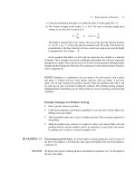
32 A 57-year-old man notices a lump on the right side of
his face that has become larger over the past year. On physical
examination, a 3- to 4-cm firm, mobile, painless mass is palpable in the region of the right parotid gland. The oral mucosa
appears normal. He does not complain of difficulty in chewing
food or talking. The mass is completely excised, and histologic
examination shows the findings in the figure. What is the most
likely diagnosis?
A
Mucoepidermoid carcinoma
B
Non-Hodgkin lymphoma
C
Pleomorphic adenoma
D
Sialolithiasis
E
Sjögren syndrome
F
Warthin tumor
ANSWERS
1 A Periodontitis becomes more prevalent with age, often
secondary to the effects of dental plaque formation driven by
oral flora. The gingival recession increases the risk for dental
caries. Regular dental cleanings to remove the plaque and
regular gentle tooth brushing help to slow the progression
of periodontitis. Some periodontitis cases arise in the setting
of systemic disease. Candidiasis is seen in immunocompromised individuals and often forms an inflammatory membrane on the tongue. Epstein-Barr virus has been associated
with development of hairy leukoplakia. Herpes simplex virus results in vesicles that can rupture and form superficial
ulcers on oral mucosa. Human papillomavirus can drive
squamous epithelial hyperplasia, dysplasia, and carcinoma.
Mucor has broad, nonseptated hyphae and can result in sinusitis, particularly in the setting of ketoacidosis.
PBD9 728 PBD8 741
2 A An aphthous ulcer is a common lesion that also is
known as a canker sore. The lesions are never large, but are annoying and tend to occur during periods of stress. Aphthous
ulcers are not infectious; they probably have an autoimmune
origin. Herpetic lesions are typically vesicles that can rupture.
Leukoplakia appears as white patches of thicker mucosa from
hyperkeratosis. It may be a precursor to squamous cell carcinoma in a few cases. The temperance ditty mentioned in the
history is a cautionary note for all young people. Oral thrush
is a superficial candidal infection that occurs in diabetic, neutropenic, and immunocompromised patients. Inflammation of
a salivary gland (sialadenitis), typically a minor salivary gland
in the oral cavity, may produce a localized, tender nodule.
PBD9 728 BP9 552 PBD8 742 BP8 580
3 B Chronic irritation is the most likely cause for an “irritation” fibroma of the buccal mucosa, which is due to connective tissue hyperplasia. Oral thrush from candidiasis
produces white-to-gray plaques on the tongue. Leukoplakia
is hyperplasia of the squamous epithelium and appears as
a white plaque or patch, and can be premalignant. A pyogenic granuloma is a reddish nodule of granulation tissue
on the gingiva, and it often ulcerates. A minor salivary gland
could become obstructed, producing a mucocele, or become
inflamed and tender (sialadenitis).
PBD9 728–729 BP9 552–553 PBD8 741–742
4 A A pyogenic granuloma may begin to enlarge abruptly
and increase in size rapidly, which can be alarming, but the
process is benign and often regresses, or resolves into fibrous
connective tissue. Though there are both acute and chronic
inflammatory cells within this granulation tissue, neither
C H A P T E R 1 6 Head and Neck
redominates. Rhabdomyosarcoma is more likely to be a
p
childhood tumor, and sarcomas in adults are more likely to occur in deep soft tissues. This reddish nodule is not leukoplakia,
which is a white plaque from squamous epithelial hyperplasia.
PBD9 729–730 BP9 553 PBD8 741–742
E The lesions of herpes simplex virus type 1 (HSV-1),
5
also known as cold sores or fever blisters, are common. Many
individuals have been infected with HSV-1, which is latent,
and the oral and perianal lesions appear during periods of
stress. Recurrence of herpes labialis is the norm. Budding
cells with pseudohyphae suggest a candidal infection with
oral thrush. A mononuclear infiltrate is nonspecific and can
be seen with aphthous ulcers. Atypical lymphocytes are seen
with infectious mononucleosis. They may be accompanied
by a rash, but do not produce vesicular lesions of the skin.
Neutrophilic abscesses suggest bacterial infection. Leukoplakia is marked by hyperkeratosis.
PBD9 729 BP9 552 PBD8 742–743 BP8 580–581
6 F Oral thrush is a common but not life-threatening
condition, resulting from oral candidiasis in immunocompromised individuals. The lesion is typically superficial.
Microscopic examination shows the typical budding cells
and pseudohyphae of Candida. Aphthous ulcers, or canker
sores, are very common in young individuals, but can a ppear
at any age; they tend to be recurrent superficial ulcerations.
Cheilosis is fissuring or cracking of the mucosa, typically
at the corners of the mouth, which may be seen with vitamin B2 (riboflavin) deficiency. Hairy leukoplakia also can
be seen with HIV infection, but it is far less common than
oral thrush. It occurs from marked hyperkeratosis, forming a
rough “hairy” surface, and is related to Epstein-Barr virus infection. Multinucleated cells suggest a herpesvirus infection,
which typically has vesicles that ulcerate. A
typical squamous
epithelial cells usually arise from areas of oral leukoplakia.
PBD9 729–730 BP9 552 PBD8 743 BP8 581
7 C Oral hairy leukoplakia is seen in immunocompromised persons. It presages AIDS in persons who are HIVpositive. Chronic alcohol and/or tobacco use are associated
with oral squamous cell carcinomas. Type 1 diabetes mellitus with ketoacidosis is associated with fungal sinusitis,
particularly with mucormycosis. Pernicious anemia from vitamin B12 deficiency is associated with glossitis that is mainly
atrophic. Sjögren syndrome leads to inflammation and atrophy of salivary glands leading to xerostomia with atrophy,
fissuring, and ulcerations in the oral cavity mucosa.
PBD9 730 BP9 554 PBD8 743 BP8 581
E This whitish, well-defined mucosal patch on the tongue
8
has the characteristic appearance of leukoplakia, a premalignant lesion that can give rise to squamous cell carcinoma. Use
of tobacco products is implicated in the development of leukoplakia. Chronic alcohol abuse also is implicated, but the
association is less strong than with tobacco. Ill-fitting dentures may lead to leukoplakia, but far less c ommonly than
259
smoking. Infections and inflammation are not recognized
risk factors for oral leukoplakia or oral squamous cell cancers. Dental caries is not a risk factor for leukoplakia, unless
the affected tooth becomes eroded and misshapen. The type
of food eaten has less of a correlation with cancer of the oral
cavity than with cancer of the esophagus.
PBD9 731 BP9 553–554 PBD8 744–745 BP8 581–582
9 F Erythroplakia is a premalignant lesion that is more
likely to progress to squamous carcinoma than leukoplakia,
but the major risk factors are the same: tobacco, alcohol, insufficient fruit intake, and betel nut. Countries of the Indian
subcontinent have the highest incidence, accounting for up
to 10% of all cancers in those populations. Of the remaining
options, dental malocclusion may lead to leukoplakia. The
oral infections listed are not premalignant, but may be found
with immunosuppression. Dietary fruit tends to mitigate the
risk, but spices have no effect either way.
PBD9 731 BP9 553–554 PBD8 744–745 BP8 581–582
10 C The raised white patches suggest leukoplakia. This is
a premalignant condition. Risk factors include tobacco use,
particularly tobacco chewing, and chronic irritation. Human
papillomavirus infection has been implicated in some l esions.
Oral thrush appears most often on the tongue of immunocompromised individuals as a yellowish plaquelike area.
Microscopic examination shows budding cells with pseudohyphae characteristic of Candida infection. Lichen planus in
the oral cavity usually appears with similar skin lesions; it
forms whitish patches that may ulcerate. The lesions have
intense submucosal chronic inflammation. A pyogenic granuloma forms a painful gingival nodule of granulation tissue.
Xerostomia, or “dry mouth,” is seen in Sjögren syndrome.
PBD9 731 BP9 553–554 PBD8 744–745 BP8 581–582
11 C Smoking and alcoholism are frequent etiologies for
oral squamous cell carcinomas, and mutations in the TP53
gene are often present. However, in nonsmokers, HPV infection may be implicated, along with overexpression of p16.
The good news: the oral carcinomas arising with HPV have
a better prognosis, though they may be multifocal and recur.
The better news: vaccination against HPV may help prevent
this disease. Oral candidiasis (thrush) may occur in immunocompromised persons. HSV causes self-limited acute
gingivostomatitis (cold sores). The genus Prevotella includes
anaerobes that are associated with periodontitis and with
buccal infections that become cellulitis (Ludwig angina).
Strep throat is an acute exudative pharyngitis that has the
immunologic complications of rheumatic heart disease or
postinfectious glomerulonephritis.
PBD9 731–733 BP9 554 PBD8 746 BP8 582–583
12 B A dentigerous cyst typically occurs in young persons
when teeth are erupting, particularly molars. It is benign and
does not recur following complete excision. Dentigerous cysts
originate around the crown of an unerupted tooth, typically
the third molar, and are lined by a thin, nonkeratinizing layer
260
U N I T I I Diseases of Organ Systems
of squamous epithelium; they contain a dense chronic inflammatory infiltrate in the stroma. An odontogenic keratocyst that
arises from rests of odontogenic epithelium within the jaw and
is benign, but can recur if inadequately excised. Ameloblastoma and odontoma are tumors arising from odontogenic epithelium. Odontoma, the most common odontogenic tumor,
shows extensive deposition of enamel and dentin. Periapical
cysts/granulomas are inflammatory lesions that develop at
the apex of teeth as complications of long-standing pulpitis.
PBD9 734 BP9 557–558 PBD8 748
13 C An odontogenic keratocyst arises from rests of odontogenic epithelium within the jaw. It is benign, but can recur
if inadequately excised. Ameloblastoma and odontoma are
tumors arising from odontogenic epithelium. Odontoma,
the most common odontogenic tumor, shows extensive deposition of enamel and dentin. Dentigerous cysts originate
around the crown of an unerupted tooth, typically the third
molar, and are lined by a thin, nonkeratinizing layer of squamous epithelium; they contain a dense chronic inflammatory
infiltrate in the stroma. Periapical cysts/granulomas are inflammatory lesions that develop at the apex of teeth as complications of long-standing pulpitis.
PBD9 734 BP9 557 PBD8 748–749
14 B Inflammatory nasal polyps can be associated with recurrent allergic rhinitis, a form of type I hypersensitivity often
called hay fever. Type I hypersensitivity is associated with high
IgE levels in the serum. The elevated hemoglobin A1c level indicates diabetes mellitus. Diabetes is not a risk factor for polyp
formation, but ketoacidosis can lead to nasopharyngeal mucormycosis. Epstein-Barr virus infection can be found in nasopharyngeal carcinomas. Autoimmune diseases are not associated
with nasal polyp formation. Staphylococcus aureus often colonizes the nasal cavity, but it usually does not cause problems.
PBD9 735–736 PBD8 749
15 C Chronic sinusitis is a common condition and may be
punctuated by episodes of acute sinusitis. Lack of smell with
nasal cavity inflammation often affects sensation of taste.
Once the cycle of inflammation, obstruction, stasis, mucociliary damage, and polymicrobial infection is established it becomes difficult to stop. Increased pressure with inflammation
in the sinus can erode into adjacent bone, causing osteomyelitis. A mucocele filled with nonpurulent secretions is more
likely to occur in frontal and ethmoid sinuses. Sinusitis is not
a risk factor for malignancy. Nasopharyngeal carcinomas are
related to Epstein-Barr virus (EBV) infection. T-cell lymphomas typically occur in men and are EBV positive. Papillomas
most often occur in men and have an exophytic growth pattern, but those that are endophytic aggressively extend into
adjacent soft tissue and bone, making removal difficult.
PBD9 735–736 PBD8 750
16 C George Washington likely succumbed to an acute bacterial epiglottitis, which is now treatable but still life-threatening,
particularly in children, in whom it is more common. Medical
care has advanced since the year 1799, but it has been little more
than a hundred years that medical care has done more good
than harm. Haemophilus influenzae may cause inflammation
with an abrupt onset of pain and possible airway obstruction,
particularly in children. In adults, the airway is typically large
enough to preclude marked obstruction. Thus, Washington’s
illness was survivable, but the treatments he received at that
time in history (bloodletting, purgatives, blistering agents) contributed to his demise. This cautionary tale supports the adage:
if you don’t know what you’re doing, then stop. Coronaviruses
are best known to cause the common cold. Corynebacterium diphtheriae is the cause of diphtheria, which produces laryngitis with
a characteristic dirty gray membrane that may slough and be aspirated. This infection is now rare because of routine childhood
immunizations. Another cause for epiglottitis is parainfluenza
virus, which has no vaccine, and is best known as the cause for
croup in children. The genus Prevotella includes anaerobes that
are associated with periodontitis and with buccal infections that
become cellulitis (Ludwig angina). Group A streptococci produce a strep throat that is an acute exudative pharyngitis.
PBD9 736 BP9 512–513 PBD8 743 BP8 537
17 E The child has croup, a laryngotracheobronchitis that
is most often caused by parainfluenza virus. The inflammation may be severe enough to produce airway obstruction.
Corynebacterium diphtheriae is the cause of diphtheria, which
produces laryngitis with a characteristic dirty gray membrane that may slough and be aspirated. This infection is now
rare because of routine childhood immunizations. EpsteinBarr virus may be associated with infectious mononucleosis
and produce pharyngitis. Epstein-Barr virus also is associated with nasopharyngeal carcinoma. Haemophilus influenzae
may cause an acute bacterial epiglottitis with an abrupt onset
of pain and possible airway obstruction. Human papillomavirus is associated with laryngeal papillomatosis. Group A
streptococci produce an exudative pharyngitis.
PBD9 739 BP9 512–513 PBD8 752 BP8 537
18 A She has a group A β-hemolytic streptococcal pharyngitis, and the feared complication is an autoimmune response from molecular mimicry to streptococcal M proteins.
Rheumatic fever results 2 to 3 weeks later from formation
of antibodies directed at endocardium, epicardium, and/
or myocardium (rheumatic heart disease). Poststreptococcal
glomerulonephritis may also occur. The pharyngitis is unlikely to spread elsewhere or produce septicemia. Streptococcus pneumoniae is more likely to produce meningitis, otitis,
and pneumonitis. Streptococci are unlikely to involve liver.
PBD9 736, 738 BP9 512–513 PBD8 750, 752 BP8 536–537
19 C Nasopharyngeal carcinoma has a strong association
with Epstein-Barr virus infection, which contributes to the
transformation of squamous epithelial cells. Allergic rhinitis is associated with development of nasal polyps, but these
do not become malignant. ANCA-associated vasculitis can
involve the respiratory tract, causing granulomatous inflammation and necrotizing vasculitis, but there is no risk of malignant transformation. Sjögren syndrome is associated with
C H A P T E R 1 6 Head and Neck
malignant lymphomas, but these typically arise in the salivary gland, not the nasal cavity. Smoking is not associated
with nasopharyngeal carcinoma, although it does contribute
to oral and esophageal cancers.
PBD9 737–738 BP9 513 PBD8 751–752 BP8 537
20 C Reactive nodules (vocal cord polyps, or singer’s nodules) occur most often in men who are heavy smokers or who
strain their vocal cords. The nodules are generally only a few
millimeters in size and have a fibrovascular core covered by hyperplastic and hyperkeratotic squamous epithelium. They are
not premalignant. Croup is an acute l aryngotracheobronchitis
that most often occurs in children and produces airway narrowing with inspiratory stridor. Epiglottitis is an acute inflammatory process that may cause airway obstruction. Squamous
cell carcinomas of the pharynx and larynx form irregular, ulcerating masses, are more common in smokers, but generally
are seen in individuals older than this patient. Squamous papillomatosis usually first appears in childhood; if it is extensive,
it can produce airway obstruction.
PBD9 739 BP9 513 PBD8 752 BP8 537
21 D Recurrent respiratory papillomatosis is caused by human papillomavirus types 6 and 11. These lesions frequently
recur after excision. They may regress after puberty. Laryngeal papillomas arising in adulthood are usually solitary and
do not recur. There is no effective antiviral therapy for human
papillomavirus. Although the lesions can arise throughout
the airways, they are benign and do not become malignant.
The occurrence of the lesions is not related to the use of the
voice, as is a laryngeal nodule, which is quite small. This is
not a congenital condition and is not part of a syndrome.
PBD9 739 BP9 513–414 PBD8 752 BP8 537–538
22 D The figure shows a large, fungating neoplasm that
has the typical appearance of a laryngeal squamous cell carcinoma. The most common risk factor is smoking, although
chronic alcohol abuse also plays a role; some patients harbor human papillomavirus sequences. Invasive cancers arise
from squamous epithelial dysplasias. Epstein-Barr virus infection is associated with nasopharyngeal carcinomas. Aspiration may result in acute inflammation, but not neoplasia.
Allergies with type I hypersensitivity may result in transient
laryngeal edema, but not neoplasia.
PBD9 739–740 BP9 514 PBD8 753 BP8 538
23 A Cholesteatomas are not true neoplasms, but they
are cystic masses lined by squamous epithelium. The desquamated epithelium and keratin degenerates, resulting in
cholesterol formation and giant cell reaction. Although their
histologic findings are benign, cholesteatomas can gradually enlarge, eroding and destroying the middle ear and surrounding structures. They occur as a complication of chronic
otitis media. Although cholesteatomas have a squamous epithelial lining, malignant transformation does not occur. An
eosinophilic granuloma of bone occasionally may be seen in
the region of the skull in young children, but it is character-
261
ized by the presence of Langerhans cells. Labyrinthitis typically is caused by a viral infection and is self-limited. Otosclerosis is abnormal bone deposition in the ossicles of the
middle ear that results in bone deafness in adults.
PBD9 740 PBD8 754
24 D Otosclerosis can be familial, particularly when it is
severe. It results from fibrous ankylosis followed by bony
overgrowth of the little ossicles (malleus, incus, stapes) of
the middle ear. A cholesteatoma is typically a unilateral process that complicates chronic otitis media in a child or young
adult. Uncomplicated otitis media is usually self-limited and
is uncommon in adults. Chondrosarcomas may involve the
skull in older adults, but are rare, solitary, bulky masses in the
region of the jaw. A schwannoma typically involves the vestibulocochlear nerve and results in a nerve conduction form of
deafness. Schwannomas are usually unilateral, although familial neurofibromatosis could result in multiple schwannomas.
PBD9 740–741 PBD8 754
25 A Branchial cysts, also known as lymphoepithelial cysts,
may be remnants of an embryonic branchial arch or a salivary gland inclusion in a cervical lymph node. They are
distinguished from thyroglossal duct cysts by their lateral
location, the absence of thyroid tissue, and their abundant
lymphoid tissue. Occult thyroid carcinoma, often a papillary carcinoma, may manifest as a metastasis to a node in
the neck, but the microscopic pattern is that of a carcinoma.
About 5% of squamous cell carcinomas of the head and neck
initially manifest as a nodal metastasis, without an obvious
primary site. This patient is quite young for such an event,
however. Mucoceles form in minor salivary glands; mucoepidermoid tumors form in salivary glands. The nodule in
this patient is in the neck. Paragangliomas are solid tumors
that may arise deep in the region of the carotid body near the
common carotid bifurcation.
PBD9 741 PBD8 755
26 E A thyroglossal duct (tract) cyst is a developmental abnormality that arises from elements of the embryonic
thyroglossal duct extending from the foramen cecum of the
tongue down to the thyroid gland. One or more remnants
of this tract may enlarge to produce a cystic mass. Although
lymphoid tissue often surrounds these cysts, malignant
transformation does not occur. Granulomatous disease is
more likely to involve lymph nodes in the typical locations
in the lateral neck regions. Salivary gland choristomas are
unlikely at this site. The cysts may contain squamous epithelium, but squamous cell carcinoma does not arise from such
a cyst. If there is a cystic lesion with lymphoid tissue and
squamous carcinoma in the neck, it is probably a metastasis
from an occult primary tumor of the head and neck.
PBD9 741 PBD8 755
27 D Paragangliomas are neuroendocrine tumors that
rarely produce sufficient catecholamines to affect blood
pressure, in contrast to their adrenal medullary counterpart,
262
U N I T I I Diseases of Organ Systems
pheochromocytoma. The microscopic appearance of these lesions does not always correlate with their biological behavior. There is a tendency for recurrence and metastasis despite
the tumor’s “bland” appearance. Metastases always should
be considered in patients this age. About 5% of squamous
cell carcinomas of the head and neck manifest initially as a
nodal metastasis, without an obvious primary site, but the
microscopic pattern here is not that of squamous cell carcinoma. Some thyroid cancers initially may manifest as a
nodal metastasis, but the microscopic pattern in this case fits
best with paraganglioma. A mucoepidermoid carcinoma or
a Warthin tumor arises in a salivary gland.
PBD9 741–742 PBD8 755–756
28 B The most common cause for dry mouth (xerostomia)
and dry eyes (xerophthalmia) is a medication effect. Anticholinergics such as trihexyphenidyl to treat the parkinsonian
tremor can be implicated, as well as antidepressants, antipsychotics, and antihistaminics. Alcohol and tobacco use are risks
for precancerous lesions and squamous cancers of the oral
cavity. The lack of saliva is unlikely to be associated with infection, which tends to be focal. Sialadenitis is unlikely to involve
all salivary glands, except in the setting of Sjögren syndrome,
which is associated with SS-A and SS-B autoantibodies, and
may be associated with some pain with inflammation.
PBD9 742–743 BP9 555 PBD8 756 BP8 583
29 E Sialadenitis is more common in older individuals, and
individuals receiving therapy for schizophrenia with “typical” antipsychotics such as haloperidol can have reduced salivary secretions, which promotes stasis and infection. Most
neuroleptic drugs are dopamine receptor blockers, but they
have extrapyramidal and anticholinergic side effects. The
dry mouth, coupled with dehydration, favors inspissation of
salivary gland secretions and stone formation to block ducts
and increase the risk of inflammation and infection. S. aureus
is the most likely organism to cause infection with suppurative inflammation. Epstein-Barr virus can be associated with
hairy leukoplakia. Human papillomavirus infection may lead
to the development of squamous dysplasias and carcinomas.
Prevotella can be found with periodontitis. Rubeola infection
with measles can cause Koplik spots at the Stensen duct.
PBD9 743 BP9 555 PBD8 756–757 BP8 583
30 D The clinical and histologic features suggest a mucocele of a minor salivary gland, which is most often the r esult
of local trauma in the very young and very old. There is e ither
rupture or blockage of a salivary gland duct. Chili peppers
contain capsaicin, which evokes a sensation of tingling and
burning pain by activating a nonselective cation channel,
called VR1, on vanilloid receptors of sensory nerve endings;
there is no significant tissue damage. Social behavior may be
a risk factor for infections such as herpes simplex virus. HIV
infection is most often associated with oral thrush (candidiasis) and with herpes simplex virus infections. Oral leukoplakia
may appear in various intraoral sites and on the lower lip border, and pipe smoking and tobacco chewing are implicated
in the development of these white patches. Irritation from
misaligned teeth or dentures also may produce leukoplakia.
In some parts of the world, the chewing of betel nut is a risk
factor for oral cancer.
PBD9 743 BP9 555 PBD8 756–757 BP8 583
31 C Pleomorphic adenoma is the most common tumor
of the parotid gland. These tumors are rarely malignant,
although they can be locally invasive. An acinic cell tumor is
composed of cells resembling the serous cells of the salivary
gland; they are generally small, but about one sixth metastasize to regional lymph nodes. Mucoepidermoid tumors are
less common than pleomorphic adenomas in major salivary
glands. They may be high-grade and aggressive. Primitive
neuroectodermal tumor, also known as an olfactory neuroblastoma, is a small, round, blue cell tumor that occurs in
childhood. It is likely to arise in the nasopharyngeal region.
Squamous cell carcinomas arise in the buccal mucosa and
are invasive. Warthin tumors are uncommon and indolent,
although they may be bilateral or multicentric.
PBD9 744–745 BP9 556–557 PBD8 758–759 BP8 584–585
32 F Warthin tumor is the second most common salivary
gland tumor, and it almost always arises within the parotid
gland. These tumors tend to be slow growing. Microscopically there are spaces lined by a double layer of superficial
columnar and basal cuboidal epithelial cells that are surrounding a lymphoid stroma. Mucoepidermoid carcinomas
are infiltrative and form mucous cysts along with a population of squamoid cells. Non-Hodgkin lymphoma may arise in
patients with long-standing Sjögren syndrome. Pleomorphic
adenomas are more common than Warthin tumors, but have a
microscopic appearance with ductal epithelial cells in a myxoid stroma containing islands of chondroid and bone. Sialolithiasis is usually accompanied by sialadenitis and is quite
painful. It may produce some gland enlargement, but usually
is not a mass effect. Sjögren syndrome can produce some salivary gland enlargement, but the process is typically bilateral.
PBD9 745 BP9 556 PBD8 759 BP8 584–585
33 B Mucoepidermoid carcinomas can arise in major and
minor salivary glands. They account for most neoplasms that
arise within minor salivary glands, particularly malignant
neoplasms. Low-grade mucoepidermoid carcinomas may
be invasive, but the prognosis is usually good, with a 5-year
survival of 90%. High-grade mucoepidermoid carcinomas
can metastasize and have a 5-year survival of only 50%. NonHodgkin lymphomas are found in adjacent cervical lymph
nodes or in the Waldeyer ring of lymphoid tissue. A primitive neuroectodermal tumor, also known as an olfactory neuroblastoma, is a small, round, blue cell tumor of childhood; it
is likely to arise in the nasopharyngeal region. Pleomorphic
adenomas are more common in the major salivary glands
than are mucoepidermoid tumors, and they are more likely
to be indolent. Squamous cell carcinomas are invasive and
arise in the buccal mucosa. Warthin tumors are uncommon
and indolent.
PBD9 745–746 BP9 557 PBD8 759–760 BP8 584
CHAPTER
Gastrointestinal Tract
17
PBD9 Chapter 17 and PBD8 Chapter 17: The Gastrointestinal Tract
BP9 Chapter 14 and BP8 Chapter 15: Oral Cavity and Gastrointestinal Tract
1 A 23-year-old primigravida gives birth at term to a boy
infant. Ultrasound examination before delivery showed polyhydramnios. A single umbilical artery is seen at the time of
birth. The infant vomits all feedings, and then develops a fever
and difficulty with respirations within 2 days. A radiograph
shows both lungs and the heart are of normal size, but there
are pulmonary infiltrates and no stomach bubble. What is the
most likely diagnosis?
A
Achalasia
B
Diaphragmatic hernia
C
Esophageal atresia
D
Hiatal hernia
E
Pyloric stenosis
F
Zenker diverticulum
2 A 24-year-old man has developed abdominal pain and
increasing fatigue over the past 6 months. On physical examination, he is afebrile and appears pale. On palpation, there is
mild pain in the right lower quadrant of the abdomen. There
are no masses, and bowel sounds are active. Laboratory studies show hemoglobin, 8.9 g/dL; hematocrit, 26.7%; MCV,
74 μm3; platelet count, 255,000/mm3; and WBC count, 7780/
mm3. His stool is positive for occult blood. Upper gastrointestinal endoscopy and colonoscopy showed no lesions. One
month later, he continues to experience the same abdominal pain. Which of the following is most likely to cause this
patient’s illness?
A
Acute appendicitis
B
Angiodysplasia
C
Celiac disease
D
Diverticulosis
E
Giardia lamblia infection
F
Meckel diverticulum
3 A 23-year-old woman, G2, P1, gave birth at term to a
boy of normal weight and length following an uncomplicated
pregnancy. The infant initially did well, but at 6 weeks, he
began feeding poorly for 1 week, and his mother noticed that
much of the milk he ingested was forcefully vomited within
1 hour. Now, on physical examination, the infant is afebrile,
and there are no external anomalies. A midabdominal mass is
palpable. Bowel sounds are active. The medical history indicates that both the mother and her first child had the same illness during infancy. Which of the following conditions is most
likely to explain these findings?
A
Annular pancreas
B
Diaphragmatic hernia
C
Duodenal atresia
D
Pyloric stenosis
E
Tracheoesophageal fistula
4 A 24-year-old woman gives birth to term infant after an
uncomplicated pregnancy. Apgar scores are 9 and 10 at 1 and
5 minutes after birth. The infant’s length and weight are at the
55th percentile. There is no significant passage of meconium.
Three days after birth, the infant vomits all oral feedings. On
physical examination, the infant is afebrile, but the abdomen
is distended and tender, and bowel sounds are reduced. An
abdominal ultrasound scan shows marked colonic dilation
above a narrow segment in the distal sigmoid region. A biopsy
specimen from the narrowed region shows an absence of ganglion cells in the muscle wall and submucosa. Which of the
following is most likely to produce these findings?
A
Colonic atresia
B
Hirschsprung disease
C
Intussusception
D
Necrotizing enterocolitis
E
Trisomy 21
F
Volvulus
263
264
U N I T I I Diseases of Organ Systems
5 A 3-year-old child has attained enough mobility, curiosity, and dexterity to explore places in the home that should
not be accessed. The child finds a bottle with a liquid under
the kitchen sink, and he drinks it. Within minutes he has chest
pain. His mother takes him to the emergency department, and
brings the bottle. Analysis of the residual contents reveals a
pH of 12. Which of the following complications is most likely
to occur following this injury?
A
Pharyngeal diverticulum
B
Esophageal stenosis
C
Gastric lymphoma
D
Duodenal ulceration
E
Megacolon
6 A 22-year-old woman has had multiple episodes of aspiration of food associated with difficulty swallowing during the
past year. On auscultation of her chest, crackles are heard at the
base of the right lung. A barium swallow shows marked esophageal dilation above the level of the lower esophageal sphincter.
A biopsy specimen from the lower esophagus shows an absence
of the myenteric ganglia. What is the most likely diagnosis?
A
Achalasia
B
Barrett esophagus
C
Plummer-Vinson syndrome
D
Sliding hiatal hernia
E
Systemic sclerosis
7 A 24-year-old woman living in eastern Bolivia has had
increasing difficulty with swallowing both liquids and solids
for the past year. She has substernal discomfort from a feeling
that foods “get stuck” going down. On examination her BMI
is 18. A barium swallow radiologically shows marked esophageal dilation. An endoscopic biopsy is obtained and microscopically shows reduced ganglion cells in myenteric plexus
along with lymphocytic infiltration. Which of the following
organisms is most likely infecting this woman?
A
Bordetella pertussis
B
Candida albicans
C
Corynebacterium diphtheriae
D
Herpes simplex virus
E
Trypanosoma cruzi
8 A 53-year-old man consumes a very large meal, washed
down with considerable alcohol. The ensuing discomfort
prompts him to take an emetic, but soon afterward he develops lower chest pain. Physical examination reveals crepitus
in subcutaneous tissue over his chest along with tachycardia
and tachypnea. Which of the following abnormalities of the
esophagus is most likely present in this man?
A
Stricture
B
Achalasia
C
Ectopia
D
Rupture
E
Varices
9 A 30-year-old man has sudden onset of hematemesis after a weekend in which he consumed large amounts of
alcohol. The bleeding stops, but he has another episode under
similar circumstances 1 month later. Upper gastroesophageal
endoscopy shows longitudinal tears at the gastroesophageal
junction. What is the most likely mechanism to cause his
hematemesis?
A
Absent myenteric ganglia
B
Autoimmune inflammation
C
Herpes simplex virus infection
D
Portal hypertension
E
Vomiting
F
Widened diaphragmatic crura
10 A 16-year-old boy who is receiving chemotherapy for
acute lymphoblastic leukemia has had pain for 1 week when
he swallows food. Physical examination shows no abnormal
findings. Upper gastrointestinal endoscopy shows 0.5- to
0.8-cm mucosal ulcers in the region of the mid to lower esophagus. The shallow ulcers are round and sharply demarcated,
and have an erythematous base. Which of the following is
most likely to produce these findings?
A
Aphthous ulcerations
B
Reflux esophagitis
C
Herpes simplex esophagitis
D
Gastroesophageal reflux disease
E
Mallory-Weiss syndrome
11 A 44-year-old woman has had increasing difficulty
swallowing liquids and solids for the past 6 months. On physical examination, her fingers have reduced mobility because
of taut, nondeforming skin. A barium swallow shows marked
dilation of the esophagus with “beaking” in the distal portion,
where there is marked luminal narrowing. A biopsy s pecimen
from the lower esophagus shows prominent submucosal
fibrosis with little inflammation. Which of the following is
most likely to produce these findings?
A
Barrett esophagus
B
Hiatal hernia
C
Iron deficiency
D
Portal hypertension
E
Systemic sclerosis
12 A 57-year-old woman has had burning epigastric
pain after meals for more than 1 year. Physical examination
shows no abnormal findings. Upper gastrointestinal endoscopy shows an erythematous patch in the lower esophageal
mucosa. A biopsy specimen shows basal zone squamous epithelial hyperplasia, elongation of lamina propria papillae, and
scattered intraepithelial neutrophils with some eosinophils.
Which of the following is the most likely diagnosis?
A
Barrett esophagus
B
Esophageal varices
C
Iron deficiency
D
Reflux esophagitis
E
Systemic sclerosis
C H A P T E R 1 7 Gastrointestinal Tract
265
13 A 51-year-old man has sudden onset of massive emesis
of bright red blood. On physical examination, his temperature is 36.9° C, pulse is 103/min, respirations are 23/min, and
blood pressure is 85/50 mm Hg. His spleen tip is palpable.
Laboratory studies show a hematocrit of 21%. The serologic
test result for HBsAg is positive. He has had no prior episodes
of hematemesis. The hematemesis is most likely to be a consequence of which of the following?
A
Barrett esophagus
B
Candida albicans infection
C
Esophageal varices
D
Reflux esophagitis
E
Squamous cell carcinoma
F
Zenker diverticulum
14 A 55-year-old man has had increasing difficulty swallowing during the past 6 months. There are no significant
findings on physical examination. Upper gastrointestinal
endoscopy shows areas of erythematous mucosa 3 cm above
the Z-line. A biopsy specimen from the lower esophagus has
changes in the mucosal epithelium illustrated in the figure.
Which of the following complications is most likely to occur as
a consequence of this patient’s condition?
A
Achalasia
B
Adenocarcinoma
C
Diverticular formation
D
Lacerations (Mallory-Weiss syndrome)
E
Squamous cell carcinoma
15 A 68-year-old man from Birmingham, England, has had
“heartburn” and substernal pain after meals for 25 years. For
the past year, he has had increased pain with difficulty swallowing both liquids and solids. On physical examination, there
are no remarkable findings. Upper gastrointestinal endoscopy
shows an ulcerated lower esophageal mass that nearly occludes
the lumen of the esophagus. A biopsy specimen of this mass is
most likely to show which of the following neoplasms?
A
Adenocarcinoma
B
Carcinoid tumor
C
Leiomyosarcoma
D
Non-Hodgkin lymphoma
E
Squamous cell carcinoma
16 A 73-year-old man with a history of chronic alcoholism
has had increasing difficulty swallowing and has noticed a
3-kg weight loss over the past 2 months. On physical examination, there are no remarkable findings. Upper gastrointestinal
endoscopy shows a 3-cm ulcerative mass in the midesophagus
that partially occludes the esophageal lumen. Esophagectomy
is performed; the gross appearance of the lesion is shown in
the figure. Which of the following is most likely to be seen on
microscopic section of this mass?
A
Adenocarcinoma
B
Dense collagenous scar
C
Dilated vascular channels
D
Multinucleated cells with intranuclear inclusions
E
Squamous cell carcinoma
17 A 66-year-old man living in Tehran, Iran, has been
bothered by difficulty swallowing for the past year. He is
now consuming liquid food. Yesterday he regurgitated food
stained with blood. On esophagoscopy, there is an ulcerated
obstructing lesion 20 cm from the lips. Biopsies are taken and
on microscopy show infiltrating nests of keratinized cells with
distinct cell borders and hyperchromatic, angulated nuclei.
Which of the following is the most likely risk factor for his
disease?
A
Genetic susceptibility
B
Autoimmunity
C
Diet
D
Infection
E
Reflux
266
U N I T I I Diseases of Organ Systems
18 A 38-year-old woman has had nausea for 6 months.
She reports no vomiting or diarrhea. On physical examination, there are no remarkable findings. Upper gastrointestinal
endoscopy shows diffuse gastric mucosal erythema with focal
mucosal erosions, but no ulcerations. The esophageal and
duodenal mucosal surfaces appear normal. Gastric biopsies
are obtained and microscopic examination shows focal mucosal hemorrhage, loss of the surface epithelium, and increased
numbers of neutrophils, lymphocytes, and plasma cells in an
edematous mucosa. No Helicobacter pylori organisms are seen.
Laboratory studies show a normal serum gastrin level. Which
of the following pharmacologic agents is most likely to produce these findings?
A
Aspirin
B
Chlorpromazine
C
Cimetidine
D
Clindamycin
E
Omeprazole
19 A 72-year-old man takes large quantities of nonsteroidal
anti-inflammatory drugs (NSAIDs) because of chronic degenerative arthritis of the hips and knees. Over the past 2 weeks,
he has had epigastric pain with nausea and vomiting and an
episode of hematemesis. On physical examination, there are
no remarkable findings. A gastric biopsy specimen is most
likely to show which of the following lesions?
A
Acute gastritis
B
Adenocarcinoma
C
Epithelial dysplasia
D
Helicobacter pylori infection
E
Hyperplastic polyp
20 A 54-year-old, previously healthy man sustained an
extensive thermal burn injury involving 70% of the total body
surface area of his skin. He was hospitalized in stable condition. Three weeks after the initial burn injury, he developed
melanotic stools. His blood pressure dropped to 80/40 mm
Hg, and his hematocrit declined to 18%. Where are gastrointestinal ulcerations most likely to be found in this man?
A
Colon
B
Duodenum
C
Esophagus
D
Ileum
E
Stomach
21 A 51-year-old woman has been feeling increasingly
tired for the past 7 months. There are no remarkable findings
on physical examination. Laboratory studies include hemoglobin, 9.5 g/dL; hematocrit, 29.1%; MCV, 124 μm3; platelet
count, 268,000/mm3; and WBC count, 8350/mm3. The reticulocyte index is low. Hypersegmented polymorphonuclear
leukocytes are found on a peripheral blood smear. The serum
gastrin is markedly increased. Antibodies to which of the following are most likely to be found in this patient?
A
Gastric H+,K+-ATPase
B
Gliadin
C
Helicobacter pylori
D
Intrinsic factor receptor
E
Tropheryma whippelli
22 A 59-year-old man has had nausea and vomiting for
5 months. He has experienced no hematemesis. On physical
examination, there is no abdominal tenderness, and bowel
sounds are present. Upper gastrointestinal endoscopy shows
erythematous areas of mucosa with thickening of the rugal
folds in the gastric antrum. The microscopic appearance of a
gastric biopsy specimen with a Steiner silver stain is shown
in the figure. Which of the following factors is most likely
responsible for this gastric mucosal pathology?
A
Cysteine proteinase
B
Cytotoxin-associated gene A
C
Heat-stable enterotoxin
D
Shiga toxin
E
Verocytotoxin
23 A 47-year-old woman with a lengthy history of heartburn and dyspepsia experiences sudden onset of abdominal
pain. On physical examination, she has severe mid epigastric
pain with guarding. Bowel sounds are reduced. An abdominal plain film radiograph shows free air under the left leaf of
the diaphragm. She is immediately taken to surgery, and a
perforated duodenal ulcer is repaired. Which of the following
organisms is most likely to have produced these findings?
A
Campylobacter jejuni
B
Cryptosporidium parvum
C
Giardia lamblia
D
Helicobacter pylori
E
Salmonella typhi
F
Shigella flexneri
G
Yersinia enterocolitica
24 A 35-year-old man has had epigastric pain for more than
1 year. The pain tends to occur 2 to 3 hours after a meal and is
relieved if he takes antacids or eats more food. He has noticed
a 4-kg weight gain in the past year. He does not smoke and
drinks 1 glass of Johannisberg Riesling daily. The result of a
urea breath test is positive, and a gastric biopsy specimen contains urease. He begins a 2-week course of antibiotics, but on
day 4, he feels better and discontinues treatment. Three weeks
later, the epigastric pain recurs. If he does not seek further
treatment, which of the following complications is he most
likely to develop?
A
Carcinoid syndrome
B
Fat malabsorption
C
Hematemesis
D
Migratory thrombophlebitis
E
Vitamin B12 deficiency
C H A P T E R 1 7 Gastrointestinal Tract
25 A 52-year-old man notes nausea with abdominal discomfort after meals. On physical examination, there are no
abnormal findings. Upper endoscopy is performed, and there
are three ovoid nodules in the fundus and antrum ranging
from 0.3 to 1.2 cm in size. They have rounded, smooth surfaces. Biopsies are taken and on microscopic examination
there are irregular, cystically dilated and elongated foveolar
glands. Which of the following treatment strategies is most
appropriate for his gastric lesions?
A
Antibiotics
B
Chemotherapy
C
Corticosteroids
D
Multivitamins
E
Total gastrectomy
F
Vagotomy
267
specimen shows a monomorphous infiltrate of lymphoid cells
microscopically. Helicobacter pylori organisms are identified
in mucus overlying adjacent mucosa. Cytogenetic analysis
shows t(11;18)(q21;q21). He receives antibiotic therapy for
H. pylori, and the repeat biopsy specimen shows a resolution
of the infiltrate. What is the most likely diagnosis?
A
Autoimmune gastritis
B
Chronic gastritis
C
Crohn disease
D
Diffuse large B-cell lymphoma
E
Gastrointestinal stromal tumor
F
Mucosa-associated lymphoid tissue tumor
26 A 49-year-old woman has a history of peptic ulcer disease
for which she has been treated with proton pump inhibitors. She
has had nausea with vomiting for the past 2 months. Upper GI
endoscopy reveals three circumscribed, round, smooth lesions
in the gastric body from 1 to 2 cm in diameter. Biopsies are taken
and microscopically show the lesions to consist of irregular
glands that are cystically dilated and lined by flattened parietal
and chief cells. No inflammation, Helicobacter pylori, metaplasia,
or dysplasia is present. What is the most likely diagnosis?
A
Fundic gland polyps
B
Gastric adenomas
C
Hyperplastic polyps
D
Hypertrophic gastropathy
27 A 53-year-old woman has had nausea, vomiting, and
midepigastric pain for 5 months. On physical examination, there
are no significant findings. An abdominal CT scan shows gastric
outlet obstruction. Upper gastrointestinal endoscopy shows an
ulcerated 2 × 4 cm bulky mass in the antrum at the pylorus. A
urease test is positive. Which of the following neoplasms is most
likely to be seen in a biopsy specimen of this mass?
30 A 26-year-old man is brought to the emergency department after sustaining abdominal gunshot injuries. At laparotomy,
while repairing the small intestine, the surgeon notices a 1-cm
mass at the tip of the appendix. The yellow-tan submucosal mass
is removed, and the microscopic appearance of the mass is shown
in the figure. Immunohistochemical staining is positive for chromogranin and synaptophysin but negative for Ki-67. Which of
the following is the most likely cell of origin of this lesion?
A
Adenocarcinoma
B
Leiomyosarcoma
C
Neuroendocrine carcinoma
D
Non-Hodgkin lymphoma
E
Squamous cell carcinoma
A
Lipoblast
B
Ganglion cell
C
Goblet cell
D
Neuroendocrine cell
E
Smooth muscle cell
28 A 67-year-old woman has experienced severe nausea,
vomiting, early satiety, and a 9-kg weight loss over the past
4 months. On physical examination, she has muscle wasting.
Upper gastrointestinal endoscopy shows that the entire gastric mucosa is eroded and has an erythematous, cobblestone
appearance. An abdominal CT scan shows that the stomach is
small and shrunken. Which of the following is most likely to be
found on histologic examination of a gastric biopsy specimen?
31 A 55-year-old man experiences episodes of diaphoresis,
dyspnea, and diarrhea for 10 months. On physical examination he has midabdominal discomfort with deep palpation,
and bowel sounds are reduced. There are no abnormal findings with upper endoscopy. Abdominal CT scan shows three
nodules in the liver, from 1 to 3 cm in size. Laboratory studies show a high level of serum 5-hydroxyindoleacetic acid
(5-HIAA). Camera endoscopy is performed, and on review of
the images, there is a midjejunal mass that partially obstructs
the lumen. At laparotomy a 5-cm submucosal jejunal mass is
resected, and on microscopy it is composed of nests and trabeculae of round cells with pink, granular cytoplasm. The cells
of this mass are most likely related to which of the following
embryologic derivatives?
A
Chronic atrophic gastritis
B
Primary gastric lymphoma
C
Gastrointestinal stromal tumor
D
Granulomatous inflammation
E
Signet ring cell adenocarcinoma
29 A 52-year-old man has had a 4-kg weight loss and nausea for the past 6 months. He has no vomiting or diarrhea.
On physical examination, there are no remarkable findings.
Upper gastrointestinal endoscopy shows a 6-cm area of irregular, pale fundic mucosa and loss of the rugal folds. A biopsy
A
Endoderm
B
Ectoderm
C
Neural crest
D
Notochord
E
Splanchnic mesoderm
268
U N I T I I Diseases of Organ Systems
32 A 61-year-old man with increasing fatigue, early satiety,
and nausea for 5 months vomited dark granular material yesterday. Endoscopy reveals a large ulcerated mass in the gastric fundus. Biopsies are taken and microscopically the mass
is composed of spindle cells that are positive for c-Kit with
immunohistochemical staining. Mitoses are frequent. Gastrectomy is performed, and the 10-cm circumscribed mass arises
from the gastric wall. Which of the following therapies is most
likely to be a useful adjunct in treatment of his disease?
A
Amoxicillin
B
Azathioprine
C
Cyclophosphamide
D
Imatinib
E
Prednisone
F
Radiation
33 A 57-year-old man from Innsbruck, Austria, goes to the
emergency department because of increasing abdominal pain
with distention that developed over the past 24 hours. On
physical examination, there is diffuse abdominal tenderness.
The abdomen is tympanitic, without a fluid wave, and bowel
sounds are nearly absent. There is a well-healed, 5-cm transverse scar in the right lower quadrant of the abdomen. There is
no caput medusa. A stool sample is negative for occult blood.
An abdominal plain film shows dilated loops of small bowel
with air-fluid levels, but there is no free air. At laparotomy,
the surgeon notices a 20-cm portion of reddish black ileum
that changes abruptly to pink-appearing bowel on distal and
proximal margins. His medical history is significant only for
an appendectomy at age 25 years. Which of the following is
most likely to have produced his findings?
A
Adenocarcinoma of the ileum
B
Adhesions
C
Crohn disease
D
Indirect inguinal hernia
E
Intussusception
F
Tuberculosis
G
Volvulus
34 An 11-month-old, previously healthy infant has not produced a stool for 1 day. The mother notices that the infant’s
abdomen is distended. On physical examination, the infant’s
abdomen is very tender, and bowel sounds are nearly absent.
An abdominal plain film radiograph shows no free air, but
there are distended loops of small bowel with air-fluid levels.
Which of the following is most likely to produce these findings?
A
Duodenal atresia
B
Hirschsprung disease
C
Intussusception
D
Meckel diverticulum
E
Pyloric stenosis
35 A 61-year-old man has had severe abdominal pain and
bloody diarrhea for the past day. On physical examination, his
abdomen is diffusely tender, and bowel sounds are absent.
Abdominal plain films show no free air. Laboratory studies
show a normal CBC and normal levels of serum amylase,
lipase, and bilirubin. His Hgb A1c is 10%. He develops shock.
A year ago he had an acute myocardial infarction. Which of
the following lesions is most likely to be found in this man?
A
Appendicitis
B
Cholecystitis
C
Pancreatitis
D
Intestinal infarction
E
Pseudomembranous colitis
36 A 71-year-old woman with a history of rheumatic heart
disease is hospitalized with severe congestive heart failure.
Four days after admission, she develops cramping lower
abdominal pain. On physical examination, she is afebrile. The
abdomen is distended and tympanitic, without a fluid wave,
and bowel sounds are absent. A stool sample is positive for
occult blood. An abdominal plain film shows no free air.
Colonoscopy shows patchy areas of mucosal erythema with
some overlying tan exudate in the ascending and descending
colon. No polyps or masses are found. What is the most likely
diagnosis?
A
Ischemic colitis
B
Mesenteric vasculitis
C
Shigellosis
D
Ulcerative colitis
E
Volvulus
37 A 60-year-old man has had increasing fatigue for the
past 8 months. On physical examination, he appears pale. On
digital rectal examination, no masses are palpable, but a stool
sample is positive for occult blood. Auscultation of the abdomen shows active bowel sounds, and on palpation there are
no masses or areas of tenderness. Laboratory studies show
hemoglobin, 8.3 g/dL; hematocrit, 24.6%; MCV, 73 μm3; platelet count, 226,000/mm3; and WBC count, 7640/mm3. Colonoscopy shows no identifiable source of the bleeding. Angiography shows a 1-cm focus of dilated and tortuous vascular
channels in the mucosa and submucosa of the cecum. What is
the most likely diagnosis?
A
Angiodysplasia
B
Collagenous colitis
C
Diverticulosis
D
Internal hemorrhoids
E
Mesenteric vein thrombosis
38 A 21-year-old man has had increasingly voluminous,
bulky, foul-smelling stools and a 7-kg weight loss for the past
year. There is no history of hematemesis or melena. He has
some bloating, but no abdominal pain. On physical examination, there are no palpable abdominal masses, and bowel
sounds are present. Which of the following laboratory findings is most likely to be present on examination of his stool?
A
Entamoeba histolytica trophozoites
B
Giardia lamblia cysts
C
Increased stool fat
D
Occult blood
E
Vibrio cholerae organisms
C H A P T E R 1 7 Gastrointestinal Tract
39 A 34-year-old woman is bothered by a low-volume,
mostly watery diarrhea associated with flatulence. The symptoms occur episodically, but they have been persistent for the
past year. She has experienced a 4-kg weight loss. She has
no fever, nausea, vomiting, or abdominal pain. On physical
examination, there are no significant findings. A stool sample
is negative for occult blood, ova, and parasites, and a stool
culture yields no pathogens. An upper gastrointestinal endoscopy is performed and a biopsy specimen from the upper part
of the small bowel shows severe diffuse blunting of villi and
a chronic inflammatory infiltrate in the lamina propria. Which
of the following serologic tests is most likely to be positive in
this patient?
A
Anticentromere antibody
B
Anti–DNA topoisomerase I antibody
C
Antimitochondrial antibody
D
Antinuclear antibody
E
Antitransglutaminase antibody
40 A 41-year-old woman has had diarrhea and fatigue with
a 3-kg weight loss over the past 6 months. On physical examination, she is afebrile and has mild muscle wasting, but her
motor strength is normal. Laboratory studies show no occult
blood, ova, or parasites in the stool. A biopsy specimen from
the upper jejunum is obtained, and microscopic findings are
reviewed. The patient begins following a special diet with no
wheat or rye grain products. The change in diet produces dramatic improvement. Which of the following microscopic features is most likely to be seen in the biopsy specimen?
A
Crypt abscesses and mucosal ulceration
B
Foamy macrophages within the lamina propria
C
Lymphatic obstruction
D
Noncaseating granulomas
E
Villous blunting and flattening
41 An epidemiologic study of children with failure to
thrive is undertaken in Guatemala. Some of these children
with ages 1 to 3 years have repeated bouts of diarrhea, but
do not improve with dietary supplements. Jejunal biopsies
show blunted, atrophic villi with crypt elongation and chronic
inflammatory infiltrates. What is the most likely factor contributing to recurrent diarrhea in these children?
A
Abetalipoproteinemia
B
Bacterial infection
C
Chloride ion channel dysfunction
D
Disaccharidase deficiency
E
NOD2 gene mutations
42 A 40-year-old man has episodic
abdominal bloating,
flatulence, and explosive diarrhea. These symptoms appear
to be related to the milk shakes that he loves to consume.
On physical examination, there are no remarkable findings.
Laboratory studies show no increase in stool fat and no occult
blood, ova, or parasites in the stool. A routine stool culture
yields no pathogens. When he does not consume milk shakes
or ice cream sodas, he is not symptomatic. Which of the following conditions best accounts for these findings?
A
Autoimmune gastritis
B
Celiac disease
C
Cholelithiasis
D
Cystic fibrosis
E
Disaccharidase deficiency
269
43 A potluck lunch party is held at the office at noon. Various meats, salads, breads, and desserts that were brought in
earlier that morning are served. Everyone has a good time,
and most of the food is consumed. By midafternoon, the
single office restroom is being used by many employees who
have vomiting and acute, explosive diarrhea accompanied by
abdominal cramping. Which of the following infectious agents
is most likely responsible for this turn of events?
A
Bacillus cereus
B
Clostridium difficile
C
Escherichia coli
D
Salmonella enterica
E
Staphylococcus aureus
F
Vibrio parahaemolyticus
44 A healthy 21-year-old woman develops a profuse, watery
diarrhea 1 day after a meal of raw oysters. On physical examination, her temperature is 37.5° C. A stool sample is negative
for occult blood. There is no abdominal distention or tenderness, and bowel sounds are present. The diarrhea subsides
over the next 3 days. Which of the following organisms is most
likely to produce these findings?
A
Cryptosporidium parvum
B
Entamoeba histolytica
C
Staphylococcus aureus
D
Vibrio parahaemolyticus
E
Yersinia enterocolitica
45 A 26-year-old man traveling to Ching Mai, Thailand,
had fever, headache, and muscle pains for a day followed by
watery diarrhea of 5 to 10 stools per day for 4 days. In the
past day, the diarrhea has been bloody and accompanied by
tenesmus. On physical examination there is diffuse abdominal
pain. Microscopic examination of the stool shows numerous
leukocytes and gram-negative curved rods. The diarrhea subsides, but 2 weeks later he has increasing weakness in his legs.
Which of the following organisms is most likely to produce his
disease?
A
Bacillus cereus
B
Campylobacter jejuni
C
Clostridium perfringens
D
Giardia lamblia
E
Rotavirus
46 A 36-year-old man experiences cramping abdominal pain with fever and watery diarrhea 2 days after eating
a chicken salad sandwich. Physical examination shows mild
diffuse abdominal pain on palpation, but there are no masses.
Bowel sounds are present. A stool sample is negative for
occult blood. He recovers completely within 5 days without
treatment. Which of the following infectious organisms is
most likely to produce these findings?
A
Bacillus cereus
B
Entamoeba histolytica
C
Escherichia coli
D
Rotavirus
E
Salmonella enterica
F
Staphylococcus aureus
G
Yersinia enterocolitica
270
U N I T I I Diseases of Organ Systems
47 In an epidemiologic study of infections of the gastrointestinal tract, cases of patients living in Haiti from whom
definitive cultures were obtained are analyzed for clinical
and pathologic findings that may be useful for diagnosis. A
group of patients is identified who initially had abdominal
pain and diarrhea during week 1 of their illness. By week 2,
these patients had splenomegaly and elevations in serum AST
and ALT levels. By week 3, they were septic and had leukopenia. At autopsy, the patients who died were found to have
ulceration of Peyer patches. Which of the following infectious
agents is most likely to produce these findings?
A
Campylobacter jejuni
B
Clostridium perfringens
C
Mycobacterium bovis
D
Salmonella typhi
E
Shigella sonnei
F
Yersinia enterocolitica
49 Over a holiday weekend, more than 100 adults at a
resort hotel develop a diarrheal illness marked by voluminous, watery stools more than 10 times per day. They also
report headache, abdominal cramping pain, and myalgias. On
physical examination they have manifestations of dehydration
and mild fever. Laboratory studies of stool samples show no
increase in leukocytes or fat, and no RBCs. Their illness lasts
just 1 to 3 days and resolves with no sequelae. Which of the
following infectious agents is the most likely cause for their
illness?
A
Cytomegalovirus
B
Clostridium botulinum
C
Norovirus
D
Staphylococcus aureus
E
Strongyloides stercoralis
F
Vibrio cholerae
50 A 5-month-old, previously healthy infant girl in Bangladesh develops a watery diarrhea that lasts for 1 week. The
infant has a mild fever during the illness, but has no abdominal pain or swelling. On physical examination, her temperature is 37.7° C. A stool sample is negative for occult blood, ova,
or parasites. Her parents are told to give her plenty of fluids,
and she recovers fully. Which of the following organisms is
most likely to produce these findings?
A
Campylobacter jejuni
B
Cryptosporidium parvum
C
Escherichia coli
D
Listeria monocytogenes
E
Norwalk virus
F
Rotavirus
G
Shigella flexneri
48 A 65-year-old woman is being treated in the hospital
for pneumonia complicated by septicemia. She has required
multiple antibiotics and was intubated and mechanically ventilated earlier in the course. On day 20 of hospitalization, she
has abdominal distention. Bowel sounds are absent, and an
abdominal radiograph shows dilated loops of small bowel
suggestive of ileus. She has a low volume of bloody stool that is
positive for Clostridium difficile toxin. Laboratory studies show
leukocytosis and hypoalbuminemia. At laparotomy, a portion
of distal ileum and cecum is resected. The gross a ppearance of
the mucosal surface is shown in the figure. What is the most
likely diagnosis?
A
Gas gangrene with myonecrosis
B
Inflammatory bowel disease
C
Ischemic bowel disease
D
Pseudomembranous enterocolitis
E
Toxic megacolon
51 A study of children living in rural Malawi in Africa
reveals a high prevalence of iron deficiency anemia. Stool
samples are positive for occult blood. Pruritus of the skin of
their feet as well as cough are additional findings in many of
these children. Which of the following parasitic infestations is
the most likely cause for these findings?
A
Ancylostoma duodenale
B
Ascaris lumbricoides
C
Cryptosporidium parvum
D
Enterobius vermicularis
E
Schistosoma mansoni
52 A 31-year-old woman had increasingly severe diarrhea
1 week after returning from a trip to Central America. Gross
examination of the stools showed mucus and streaks of blood.
The diarrheal illness subsided within 4 weeks, but now she
has become febrile and has pain in the right upper quadrant of
the abdomen. An abdominal ultrasound scan shows a 10-cm,
irregular, partly cystic mass in the right hepatic lobe. Which of
the following infectious organisms is most likely to produce
these findings?
A
Clostridium difficile
B
Cryptosporidium parvum
C
Giardia lamblia
D
Entamoeba histolytica
E
Strongyloides stercoralis
C H A P T E R 1 7 Gastrointestinal Tract
271
A
Cystic fibrosis
B
Diverticular disease
C
Inflammatory bowel disease
D
Irritable bowel syndrome
E
Viral gastroenteritis
53 A 27-year-old man has sudden onset of marked abdominal pain. On physical examination, his abdomen is diffusely
tender and distended, and bowel sounds are absent. He
undergoes surgery, and a 27-cm segment of terminal ileum
with a firm, erythematous serosal surface is removed. The
microscopic appearance of a section through the excised ileum
is shown in the figure. Which of the following additional complications is the patient most likely to develop as a result of
this disease process?
A
Adenocarcinoma
B
Enterocutaneous fistula
C
Intussusception
D
Liver abscess
E
Mesenteric artery thrombosis
54 A 30-year-old woman has a 5-year history of recurrent
episodes marked by days of abdominal bloating with alternating constipation and diarrhea. She notes hard stools of narrow caliber, low volume mucous diarrhea, and pain in the left
lower quadrant. Her symptoms are relieved by defecation,
which occurs more frequently now. On physical examination
there are no abnormal findings. Laboratory studies including
stool for ova and parasites, bacterial pathogens, and fat show
no abnormalities. An abdominal CT scan is unremarkable.
What is the most likely diagnosis?
55 A 49-year-old woman has had abdominal cramps and
diarrhea with six stools per day for the past month. She has a
history of similar episodes of self-limited pain and diarrhea,
which have occurred multiple times during the past 20 years.
Each episode lasts about 2 weeks and resolves without treatment. Findings on physical examination are unremarkable, but
a stool sample is positive for occult blood. Laboratory studies
show no ova or parasites in the stool. Colonoscopy shows diffuse and uninterrupted mucosal inflammation and superficial
ulceration extending from the rectum to the ascending colon.
Colonic biopsy specimens from the area show the findings in
the figure. She is at greatest risk for developing which of the
following complications?
A
Adenocarcinoma
B
Diverticulitis
C
Fat malabsorption
D
Perirectal fistula formation
E
Primary biliary cirrhosis
F
Pseudomembranous colitis
272
U N I T I I Diseases of Organ Systems
58 A 26-year-old man has had intermittent cramping
abdominal pain and low-volume diarrhea for 3 weeks. On
physical examination, he is afebrile; there is mild lower
abdominal tenderness but no masses, and bowel sounds are
present. A stool sample is positive for occult blood. The symptoms subside within 1 week. Six months later, the abdominal
pain recurs with perianal pain. On physical examination, there
is now a perirectal fistula. Colonoscopy shows many areas of
mucosal edema and ulceration and some areas that appear
normal. Microscopic examination of a biopsy specimen from
an ulcerated area shows a patchy acute and chronic inflammatory infiltrate, crypt abscesses, and noncaseating granulomas. Which of the following underlying disease processes best
explains these findings?
A
Amebiasis
B
Crohn disease
C
Sarcoidosis
D
Shigellosis
E
Ulcerative colitis
56 A 35-year-old woman has had increasing lower back
pain for 5 years. During the past year she also has had arthritic
pain involving the knees, hips, and wrists. A stool sample is
positive for occult blood. A pelvic radiograph shows changes
consistent with sacroiliitis. A colonoscopy is performed, and
she undergoes a total colectomy. The figure shows the gross
appearance of the colectomy specimen. What is the most likely
underlying mechanism of the illustrated condition?
A
Development of autoantibodies directed against
tropomyosin
B
Development of antimicrobial antibodies that cross
react with colonic mucosa
C
Development of TH17 immune responses
D
Germline inheritance of the APC gene mutation
E
Mutations in the NOD2 gene
57 A 30-year-old woman has suffered intermittent bouts of
lower abdominal pain and low-volume diarrhea for the past
2 years. On colonoscopy there is friable mucosa from the rectum to the ascending colon, and a perianal fistula is noted. Biopsies are taken and on microscopic examination show acute and
chronic mucosal inflammation with focal erosion. Her stool is
negative for ova, parasites, and bacterial pathogens. Which of
the following ongoing testing procedures is most useful for
long-term follow-up of this woman?
A
Abdominal CT scanning
B
Biopsy screening for dysplasia
C
Genetic mutational analysis for NOD2
D
Serologic titers for Saccharomyces
E
Stool cultures for microbiota
59 A clinical study of adult patients with chronic bloody
diarrhea is performed. One group of these patients is found
to have a statistically increased likelihood for the following:
antibodies to Saccharomyces cerevisiae but not anti–neutrophil
cytoplasmic autoantibodies, NOD2 gene polymorphisms, TH1
and TH17 immune cell activation, vitamin K deficiency, megaloblastic anemia, and gallstones. Which of the following diseases is this group of patients most likely to have?
A
Angiodysplasia
B
Crohn disease
C
Diverticulitis
D
Ischemic enteritis
E
Ulcerative colitis
60 A 65-year-old woman has a routine health maintenance
examination. A stool sample is positive for occult blood.
CT scan of the abdomen shows numerous air-filled, 1-cm outpouchings of the sigmoid and descending colon. Which of
the following complications is most likely to develop in this
patient?
A
Adenocarcinoma
B
Bowel obstruction
C
Pericolic abscess
D
Malabsorption
E
Toxic megacolon
61 The mother of a 4-year-old child notes blood when laundering his underwear. Physical examination reveals a rectal
mass. On proctoscopy, there is a smooth-surfaced, pedunculated, 1.5-cm polyp. It is excised and microscopically shows
cystically dilated crypts filled with mucin and inflammatory
debris, but no dysplasia. What is the most likely diagnosis?
A
Familial adenomatous polyposis
B
Gardner syndrome
C
Juvenile polyp
D
Lynch syndrome
E
Peutz-Jeghers syndrome
C H A P T E R 1 7 Gastrointestinal Tract
273
A
B
62 A 53-year-old woman undergoes a routine checkup.
The only abnormal finding is a stool specimen that contains
occult blood. Colonoscopy shows a 1.5-cm, solitary, rounded,
erythematous polyp on a 0.5-cm stalk at the splenic flexure.
The polyp is removed; its histologic appearance is shown in
the figure at low (A) and high (B) magnifications. Her colonic
lesion is most likely associated with which of the following?
A
Low risk for development of carcinoma
B
Inheritance of an abnormal tumor suppressor gene
C
Presence of similar lesions in the small intestine
D
History of iron deficiency anemia
E
Risk for development of endometrial carcinoma
63 A 70-year-old man has a routine health maintenance
examination. On physical examination, there are no remarkable findings, but a stool sample is positive for occult blood.
A colonoscopy is performed and shows a 5-cm sessile mass in
the upper portion of the descending colon at 50 cm from the
anal verge. The histologic appearance at low power of a biopsy
specimen of the lesion is shown in the figure. The patient
refused further workup and treatment. Five years later, he has
constipation, microcytic anemia, and a 5-kg weight loss over
6 months. On surgical exploration, there is a 7-cm mass encircling the descending colon. Which of the following neoplasms
is he now most likely to have?
A
Adenocarcinoma
B
Non-Hodgkin lymphoma
C
Carcinoid tumor
D
Leiomyosarcoma
E
Mucinous cystadenoma
F
Villous adenoma
274
U N I T I I Diseases of Organ Systems
64 A 19-year-old man is advised to see his physician because
genetic screening has detected a disease in other family members. On physical examination, a stool sample is positive for
occult blood. A colonoscopy is performed, followed by a colectomy. The figure shows the gross appearance of the mucosal
surface of the colectomy specimen. Microscopic examination
shows these lesions are tubular adenomas. Molecular analysis of this patient’s normal fibroblasts is most likely to show a
mutation in which of the following genes?
A
APC
B
MLH1
C
KRAS
D
NOD2
E
p53
66 A 33-year-old man has a routine health maintenance
examination. A stool sample is positive for occult blood. On
colonoscopy, a 6-cm ulcerative lesion is seen projecting into
the cecum. There are three smaller sessile lesions from 1 to
3 cm in size. The microscopic appearance of a section of the
ulcerated lesion is shown in the figure. The smaller lesions are
reported as sessile serrated adenomas. Which of the following
molecular biological events is thought to be most c ritical in the
development of such lesions?
A
Amplification of ERBB2 gene
B
Defective DNA mismatch repair gene
C
Germline transmission of a defective RB1 gene
D
Overexpression of E-cadherin gene
E
Translocation of retinoic acid receptor alpha gene
65 A 44-year-old woman has had increasing abdominal distention for the past 6 weeks. On physical examination, there is
an abdominal fluid wave, and bowel sounds are present. Paracentesis yields 1000 mL of slightly cloudy serous fluid. Cytologic examination of the fluid shows malignant cells consistent
with adenocarcinoma. Molecular analysis of these cells shows
an MSH2 gene mutation with microsatellite instability. Her
medical history indicates that she has had no major medical
illnesses and no surgical procedures. Her sister was diagnosed
with endometrial cancer and her brother had carcinoma of the
stomach. Which of the following conditions is the most likely
cause of this patient’s symptoms?
A
Angiodysplasia
B
Crohn disease
C
Diverticulosis
D
Lynch syndrome
E
Peptic ulcer disease
67 A 73-year-old man has noted a change in bowel habits
for the past year. Defecation is more difficult and the caliber
of stools has decreased. On physical exam, there are no abnormal findings except for stool positive for occult blood. Colonoscopy is performed for the first time in this man, followed
by colonic resection with the gross appearance shown in the
figure. Which of the following molecular abnormalities has
most likely led to these findings?
A
Acquired APC gene mutation
B
Homozygous loss of PTEN gene
C
Inactivation of the RB1 protein by HPV-16
D
Mutation in a DNA mismatch repair gene
E
Tyrosine kinase activation with KIT mutation
C H A P T E R 1 7 Gastrointestinal Tract
68 A 20-year-old woman in her ninth month of pregnancy
has increasing pain on defecation and notices bright red blood
on the toilet paper. She has had no previous gastrointestinal
problems. After she gives birth, the rectal pain subsides, and
there is no more bleeding. Which of the following is the most
likely cause of these findings?
A
Angiodysplasia
B
Hemorrhoids
C
Intussusception
D
Ischemic colitis
E
Volvulus
69 A 20-year-old woman has had nausea and vague lower
abdominal pain for the past 24 hours, but now the pain has
become more severe. On physical examination, the pain is
worse in the right lower quadrant, and there is rebound tenderness. A stool sample is negative for occult blood. Abdominal
plain film radiographs show no free air. The result of a serum
pregnancy test is negative. Which of the following laboratory
findings is most useful to aid in the diagnosis of this patient?
A
Entamoeba histolytica cysts in the stool
B
Hyperamylasemia
C
Hypernatremia
D
Increased serum alkaline phosphatase
E
Increased serum carcinoembryonic antigen
F
Neutrophilia with left shift
275
70 A 53-year-old woman has increasing abdominal girth
for the past 2 years. On physical examination she has abdominal distension. An abdominal CT scan shows multiple nodules
on peritoneal surfaces along with low attenuation mucinous
ascites. Paracentesis is performed and cytologic examination
of the fluid obtained shows well-differentiated columnar cells
with minimal nuclear atypia. Where did this proliferative
process most likely arise in this woman?
A
Appendix
B
Jejunum
C
Ileum
D
Pancreas
E
Stomach
71 A 59-year-old man with a lengthy history of chronic
alcoholism has noticed increasing abdominal girth for the past
6 months. He has had increasing abdominal pain for the past
2 days. On physical examination, his temperature is 38.2° C.
Examination of the abdomen shows a fluid wave and prominent caput medusae over the skin of the abdomen. There is
diffuse abdominal tenderness. An abdominal plain film radiograph shows no free air. Paracentesis yields 500 mL of cloudy
yellow fluid. Gram stain of the fluid shows gram-negative
rods. Which of the following is the most likely diagnosis?
A
Appendicitis
B
Collagenous colitis
C
Diverticulitis
D
Ischemic colitis
E
Pseudomembranous colitis
F
Spontaneous bacterial peritonitis
ANSWERS
1 C An esophageal atresia is often combined with a fistula
between the esophagus and trachea. Gastrointestinal obstruction in utero can lead to polyhydramnios. The presence of a
single umbilical artery suggests additional anomalies are present. Vomiting in an infant risks aspiration with development
of pneumonia. Achalasia is incomplete relaxation of the lower
esophageal sphincter and is usually not manifested at birth.
Absence of a diaphragmatic leaf, usually on the left, results in
herniation of abdominal contents into the chest and functional
gastrointestinal obstruction, but in this case normal-sized lungs
suggest no herniated contents were present. A hiatal hernia
from widened diaphragmatic muscular crura predisposes to
gastroesophageal reflux, and obstruction is not a typical complication. Pyloric stenosis is a cause for gastric outlet obstruction
in an infant, but the onset is usually in the second or third week
of life. A pharyngoesophageal (Zenker) diverticulum above the
upper esophageal sphincter is usually a disease of adults.
PBD9 750 BP9 558 PBD8 765 BP8 600
2 F About 2% of individuals have a Meckel diverticulum, an
embryologic remnant of the omphalomesenteric duct, but only
a small subset of these individuals have ectopic gastric mucosa
within it, which causes intestinal ulceration. The symptoms
may mimic acute appendicitis, but appendicitis should not last
for 1 month or cause significant blood loss. Angiodysplasia
may be difficult to detect, and it is almost always seen in
patients older than 70 years, but can cause significant blood
loss. Celiac disease can occur in young individuals, but it
does not produce significant hemorrhage. Diverticulosis can
be associated with hemorrhage, but the diverticula are almost
always in the colon of older persons. Giardiasis produces a
self-limited, watery diarrhea without hemorrhage.
PBD9 751 PBD8 765–766 BP8 600
3 D The infant’s condition occurred several weeks after
birth because of hypertrophy of pyloric smooth muscle.
Pyloric stenosis has features of multifactorial inheritance
with a “threshold of liability,” above which the disease is
manifested when more genetic risks are present, such as
family history and twin gestation. The incidence in males is
1 in 200 and in females is 1 in 1000, reflecting the fact that
more risks must be present in females for the disease to
occur. Annular pancreas is a rare anomaly that can also cause
obstruction of the duodenum, and has variable age of onset,
but a palpable mass would not be expected. Tracheoesophageal fistula, diaphragmatic hernia, and duodenal atresia are
serious conditions that are manifested at birth and are often
associated with multiple anomalies. Pyloric stenosis is an isolated condition that typically occurs without other anomalies.
PBD9 751 PBD8 766 PBD8 766 BP8 592
276
U N I T I I Diseases of Organ Systems
4 B In Hirschsprung disease, seen in 1 in 5000 live births,
the aganglionic segment (either a short or long segment) of
the bowel wall produces a functional obstruction with proximal distention. Most familial cases and some sporadic cases
have RET gene mutations affecting neural crest cell migration. Atresias are congenitally narrowed segments of bowel
(usually the small intestine) that occur with other anomalies. Patients with trisomy 21 may have intestinal (usually
duodenal) atresias. Complete absence of the colonic lumen
at a point of atresia is a rare congenital anomaly and is not
associated with loss of ganglion cells. Intussusception also is
a cause of bowel obstruction in infants, but it is not caused
by an aganglionic segment of bowel. Necrotizing enterocolitis is a complication of prematurity. Volvulus is a form of
mechanical obstruction that occurs from twisting of the small
bowel on the mesentery or twisting of a segment of the colon
(sigmoid or cecal regions).
PBD9 751–752 BP9 573–574 PBD8 766–767 BP8 600–601
5 B Caustic alkaline solutions tend to damage the esophagus, and may not even get as far as the stomach. If the
esophagus is perforated, a severe mediastinitis may occur.
The inflammation can resolve with scarring and stenosis, and
that tends to affect swallowing of solids more than liquids,
typical for mechanical obstruction. A pharyngeal Z
enker
diverticulum occurs at a point of weakness in the hypopharynx, most often between the inferior constrictor muscle and
cricopharyngeus muscle; it is a pulsion diverticulum from
motility problems. Gastric lymphomas may be related to
Helicobacter pylori infection (MALTomas) and to immune
dysregulation. Duodenal ulcerations are predominantly
related to H. pylori infection. Megacolon results from marked
colonic inflammation or motor disturbances, and swallowed
substances are not likely to reach the colon unaltered.
PBD9 754 BP9 558 PBD8 767 BP8 588
6 A In achalasia, there is incomplete relaxation of the lower
esophageal sphincter with lack of peristalsis. Most cases are
“primary” or of unknown origin. They may be caused by
degenerative changes in neural innervation; the myenteric
ganglia are usually absent from the body of the esophagus.
There is a long-term risk of development of squamous cell
carcinoma. In Barrett esophagus, there is columnar epithelial
metaplasia, but the myenteric plexuses remain intact. Reflux
esophagitis may be associated with hiatal hernia, but myenteric ganglia remain intact. Plummer-Vinson syndrome is a
rare condition caused by iron deficiency anemia; it is accompanied by an upper esophageal web. Systemic sclerosis
(scleroderma) is marked by fibrosis with stricture.
PBD9 753–754 BP9 558 PBD8 768 BP8 585–586
7 E Chronic Chagas disease can lead to damage to not only
myocardium but also tubular structures of the GI tract, especially the esophagus with secondary achalasia. The organisms
are hard to find microscopically, but they elicit the inflammatory response that damages neurons to produce the motility
problems. Pertussis is whooping cough, typically a childhood
disease affecting the upper airways. Candidiasis tends to
produce surface plaques with minimal erosion in immunocompromised persons. Diphtheria is most often a childhood
disease of upper airways, and there can be toxin-mediated systemic disease, including myocarditis, but there is no chronic
infection. Herpetic ulcers are sharply demarcated, and infection is most often found in immunocompromised persons.
PBD9 395, 754 BP9 558
8 D Grand Admiral Baron Jan Gerrit van Wassenaer
was attended by Dr. Herman Boerhaave in 1724, who then
described esophageal rupture. Boerhaave syndrome may
follow forceful vomiting, or may occur as a complication of
instrumentation. Dissection of air from the rupture extends
into soft tissue, producing the subcutaneous emphysema.
There is no serosal barrier above the diaphragm, so esophageal contents spill into the chest cavity, producing marked
mediastinitis that is hard to treat. A stricture is likely to occur
with long-standing inflammation or from the fibrosis associated with systemic sclerosis (scleroderma). Achalasia is a
functional obstruction from failure of inhibitory neurons that
relax the lower esophageal sphincter. Ectopia refers to tissue
that is out of place, most often gastric mucosa that is in the
esophagus, which can lead to esophagitis. Varices present a
risk for marked bleeding.
PBD9 754 BP9 559 PBD8 768
9 E Mallory-Weiss syndrome with esophageal tears results
from severe vomiting. Most cases occur in the context of
alcohol abuse. The bleeding is usually not as life-threatening
as varices. Absent myenteric ganglia occur with achalasia.
Autoimmunity underlies scleroderma with fibrosis and
esophageal obstruction, but there is typically no bleeding.
Herpes simplex virus infection causes ulcerations that are
usually superficial and cause pain, but do not bleed significantly. Portal hypertension leads to dilation of esophageal
submucosal veins, which can bleed profusely; in this case,
the patient’s age argues against the presence of cirrhosis
from alcohol abuse. Widened diaphragmatic crura are present with hiatal hernia that predisposes to gastroesophageal
reflux, and this is not associated with alcohol abuse.
PBD9 754 BP9 559 PBD8 768 BP8 586–587
10 C The “punched-out” ulcers described result from rupture of the herpetic vesicles. Herpesvirus and Candida infections typically occur in immunocompromised patients, and
both can involve the esophagus. Aphthous ulcers (canker
sores) also can be found in immunocompromised patients,
but these shallow ulcers occur most frequently in the oral
cavity. Candidiasis has the gross appearance of tan-to-
yellow plaques. Gastroesophageal reflux disease (GERD)
can produce acute and chronic inflammation with some
erosion, although typically not in a sharply demarcated
pattern; GERD has no relationship with immune status.
Mallory-Weiss syndrome results from mucosal tears of the
esophagus, and laceration of the esophagus can occur with
severe vomiting and retching.
PBD9 754–755 BP9 560 PBD8 768–769 BP8 580–581
C H A P T E R 1 7 Gastrointestinal Tract
11 E Esophageal dysmotility is the E in CREST, a mnemonic
that details the key findings with the limited form of systemic
sclerosis (scleroderma): C = calcinosis; R = Raynaud phenomenon; E = esophageal dysmotility; S = sclerodactyly; T = telangiectasias. Although scleroderma is an autoimmune disorder
that often includes formation of anticentromere antibodies,
little inflammation is seen by the time the patient seeks clinical
attention. There is increased collagen deposition in gastrointestinal submucosa and muscularis. Fibrosis may affect any part
of the gastrointestinal tract, but the esophagus is the site most
often involved. For a diagnosis of Barrett esophagus, columnar metaplasia must be seen histologically, and there is often
a history of gastroesophageal reflux disease. Hiatal hernia is
frequently diagnosed in individuals with reflux esophagitis
and can lead to inflammation, ulceration, and bleeding, but
formation of a stricture is uncommon. An upper esophageal
web associated with iron deficiency anemia might produce difficulty in swallowing, but this condition is rare. Portal hypertension gives rise to esophageal varices, not fibrosis.
PBD9 750, 753 BP9 560–561 PBD8 223–225 BP8 150
12 D Her ongoing inflammatory process results from
r eflux of acid gastric contents into the lower esophagus. Gastroesophageal reflux disease (GERD) is a common problem
that stems from a variety of causes, including sliding hiatal
hernia, decreased tone of the lower esophageal sphincter,
and delayed gastric emptying. Patients may have a history of
heartburn after eating. Barrett esophagus is a complication
of long-standing GERD and is characterized by columnar
metaplasia of the squamous epithelium that normally lines
the esophagus. There may be inflammation and mucosal
ulceration overlying varices, but this condition usually does
not have heartburn as the major feature. Esophageal varices
from portal hypertension can lead to marked hematemesis.
A rare complication of iron deficiency is the appearance of
an upper esophageal web (Plummer-Vinson syndrome).
Progressive fibrosis with stenosis is found in scleroderma.
PBD9 755–756 BP9 560–561 PBD8 769–770 BP8 588
13 C Variceal bleeding is a common complication of
epatic cirrhosis, which can be an outcome of chronic hepah
titis B infection. Portal hypertension leads to dilated submucosal esophageal veins that can erode and bleed profusely.
Barrett esophagus is a columnar metaplasia that results from
gastroesophageal reflux disease (GERD). Bleeding is not a
key feature of this disease. Esophageal candidiasis may be
seen in immunocompromised patients, but it most often produces raised mucosal plaques and is rarely invasive. GERD
may produce acute and chronic inflammation and, rarely,
massive hemorrhage. Esophageal carcinomas may bleed,
but not enough to cause massive hematemesis. A Zenker
diverticulum is located in the upper esophagus and results
from cricopharyngeal motor dysfunction; it presents a risk
for aspiration, but not for hematemesis.
PBD9 756–757 BP9 559 PBD8 771–772 BP8 587–588
14 B The biopsy specimen shows residual ulcerated
squamous epithelium along with columnar metaplasia and
277
focal dysplasia, typical of Barrett esophagus. Patients with
a focus of Barrett esophagus have a higher risk of developing adenocarcinoma than the general population, particularly
when high-grade dysplasia is present. Achalasia refers to the
failure of the lower esophageal sphincter to relax, which gives
rise to dilation of the proximal portion of the esophagus. An
epiphrenic diverticulum in the lower esophagus is not associated with Barrett mucosa, but arises from increased intraluminal pressure against lower esophageal sphincter obstruction.
Mallory-Weiss syndrome is associated with vertical lacerations
in the esophagus that may occur with severe vomiting and
retching. Squamous cell carcinomas occur in the midesophagus, but they do not arise in association with Barrett esophagus.
Instead, they are linked to smoking and alcohol consumption.
PBD9 757–758 BP9 561–562 PBD8 770–771 BP8 588–589
15 A Adenocarcinomas of the esophagus are typically
located in the lower esophagus, where Barrett esophagus
develops at the site of long-standing gastroesophageal reflux
disease. Barrett esophagus is associated with an increased risk
of developing adenocarcinoma, particularly when high-grade
dysplasia is present. Columnar metaplasia may progress to
dysplasia, then adenocarcinoma. Carcinoid tumors occur in
different parts of the gut, including the appendix, ileum, rectum, stomach, and colon. Leiomyosarcoma of the esophagus
is rare and is unrelated to a history of heartburn. Malignant
lymphomas of the gastrointestinal tract do not commonly
occur in the esophagus and are not related to reflux esophagitis. Squamous cell carcinomas of the esophagus are most often
associated with a history of chronic alcoholism and smoking.
PBD9 758–759 BP9 562 PBD8 772–773 BP8 589–591
16 E This large, ulcerated lesion with heaped-up margins
is a malignant tumor of the esophageal mucosa. There are two
main histologic types of esophageal carcinomas—squamous
cell carcinoma and adenocarcinoma—with distinct risk factors. Smoking and alcoholism are the primary risk factors for
esophageal squamous cell carcinoma in the Western world.
Adenocarcinoma is most likely to arise in the lower third
of the esophagus and to be associated with Barrett esophagus. Chronic inflammation may lead to stricture and not to a
localized mass. Dilated veins occur in esophageal varices; they
do not produce an ulcerated mass. A dense, collagenous scar
of the mid esophagus is uncommon, but it may occur after
injury from ingestion of a caustic liquid. Intranuclear inclusions suggest infection with herpes simplex virus or cytomegalovirus, both of which are more likely to produce ulceration
without a mass; both occur in immunocompromised patients.
PBD9 758–759 BP9 562–563 PBD8 773–774 BP8 589–591
17 C The Turkmen population around the Caspian Sea
has the highest rate of esophageal cancer on earth, and most
of these are squamous cell carcinomas arising in the midesophagus. Consuming hot tea, contamination with silicates
in consumed food, micronutrient deficiencies, and family history have been implicated, as well as human papillomavirus
infection. There are no specific gene mutations known to be
associated with esophageal carcinoma in this population. In