Paraconsistent neurocomputing and brain signal analysis
Bạn đang xem bản rút gọn của tài liệu. Xem và tải ngay bản đầy đủ của tài liệu tại đây (1.68 MB, 12 trang )
Vietnam J Comput Sci (2014) 1:219–230
DOI 10.1007/s40595-014-0022-9
POSITION PAPER
Paraconsistent neurocomputing and brain signal analysis
Jair Minoro Abe · Helder F. S. Lopes ·
Kazumi Nakamatsu
Received: 24 November 2013 / Accepted: 4 May 2014 / Published online: 8 July 2014
© The Author(s) 2014. This article is published with open access at Springerlink.com
Abstract In this work we summarize some of our studies
on paraconsistent artificial neural networks (PANN) applied
to electroencephalography. We give attention to the following
applications: probable diagnosis of Alzheimer disease and
attention-deficit /hyperactivity disorder (ADHD). PANNs are
well suited to tackle problems that human beings are good at
solving, like prediction and pattern recognition. PANNs have
been applied within several branches and among them, the
medical domain for clinical diagnosis, image analysis, and
interpretation signal analysis, and interpretation, and drug
development. For study of ADHD, we have a result of recognition electroencephalogram standards (delta, theta, alpha,
and beta waves) with a median kappa index of 80 %. For
study of the Alzheimer disease, we have a result of clinical
diagnosis possible with 80 % of sensitivity, 73 % of specificity, and a kappa index of 76 %.
Keywords Artificial neural network · Paraconsistent
logics · EEG analysis · Pattern recognition · Alzheimer
disease · Dyslexia
J. M. Abe
Graduate Program in Production Engineering,
ICET-Paulista University, R. Dr. Bacelar, 1212,
São Paulo, SP CEP 04026-002, Brazil
J. M. Abe (B) · H. F. S. Lopes
Institute For Advanced Studies, University of São Paulo,
São Paulo, Brazil
e-mail:
H. F. S. Lopes
e-mail:
K. Nakamatsu
School of Human Science and Environment/H.S.E.,
University of Hyogo, Kobe, Japan
e-mail:
1 Introduction
Generally speaking, artificial neural network (ANN) can be
described as a computational system consisting of a set of
highly interconnected processing elements, called artificial
neurons, which process information as a response to external
stimuli. An artificial neuron is a simplistic representation that
emulates the signal integration and threshold firing behavior
of biological neurons by means of mathematical structures.
ANNs are well suited to tackle problems that human beings
are good at solving, like prediction and pattern recognition.
ANNs have been applied within several branches, among
them, in the medical domain for clinical diagnosis, image
analysis, and interpretation signal analysis and interpretation,
and drug development.
So, ANN constitutes an interesting tool for electroencephalogram (EEG) qualitative analysis. On the other hand,
in EEG analysis we are faced with imprecise, inconsistent
and paracomplete data.
The EEG is a brain electric signal activity register, resultant of the space-time representation of synchronic postsynaptic potentials. The graphic registration of the sign of
EEG can be interpreted as voltage flotation with mixture of
rhythms, being frequently sinusoidal, ranging 1–70 Hz [1].
In the clinical-physiological practice, such frequencies are
grouped in frequency bands as can see in Fig. 1.
EEG analysis, as well as any other measurements devices,
is limited and subjected to the inherent imprecision of the several sources involved: equipment, movement of the patient,
electric registers, and individual variability of physician
visual analysis. Such imprecision can often include conflicting information or paracomplete data. The majority of theories and techniques available are based on classical logic and
so they cannot handle adequately such set of information, at
least directly.
123
220
Vietnam J Comput Sci (2014) 1:219–230
Fig. 1 Frequency bands
clinically established and
usually found in EEG
In this paper we employ a new kind of ANN based on paraconsistent annotated evidential logic Eτ , which is capable of
manipulating imprecise, inconsistent, and paracomplete data
to make a first study of the recognition of EEG standards.
The studies about recognition of EEG standards have
application in two clinical areas: attention-deficit/
hyperactivity disorder (ADHD) and Alzheimer disease (AD).
Recent researches reveal that 10 % of the world population
in school age suffer of learning and/or behavioral disorders
caused by neurological problems, such as ADHD, dyslexia,
and dyscalculia, with predictable consequences in those students insufficient performance in the school [2–7]. EEG alterations seem to be associated those disturbances. Thus, some
authors have proposed that there is an increase of the delta
activity in EEG in those tasks that demand a larger attention
to the internal processes.
Several studies on behavioral and cognitive neurology
have been conducted to characterize dementias through biological and functional markers, for instance, the EEG activity, aimed at understanding the evolution of AD, following
its progression, as well as leading toward better diagnostic
criteria for early detection of cognitive impairment [8,9]. At
present, there is no method able to determine a definitive
diagnosis of dementia, where a combination of tests would
be necessary to obtain a probable diagnosis [10].
Let us now make some considerations of how to apply
paraconsistent artificial neural network (PANN) to analyze
probable diagnosis for ADHD and AD.
2 Background
PANN is a new artificial neural network [11]. Its basis leans
on paraconsistent annotated logic Eτ [12]. Let us present it
briefly.
The atomic formulas of the logic Eτ are of the type p(μ,λ) ,
where (μ, λ) ∈ [0, 1]2 and [0, 1] is the real unitary interval
( p denotes a propositional variable). p(μ,λ) can be intuitively
read: “it is assumed that p’s favorable evidence is μ and
contrary evidence is λ”. Thus
•
•
•
•
p(1.0,0.0) can be read as a true proposition.
p(0.0,1.0) can be read as a false proposition.
p(1.0,1.0) can be read as an inconsistent proposition.
p(0.0,0.0) can be read as a paracomplete (unknown) proposition.
123
Table 1 Extreme and non-extreme states
Extreme states
Symbol
Non-extreme states
Symbol
True
V
QV → T
False
F
Inconsistent
T
Paracomplete
⊥
Quasi-true tending
to inconsistent
Quasi-true tending
to paracomplete
Quasi-false tending
to inconsistent
Quasi-false tending
to paracomplete
Quasi-inconsistent
tending to true
Quasi-inconsistent
tending to false
Quasi-paracomplete
tending to true
Quasi-paracomplete
tending to false
QV → ⊥
QF → T
Qf → ⊥
QT → V
QT → F
Q⊥ → V
Q⊥ → F
• p(0.5,0.5) can be read as an indefinite proposition.
We introduce the following concepts (all considerations are
taken with 0 ≤ μ, λ ≤ 1):
• Uncertainty degree : G un (μ, λ) = μ + λ − 1
(2.1)
• Certainty degree : G ce (μ, λ) = μ − λ
(2.2)
Intuitively, G un (μ, λ) show us how close (or far) the annotation constant (μ, λ) is from inconsistent or paracomplete
state. Similarly, G ce (μ, λ) show us how close (or far) the
annotation constant (μ, λ) is from true or false state. In this
way we can manipulate the information given by the annotation constant (μ, λ). Note that such degrees are not metrical
distance.
An order relation is defined on [0, 1]2 : (μ1 , λ1 ) ≤
(μ2 , λ2 ) ⇔ μ1 ≤ μ2 , and λ2 ≤ λ1 , constituting a lattice
that will be symbolized by τ .
With the uncertainty and certainty degrees we can get the
following 12 output states (Table 1): extreme states and nonextreme states:
Some additional control values are:
•
•
•
•
Vscct = maximum value of uncertainty control = Ftun
Vscc = maximum value of certainty control = Ftce
Vicct = minimum value of uncertainty control = −Ftun
Vicc = minimum value of certainty control = −Ftce
Vietnam J Comput Sci (2014) 1:219–230
221
Fig. 2 Extreme and non-extreme states
Such values are determined by the knowledge engineer,
depending on each application, finding the appropriate control values for each of them.
All states are represented in the next figure (Fig. 2).
Fig. 3 Basic cell of PANN
3 The main artificial neural cells
In the PANN, the certainty degree G ce indicates the ‘measure’
falsity or truth degree.
The uncertainty degree G un indicates the ‘measure’ of the
inconsistency or paracompleteness. If the certainty degree in
module is low or the uncertainty degree in module is high, it
generates a paracompleteness.
The resulting certainty degree G ce is obtained as follows:
• If: Vcfa = G ce = Vcve or −Ftce = G ce = Ftce ⇒ G ce =
indefiniteness
• For: Vcpa = G un = Vcic or −Ftun = G un = Ftun
• If: G ce = Vcfa = −Ftce ⇒ G ce = false with degree
G un
• If: Ftce = Vcve = G ce ⇒ G ce = true with degree G un
A paraconsistent artificial neural cell (PANC) is called
basic PANC (Fig. 3) when given a pair (μ, λ) is used as
input and resulting as output:
• S2a = G un = resulting uncertainty degree
• S2b = G ce = resulting certainty degree
• S1 = X = constant of indefiniteness.
The uncertainty degree G un indicates the ‘measure’ of the
inconsistency or paracompleteness. If the certainty degree in
module is low or the uncertainty degree in module is high, it
generates an indefiniteness.
The resulting certainty degree G ce is obtained as follows:
• If: Vcfa = G ce = Vcve or −Ftce = G ce = Ftce ⇒
G ce = indefiniteness
• For: Vcpa = G un = Vcic or −Ftun = G un = Ftun
• If: G ce = Vcfa = −Ftce ⇒ G ce = false with degree
G un
• If: Ftce = Vcve = G ce ⇒ G ce = true with degree G un
A PANC is called basic PANC (Fig. 3) when given a pair
(μ, λ) is used as input and resulting as output:
• S2a = G un = resulting uncertainty degree
• S2b = G ce = resulting certainty degree
• S1 = X = constant of Indefiniteness.
Using the concepts of basic PANC, we can obtain the
family of PANC considered in this work: analytic connection (PANCac), maximization (PANCmax), and minimization (PANCmin) as described in Table 2 below:
To make easier the understanding on the implementation
of the algorithms of PANC, we use a programming language
Object Pascal, following logic of procedural programming
in all samples.
3.1 Paraconsistent artificial neural cell of analytic
connection (PANCac)
The PANCac is the principal cell of all PANN, obtaining the
certainty degree (G ce ) and the uncertainty degree (G un ) from
the inputs and the tolerance factors.
123
222
Table 2 Paraconsistent artificial
neural cells
Vietnam J Comput Sci (2014) 1:219–230
PANC
Inputs
Calculations
Output
Analytic connection: PANCac
μ
λc = 1 − λ
If |G ce | > Ftce then S1 = μr and S2 = 0
λ
G un G ce ,
If |G un | > Ftct and |G un | > |G ce | then
Ftun
μr = (G ce + 1)/2
Maximization: PANCmax
Minimization: PANCmin
S1 = μr and S2 = |G un |
If not S1 = 1/2 and S2 = 0
Ftun
μ
G ce
If μr > 0.5, then S1 = μ
λ
μr = (G ce + 1)/2
If not S1 = λ
μ
G ce
If μr < 0.5, then S1 = μ
λ
μr = (G ce + 1)/2
If not S1 = λ
Table 3 PANCac implementation
Fig. 4 Representation of PANCac
This cell is the link which allows different regions of
PANN perform signal processing in distributed and through
many parallel connections [11].
The different tolerance factors certainty (or contradiction)
acts as inhibitors of signals, controlling the passage of signals
to other regions of the PANN, according to the characteristics
of the architecture developed (Fig. 4).
In Table 3, we have a sample of implementation made in
Object Pascal.
Table 4 PANCmax implementation
3.2 Paraconsistent artificial neural cell of maximization
(PANCmax)
The PANCmax allows selection of the maximum value
among the entries.
Such cells operate as logical connectives OR between
input signals. For this is made a simple analysis, through
the equation of the degree of evidence (Table 4) which thus
will tell which of the two input signals is of greater value,
thus establishing the output signal [11] (Fig. 5).
In Table 4, we have a sample of implementation made in
Object Pascal.
3.3 Paraconsistent artificial neural cell of minimization
(PANCmin)
The PANCmin allows selection of the minimum value among
the entries.
123
Such cells operate as logical connectives AND between
input signals. For this it is made a simple analysis, through
the equation of the degree of evidence (Table 5) which thus
will tell which of the two input signals is of smaller value,
thus establishing the output signal [11].
In Table 5, we have a sample of implementation made in
Object Pascal.
3.4 Paraconsistent artificial neural unit
A PANU is characterized by the association ordered PANC,
targeting a goal, such as decision making, selection, learning,
or some other type of processing.
Vietnam J Comput Sci (2014) 1:219–230
Fig. 5 Representation of
PANCmax
223
A control database is composed by waves presenting 256
positions with perfect sinusoidal morphology, with 0.5 Hz of
variance, so taking into account delta, theta, alpha, and beta
(of 0.5–30.0 Hz) wave groups.
In other words, morphological analysis checks the similarity of the passage of the examination of EEG in a reference
database that represents a wave pattern.
4.1 Data preparation
Table 5 PANCmin implementation
The process of wave analysis by PANN consists previously
of data capturing, adaptation of the values for screen examination, elimination of the negative cycle, and normalization
of the values for PANN analysis.
As the actual EEG examination values can vary highly, in
module, something 10–1,500 µV, we make a normalization
of the values between 100 and −100 µV by a simple linear
conversion, to facilitate the manipulation the data:
x=
When creating a PANU, one obtains a data processing
component capable of simulating the operation of a biological neuron.
3.5 Paraconsistent artificial neural system
Classical systems based on binary logic are difficult to
process data or information from uncertain knowledge. These
data are captured or received information from multiple
experts usually comes in the form of evidences.
Paraconsistent artificial neural system (PANS) modules
are configured and built exclusively by PANU, whose function is to provide the signal processing ‘similar’ to processing
that occurs in the human brain.
4 PANN for morphological analysis
The process of morphological analysis of a wave is performed
by comparing with a certain set of wave patterns (stored in
the control database). A wave is associated with a vector
(finite sequence of natural numbers) through digital sampling. This vector characterizes a wave pattern and is registered by PANN. Thus, new waves are compared, allowing
their recognition or otherwise.
Each wave of the survey examined the EEG corresponds
to a portion of 1 s examination. Every second of the exam
contains 256 positions.
The wave that has the highest favorable evidence and lowest contrary evidence is chosen as the more similar wave to
the analyzed wave.
100 · a
,
m
(4.1)
where m is the maximum value of the exam; a is the current
value of the exam; x is the current normalized value.
The minimum value of the examination is taken as zero
value and the remaining values are translated proportionally.
It is worth observing that the process above does not allow
the loss of any wave essential characteristics for our analysis.
4.2 The PANN architecture
The architecture of the PANN used in decision making is
based on the architecture of PANS for treatment of contradictions.
Such a system performs a treatment of the contradictions
continuously if presented by the three information signal
inputs, presenting as an output a resulting signal that represents a consensus among the three information. This is made
by analyzing the contradiction between two signals, and by
adding a third one; the output is chosen by dominant majority. The analysis is instantly carrying all processing in real
time, similar to the functioning of biological neurons.
This method is used primarily for PANN (Fig. 8) to
balance the data received from expert systems. After this
the process uses a decision-making lattice to determine the
soundness of the recognition (Table 6; Fig. 6).
A sample of morphological analysis implementation using
Object Pascal is showed in Table 7.
The definition of regions of the lattice decision-making
was done through double-blind trials, i.e., for each battery of tests, a validator checked the results and returned
only the percentage of correct answers. After testing several
different configurations, set the configuration of the lattice
123
224
Vietnam J Comput Sci (2014) 1:219–230
Table 6 Lattice for decision-making used in the morphological analysis
(Fig. 7)
Limits of areas of lattice
True
Fe > 0.61 Ce < 0.40 G ce > 0.22
False
Fe < 0.61 Ce > 0.40 G ce ≤ 0.23
Ce contrary evidence, Fe favorable evidence, G ce certainty degree
Fig. 6 Lattice for decision-making used in morphological analysis
used after making PANN; F logical state false (it is interpreted as wave
not similar); V logical state true (it is interpreted as wave similar)
Table 7 The architecture for morphological analysis implementation
(Fig. 8)
Fig. 7 Representation of
PANCmin
regions whose decision-making had a better percentage of
success.
For an adequate PANN wave analysis, it is necessary that
each input of PANN is properly calculated. These input vari-
123
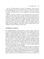
Fig. 8 The architecture for morphological analysis. Three expert systems operate: PA for check the number of wave peaks; PB for checking
similar points, and PC for checking different points The 1st layer of
the architecture: C1–PANC which processes input data of PA and PB;
C2–PANC which processes input data of PB and PC; C3–PANC which
processes input data of PC and PA. The 2nd layer of the architecture:
C4–PANC which calculates the maximum evidence value between cells
C1 and C2; C5–PANC which calculates the minimum evidence value
between cells C2 and C3; The 3rd layer of the architecture: C6–PANC
which calculates the maximum evidence value between cells C4 and C3;
C7–PANC which calculates the minimum evidence value between cells
C1 and C5. The 4th layer of the architecture: C8 analyzes the experts
PA, PB, and PC and gives the resulting decision value. PANC A = paraconsistent artificial neural cell of analytic connection. PANCLsMax =
paraconsistent artificial neural cell of simple logic connection of maximization. PANCLsMin = paraconsistent artificial neural cell of simple
logic connection of minimization. Ftce = certainty tolerance factor;
Ftun = uncertainty tolerance factor. Sa = output of C1 cell; Sb = output of C2 cell; Sc = output of C3 cell; Sd = output of C4 cell; Se =
output of C5 cell; Sf = output of C6 cell; Sg = output of C7 cell. C
= complemented value of input; μr = value of output of PANN; λr =
value of output of PANN
ables are called expert systems as they are specific routines
for extracting information.
In analyzing EEG signals, one important aspect to take
into account is the morphological aspect. To perform such
a task, it is convenient to consider an expert system which
analyzes the signal behavior verifying which band it belongs
to (delta, theta, alpha and beta).
The method of morphological analysis has three expert
systems that are responsible for feeding the inputs of PANN
with information relevant to the wave being analyzed: number of peaks, similar points, and different points.
Vietnam J Comput Sci (2014) 1:219–230
225
Table 8 Checking the number of wave peaks function implementation
Table 9 Checking similar points function implementation
4.4 Expert system 2: checking similar points
The aim of the expert system 2 is to compare the waves and
analyze their differences regarding to similar points.
When we analyze the similar points, it means that we are
analyzing how one approaches the other point.
It is worth remembering that, because it is biological signal, we should not work with absolute quantification due to
the variability characteristic of this type of signal. Therefore, one should always take into consideration a tolerance
factor.
A sample of checking similar points function implementation using Object Pascal is shown in Table 9.
4.3 Expert system 1: checking the number of wave peaks
The aim of the expert system 1 is to compare the waves and
analyze their differences regarding the number of peaks.
In practical terms, one can say that when we analyzed the
wave peaks, we are analyzing the resulting frequency of wave
(so well rudimentary).
It is worth remembering that, because it is biological signal, we should not work with absolute quantification due to
the variability characteristic of this type of signal. Therefore, one should always take into consideration a tolerance
factor.
A sample of checking the number of wave peaks function
implementation using Object Pascal is show in Table 8.
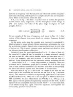
Se1 = 1 −
(|bd − vt|)
,
(bd + vt)
(4.2)
where vt is the number of peaks of the wave, bd is the number
of peaks of the wave stored in the database, Se1 is the value
resulting from the calculation.
Se2 =
n
j=1
n
xj
,
(4.3)
where n is the total number of elements, x is the element
of the current position, a j is the current position, Se2 is the
value resulting from the calculation.
4.5 Expert system 3: checking different points
The aim of the expert system 3 is to compare the waves and
analyze their differences regarding of different points.
When we analyze the different points, it means that we
are analyzing how a point more distant from each other, so
the factor of tolerance should also be considered.
A sample of checking different points function implementation using Object Pascal is shown in Table 10.
⎛
Se3 = 1 − ⎝
n
j=1
|x j −y j | ⎞
a
⎠,
n
(4.4)
where n is the total number of elements, a is the maximum
amount allowed, j is the current position, x is the value of
123
226
Vietnam J Comput Sci (2014) 1:219–230
Table 12 Statistical results—sensitivity and specificity: delta waves
Table 10 Checking different points function implementation
Visual analysis
Delta
Not delta
Total
True
31
124
155
False
22
3
25
Total
53
127
180
PANN
Sensitivity = 58 %; specificity = 97 %
Table 13 Statistical results—sensitivity and specificity: theta waves
Visual analysis
True
Visual analysis
Theta
Not theta
Total
88
65
153
PANN
Table 11 Contingency table
Delta
Theta
Alpha
Beta
Unrecognized
Total
False
10
17
27
Total
98
82
180
Sensitivity = 89 %; specificity = 79 %
PANN Analysis
Delta
31
3
0
0
0
34
Theta
15
88
1
1
0
105
Alpha
0
5
22
0
0
27
Beta
0
0
1
3
0
4
N/D
7
2
1
0
0
10
Total
53
98
25
4
0
180
Index kappa = 0.80
wave 1, y is the value of wave 2, Se3 is the value resulting
from the calculation.
Table 14 Statistical results—sensitivity and specificity: alpha waves
Visual analysis
Alpha
Not alpha
Total
True
22
150
172
False
3
5
8
Total
25
155
180
PANN
Sensitivity = 88 %; specificity = 96 %
Table 15 Statistical results—sensitivity and specificity: beta waves
Visual analysis
5 Experimental procedures: differentiating frequency
bands
Beta
Not beta
Total
True
3
175
178
False
1
1
2
Total
4
176
180
PANN
In our work we have studied two types of waves, specifically
delta and theta waves band, where the size of frequency established clinically ranges (Fig. 1).
Seven examinations of different EEG were analyzed,
being two examinations belonging to adults without any
learning disturbance and five examinations belonging to children with learning disturbance [5,6,13].
Each analysis was divided into three rehearsals; each
rehearsal consisted of 10 s of the analyzed, free from visual
analysis of spikes and artifacts regarding the channels T3 and
T4.
In the first battery of tests, a wave recognition filter belonging to the delta band was considered. In the second one, a
wave recognition filter belonging to the theta band was considered. In the third one, none of the filters were considered
for recognition (Tables 11, 12, 13, 14, 15, 16).
123
Sensitivity = 75 %; specificity = 99 %
Table 16 Statistical results—sensitivity and specificity: unrecognized
waves
Visual analysis
Unrecognized
Recognized
Total
True
0
170
170
False
0
10
10
Total
0
180
180
PANN
Sensitivity = 100 %; specificity = 94 %
Vietnam J Comput Sci (2014) 1:219–230
227
Table 17 Lattice for decision-making (Fig. 9) used in diagnostic analysis used after making PANN analysis (Fig. 10)
Characterization of the lattice
Area 1
Area 2
Area 3
Area 4
G ce ≤ 0.1999 and G ce ≥ 0.5600 and |G un | < 0.3999
and |G un | ≥ 0.4501
0.2799 < G ce < 0.5600 and 0.3099 ≤ |G un | < 0.3999
and Fe < 0.5000
0.1999 < G ce < 0.5600 and 0.3999 ≤ |G un | < 0.4501
and Fe > 0.5000
G ce > 0.7999 and |G un | < 0.2000
Ce contrary evidence, Fe favorable evidence, G ce certainty degree, G un
uncertainty degree
6 Experimental procedures: applying in Alzheimer
disease
It is known that the visual analysis of EEG patterns may be
useful in aiding the diagnosis of AD and indicated in some
clinical protocols for diagnosing the disease [14,15]. The
most common findings on visual analysis of EEG patterns
are slowing of brain electrical activity based on predominance of delta and theta rhythms and decrease or absence of
alpha rhythm. However, these findings are more common and
evident in patients in moderate or advanced stages of disease
[8,16,17].
In this study we have 67 analyzed EEG records, 34 normal
and 33 probable AD ( p value = 0.8496) during the awake
state at rest.
All tests were subjected to morphological analysis methodology for measuring the concentration of waves. Later
this information is submitted to a PANN unit responsible for assessing the data and arriving at a classification
of the examination in normal or probable AD (Table 17;
Fig. 9).
Fig. 9 The architecture for diagnosis analysis
6.2 Expert system 2: high-frequency band concentration
The role of the expert system 2 is to analyze alpha band
concentration. For this, we consider the quotient of the sum
of fast alpha and beta waves over slow delta and theta waves
(Eq. 6.2) as first output value. For the second output value
(contrary evidence λ) is used Eq. 6.1.
μ=
6.1 Expert system 1: detecting the diminishing average
frequency level
The aim of the expert system 1 is to verify the average frequency level of alpha band waves and compare them with a
fixed external parameter wave.
Such external parameter can be, for instance, the average
frequency of a population or the average frequency of the
last examination of the patient. This system also generates
two outputs: favorable evidence μ normalized values ranging
from 0 (corresponds to 100 %—or greater frequency loss) to
1 (which corresponds to 0 % of frequency loss) and contrary
evidence λ (Eq. 6.1).
The average frequency of population pattern used in this
work is 10 Hz.
λ=1−μ
(6.1)
(A + B)
,
(D + T )
(6.2)
where A is the alpha band concentration; B is the beta band
concentration, D is the delta band concentration; T is the
theta band concentration; and μ is the value resulting from
the calculation.
6.3 Expert system 3: low frequency band concentration
The role of the expert system 3 is to analyze theta band concentration. For this, we consider the quotient of the sum of
slow delta and theta waves over fast alpha and beta waves
(Eq. 6.3) as first output value. For the second output value
(contrary evidence λ) is used Eq. 6.1.
μ=
(D + T )
(A + B)
(6.3)
123
228
Vietnam J Comput Sci (2014) 1:219–230
•
•
Fig. 10 Lattice for decision-making used in diagnostic analysis (Fig.
9). Area 1 state logical false (AD likely below average population), area
2 state logical Quasi-true (AD likely than average population); area 3
state logical Quasi-false (normal below average population); area 4 state
logical true (normal above average population); area 5 logical state of
uncertainty (not used in the study area)
where A is the alpha band concentration; B is the beta band
concentration. D is the delta band concentration; and T is
the theta band concentration. μ is the value resulting from
the calculation.
6.4 Results
See Table 18.
7 Experimental procedures: applying in
attention-deficit/hyperactivity disorder (ADHD)
A similar architecture using PANN was built to study some
cases in ADHD. Recent researches reveal that 10 % of the
world population in school age suffer of learning and/or
behavioral disorders caused by neurological problems, such
as ADHD, dyslexia, and dyscalculia, with predictable consequences in those students’ insufficient performance in the
school [2–6,13].
Concisely, a child without intellectual lowering is characterized as bearer of ADHD when it presents signs of
• Inattention: difficulty in maintaining attention in tasks
or games; the child seems not to hear what is spoken;
difficulty in organizing tasks or activities; the child loses
Table 18 Diagnosis: normal × probable AD patients
Gold standard
AD patient (%)
Normal patient (%)
Total (%)
35.82
14.93
8.96
40.30
49.25
44.78
55.22
100.00
PANN
AD patient
Normal patient
Total
50.75
Sensitivity = 80 %; specificity = 73 ; index of coincidence (kappa)
76 %
123
•
•
things; the child becomes distracted with any incentive,
etc.
Hyperactivity: frequently the child leaves the class room;
the child is always inconveniencing friends; the child runs
and climbs in trees, pieces of furniture, etc; the child
speaks a lot, etc.
Impulsiveness: the child interrupts the activities of colleagues; the child does not wait his time; aggressiveness
crises, etc.
Dyslexia: the child begins to present difficulties to recognize letters or to read them and to write them although
the child has not a disturbed intelligence, that is, a normal
IQ;
Dyscalculia: the child presents difficulties to recognize
amounts or numbers and/or to figure out arithmetic calculations.
A child can present any combination among the disturbances above. All those disturbances have their origin in a
cerebral dysfunction that can have multiple causes, many
times showing a hereditary tendency.
Since from the first discoveries, those disturbances have
been associated with cortical diffuse lesions and/or more specific, temporal-parietal areas lesions in the case of dyslexia
and dyscalculia [2,5,13].
The disturbances of ADHD disorder seem to be associated with an alteration of the dopaminergic system, that is,
it is involved with mechanisms of attention and they seem
to involve a frontal-lobe dysfunction and basal ganglia areas
[3,13].
EEG alterations seem to be associated with those disturbances. Thus, some authors have proposed that there is
an increase of the delta activity in EEG in those tasks that
demand a larger attention to the internal processes.
Other authors [1] have described alterations of the delta
activity in dyslexia and dyscalculia children sufferers.
Klimesch [18] has proposed that a phase of the EEG component would be associated with the action of the memory
work. More recently, Kwak [19] has showed delta activity is
reduced in occipital areas, but not in frontals, when dyslexic
children were compared with normal ones.
In this way, the study of the delta and theta bands becomes
important in the context of the analysis of learning disturbances.
So, in this paper we have studied two types of waves,
specifically delta and theta wave bands, where the size of
frequency established clinically ranges 1.0–3.5 and 4.0–7.5
Hz, respectively.
Seven exams of different EEG were analyzed, being
two exams belonging to adults without any learning disturbance and five exams belonging to children with learning disturbances (exams and respective diagnoses given by
Vietnam J Comput Sci (2014) 1:219–230
229
ENSCER—Teaching the Brain, EINA—Studies in Natural
Intelligence and Artificial Ltda).
Each analysis was divided into three rehearsals, and each
rehearsal consisted of 10 s of the analyzed, free from visual
analysis of spikes and artifacts regarding the channels T3
and T4. In the first battery of tests, a delta recognition filter
wave was considered. For second battery of tests, a theta
recognition wave was considered. For the third battery of
tests, none of the filters were considered for recognition, i.e.,
the system worked freely for any wave type recognition. The
total number of exams is 180 (Tables 19, 20, 21, 22, 23, 24).
Table 22 Statistical results—sensitivity and specificity: alpha waves
Visual analysis
Alpha
Not alpha
Total
True
22
150
172
False
3
5
8
Total
25
155
180
PANN analysis
Sensitivity = 88 %; specificity = 96 %
Table 23 Statistical results—sensitivity and specificity: beta waves
Visual analysis
8 Conclusions
We believe that a process of the examination analysis using
a PANN attached to EEG findings, such as relations between
Table 19 Contingency table
Beta
Not beta
Total
True
3
175
178
False
1
1
2
Total
4
176
180
PANN analysis
Sensitivity = 75 %; specificity = 99 %
Visual analysis
Delta
Theta
Alpha
Beta
Unrecognized
Total
Table 24 Statistical results—sensitivity and specificity: unrecognized
waves
PANN analysis
Visual analysis
Delta
31
3
0
0
0
34
Theta
15
88
1
1
0
105
Alpha
0
5
22
0
0
27
Beta
0
0
1
3
0
4
Unrecognized
Recognized
Total
True
0
180
180
0
0
0
0
180
180
PANN analysis
N/D
7
2
1
0
0
0
False
Total
53
98
25
4
0
180
Total
Sensitivity = 100 %; specificity = 100 %
Index kappa = 0.80
Table 20 Statistical results—sensitivity and specificity: delta waves
Visual analysis
Delta
Not delta
Total
True
31
124
155
False
22
3
25
Total
53
127
180
PANN analysis
Sensitivity = 58 %; specificity = 97 %
Table 21 Statistical results—sensitivity and specificity: theta waves
Visual analysis
Theta
Not theta
Total
True
88
65
153
False
10
17
27
Total
98
82
180
PANN analysis
Sensitivity = 89 %; specificity = 79 %
frequency bandwidth and inter hemispheric coherences, can
create computational methodologies that allow the automation of analysis and diagnosis.
These methodologies could be employed as tools to aid in
the diagnosis of diseases such as dyslexia or Alzheimer, provided they have defined electroencephalographic findings.
In the case of Alzheimer’s disease, for example, the studies carried out previously have shown satisfactory results
[20] (but still far from being a tool to aid clinical) that
demonstrated the computational efficiency of the methodology using a simple morphological analysis (only paraconsistent annotated logic Eτ ). These results encouraged us to
improve the morphological analysis of the waves and try to
apply the method in other diseases besides Alzheimer’s disease.
With the process of morphological analysis using the
PANN, it becomes possible to quantify the frequency average of the individual without losing its temporal reference.
This feature becomes a differential, compared to traditional
analysis of quantification of frequencies, such as fast Fourier
123
230
transform, aiming at a future application in real-time analysis, i.e., at the time of acquisition of the EEG exams.
Regarding the specificity, the method showed more reliable results. Taking into account an overall assessment in the
sense we take the arithmetic mean of sensitivity (75.50 %)
and specificity (92.75 %), we find reasonable results that
encourage us to seek improvements in this study.
The consideration of morphological analysis of the main
brain waves by employing PANN showed be effective,
allowing interesting quantitative and qualitative examinations of EEG data. PANN has been applied in other branches:
MICR automated recognition [16], computer-aided diagnosis (breast cancer) [17], and many other themes.
Open Access This article is distributed under the terms of the Creative
Commons Attribution License which permits any use, distribution, and
reproduction in any medium, provided the original author(s) and the
source are credited.
References
1. Niedermeyer, E., da Silva, F.H.L.: Electroencephalography, 5th
edn. Lippincott Williams & Wilkins, Philadelphia (2005)
2. Ansari, D., Karmiloff-Smith, A.: Atypical trajectories of number
development: a neuroconstructivist perspective. Trends Cogn. Sci.
12, 511–516 (2002)
3. Blonds, T.A.: Attention-deficit disorders and hyperactivity. In
developmental disabilities in infancy and Ramus, F., developmental dyslexia: specific phonological deficit or general sensorimotor
dysfunction? Curr. Opin. Neurobiol. 13, 1–7 (2003)
4. Hynd, G.W., Hooper, R., Takahashi, T.: Dyslexia and languagebased disabilities. In: Coffey, C.E., Brumbak, R.A. (eds.) Text Book
of Pediatric Neuropsychiatry, pp. 691–718. American Psychiatric
Press, Washington, DC (1985)
5. Lindsay, R.L.: Dyscalculia. In: Capute, A.J., Accardo, P.J. (eds.)
Developmental Disabilities in Infancy and Childhood, pp. 405–
415. Paul Brookes Publishing Co, Baltimore (1996)
6. Temple, E.: Brain mechanisms in normal and dyslexic readers.
Curr. Opin. Neurobiol. 12, 178–183 (2002)
7. Kwak, Y.T.: Quantitative EEG findings in different stages of
Alzheimer’s disease. J. Clin. Neurophysiol. 23(5), 456–461 (2006)
8. Duffy, F.H., Albert, M.S., Mcnulty, G., Garvey, A.J.: Age differences in brain electrical activity of healthy subjects. Ann. Neural
16, 430–438 (1984)
123
Vietnam J Comput Sci (2014) 1:219–230
9. Nuwer, M.R., Comi, G., Emerson, R., Fuglsang-Frederiksen, J.,
GuériT, M., Hinrichs, H., Ikeda, A., Luccas, F.J.C., Rappelsberger,
P.: IFCN standards for digital recording of clinical EEG. Electroencephalogr. Clin. Neurophysiol. 106, 259–261 (1998)
10. Nitrini, R., Caramelli, P., Bottino, C.M., Damasceno, B.P., Brucki,
S.M., Anghinah, R.: Academia Brasileira de Neurologia. Diagnosis
of Alzheimer’s disease in Brazil: diagnostic criteria and auxiliary
tests. Recommendations of the Scientific Department of Cognitive
Neurology and Aging of the Brazilian Academy of Neurology. Arq
Neuropsiquiatr. 63(3A), 9–713 (2005)
11. Da Silva Filho, J.I., Torres, G.L., Abe, J.M.: Uncertainty Treatment
Using Paraconsistent Logic—Introducing Paraconsistent Artificial
Neural Networks, vol. 211. IOS Press, Netherlands (2010). ISBN
978-1-60750-557-0. doi:10.3233/978-1-60750-558-7-I
12. Abe, J.M.: Foundations of annotated logics. PhD thesis (in Portuguese) USP, Brazil (1992)
13. Voeller, K.K.S.: Attention-deficit/hyperactivity: neurobiological
and clinical aspects of attention and disorders of attention. In:
Coffey, C.E., Brumbak, R.A. (eds.) Text Book of Pediatric Neuropsychiatry, pp. 691–718. American Psychiatric Press, Washington, D.C (1998)
14. Claus, J.J., Strijers, R.L.M., Jonkman, E.J., Ongerboer De Visser,
B.W., Jonker, C., Walstra, G.J.M., Scheltens, P., Gool, W.A.: The
diagnostic value of EEG in mild senile Alzheimer’s disease. Clin.
Neurophysiol. 18, 15–23 (1999)
15. Crevel, H., Gool, W.A., Walstra, G.J.M.: Early diagnosis of dementia: which tests are indicated? What are their costs. J. Neurol. 246,
73–78 (1999)
16. Souza, S., Abe, J.M., Nakamatsu, K.: MICR Automated Recognition Based on Paraconsistent Artificial Neural Networks, Procedia
Computer Science, vol. 22, pp. 170–178. Elsevier, London (2013)
17. Amaral, F.V.: Paraconsistent mammography image attributes classifier in breast cancer diagnosis: based on paraconsistent artificial
neural network. PhD thesis (in Portuguese) UNIP, Brazil (2013)
18. Klimeshc, W.: EEG alpha and theta oscillations reflect cognitive
and memory performance: a review and analysis. Brain Res. Ver.
29, 169–195 (1999)
19. Klimesch, W., Doppelmayr, H., Wimmer, J., Schwaiger, D., Rôhm,
D., Bruber, W., Hutzler, F.: Theta band power changes in normal
and dyslexic children. Clin. Neurophysiol. 113, 1174–1185 (2001)
20. Lopes, H.F.S.: Aplicação de redes neurais artificiais paraconsistentes como método de auxílio no diagnóstico da doença
de Alzheimer. MSc Dissertation (in Portuguese), Faculdade de
Medicina-USP, São Paulo (2009)