Bidirectional glenn operation without cardiopulmonary bypass: Operative protocol and early results
Bạn đang xem bản rút gọn của tài liệu. Xem và tải ngay bản đầy đủ của tài liệu tại đây (328.81 KB, 10 trang )
JOURNAL OF MEDICAL RESEARCH
BIDIRECTIONAL GLENN OPERATION WITHOUT
CARDIOPULMONARY BYPASS: OPERATIVE PROTOCOL
AND EARLY RESULTS
Nguyen Tran Thuy¹, Ngo Thi Hai Linh¹, Doan Quoc Hung²
¹Cardiovascular Center, E Hospital
²Hanoi Medical University
The bidirectional Glenn (BDG) shunt operation serves as temporary treatment of single-ventricle physiology before the eventual Fontan procedure. Some cases can be performed without the support of a
cardiopulmonary bypass (CPB) machine. In this study, we present the surgical outcomes of off-pump
BDG operations with the use of temporary veno-atrial shunt to decompress the superior vena cava
(SVC) during clamping. From June 2013 to June 2015, 23 patients underwent off-pump BDG operations at Cardiovascular Center, E Hospital. All patients were operated on using a venoatrial shunt to
decompress the SVC. Satisfactory results with mean oxygen saturation increased from 79.6 ± 11.2 %
to 87.2 ± 4.7 %. The superior vena cava (SVC) clamping time was 14 ± 2.4 minutes (ranging from 12
to 21 minutes). No neurological complications or deaths occurred after the surgery and the postoperative period was uneventful. In conclusion, the use of venoatrial shunt to decompress SVC during
the off-pump BDG operation is safe and produces good surgical outcomes. Its wider adoption can the
deleterious effects associated with CPB. The operation is easily reproducible at low cost and overcome.
Keywords: congenital heart disease, bidirectional Glenn operation, without
cardiopulmonary bypass
I. INTRODUCTION
Bidirectional Glenn shunt operation is
performed as the initial step in the treatment of functional single-ventricle physiology before the completion of the Fontan
procedure. The purpose of this surgery is
to provide balanced venous blood flow into
two pulmonary arteries for oxygenation, as
Corresponding author: Nguyen Tran Thuy, E Hospital
Email:
Received:09 May 2017
Accepted: 16 November 2017
JMR 111 E2 (2) - 2018
oppoed to providing mixed ateriovenous
blood, as in the Blalock – Taussig shunt surgery (aortopulmonary shunt) [1 - 3].
Off-pump BDG operations without a
temporary shunt to decompress the SVC
will cause an elevation in the cerebral blood
volume, leading to increased intracranial
pressure and eventually, thereby, brain reduced blood flow to the brain and damage
[3; 4]
The BDG operation is conventionally
performed with the support of CPB at the
expense of higher cost and disadvantages
75
JOURNAL OF MEDICAL RESEARCH
of CPB. Therefore, globally, there has been
a variety of reports on BDG operations without CPB [1; 5; 6]. The have show that in offpump BDG operation, pulmonary arterial
pressure is lower and the hospital length of
stay of off-pump group is shorter than that
of the on-pump group [7; 8].
However, there have been no official reports on this issue in Vietnam. In this study,
we present the surgical protocol to perform
off-pump BDG operation using the SVC-RA
pressure lowering system and present early
outcomes of this newly applied technique
[9], [10].
II. SUBJECTS AND METHODS
1. Subjects
Subjects were patients who had attributes suitable for BDG operation, without
any intracardiac defects requiring correction including: pulmonary artery-plasty, atrial septal extension, atrioventricular valvuloplasty, etc.
2. Methods
The study disign was a retrospective observational study
Patients were prepared for the survey
through the following steps:
- Physical examination: Clinical symptoms (evaluating the severity of heart failure, using the NYHA classification, and the
level of cyanosis), SpO2, and medical history.
- Laboratory tests:
+ Routine blood tests, electrocardiography, and chest x-ray.
+ Echocardiography: evaluate left
ventricular function, abnormal wall motion,
76
chamber size, the functional status of the
heart valves, pulmonary artery (PA) size.
+ Cardiac catheterization: measure
PA size, anatomy, pressure and resistance.
- Definitive diagnosis was established
based on the following: physical examination, Doppler echocardiography, cardiac
catheterization, blood tests, electrocardiography and chest x-ray.
- Surgical consultation, hospital admission, and preoperative medical therapy.
- When all conditions had been assured,
the patients underwent surgery according to
the same protocol in anesthesia, operative
techniques, and postoperative resuscitation. In the operating room, hemodynamic
parameters were recorded.
- Technical procedure:
+ General anesthesia, intubation.
Premedication with Midazolam, Fentanyl,
Rocuronium. Patients were on controlled
mechanical ventilation with Vt = 150 ml and
the respiratory rate of 18 per minute. The
anesthesia was maintained by Isoflurane,
Fentanyl, and Rocuronium. A femoral vein
catheter was placed for drug distributions
and monitoring of the right atrial pressure.
A right internal jugular vein catheter was
inserted for SVC pressure monitoring. An
invasive arterial pressure line was also
placed.
- Surgical steps:
+ Whole body antiseptic application, from the chest to the legs;
+ Median sternotomy;
+ Dissect the SVC and ligate the
azygos vein;
+ Dissect the right branch of PA,
and measure PA pressure;
JMR 111 E2 (2) - 2018
JOURNAL OF MEDICAL RESEARCH
+ Set up the system to decrease
SVC-PA pressure
+ Trial right PA clamp for several
minutes to check the changes in transcutaneous oxygen saturation (SpO2). Systemic
heparin with the dose of 1 mg/kg to achieve
the ACT of more than 200 seconds. Set up
the system to decrease SVC-PA pressure
with the head of the patients elevated 15
degrees, inject methylprenisolone (20 mg/
kg) intravenously, SVC clamp to anstomose
with right PA, maintain the difference between mean arterial pressure and mean
SVC pressure during clamping higher than
40 mmHg.
During surgery, hemodynamic stability
was maintained by fluid replacement and
inotropes: adrenaline 0.1 mcg/kg/min and
Milrinone 0.3 mcg/kg/min.
+ Make end-to-side SVC-PA anastomosis by 7.0 prolene suture
+ Remove cannulae, achieve hemostasis, insert drains, electrodes, close
the pericardium if possible
+ Close the sternotomy by steel suture, soft tissue was closed using running
suture or interrupted absorbable suture in
patients with high risks of infection.
+ In the intensive care unit, an
echocardiography, routine laboratory tests
(complete blood count, electrolytes, arterial
blood gases, ...) were done. All complications and actions taken were recorded.
+ After the ICU stay, patients were
transferred to Pediatric Cardiology Department for further treatment until discharge.
3. Ethics
All study procedures complied with the
ethical principles of biomedical research.
Participants consented to take part in the
study and were told that they could withdraw at any time. Participants’ information
was kept secure and confidential.
III. RESULTS
From June 2013 to June 2015, we performed off-pump BDG operation on 23 patients. The mean SVC clamp time was 14 ±
2.4 minutes (ranged from 12 - 21 minutes).
During clamping, the mean central venous
pressure ranged from 24 to 40 mmHg (average 31.5 ± 6.1 mm Hg). Preoperative PA
pressure ranged 11 - 25 mmHg (average
16.3 ± 3.2 mmHg). There was no conversion to CPB machine.
Indications of patients undergoing BDG
operations are summarized in Table 1.
Table 1. Indications of patients undergoing BDG operations
Other surgeries
Patients (n)
Percent (%)
Single-ventricle physiology
11
47.8
Double outlet right ventricle with transposition of the great
arteries
5
21.7
Transposition of the great arteries, pulmonary stenosis,
large ventricular septal defect
6
26.2
Atrioventricular disassociation, double outlet right ventricle
1
4.3
JMR 111 E2 (2) - 2018
77
JOURNAL OF MEDICAL RESEARCH
Early results
The mean ventilator time after surgery was 2.6 ± 1.2 hours (1 - 6 hours), the ICU length
of stay was 13.2 ± 3.1 (10 - 18 hours); no death occurred. Echocardiography evaluation at
discharge showed no anastomosis stenosis, and postoperative electrocardiography (ECG)
revealed no arrhythmia.
Mean postoperative PA pressure was 13.6 ± 2.5 mmHg.
Table 2. Postoperative complications
Complications
Patient (n)
Percent (%)
Chylothorax
1
4.3
Pneumonia
2
8.6
Pulmonary effusion requires drainage
1
4.3
Surgical wound infection
1
4.3
Reoperation
1
4.3
Neurological deficits
0
0
Reoperation due to thrombus at the Glenn anastomosis
Table 3. Pre and postoperative Hct, SpO2
Parameters
Preoperative
Postoperative
p
Hct (%)
0.53 ± 0.11
0.43 ± 0.05
0.001
SpO2 (%)
79.96 ± 11.2
87.2 ± 4.7
0.011
The hospital length of stay ranged from
6 to 9 days (average 7.1 ± 1.3 days). Echocardiography showed no significant pressure gradient through the SVC-RPA anastomosis and also showed good velocity
of blood flow; ECG showed normal sinus
rhythm in all patients, and no neurological
complications were recorded.
IV. DISCUSSION
Several studies have documented the
decrease in oxyhemoglobin in brain tissue,
a 50% reduction in blood flow in the middle cerebral artery with significant changes
78
in encephalography. Rodriguez found that
clamping the SVC decreases the systolic
pressure of cerebral arteries and subsequently decreases the brain's oxygen supply [2 - 4]. To avoid these complications
many studies have reports on the used a
temporary shunt to decompress the SVC
and improve perfusion of the brain.
Table 4 is summary of all studies in the
past 15 years examining BDG operations
without CBP. Lamberti polished his research
on seven patients in 1990 and subsequently, there was a series of other studies examining off-pump BDG surgery [1; 5; 9].
JMR 111 E2 (2) - 2018
JOURNAL OF MEDICAL RESEARCH
Table 4. Studies on off-pump BDG surgery
Study
Year
Number of study
patients
Temporary shunt
Lamberti
1990
7
SVC – RA
Lal
1996
6
SVC – RA
Murthy K S
1999
5
SVC – PA
Jahangiri
1999
6
No
Villagra F
2000
5
No
Tiereli
2003
30
SVC – RA/PA
Maddali
2003
2
SVC – RA
Liu
2004
20
SVC – RA/PA
Luo
2004
36
SVC – RA
Maeba
2006
18
SVC – RA/PA
Kotani
2006
14
SVC – RA
Hussain
2007
22
No
Kandakure
2010
218
SVC – RA
13 studies
389
Total
RA: right atrium; PA: pulmonary artery; SVC: superior vena cava
(Until now, there have been no official reports on this technique in Vietnam).
In the study of Ulisses Alezandre Crotti, the mean age of on-pump group was 66 months
and that of off-pump group was 50 months (p = 0.17 using Mann-Whitney test). This suggests the differences in age, gender, weight, types of defects between on-pump and off-pump
group are not important factors in choosing the use of peripheral circulation.
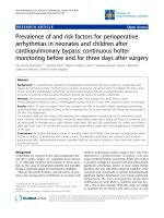
The choice of a temporary shunt depends on the experience and ability of the surgeons,
and anesthegist, as well as the conditions of the surgical center. Our technique uses a temporary veno-atrial shunt with the following steps: placing a venous graft at the junction of SVC
and azygos vein, which effectively decreases the pressure of the clamped SVC and avoids
the possibility of SVC stenosis. In addition, the head-elevated position during operation facilitates the adequate decompression of SVC and provides enough space surgical.
JMR 111 E2 (2) - 2018
79
JOURNAL OF MEDICAL RESEARCH
Figure 1. Description of a veno-atrial shunt used in our technique
SVC: superior vena cava; RPA: right pulmonary artery; LPA: left pulmonary artery; RA:
right atrium.
According to our experiences, with
the veno-atrial shunt, SVC pressure after
clamping did not exceed 40 mmHg. Postoperative chylothorax and dysfunction of
the diaphragm occured at low incidences
because, with our technique, the dissect on
field of SVC was short; avoids and the injury
to phrenic nerve and the refocus surrounding lymphatics. Performing Glenn operation
on patients who already have a Blalock –
Taussig shunt or patent arteriosus ductus
(PAD) is more convenient as the aortopulmonary shunt continuously supplies blood
for the lungs during the reconstruction of
Glenn anastomosis and maintains the good
stable oxygen saturation. The choice of
SVC and atrial cannula size were based on
the size of the patients SVC and right atrium, and patient’s weight, skin area in CPB.
During surgery, the cooperation between
80
surgeons and anesthetist is key to a successful off-pump BDG operation [6].
During SVC clamping, the blood flow to
the brain is reduced; therefore, to maintain
good cerebral perfusion during off-pump
BDG surgery, the authors proposed the
concept of transcranial pressure, which is
the difference between mean aterial pressure and mean SVC pressure during SVC
clamping (transcranial pressure = mean arterial pressure – central venous pressure)
[7]. This pressure has to be maintained at a
minimum of 30 mmHg during SVC clamping
to assure adequate cerebral perfusion. Veno-atrial shunt reduced SVC pressure and
improved cerebral perfusion [3; 4].
Monitoring of parameters of brain function to provide additional information about
hemodynamic effects of SVC clamping
on brain tissue transcranial Doppler ultra-
JMR 111 E2 (2) - 2018
JOURNAL OF MEDICAL RESEARCH
sound, near-infrared spectroscopy, and
encephalography [6]. However, these tests
were not available during this study so the
authors monitored brain function by assessing mean arterial pressure and central venous pressure.
Corticosteroid was used to minimalize
brain edema and neurological insults. Body
temperature was kept at approximately 33
- 34⁰C in order to reduce the metabolism
of brain cells and adjust for the reduced
pressure of cerebral blood flow during SVC
clamping. Inotropes and crystalloid replacement were used to maintain adequate cerebral blood flow and a transcranial pressure
higher than 30 mmHg during SVC clamping
[8].
Hypoxia was regulated by increasing
fraction of inspiratory oxygen (FiO2), increasing mean arterial pressure by using
inotropes, and providing enough circulating
fluid and to improve blood flow.
Postoperative treatment to the lung decreas pulmonary vascular resistance and
increas blood return to the SVC. Pulmonary
dilation medications (milrinone, iloprost
...) helped to decrease pulmonary arterial
pressure and end-diastolic left ventricular
pressure [1]. Prolonged mechanical ventilation time resulted in increased intrathoracic
pressure and negatively affected the blood
return to the SVC and blood flow through
the shunt, early weaning and extubation
helped circument these problems. The
mean time on ventilator in our study was
2.6 ± 1.2 hours (1 - 6 hours), which is comparable to other studies [1]. Short ventilatory time is also a big advantage of off-pump
BDG operation compared to conventional
JMR 111 E2 (2) - 2018
BDG surgery with CPB [2; 3; 12]. The different factors in our study are comparable
to those Crotti.The mean duration to extubation of the off-pump group was 3 hours
and that of on-pump group was 11 hours (p
= 0.83). The mean length of stay in the ICH
was 3 days and 5 days in the on-pump and
off-pump group, respectively (p = 0.29). The
average hospital length of stay of the former
group was 9 days, of the latter group was
5 days, and of the whole study group was
7 days. In Mohamed, the off-pump group
were extubated earlier, and had shorter
length of stay in the ICU and shorter hospital length of stay than on-pump group.
In our study, all cases had shunts that
supplied blood to the lungs; and the patent
arteriosus ductus, collaterals, aortopulmonary shunt (Blalock-Taussig) had the shunt
ligated to avoid the increased left ventricular after load, improve cardiac function, and
decrease the severity of atrioventricular
valve regurgitation [9]. Mean postoperative
pulmonary arterial pressure was 13.6 ± 2.5
mmHg, which was the ideal pressure after
BDG operation. According to Tables 3 and
4, the oxygen saturation was significantly
improved after surgery (p < 0.011) and the
hematocrit decreased substantially postoperatively (p < 0.001). In 23 study participants, there were six cases with early postoperative complications, which accounted
for 26.1% of the total sample (Table 2), and
only one cases with more than one complication. According to Chang [9], the incidences of postoperative complications, such as
superior vena cava syndrome, low cardiac
output syndrome, arrhythmia, were high,
while in research in our center and by oth81
JOURNAL OF MEDICAL RESEARCH
er authors [10], the incidences of the above
mentioned complications were very low.
There were no case requiring reoperation in
our study; in other research the rate of this
complication was 6%. There was a case requiring reoperation; especially three days
after BDG surgery, facial edema occurred
and echocardiography revealed thrombi
inside SVC. In reoperation, we found that
there were thrombi along the central venous
catheter and at the Glenn anastomosis. The
thrombi were removed and the central venous catheter was replaced. The reason
for this thrombi formation may be from in
the previous surgery, during the separation
of SVC when we cut a part of the central
venous catheter that lies in right atrium
(Catheter which is too long will cause the
difficulty for operation and cannot measure
SVC pressure), In general, the incidences
of postoperative complications in our study
are comparable or lower than other studies
[11; 12].
In our study, there were no deaths in offpump group and two deaths in the on-pump
group. There were no cases with chylothorax in the off-pump group, but eight patients
in the on-pump group suffered from this
complication. Only two patients in off-pump
group had early complications, while 14 patients in the on-pump group did. One advantage of the Glenn procedure without peripheral circulation is the significant reduction
in post surgical complications compared to
on-pump group. Our results are comparable to those of Mohamed’s study. The rates
of hemorrhage requiring reoperation in two
groups are significantly different (p = 0.044);
the rate of chylothorax in on-pump group is
82
significantly higher than that of the off-pump
group (p < 0.01). The early mortality rates
of on-pump and off-pump groups are 0%
and 4%, respectively. The causes of death
in on-pump group were low cardiac output
syndrome, heart failure, and neurological
complications. Comparing the results from
this study, to ours the off-pump group had
better postoperative recovery, shorter time
on mechanical ventilator, shorter length of
stay in the ICU and hospital, and fewer post
surgical complications compared to those
undergroing the on-pump Glenn procedure.
Without the CPB machine, patients can
avoid unwanted effect including: increased
pulmonary vascular resistance, blood dilution, air embolism and a host of other undesirable effects. Tireli [13] 2003, confirmed
that in the off-pump BDG operation, pulmonary arterial pressure was lower and the
hospital length of stay of off-pump group
was shorter than the those of on-pump
group. All patients were on heparin in the
first 24 hours, and aspirin was used subsequently. Patients were monitored regularly,
and all of them maintained good oxygen
saturation; no neurological complications
occurred.
Reducing medical cost a global priority.
According to Hussain (2007), the cost of an
on-pump BDG surgery is 1200 USD and that
of an off-pump BDG operation is only 250
USD [8]. To date, the cost of a BDG shunt
institution with CPB (49 million VND) is 7
times higher than that of the same operation
without CPB (7 million VND) at our Cardiovascular Center. The off-pump BDG operation technique reduced cost by omitting use
of CPB, reducing use of blood products and
JMR 111 E2 (2) - 2018
JOURNAL OF MEDICAL RESEARCH
reducing the suctioning system after sterilization. Postoperative period and hospital
length of stay were shorter, and the rates
of pulmonary effusion, chylothorax and diaphragm paralysis were lower. Lastly no neurological complications were documented.
V. CONCLUSION
After performing off-pump BDG shunt
institution in 23 patients from June 2013 to
June 2015, at Cardiovascular Center - E
Hospital, we concluded that off-pump BDG
operation using veno-atrial shunt to decompress the SVC was safe, and produced satisfactory surgical outcomes. This technique
can avoid the disorders caused by CPB,
significantly improve oxygen saturation, and
the quality of life, and reduce mortality rate
after Fotan procedure.
Acknowledgements
I would like to express my deepest gratitude to the Cardiovascular Center, E Hospital for supporting us in the data collection
process.
REFERENCES
1. A. K. D. P R Kandakure (2012). Venoatrial Shunt-Assisted Cavopulmonary
Anastomosis. Asian Cardiovasc Thorac
Ann, 18, 569 – 573.
2. L. Y. Liu J, Chen H, Shi Z, Su Z, Ding
W (2004). Bidirectional Glenn procedure
without cardiopulmonary bypass. Ann Thorac Surg, 77, 1349 – 1352.
3. W. N. Rodriguez RA, Cornel G.
(2000). Should the bidirectional Glenn procedure be better performed through the
JMR 111 E2 (2) - 2018
support of cardiopulmonary bypass? J Thorac Cardiovasc Surg, 119, 634 - 635.
4. C. G. Rodriguez RA, Semelhago
L, Splinter WM, (1997). Cerebral effects
in superior vena cava obstruction: the role
of brain monitoring. Ann Thorac Surg, 64,
1820 - 1822.
5. M. G. Mahadev Dixit, M.Ch., Anuradha Dubey, M.Ch., (2007). Off Pump Bidirectional Glenn performed through a thoracotomy. Ind J Thorac Cardiovasc Surg,
23, 180 - 183.
6. N. R. F Onyekwulu, P Kandakure
(2011). Anesthesia For Off Pump Bidirectional Glenn Shunt Surgery: Case Report.
The Internet Journal of Anesthesiology, 30
1 - 3,
7. K. B. Jahangiri M, Shinebourne EA,
Lincoln C (1999). Should the bidirectional
Glenn procedure be performed through a
thoracotomy without cardiopulmonary bypass?. J ThoracCardiovasc Surg, 118, 367
– 368.
8. A. B. Syed Tarique Hussain, Savita
Sapra, Rajnish Juneja (2007). The bidirectional cavopulmonary (Glenn) shunt without
cardiopulmonary bypass: is it a safe option?
Interact CardioVascThorac Surg, 6, 77 - 82.
9. C. A. e. al (1993). Early bidirectional cavopulmonary shunt in young infants:
Postoperative course and early results. Circulation, 88, 149 - 158.
10. C. J. e. al (2003). Effects of controlled antegrade pulmonary blood flow on
cardiac function after Bidirectional cavopulmonary anastomosis. Ann Thorac Surg, 76,
1917 - 1921.
11. R. C. Kona Samba Murthy,
Shivaprakasha K. Naik (1999). Novel
83
JOURNAL OF MEDICAL RESEARCH
Techniques of Bidirectional Glenn Shunt
Without Cardiopulmonary Bypass. Ann
Thorac Surg, 67, 1771 - 1774.
12. Z. J. F. Xie Bin, Devi Prasad Shetty
(2001). Bidirectional Glenn Shunt: 170 Cases. Asian Cardiovasc Thorac Ann, 9, 196 -
84
199.
13. B. M. Tireli E, Kafali E, et al (2003).
Peri-operative comparison of different transient external shunt techniques in bidirectional cavo-pulmonary shunt. Eur J Cardiothoracic Surg, 23, 518 – 524.
JMR 111 E2 (2) - 2018