Ebook CT and MRI of the abdomen and pelvis - A teaching file (3rd edition): Part 1
Bạn đang xem bản rút gọn của tài liệu. Xem và tải ngay bản đầy đủ của tài liệu tại đây (26.42 MB, 115 trang )
CT and MRI of the
Abdomen and Pelvis
A Teaching File
EDITION
CT and MRI of the
Abdomen and Pelvis
A Teaching File
•
DITION
Editors
Pablo R. Ros, MD, MPH, FACR
Theodore J. Castele University
Professor and Chair
Department of Radiology
University Hospital.9 Case Medical Center
Case Western Reserve University
Radiologist-in-Chief
University Hospitals Health System
Cleveland, Ohio
Koenraad J. Mortele, MD
Associate Professor of Radiology
Harvard Medical School
Director, Division of Clinical MRI
Staff Radiologist. Divisions of Abdominal Imaging and Body MRI
Department of Radiology
Beth Israel Deaconess Medical Center
Boston. Massachusetts
Associate Editors
Vincent Pelsser, MD
Assistant Professor of Radiology
McGill University
Staff Radiologist
Jewish General Hospital
Montreal, Quebec, Canada
Smitha Thomas, MD
Clinical Instructor, Abdominal Imaging
University Hospitals Case Medical Center
Case Western Reserve University
Cleveland, Ohio
I
®.Wolters Kluwer Lippincott Williams & Wilkins
HMith
l'h18cltllhill • Belli'nOtt • Ntw"'"' •I.Mion
a-Am •Hare l(q• Syfty • Takyo
Senior Executive Editor: Jonathan W. Pine, Jr.
Product Manager: Amy G. Dinkel
Vendor Manager: Bridgett Dougherty
Senior Manufacturing Coordinator: Beth Welsh
Senior Marketing Manager: Kimberly Schonberger
Senior Designer: Stephen Druding
Production Service: Integra Software Services Pvt. Ltd.
© 2014 by LIPPINCOTT WILLIAMS & WILKINS, a WOLTERS KLUWER business
Two Commerce Square
2001 Market Street
Philadelphia, PA 19103 USA
LWW.com
Second Edition© 2007 by LIPPINCO'IT WllLIAMS & WILKINS, a WOLTERS KLUWER business
First Edition© 1997 by LIPPINCOTT WILLIAMS & WILKINS, a WOLTERS KLUWER business
All rights reserved. This book is protected by copyright. No part of this book may be reproduced in any
form by any means, including photocopying, or utilized by any information storage and rettieval system
without written permission from the copyright owner, except for brief quotations embodied in critical
articles and reviews. Materials appearing in this book prepared by individuals as part of their official
duties as U.S. government employees are not covered by the above-mentioned copyright.
Printed in China
Library of Congress Cataloging-in-Publication Data
CT and MRI of the abdomen and pelvis : a teaching file I editors, Pablo R. Ros, Koenraad J. Mortele;
associate editors, Vincent Pelsser, Smitha Thomas. -Third edition.
p.;cm.
Includes bibliographical references and index.
ISBN-13: 978-1-4511-1352-5
ISBN-10: 1-4511-1352-8
I. Ros, Pablo R., editor of compilation. IT. Mortele, Koenraad J., editor of compilation. ill. Pelsser,
Vincent, editor of compilation. IV. Thomas, Smitha, editor of compilation.
[DNLM: 1. Abdomen-pathology-Atlases. 2. Diagnosis, Differential-Atlases. 3. Digestive
System Diseases--diagnosis-Atlases. 4. Magnetic Resonance Imaging-Atlases. 5. Pelvispathology-Atlases. 6. Tomography, X-Ray Computed-Atlases. Wl17]
RC944
617 .5'50754S--dc23
2013030751
Care has been taken to confirm the accuracy of the information presented and to describe generally
accepted practices. However, the authors, editors, and publisher are not responsible for errors or
omissions or for any consequences from application of the information in this book and make no
warranty, expressed or implied, with respect to the currency, completeness, or accuracy of the contents
of the publication. Application of the information in a particular situation remains the professional
responsibility of the practitioner.
The authors, editors, and publisher have exerted every effort to ensure that drug selection and
dosage set forth in this text are in accordance with current recommendations and practice at the time of
publication. However, in view of ongoing research, changes in government regulations, and the constant
flow of information relating to drug therapy and drug reactions, the reader is urged to check the package
insert for each drug for any change in indications and dosage and for added warnings and precautions.
This is particularly important when the recommended agent is a new or infrequently employed drug.
Some drugs and medical devices presented in the publication have Food and Drug Administration
(FDA) clearance for limited use in restricted research settings. It is the responsibility of the health care
provider to ascertain the FDA status of each drug or device planned for use in their clinical practice.
To purchase additional copies of this book, call our customer service department at (800) 638-3030 or fax
orders to (301) 223-2320. International customers should call (301) 223-2300.
Visit Lippincott Williams & Wilkins on the Internet at: LWW.com. Lippincott Williams & Wilkins
customer service representatives are available from 8:30 am to 6 pm, EST.
10 9 8 7 6 54 3 2 1
To all the residents I had the opportunity to show cases to during my career. They are the
inspiration for this book.
Pablo Ros, MD
To Dejana, for her bottomless love.
To Charlotte, Christophe, Mabel and Mila-my four extraordinary children-for the jay
they give me every day.
To my family and friends, for all their help and support over the years.
To my mentors, colleagues and trainees, for keeping abdominal imaging exciting and fun
to practice!
Koenraad J. Mortele, MD
To my parents, Albert and Odile, for instilling in me their values.
To my brother Bernard, for his invaluable advice.
To Chantal, for her love and unconditional support.
Vincent Pelsser, MD
To Binu, Matthew, and Irene, for your support and understanding.
Smitha Thomas, MD
Teaching Files are one of the hallmarks of education in radiology. When there was a need for a comprehensive series of books to provide the resident and practicing radiologist with the kind of personal consultation with the experts normally found only in the setting of a teaching hospital, Lippilloott Williams
& Wilkins is proud to have created a series that answers this need.
Actual cases have been culled from extensive teaching files in major medical centers. The discussions presented mimic those performed on a daily basis between residents and faculty members in all
radiology departments.
This series is designed so that each case can be studied as an unknown. A consistent format is used to
present each case. A brief clinical history is given, followed by several images. Then, relevant findings,
differential diagnosis, and final diagnosis are given, followed by a discussion of the case. The authors
thereby guide the reader through the interpretation of each case.
This year we have made additional changes to the series. Cases have been randomized to better prepare the reader for the challenges of the clinical setting. In addition, to answer the growing demand for
Web-based product, we have included more cases online, which has left us, in tum, able to offer a more
cost-effective product.
We hope that this series will continue to be a trusted teaching tool for radiologists at any stage of
training or practice, and that it will also be a benefit to clinicians whose patients undergo these imaging
studies.
The Publisher
vi
I
rHE THIRD EDITION
As the saying goes, the only constant in life is change. Obviously, we are delighted that the Third
Edition of CT and MRJ of the Abdomen and Pelvis: A Teaching File is seeing the light in its third
incarnation.
This book., which started as a coll.ection of interesting cases in the First Edition, became a more
robust textbook in the Second Edition with 470 cases and almost 2,000 illustrations in a 500-page hardcover volume. Fortunately, the Second Edition did well, and as with the First Edition, our publishers
started to lobby for a new edition. However, as mentioned, things had changed. The changes were obvious, both in our team and in the publishing world. In the time that since the Second Edition (2007), the
publishing environment has changed. Radiologists throughout the world, like any other walk. of life, are
living not only in the world of hard copy material but also in the world of digital and Web-based media.
1bis is an incredibly attractive alternative for a specialty such as ours that is primarily based on images.
The possibilities of Web-based publications are endless. Therefore, the Third Edition of CT and MRI
ofthe Abdomen and Pelvis: A Teaching File is a hybrid publication. The hard copy version is limited
to 150 cases, and it bas a soft cover for better ease of transportation and immediate access for consultation. Of interest is that the 150 cases contain completely new material either with totally new cases not
presented in the previous editions or with completely new and current images of well-established and
classic entities. Our beloved textbook is becoming a leaner, meaner textbook.
The beauty of the hybrid approach is that the publ.i.shers have made available to all of the patrons of
this Third Edition 416 cases that can be accessed on the Web.
Our team also bas undergone changes. What was originally the effort of a small team of two people
in the late 1990s, became a larger team for the Second Edition but still primarily based in a single
center where Vincent, Koenraad, and Pablo were working in the mid-2000s. The Third Edition is now
based in three different centers in two countries. Vincent returned to his native Canada and therefore
his contributions are cases from McGill University in Montreal; Koenraad moved into town to the Beth
Israel-Deaconess Medical Center; and Pablo moved to Case Western Reserve and therefore enlisted
the help of Smitha. In short, we are delighted that the Third Edition is now a reality, despite publication
and personnel changes.
1bis volume is faithful to its basic principles: it is composed of great clinical cases with exquisite
illustrations of the highest quality. Another change from prior editions is that all the cases are now
randomized and not presented by chapters following organ/system divisions.
We hope our readers will also have fun with the Third Edition of CT and MRI of the Abdomen and
Pelvis: A Teaching File and will be infected by the enthusiasm of the authors for teaching Abdominal
Imaging using a case format. We will be looking forward to receiving feedback regarding this hybrid
publication combining hard copy printed material and access to hundreds of Abdominal Imaging cases
on the Web.
Pablo R. Ros, MD, MPH, FACR
Koenraad J. Mortele, MD
Vincent Pelsser, MD
Smitha Thomas, MD
vii
I
~HE
SECOND EDITION
Although it is said that sequels rarely improve on the original movie, we hope our readers will agree
that this Second Edition of our book, CT and MRI of the Abdomen and Pelvis: A Teaching File, is
clearly better than its predecessor. The images are technically better, there is an increased number of
cases illustrating more entities; it includes advanced technology, such as three-dimensional reformatted
images; and it has more collaborators with specialized expertise than the First Edition.
This project started a few years ago when we kept receiving emails and verbal comments from radiologists asking if they could get a copy of the First Edition since it was out of print. Because we did not
have additional copies of the book on hand, we started to think about writing a Second Edition. Because
we had a professional relationship of over 10 years and understood each other very well, it was natural
to decide to pool our efforts and talents to tackle this Second Edition.
We initially thought we could keep 80% of the old cases, add 20% of new ones, and update a few
of the older images. Doing that would have taken only a few months. We really underestimated the
amount of work to be done. Because we wanted to offer to our readers a complete, modern collection
of outstanding cases, we ended up adding many more cases, changing almost all of the images, and
making this Second Edition a more robust and complete teaching atlas. We selected the best possible
cases out of our daily practice at the Brigham and Women's Hospital and Dana-Farber Cancer Institute
in Boston, Massachusetts, and put them in an unknown case format, as we would present them in our
routine case conferences. We also tried to incorporate cases that one of us has had the chance to see
during our visits to other departments, particularly the Armed Forces Institute of Pathology in Washington, DC, and the University Hospital in Ghent, Belgium, or received in consultation from the United
States and abroad. We have kept the best material from the First Edition because we realized that some
cases were so unique that we could not replace them.
At the end, we had trouble limiting the number of cases we wanted to include from our pool and
staying within the space allowed by this single volume. We enjoyed meeting weekly with Vincent and
viii
Preface to the Second Edition
ix
trying to convince each other to include '1ust one more case" in a particular section, constantly updating
differential diagnoses and making sure we had the best and most updated discussions and references
for each case.
The structure of the book is similar to the First Edition. We have divided the cases according to
the traditional abdominal sections: Liver and Biliary System; Pancreas; Gastrointestinal Tract; Spleen;
Mesentery, Omentum, and Peritoneum; Kidney, Ureter, and Bladder; Pelvis; Retroperitoneum and
Adrenal Glands; and Abdominal Wall. For each case, after a brief history, up to four images follow,
which by definition are CI' and/or MRI. A brief description of the findings, the differential diagnosis,
the final diagnosis, and a short discussion complete the case. This format allows the readers to take these
cases as unknowns, thereby simulating the daily clinical practice of a radiologist.
We hope our readers will have fun with the Second Edition of CT and MRI of the Abdomen and
Pelvis: A Teaching File and sense the enthusiasm of the authors for teaching abdominal imaging using
a case format. If our readers gain even a small nugget of additional knowledge, we will feel satisfied that
the educational goal of this book has been achieved.
Pablo R. Ros, MD, MPH, FACR
Koenraad J. Mortele, MD
I
~ HE
FIRST EDITION
This book is the fruit of our collaboration that spans at least 6 years, when both of us were in the
Department of Radiology at the University of Florida College of Medicine, Gainesville. Although
the point of view of one of us (Sly) changed from medical student to radiology resident. fellow, and
finally attending, we tried to duplicate the experience of reviewing interesting cases presented in the
Abdominal Imaging divisional conferences, in "hot seat'' sessions for the senior residents, and most
importantly, .in daily read-out sessions.
Our goal was to select the "best in show'' material out of an archive of over 5,000 teaching file cases
and put it in book format. The Abdominal Imaging teaching file at the University of Florida College
of Medicine contains primarily cases that have been performed at Shands Hospital at the University
of Florida and also cases originating from the Armed Forces Institute of Pathology, Washington. DC.
It also includes cases collected in visiting professorships in the United States and Canada, as well as
other countries in Europe, Central and South America, and Asia, cases that have been presented in
film-reading panels in national and international meetings, and cases that have been brought by visitors
to the department. All cases entered into the teaching file have to be proven by either surgery, biopsy,
laboratory data, or cl.inical and/or radiologic follow-up. Cases with an obvious pathognomonic imaging diagnosis (e.g., pneumoperitoneum) are also included. From this pool, we selected the best ones
and divided them into chapters according to the traditional abdominal sections: liver and biliary tree,
pancreas, spleen, gastrointestinal tract. kidney, retroperitoneum and adrenal, mesentery and omentum,
and pelvis. We also added a chapter called "Unknowns and Aunt Minnies," which contains a potpourri
of cases with a short differential diagnosis.
The format for each case is the same. A brief clinical history is followed by two to four images, which
by definition are either computed tomography, magnetic resonance imaging scans, or a combination
of both. Then, pertinent findings, differential diagnosis, final diagnosis, and a brief discussion follow.
This format is designed so that cases can be taken as unknowns. A simple piece of paper will cover
the entire infolmation given on each case. If the reader wants to know the findings or the differential
diagnosis before knowing the final diagnosis, this can be easily accomplished by removing the paper.
To make this book reflect real life, we took actual cases from an extensive teaching file and reaeated
the discussions performed on a daily basis in hundreds of departments of radiology between residents
and faculty members. We duplicated our discussions at the viewbox, emphasizing a practical approach.
The cases in each chapter are not presented with traditional divisions (congenital, infiammatory, neoplastic, vascular, etc.), but are all mixed up, again mimicking real life. The end result, we hope, is that
radiologists at any stage in their training or careers will benefit from reading this book.
We had fun selecting the cases, going over differential diagnosis lists, and trying to summarize a
pertinent discussion for each case. We hope the reader will also enjoy going over them and learning
more about diseases of the abdomen and pelvis using computed tomography and magnetic resonance
imaging as diagnostic tools.
Pablo R. Ros, MD, MPH, FACR
Sylvester Lee, MD
X
The Publication of any book iB possible with the efforts of many contributors. The same happened with
this Third Edition of our book.
Like in prior editions, we like to acknowledge the residents, fellows, and supporting staff of the
division of Abdominal Imaging at Case Western reserve University/University Hospitals Case Medical Center and the Divisions of Body MRI and Abdominal Imaging of Beth IsraeiiDeaconess Medical
Center/Harvard Medical School. All of our trainees and support staff directly and indirectly contributed
to this book by either donating cases, presenting differential diagnosis when the cases were originally
read, or producing the images that now constitute this volume. We also wish to extend our gratitude to
many clinical colleagues around the world who also provided cases.
A special thanks goes to the staff of Wolters K.luwer-Lippincott Williams and Wilkins. Without the
help of Jonathan Pine, Sarah Granlund, Amy Dinkel, and Jeffrey Gunning, this edition would not have
come to fruition.
Special appreciation also goes to our .Administrative and Executive Assistants, Molly McGinnis
and Marianne Chaloupek from University Hospitals Case Medical Center, and Lois Gilden from Beth
Israel Deaconess Medical Center who helped us prepare and send materials to the publisher and also
made our days go smoothly so we could dedicate time to the preparations of the materials.
Pablo R. Ros, MD, MPH, FACR
Koenraad J. Mortele, MD
Vincent Pelsser, MD
Smitha Thomas, MD
xl
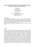
CLINICAL HISTORY 28-year-old man presenting with persistently elevated liver function tests
post-ERCP for stone extraction.
FIGURE
1C
FIGURE
10
FIGURE 1A
DIAGNOSIS Pneumobilia.
FIGURE
18
FINDINGS Thick-slab MRCP image (A) demonstrates
absence of fluid signal in the common hepatic duct (CHD)
and apparent strictures of left-sided intrahepatic bile ducts.
Note the mild bile duct dilatation. Axial fat-suppressed
T2-WI (B) shows nondependent dark signal in the CHD
with dependent layering :fluid. Out-of-phase Tl-WI (C) and
gadolinium..enhanced, fat-suppressed axial Tl-WI (D) demonstrate a larger signal ''void" in the central bile ducts and in
the intrahepatic ducts.
DIFFERENTIAL DIAGNOSIS Retained stones, flow artifact.
2
DISCUSSION Pneumobilia or gas in the bile ducts is usually the result of a previous ERCP or choledocho-enteric
anastomosis. Rare causes include fistulas with bowel due to
stone disease, or from tumor eroding into bowel. As gas is
less dense than :fluid, it forms a nondependent layer above the
fluid in the bile ducts, and during an MRI examination. it will
shift preferentially in the left-sided intrahepatic bile ducts, as
they are the most nondependent ducts in the supine position.
On the gradient echo sequences with longer echo times, due
to magnetic susceptibility, gas tends cause blooming artifacts
and may even obscure residual bile signal, as seen in this case.
Other causes of filling defect in the bile ducts are stones, but
these are usually in the extrahepatic ducts, and will be dependent as they are denser than bile. Flow artifact can also be
seen in the extrahepatic bile ducts, in the form of a dark signal, but this signal is centrally located and entirely surrounded
by fluid signal, allowing distinction from a true filling defect.
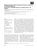
CLINICAL HISTORY 22-year-oldfemale presenting withfrver.
FIGURE
fiGURE
FIGURE
2C
FIGURE
20
2A
28
FINDINGS Coronal (A and B) CEcr images of the lung
demonstrates hypoarterial branching pattern of bilateral
bronchi and bilobar lungs. Axial CEC'f images (C and D)
demonstrate centrally located liver and stomach, polysplenia
and interrupted IVC with azygous continuation.
DIFFERENTIAL DIAGNOSIS None.
DIAGNOSIS Heterotaxy syndrome.
DISCUSSION Heterotaxy is defined as an abnormality
where the internal thoracoabdo.minal organs demonstrate
abnormal arrangement across the left-right axis of the
body. They can be divided into two subgroups: asplenia
syndrome and polysplenia syndrome. Polysplenia is more
common in females. In general, cardiac anomalies are less
common in this subgroup. The critical structures evaluated with imaging in determining situs include position of
atria, position of venous drainage below the diaphragm,
position of liver and gallbladder, presence, appearance,
and number of spleen, and presence of bi- or trilobed
lungs. Malrotation of the bowel is a frequent finding in
heterotaxy syndrome. The common findings in polysplenia syndrome include bilateral bilobar lungs and IVC
interruption. The spleen or spleens are always on the same
side of the stomach, typically along the greater curvature.
The splenic function may be abnormal and patients can
even present with biliary atresia. The mortality rate in the
first year of life is about 60% in polysplenia and 85% in
asplenia syndrome.
3
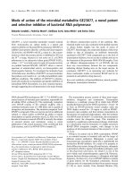
CLINICAL HISTORY 64-year-old man presenting with left lower quadrant pain.
FIGURE 3A
FIGURE
3C
38
FIGURE
30
FIGURE
FINDINGS Axial T2-WI (A) and fat-suppressed T2-WI
(B) show a 3.5-<:m hypointense exophytic renal mass. It is
isointense to the renal cortex on the out-of-phase Tl-WI
(C). Gadolinium-enhanced, fat-suppressed axial Tl-WI (D)
shows only mild enhancement of the mass.
DIFFERENTIAL DIAGNOSIS Clear cell renal carcinoma,
angiomyolipoma with minimal fat, oncocytoma.
DIAGNOSIS Papillary renal cell carcinoma.
DISCUSSION Papillary renal carcinoma is the second most
common (15% to 20%) pathologic subtype of primary renal
neoplasm, clear cell (conventional) being the most common
(70%). The 5-year survival rate is also much better with the
papillary subtype (80% to 90%) when compared to the conventional type (55% to 60%). Although papillary renal cell
cancer may be hypcrdense on NECT, the attenuation value
of the mass on NECT is not a reliable differentiating factor. However, on CEcr, papillary renal carcinoma enhances
4
less than the clear cell subtype. Tumors that enhance more
than 84 HU in the corticomedullary phase are more likely
to be of the clear cell variant. Papillary renal carcinomas are
considered hypovascular, as seen in this case. Calcifications
in a renal mass are also more common in the papillary variant (32%), than in the conventional type (11%). On T2-WI,
the signal intensity of the papillary subtype tends to be
less than that of the renal parenchyma. On the gadoliniumenhanced gradient echo sequence, the papillary tumors may
appear hypointense, as seen in this case, likely due to artifacts caused by hemosiderin within the mass. This subtype
of renal cell carcinoma can be suggested based on imaging
characteristics, but histologic confirmation is often needed to
differentiate the lesion for an angiomyolipoma with minimal
fat or an oncocytoma. Both may be hyperdense on NECI
and hypointense on T2-WI, but are expected to enhance
more vividly following contrast administration.
Case images courtesy of Dr. Dejana Radulovic, St-Boniface
Hospital, University of Manitoba, Winnipeg, Canada.
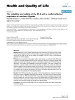
CLINICAL HISTORY 46-year-old woman presenting with acute right upper quadrant pain and
fever.
FIGURE
FIGURE
4A
FIGURE
4C
FIGURE
40
48
FINDINGS Axial CECT (A) and axial T2-weighted MRI
(C) demonstrate the dilated gallbladder, thickened gallbladder wall, pericholecystic fat stranding, and a IJlrge gallstone.
Reformatted sagittal CECf image (B) demonstrates an illdefined bypodensity adjacent to the gall bladder, which
could represent an abscess. Post-gadolinium reformatted
sagittal image (D) indicates the presence of tract between the
collection and the gallbladder.
DIFFERENTIAL DIAGNOSIS None.
DIAGNOSIS Acute cholecystitis with gallbladder perforation and abscess formation.
DISCUSSION Acute cholecystitis is the fourth most common cause of hospital admissions for patients with acute
abdomen pain. It typically results from obstruction of the
cystic duct or gallbladder (GB) neck due to cholelithiasis
with resulting infiammation of the gallbladder wall. Acute
cholecystitis is caused by gallstones in most patients, with
acalculous cholecystitis occurring in approximately 5% to
!5
6
Case 4
10%. In a patient with suspected acute cholecystitis, ultrasonography is typically the imaging procedure of choice.
The typical CT findings in acute cholecystitis include
gallstones, gallbladder distention (>5 em in anteroposterior diameter), mural thickening (>3 mm), pericholecystic
fluid, poor definition of the gallbladder wall at the interface
with the liver, inflammatory stranding in the pericholecystic fat, and hyperemia of the adjacent liver parenchyma. Of
all these findings, the presence of pericholecystic inflammatory change is assumed to be the most specific because
other findings, such as gallbladder wall thickening and distention, do not necessarily indicate cholecystitis. The complications of acute cholecystitis include massive dilation of
the gallbladder, rupture, and abscess formation as in this
case. This case also illustrates the important role of MRI in
evaluating complications of cholecystitis.
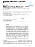
CLINICAL HISTORY 32-year-old man with a history ofulcerative colitis presenting with
increased liver function tests.
FIGURE
5A
FIGURE
5C
FIGURE
58
FIGURE
50
FINDINGS Thick-slab MRCP images (A and B) demonstrate alternating strictures-narrowing and dilatation of the
intrahepatic and upstream extrahepatic bile ducts. Heavily weighted axial T2-WI (C) and gadolinium-enhanced,
fat-suppressed axial Tl-WI (D) demonstrate focally
dilated peripheral intrahepatic bile ducts without abnormal
enhancement.
DIFFERENTIAL DIAGNOSIS Recurrent pyogenic cholangitis (oriental cholangiohepatitis), acute cholangitis.
DIAGNOSIS Primary sclerosing cholangitis (PSC).
DISCUSSION Primary sclerosing cholangitis is an autoimmune disorder characterized by obliterative fibrotic inflammation of the bile ducts that eventually leads to cholestasis, and
biliary cirrhosis. It usually presents in patients younger than
40 years and is more common in males. Typically, intra- and
extrahepatic bile ducts are involved simultancously (80%).
A strong association with inflammatory bowel disease, especially ulcerative colitis, is noted (70%). Although patients may
be asymptomatic, 75% have progressive fatigue, intermittent
obstructive jaundice, and pruritus. On MRI, the characteristic
appearance of the disease is multiple stenoses, minor dilar.ations (because of the periductal fibrosis), and beaded appearance of the bile ducts. In patients with cilrhosis, associated
imaging findings, such as splenomegaly, confluent hepatic
fibrosis, and especially caudate lobe hypertrophy, are present.
On MRCP, by using heavily T2-WI sequences, the signal of
static or slow-moving :fluid-filled structures, such as the bile
ducts, is greatly increased, resulting in increased duct-to-hackground contrast and confidence in diagnosis.
7
CLINICAL HISTORY 50-year-old woman presenting with pulmoMry embolism.
FIGURE
6A
FIGURE
6C
FIGURE
68
FIGURE
60
FINDINGS Coronal post-gadolinium MRI sequences
(A-C) through the retroperitoneum demonstrate the confiuence of bilateral common iliac veins to form the IVC in
the lower abdomen. The IVC is not visualized in the midabdomen and is reconstituted by multiple collateral vessels
as depicted on axial sequence (D). There is reformation of
the suprarenal IVC by the collaterals.
8
DIFFERENTIAL DIAGNOSIS IVC tumor.
DIAGNOSIS IVC thrombosis.
DISCUSSION Predisposing factors for thrombus formation include alterations in blood :How (stasis), injury to the
vascular endothelium, and abnormalities in the constitution
Case 6
of blood hypercoagulability (Virchow's Triad). Endothelial damage is invariably an acquired phenomenon
whereas hypercoagulability may result from both congenital and acquired risk factors (especially in the perioperative period). The classical presentation of IVC thrombus
varies according to the level of the thrombosis with up to
50% of patients presenting with bilateral lower extremity swelling and dilatation of superficial abdominal vessels. Duplex ultrasound scanning has become an accurate
noninvasive method of diagnosing IVC thrombosis, but is
9
operator dependent and can be limited by body habitus or
the presence of bowel gas CT imaging is a rapid noninvasive method which can accurately diagnose and assess
the extent of thrombus as well as delineate any associated
abdominal or pelvic abnormality. MRI is now replacing CT as the optimal investigative tool avoiding radiation and giving more accurate delineation of thrombus as
well as any IVC anomaly. MRI is also used to follow-up
patients to determine morphologic changes in the thrombus following therapy.
CLINICAL HISTORY 45-year-old man presenting with acute left lower quadrant pain.
FIGURE
7A
FIGURE
7C
FIGURE
78
FIGURE
70
FINDINGS Axial (A and B), coronal reformatted (C), and
sagittal reformatted (D) CECT images demonstrate a fatcontaining lesion on the anti-mesenteric side of the distal
descending colon, with a hyperdense surrounding rim and
adjacent fat stranding. A central hyperdense dot (B) is present. The adjacent colonic wall is not thickened.
DIFFERENTIAL DIAGNOSIS Omental infarct, acute diverticulitis, liposarcoma.
DIAGNOSIS Epiploic appendagitis.
DISCUSSION Epiploic appendages are fat-containing
outpouchings arising from the anti-mesenteric side of the
colon. They can measure up to 5 em. Acute inflammation
10
of one of them, called epiploic appendagitis, results either
from torsion or venous occlusion. Patients present acutely
with abdominal pain. Classically, appendagitis occurs
either in the sigmoid or descending colon, but can be found
in any colonic segment. The lesion has a central fatty core,
with a surrounding hyperdense rim and a central dot representing the occluded central vessel. This central dot is only
seen in 54% of cases, however. The adjacent colonic wall
is rarely thickened; this is a useful differentiating factor
as the colonic wall is almost invariably thickened in acute
diverticulitis. A hyperdense rim is also not seen in cases of
omental torsion, which excludes this diagnosis. Liposarcomas are exceedingly rare intraperitoneally and occur in the
retroperitoneum. The imaging findings typically resolve
completely after 6 months.
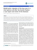
CLINICAL HISTORY 46-year-old man presenting with abdominal pain and constipation.
FIGURE
FIGURE
8A
BB
FINDINGS Coronal 1'2-weighted MRI (A) demonstrates
diffuse segmental wall thickening of the ascending colon.
There is a small focal area of narrowing involving the midascending colon. There is increased fat proliferation within
FIGURE
8C
FIGURE
80
the right pericolic region. Post-gadolinium sagittal (B) and
coronal (C) sequences demonstrate diffuse mural enhancement. Note the relative sparing of the left hemicolon. There
is thickening of the ileocecal valve and terminal ileum. An
additional small bowel segment is involved on the left. Axial
post-gadolinium sequences (D) demonstrate the concentric
mural thickening and pericolonic fat stranding.
DIFFERENTIAL DIAGNOSIS illcerative colitis, pseudo-
membranous colitis.
,
12
Case B
DIAGNOSIS Crohn colitis.
DISCUSSION Crohn disease is of unknown etiology
and characterized by transmural inflammation in a discontinuous fashion (skip lesions) usually involving the terminal ileum. Involvement of the colon and terminal ileum is
seen in approximately 40% to 45% of patients, and colonic
involvement alone is seen in 30% of patients. Complications
include fistula and abscess formation and adenocarcinoma.
Changes of Crohn colitis include aphthous ulcers, thickening
of the bowel wall, deep ulcers, rigidity of the bowel wall,
and a "cobblestone" appearance of the mucosa. The cobble-
stone appearance is due to linear ulcers separated by areas
of edematous mucosa. Finally, there is stricture formation
of the bowel. ''Creeping fat" represents fibrofatty proliferation typically seen in patients with Crohn disease, as demonstrated in this case. It is thought to occur as a response to
repeated episodes of inflammation resulting in separation of
loops of bowel; it is most frequently seen in the small bowel
mesentery but can occur in the colon as well. Pseudomembranous colitis typically involves the entire colon, and there
is a greater degree of bowel wall thickening. Ulcerative colitis begins in the rectum, progresses proximally without skip
lesions, and does not involve the terminal ileum.