Ebook Ultrasonography of the pancreas (edition): Part 1
Bạn đang xem bản rút gọn của tài liệu. Xem và tải ngay bản đầy đủ của tài liệu tại đây (5.14 MB, 88 trang )
Ultrasonography of the Pancreas
Mirko D’Onofrio
Ultrasonography
of the Pancreas
Imaging and Pathologic Correlations
Foreword by
Claudio Bassi
Paolo Pederzoli
123
uploaded by: [UnitedVRG]
Editor
Mirko D’Onofrio
Department of Radiology
G.B. Rossi University Hospital
Verona, Italy
ISBN 978-88-470-2378-9
e-ISBN 978-88-470-2379-6
DOI 10.1007/978-88-470-2379-6
Springer Milan Dordrecht Heidelberg London New York
Library of Congress Control Number: 2011939492
© Springer-Verlag Italia 2012
This work is subject to copyright. All rights are reserved, whether the whole or part of the material is concerned, specifically
the rights of translation, reprinting, reuse of illustrations, recitation, broadcasting, reproduction on microfilm or in any other
way, and storage in databanks. Duplication of this publication or parts thereof is permitted only under the provisions of the
Italian Copyright Law in its current version, and permission for use must always be obtained from Springer. Violations are liable to prosecution under the Italian Copyright Law. The use of general descriptive names, registered names, trademarks, etc.
in this publication does not imply, even in the absence of a specific statement, that such names are exempt from the relevant
protective laws and regulations and therefore free for general use. Product liability:The publishers cannot guarantee the accuracy of any information about dosage and application contained in this book. In every individual case the user must check
such information by consulting the relevant literature.
9 8 7 6 5 4 3 2 1
2012 2013 2014
Cover design: Ikona S.r.l., Milan, Italy
Typesetting: Ikona S.r.l., Milan, Italy
Printing and binding: Grafiche Porpora S.r.l., Segrate, Milan
Printed in Italy
Springer-Verlag Italia S.r.l. – Via Decembrio 28 – I-20137 Milan
Springer is a part of Springer Science+Business Media
uploaded by: [UnitedVRG]
To my Family
“…l’amor che move il sole e l’altre stelle.”
Dante Alighieri. Divina Commedia, Paradiso, XXXIII Canto.
To my Friends
“If the doors of perception were cleansed everything
would appear to man as it is, infinite.”
William Blake. The marriage of heaven and hell.
Foreword
It is with great enthusiasm and true pleasure that we commend this book to the
attention of our colleagues.
In high-volume institutions, such as the Pancreas Center in Verona, ultrasonography
plays an extremely important role in the study of pancreatic pathologies. This carefully
assembled and up-to-date work on the topic will be very useful not only for radiologists
but also for gastroenterologists, surgeons, oncologists and intensive care doctors!
In many applications, ultrasonography findings are now comparable to the results
achieved with multidetector computed tomography (MCT); furthermore, in some
specific applications, such as guidance of diagnostic interventional procedures, ultrasonography is preferable to both MCT and magnetic resonance imaging because it is
faster, easier and cheaper to carry out.
Ultrasonography performed upon hospital admission or during consultation allows
immediate confirmation of the presence of a pancreatic disease (in particular a tumour
mass), assessment of surgical resectability and detection of liver involvement. Moreover, in non-resectable masses, ultrasound-guided percutaneous fine-needle aspiration
with immediate cytological reading will give a definitive diagnosis within a few
hours, and it is to be kept in mind that in experienced hands more than ten such procedures can be performed each half day.
Mirko D’Onofrio from the Radiological Department of our University Hospital is
a skilled radiologist who focusses in particular on the use of ultrasonography. The
work he carries out in this field is of extreme importance in planning our clinical
pathways for the diagnosis and therapy of pancreatic diseases. On account of his enthusiasm and his continuous efforts to exploit the new technologies applicable in ultrasonography (in particular the use of ultrasound contrast media), the above-mentioned
key features of ultrasonography are determinant factors in meeting our everyday
needs, as surgeons, in staging patients suffering from pancreatic tumours.
This book presents the results that can now be achieved with ultrasonography of
the pancreas in the hope that it will encourage wider use of this readily available and
accurate imaging method for the study of pancreatic pathology.
Prof. Claudio Bassi
Prof. Paolo Pederzoli
Department of Surgery
G.B. Rossi University Hospital
Verona, Italy
VII
Preface
Ultrasonography (US) of the pancreas is, in many cases, the initial imaging modality
in most institutions to evaluate pancreatic pathologies and clinical symptoms which
may be related to pancreatic diseases. However, the role of US of the pancreas is often
questioned because the results of this examination are quite variable and not reproducible by different operators. The main reasons for this disagreement are variable operator experience, patient-related problems, e.g. meteorism and obesity, and/or low
contrast and spatial resolution. However, many of these limitations have been overcome
by technological advances in US which have had an extremely positive impact on the
study of the pancreas, as in other organs.
Significant advances have been achieved in conventional, harmonic and Doppler
imaging. Nowadays all portions of the normal pancreas can be visualized in the great
majority of cases. Peri-pancreatic vessels are adequately visualized with conventional
and Doppler imaging or with new advanced techniques. Therefore pancreatic pathologies can be adequately examined and pancreatic tumours, even if very small in diameter
(e.g. insulinoma), can be detected with increased accuracy.
Contrast media have received growing attention in ultrasonography, with special
emphasis on liver studies, where contrast-enhanced ultrasonography (CEUS) has become a well-established imaging modality. In the pancreas the contribution of contrast
media in detecting and characterizing both solid and cystic exocrine or endocrine pancreatic neoplasms is increasing.
Furthermore, the applications of and indications for interventional, endoscopic and
intraoperative US have increased significantly in recent years owing to technological
advances.
All these new applications of US are extensively reviewed in this book in order to
provide the reader with an up-to-date overview of modern imaging of the pancreas.
The book is organized into 14 chapters. Technical issues concerning modern US
imaging, image-guided biopsy, endoscopic US, interventional US-guided procedures
and intraoperative US are first addressed. An interesting chapter is then included on
normal anatomy, including variants and pseudolesions of the pancreas. Thereafter a series of chapters are dedicated to pancreatic pathologies, namely pancreatitis, solid and
cystic tumours, and rare pancreatic tumours, which are presented with emphasis on the
imaging and pathologic correlation. Finally the role of US is discussed in the different
flowcharts.
IX
X
The book is supported by a large number of figures of excellent quality obtained
with up-to-date US equipment and correlated with the findings of other imaging modalities, providing a complete overview of the present status and the real possibilities of
modern US of the pancreas.
Prof. Roberto Pozzi Mucelli
Department of Radiology
G.B. Rossi University Hospital
Verona, Italy
Preface
Contents
1
Ultrasound Imaging . . . . . . . . . . . . . . . . . . . . . . . . . . . . . . . . . . . . . . . . . . . . . . . . . . . . . . . . . . . . . . . . . . . . . . . . .
Anna Gallotti and Fabrizio Calliada
1
2
Transabdominal Ultrasonography of the Pancreas . . . . . . . . . . . . . . . . . . . . . . . . . . . . .
Elisabetta Buscarini and Salvatore Greco
17
3
Endoscopic Ultrasonography of the Pancreas. . . . . . . . . . . . . . . . . . . . . . . . . . . . . . . . . . . . .
Elisabetta Buscarini and Stefania De Lisi
31
4
Percutaneous Ultrasound Guided Interventional Procedures
in Pancreatic Diseases . . . . . . . . . . . . . . . . . . . . . . . . . . . . . . . . . . . . . . . . . . . . . . . . . . . . . . . . . . . . . . . . . . . . . . .
Elisabetta Buscarini and Guido Manfredi
47
5
Intraoperative Ultrasonography of the Pancreas . . . . . . . . . . . . . . . . . . . . . . . . . . . . . . . .
Mirko D’Onofrio, Emilio Barbi, Riccardo De Robertis,
Francesco Principe, Anna Gallotti and Enrico Martone
55
6
Pancreatic Anatomy, Variants and Pseudolesions of the Pancreas . . . . . . .
Emilio Barbi, Salvatore Sgroi, Paolo Tinazzi, Stefano Canestrini,
Anna Gallotti and Mirko D’Onofrio
63
7
Pancreatitis and Pseudocysts. . . . . . . . . . . . . . . . . . . . . . . . . . . . . . . . . . . . . . . . . . . . . . . . . . . . . . . . . . . . .
Steffen Rickes and Holger Neye
83
8
Solid Pancreatic Tumors . . . . . . . . . . . . . . . . . . . . . . . . . . . . . . . . . . . . . . . . . . . . . . . . . . . . . . . . . . . . . . . . . . . 93
Christoph F. Dietrich, Michael Hocke, Anna Gallotti and Mirko D’Onofrio
9
Cystic Pancreatic Tumors . . . . . . . . . . . . . . . . . . . . . . . . . . . . . . . . . . . . . . . . . . . . . . . . . . . . . . . . . . . . . . . . . 111
Mirko D’Onofrio, Paolo Giorgio Arcidiacono and Massimo Falconi
10 Rare Pancreatic Tumors . . . . . . . . . . . . . . . . . . . . . . . . . . . . . . . . . . . . . . . . . . . . . . . . . . . . . . . . . . . . . . . . . . . 135
Roberto Malagò, Ugolino Alfonsi, Camilla Barbiani, Andrea Pezzato
and Roberto Pozzi Mucelli
11 Imaging Correlation . . . . . . . . . . . . . . . . . . . . . . . . . . . . . . . . . . . . . . . . . . . . . . . . . . . . . . . . . . . . . . . . . . . . . . . . . 147
Marie-Pierre Vullierme and Enrico Martone
XI
XII
12 Pancreatic Lesions: Pathologic Correlations . . . . . . . . . . . . . . . . . . . . . . . . . . . . . . . . . . . . . . 165
Paola Capelli and Alice Parisi
13 Clinical and Imaging Scenarios . . . . . . . . . . . . . . . . . . . . . . . . . . . . . . . . . . . . . . . . . . . . . . . . . . . . . . . . . 187
Anna Gallotti and Riccardo Manfredi
14 Flowcharts in Pancreatic Diseases . . . . . . . . . . . . . . . . . . . . . . . . . . . . . . . . . . . . . . . . . . . . . . . . . . . . . 191
Elisabetta Buscarini
Subject Index . . . . . . . . . . . . . . . . . . . . . . . . . . . . . . . . . . . . . . . . . . . . . . . . . . . . . . . . . . . . . . . . . . . . . . . . . . . . . . . . . . . . . . . . . 199
Contents
Contributors
Ugolino Alfonsi Department of Radiology, G.B. Rossi University Hospital, Verona,
Italy
Paolo Giorgio Arcidiacono Gastroenterology and Gastrointestinal Endoscopy Unit,
Vita Salute San Raffaele University, San Raffaele Scientific Institute, Milan, Italy
Emilio Barbi Department of Radiology, Hospital “Casa di Cura Pederzoli”, Peschiera
del Garda (VR), Italy
Camilla Barbiani Department of Radiology, G.B. Rossi University Hospital, Verona,
Italy
Elisabetta Buscarini Department of Gastroenterology, Maggiore Hospital, Crema,
Italy
Fabrizio Calliada Department of Radiology, IRCCS Policlinico S. Matteo, Pavia,
Italy
Stefano Canestrini Department of Radiology, G.B. Rossi University Hospital,
Verona, Italy
Paola Capelli Department of Pathology, G.B. Rossi University Hospital, Verona, Italy
Stefania De Lisi Department of Endoscopy, European Institute of Oncology (IEO),
Milan, Italy
Riccardo De Robertis Department of Radiology, G.B. Rossi University Hospital,
Verona, Italy
Christoph F. Dietrich Department of Clinical Medicine, Caritas-Krankenhaus, Bad
Mergentheim, Germany
Mirko D’Onofrio Department of Radiology, G.B. Rossi University Hospital, Verona,
Italy
Massimo Falconi Department of Surgery, G.B. Rossi University Hospital, Verona,
Italy
Anna Gallotti Department of Radiology, IRCCS Policlinico S. Matteo, Pavia, Italy
XIII
XIV
Salvatore Greco Department of Gastroenterology, Riuniti Hospital, Bergamo, Italy
Michael Hocke Department of Clinical Medicine, Caritas-Krankenhaus, Bad
Mergentheim, Germany
Roberto Malagò Department of Radiology, G.B. Rossi University Hospital, Verona,
Italy
Guido Manfredi Department of Gastroenterology, Maggiore Hospital, Crema, Italy
Riccardo Manfredi Department of Radiology, G.B. Rossi University Hospital,
Verona, Italy
Enrico Martone Department of Radiology, G.B. Rossi University Hospital, Verona,
Italy
Holger Neye Department of Internal Medicine, AMEOS Hospital St. Salvator,
Halberstadt, Germany
Alice Parisi Department of Pathology, G.B. Rossi University Hospital, Verona, Italy
Andrea Pezzato Department of Radiology, G.B. Rossi University Hospital, Verona,
Italy
Roberto Pozzi Mucelli Department of Radiology, G.B. Rossi University Hospital,
Verona, Italy
Francesco Principe Department of Radiology, G.B. Rossi University Hospital,
Verona, Italy
Steffen Rickes Department of Internal Medicine, AMEOS Hospital St. Salvator,
Halberstadt, Germany
Salvatore Sgroi Department of Radiology, Hospital “Casa di Cura Pederzoli”,
Peschiera del Garda (VR), Italy
Paolo Tinazzi Department of Radiology, Hospital “Casa di Cura Pederzoli”, Peschiera
del Garda (VR), Italy
Marie-Pierre Vullierme Department of Radiology, Beaujon Hospital, Clichy, Paris,
France
Contributors
1
Ultrasound Imaging
Anna Gallotti and Fabrizio Calliada
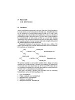
1.1
Introduction
Ultrasonography (US) is usually the first imaging modality chosen for the primary evaluation of the pancreas.
The pancreatic gland can almost always be visualized
by US. Even though there are well-known and sometimes
over-emphasized limitations, the pancreatic gland can
be adequately visualized by using correct US techniques,
imaging and settings. Conventional US is a noninvasive
and relatively low cost imaging method which is widely
available and easy to perform. Tissue harmonic imaging
(THI) and Doppler imaging are well known technologies
that provide significant complementary information to
the conventional method, playing an important role in
the diagnosis and staging of pancreatic diseases. In recent
decades, new interesting US methods have been developed focused on the evaluation of mechanical strain
properties of tissues, such as elastography and sonoelasticity. Acoustic radiation force impulse (ARFI) imaging
is a promising new US method that allows the evaluation
of mechanical strain properties of deep tissues with the
potential to characterize tissue without the need for external compression. Contrast-enhanced ultrasonography
(CEUS) advances the accuracy of this first line examination by characterizing focal solid and cystic lesions
and providing an accurate real-time evaluation of macroand microcirculation in and around a focal mass.
The aim of this chapter is to describe the US imaging
methods and implementations now available for study-
A. Gallotti ( )
Department of Radiology
IRCCS Policlinico S. Matteo, Pavia, Italy
e-mail:
ing the pancreatic gland. Advantages and disadvantages
of the US imaging methods are also mentioned. US
approaches, such as transabdominal, endoscopic, laparoscopic and intraoperative procedures will be accurately illustrated in a dedicated chapter.
1.2
Conventional Imaging
Conventional US is a well-known, relatively low cost
noninvasive imaging method which is widely available
and easy to perform compared to computed tomography
(CT) and magnetic resonance imaging (MRI), modalities
which are usually used as second-line examinations. It
is also free from side effects (i.e. lack of ionizing radiation) or contraindications, so is largely applicable also
in young people. Two other important aspects are its
real-time and multiplanar capabilities [1]. According to
the literature, the pancreatic gland can almost always be
visualized by US, even though in some cases this can be
difficult due to the limited contrast between the pancreas
and surrounding fat [2, 3]. In some overweight patients
the visualization of the gland may also be difficult or
unfeasible, despite several attempts. Examining the patient in different positions, such as erect or supine, with
upleft or upright rotation, with suspended inspiration or
expiration, may be suitable for achieving better pancreatic
visualization. In the presence of abundant gas distension
of the digestive tract, moving the transducer and applying
compression can be useful to displace the bowel loops
and visualize the pancreatic gland [2, 4]. Filling the
stomach with degassed water (100-300 mL) or simethicone-water mixture may be used as a last option to
improve US visualization of the pancreas since air bubble
that cause artifacts will also be introduced into the stomach and a filled stomach is less compressible.
M. D’Onofrio (ed.), Ultrasonography of the Pancreas, © Springer-Verlag Italia 2012
1
2
A. Gallotti, F. Calliada
a
c
b
d
Fig. 1.1 a-d Pancreas. a B-mode image (4.0 Mhz). b Vascular enhancement image (4.0 Mhz). c Spatial compound image (4.0 Mhz).
d Harmonic compound image (3.0 Mhz)
The US examination of the pancreas requires the use
of multifrequency transducers (at least from 3 to 4 MHz)
to study the entire gland with the proper frequencies for
any depth (Fig. 1.1). The anatomic location, the bodysize of the patient and the respiration phase may influence the depth of the pancreas, which is not a completely
fixed retroperitoneal gland (see Chapter 6). Conventional
US utilizes the same frequency bandwidth for both the
transmitted and the received signal. The choice of frequency is mainly based on a compromise between the
spatial resolution, which depends on the wavelength,
and higher frequencies, which provide higher spatial
resolution but which suffer greater tissue attenuation
[5]. The basic US wave is a simple sinusoidal wave
with a spectrum characterized by a single line and just
one frequency of energy (f0), also called the fundamental
frequency or first harmonic. Furthermore, new technologies based on both the amplitude and the phase in-
formation of the return echo (e.g. coherent image formation, Acuson, Siemens) to create images are able to
produce images with more information and detailed resolution [2].
The US study should be performed after a minimum
fast of 6 hours to improve the visualization of the pancreas, creating the best situation for the evaluation of
the gland. Through transverse, longitudinal and angled
oblique scan planes (multiplanar view), the entire pancreatic gland should be recognizable. Beginning with
the patient in the supine position, the probe should be
slightly moved to the right of the midline to visualize
the head and neck of the pancreas descending a little
above the umbilical line for the uncinate process. To
adequately study the body and tail of the pancreas the
operator should move the transducer to the left of the
midline with the end (right part) of the probe rotated
slightly cranially. This positioning obviously reflects
1 Ultrasound Imaging
the most common location of the pancreatic gland, with
the head at a more caudal plane than the tail [1]. The
left lateral approach may also be useful for the evaluation of the pancreatic tail, which can be visualized between the spleen and the left kidney (see Chapter 6).
An accurate US study of the pancreas consists of
the evaluation of the morphology, size, contour and
echotexture of all the portions of the gland, the latter
being comparable to the normal liver. The main pancreatic duct and the common bile duct, together with
the main peri-pancreatic vascular structures, such as
the celiac, superior mesenteric, hepatic and splenic arteries and the portal, superior mesenteric and splenic
veins should be assessed. Lastly, the evaluation of the
adjacent organs, in particular the liver, is always required for a complete study.
As reported in the literature, conventional B-mode
US has a high sensitivity in detecting focal pancreatic
disease due to differences in acoustic impedance between
diseases and surrounding parenchyma. The teardrop
sign, which is highly suggestive of vascular encasement
in the presence of a neoplastic lesion, can only be detected in B-mode, which is also able to identify a dilation
of the main pancreatic duct, parenchymal or ductal calcifications and potentially present peri-pancreatic fluid
collections with great confidence [2].
Technical developments in recent years have led to
image fusion, which is now currently available. This
technology may help in diagnostic and interventional
procedures by making the comparison between US and
other imaging modalities more immediate. In interventional pancreatic procedures the advantages of US guidance, such as its dynamism and the possibility of innumerable manual scanning planes, would be maintained
and it would also overcome the technical limitations of
the technique, such as tympanites and obesity, through
the simultaneous visualization of the previously acquired CT images matched and synchronized with the
US images.
1.3
Harmonic Imaging
Tissue harmonic imaging (THI) is a well know technology that improves conventional US by providing
images of higher quality [5-7]. While conventional US
utilizes the same frequency bandwidth for both the
transmitted and the received signal (f0), THI uses low
frequency for the transmitted signal and higher har-
3
monic frequencies for the received signal. In other
words, by using a Gaussian shaped transmit pulse the
harmonic component can be separated from the returning echo without overlapping with fundamental reflections. In fact, nonlinear harmonic frequencies, generated
by propagation of the US wave through the tissue, occur
as whole-numbered multiples of the fundamental or
transmitted sonographic frequency [5]. Therefore, the
waveform changes compared to the basic US wave, resulting in a distorted wave with a complex form owing
to the presence of both the fundamental and multiple
harmonic frequencies [8].
THI takes advantages of nonlinear harmonic frequencies to correct the defocusing effects and to extensively reduce artifacts caused by low amplitude
pulses [8]. As a consequence, THI produces images
with improved lateral resolution by reducing side-lobe
artifacts and improved signal-to-noise ratio compared
with conventional US, thus resulting in an enhanced
overall image quality [9]. The primary advantage is
fewer artifacts in cavities, such as vascular structures,
which can therefore be better evaluated. There are also
advantages in fluid-solid differentiation, with the finely
detailed depiction of anatomy such as the main pancreatic duct [7]. The physical basis depends on three
main factors: (1) the contraction of the width of the
harmonic wave; (2) the reduction of side-lobe artifacts;
and (3) a received signal free of the original frequency
transmitted.
Lateral resolution mostly depends on the width of
the US wave. Since nonlinear harmonic waves are narrower than the fundamental, they also have lower sidelobe levels, thus improving lateral resolution which is
most evident in fluid-filled structures (Fig. 1.2). The signal-to-noise ratio is consequently enhanced, with higher
contrast resolution, resulting in images characterized by
brighter tissues and darker cavities (e.g. main pancreatic
duct, vascular structures, cystic lesions). Therefore, a
narrow-bandwidth low-frequency pulse is transmitted, a
filter automatically processes the received signal, and
only the returning echo, characterized by high-frequency
harmonic signal is used to generate the image.
THI has been incorporated in all state-of-the-art systems. By pushing the specific button on the US scanner,
the receiver automatically is regulated on a frequency
higher than the fundamental, with little or no overlap
between them, and all the components that are in the
transmitted pulses are rejected. Harmonic band filtering
and phase inversion are the two main methods used for
4
a
A. Gallotti, F. Calliada
b
Fig. 1.2 a,b Pancreatic mucinous cystic neoplasm. Better definition of the cystic wall and intralesional septa moving from conventional US (a) to harmonic US (b) imaging
the generation of harmonic images [8]. In harmonic
band filtering, there is little or no overlap between the
transmitted and received pulses, but through a highpass filter to the received signal, just the higher harmonic frequencies should be used. However, to separate
them a fine bandwidth of the fundamental transmitted
frequency must be selected and, as a consequence, decreased spatial resolution is the result. The same
processes are also applied to the receiver, with a consequent decrease in contrast resolution [10]. These
shortcomings can be overcome with the phase inversion
method. This uses two sequential pulses, the second of
which is phase reversed, and is able to remove the fundamental frequency by electronically storing the reflected signal following the first pulse and adding it to
the second one, leaving only the harmonic waves [8].
The disadvantages are that the frame rate is halved and
motion artifacts can occur.
The pancreatic examination requires the use of the
same multifrequency curved array transducers (at least
from 3 to 4 MHz) used for conventional US. Typically,
the frequency setting consists of a transmitted frequency
of 2.0 MHz and a received frequency of 4.0 MHz (second harmonic). The examination protocol is similar to
that reported above for conventional US.
As reported in the literature, an accurate pancreatic
THI examination is characterized by a higher sensitivity
than conventional B-mode US regarding the detection
of focal solid and cystic pancreatic lesions [8, 11]. THI
is able to more clearly delineate lesion margins as well
as internal solid components of a mass with more confi-
dence [7]. Compared to conventional US, THI provides
a higher soft tissue differentiation, allowing both the detection of even small lesions with little changes in
echogenicity with respect to the surrounding parenchyma
and the identification of calcifications [11, 12]. Moreover,
other important advantages consist of the ability to clearly
study deep structures and overweight patients, due to
the rejection of low-amplitude pulses which generate artifacts in the conventional examination [8]. In a nutshell,
in the study of the pancreas and compared to conventional
B-mode US, THI can increase both spatial and contrast
resolution, providing an enhanced overall image quality,
better lesion conspicuity, and advantages in fluid-solid
differentiation, thus achieving a better detection of pancreatic cancer.
1.4
Compound and Volumetric
Imaging
State-of-the-art systems provide images with high detail
resolution owing to both amplitude and phase information of the return echo and compound technology.
Compounding is able to improve contrast and spatial
resolution in the B-mode image (Fig. 1.1), reducing
the intrinsic acoustic noise of US imaging (speckle) by
generating several independent frames of data and then
averaging them [2]. There are different types of compounding technology available, such as frequency compounding and spatial compounding (Fig. 1.1).
The introduction of volumetric image acquisition,
1 Ultrasound Imaging
Fig. 1.3 Solid focal pancreatic lesion. Volumetric imaging of a
solid focal hypoechoic (arrow) pancreatic head lesion
which maintains the real-time and multiplanar capabilities of conventional US, opens up new clinical opportunities for a more complete evaluation of the pancreatic gland [1]. Volumetric US imaging is a relatively
new technique based on the acquisition of a volume
dataset of anatomic structures (Fig. 1.3). Automated
volumetric imaging is able to overcome the low reproducibility of the previous volume freehand sweep acquisition, owing to the possibility of a standardized
and objective acquisition during the study. The whole
volume of a region of interest is automatically acquired
during a breath hold of a few seconds without moving
the probe (Fig. 1.4). With the volumetric electromechanical transducers, such as 4D3C (GE Healthcare, Waukesha, WI, USA), the acquisition is related to the internal
movement of the piezoelectric elements inside the probe
with an angle of acquisition from 40° to 60°. Therefore
the entire volume is uniformly and automatically acquired, and then reviewed and studied by means of different applications: volume review for reviewing the
whole volume acquired to obtain a virtual scan of the
pancreas; tomographic imaging for allowing the multiplanar vision of the region of interest; volume rendering for allowing the volumetric visualization of a
pancreatic lesion. Moreover, when studying a pancreatic
mass the evaluation of the involvement of the peri-pancreatic vessels can be improved by using multiplanar
reconstruction (Fig. 1.5). In general, the correct application of these new technologies in the US study of the
pancreas results in a conventional imaging of the gland
with very high spatial and contrast resolution.
5
Fig. 1.4 Pancreatic mucinous cystic neoplasm. Volumetric imaging of a cystic pancreatic mass completely included in the automated acquisition scan
1.5
Doppler Imaging
Doppler imaging is a well-known technology that advances and completes the conventional US examination,
providing significant complementary information about
the vascular structures. Since its high sensitivity in evaluating flow in all the main peri-pancreatic arterial (i.e.
celiac, superior mesenteric, hepatic and splenic arteries)
and venous (i.e. portal, superior mesenteric and splenic
veins) structures, together with its increased sensitivity
in recognizing smaller intrapancreatic and intratumoral
vessels, this technology plays an important role in diagnosing and staging pancreatic diseases [6, 13].
While conventional US is based on short pulses of
US, Doppler signals derive from both continuous and
pulsed waves and are mostly due to scattering from red
blood cells. Some special methods have been developed
for Doppler study. Continuous-wave technique, which
is very sensitive to small vessels, enables measurements
of a wide velocity range, but is unable to obtain information about the source of the Doppler signal because
any moving object produces a signal. To overcome this
shortcoming, the pulsed-wave technique, which is based
on the pulse length and the duty cycle, enables the selective measurement of the wave speed at precise locations in the beam, even though the exact source of the
Doppler signal remains difficult to determine because
an image of the subsurface anatomy is not reported and
is prone to false velocity indications (i.e. aliasing). The
real advance in the application of Doppler technology is
6
A. Gallotti, F. Calliada
Fig. 1.5 Solid focal
pancreatic lesion. Sagittal
views of a solid focal
hypoechoic (arrow)
pancreatic head lesion after
automated volumetric
acquisition scan
duplex Doppler imaging. This is more complex and expensive as it combines both previous techniques, but it
does enable the precise location of the signal; image and
both peak velocity and velocity distribution are provided
in real-time together with indications of the sample size.
Lastly, color-flow Doppler imaging, which combines
both anatomic and velocity data, provides qualitative
and quantitative information adding velocity information
to the conventional images as color data: red represents
blood moving toward the transducer, whereas blue represents blood moving away. Variation of the velocity is
also reproduced as a different color intensity. Typically,
the lighter the color is, the higher the velocity (i.e. aliasing
in the presence of improper velocity range) [14].
Doppler technology has been incorporated in all
state-of-the-art systems. The pancreatic examination requires the use of the same multifrequency curved array
transducers (at least from 3 to 4 MHz) used for conventional US and is based on an adequate visualization of
the gland and of the targeted vascular structures at Bmode US. Color gain and velocity settings are tuned to
provide good color filling of the vascular structures
avoiding the generation of artifacts [15]. Typically, the
frequency setting varies from 1 to 4 MHz, mostly depending on two factors: first, the targeted vascular structures, since lower frequencies allow an adequate evaluation of the peri-pancreatic main vessels owing to their
higher penetration, while higher frequencies allow the
1 Ultrasound Imaging
7
evaluation of smaller vessels characterized by slower
flows or vascular structures in thin patients whose pancreas is less deep; second, the patient’s habitus. An accurate velocity measurement requires: (1) a correct angle
between the vessel, the Doppler angle and the axis of
the US beam, which should be as small as possible to
generate signals with high signal-to-noise ratios; (2) the
gate has to be located in the vessel center, with a size as
small as possible; and (3) a correct angle for the velocity
measurement has to be chosen, usually less than 60°.
High-pass filters are used to reduce the influence of
vessel wall and other non-vascular movements [14].
The examination protocol is similar to that reported
above for conventional US.
Doppler technology implements conventional US in
studying vascular structures, providing useful anatomic
information and an accurate evaluation of patency
(color-power study) and blood flow (color-Doppler
study). At color-power imaging, a patent vessel of
course appears colored. The color study offers an adequate evaluation of large vessels, providing information
about the direction of flow, but it is dependent on the
angle and is potentially affected by aliasing due to the
difficulty in separating background noise from true
flow in slow-flow states. Smaller vascular structures
are better identified by the power study, which along
with being relatively angle independent and unaffected
by aliasing is characterized by higher signal persistence
with better definition of vessel margins. However, it
also suffers from increased movement artifacts and is
unable to demonstrate flow direction or to estimate
flow velocity [16]. Moreover, both technologies may
provide useful information about the vascular network
of focal lesions which may be present. Therefore, spectral waveform changes in peri-pancreatic vessels may
depend on the effect of pancreatic diseases on the vascular structures [13].
As reported in the literature, an accurate pancreatic
Doppler examination is based on the evaluation of all
peripancreatic, intrapancreatic and intratumoral vessels.
The most important applications are the identification
of the vascular nature of an anechoic lesion (Fig. 1.6)
detected at conventional US (i.e. pseudoaneurysm) and
the differentiation between resectable (Fig. 1.7) and nonresectable (Fig. 1.7) pancreatic tumor (i.e. localized aliasing with reverse flow, mosaic pattern and accelerated
flow velocity are detected at the site of stenosis, while
parvus et tardus flow is observed downstream from an
infiltrated tract) [17-19] with a reported accuracy of 8590.5% [19]. As well described in the literature, a locally
advanced pancreatic mass is defined by the extended invasion of a main arterial or venous vessel, by the encasement of a main arterial structure and/or by the occlusion of a main venous structure [19, 20]. Splenic
arterial or venous encasement is not a contraindication
for surgical resection [6]. If both a dilation of small peripancreatic veins and a tumor surrounding three quarters
of a main vessel lumen allow the diagnosis of a vascular
infiltration, while the teardrop sign, due to a tumor surrounding more than a half but less than three quarters of
a main vessel lumen is highly suggestive of vascular encasement, a simple contiguity (less than a half of the
vessel circumference) between tumor and vessel does
not necessary correspond to vascular invasion [20].
a
b
Fig. 1.6 a,b Pseudoaneurysm. Cystic lesion (asterisk) in the pancreatic tail at conventional imaging (a) in patient with chronic pancreatitis with final diagnosis of pseudoaneurysm at Doppler study (b)
8
A. Gallotti, F. Calliada
a
b
c
d
Fig. 1.7 a-d Pancreatic mass resectability. a Schematic representation of a resectable pancreatic head mass. b US detection of a resectable hypoechoic mass (arrow) of the pancreatic head. c Pancreatic head solid mass infiltrating the superior mesenteric vein at
conventional imaging and confirmed at Doppler study. d Pancreatic head solid mass infiltrating the superior mesenteric artery at
conventional imaging and confirmed at Doppler study
Some new technologies have been developed: wideband Doppler, which improves both spatial and temporal resolution of the color-Doppler signal with decreased artifacts [13]; power-like flow systems such as
B-flow (General Electric) and e-flow (Aloka) imaging
which are able to suppress tissue clutter and improve
sensitivity to directly visualize blood reflectors and
consequently provide images characterized by better
spatial resolution [13]; color flow imaging (CFI), mostly
used to image the blood movement through arteries
and veins, but also to represent the motion of solid tissues [21]. The weak signals from blood echoes are enhanced and correlated with the corresponding signals
of the adjacent frames to suppress non-moving tissues.
The remaining aspects of the data processing are essentially the same as in conventional grey-scale imaging. In comparison with Doppler techniques these new
Fig. 1.8 Superior mesenteric artery. Doppler based US imaging
of superior mesenteric artery shows flow only inside the lumen
of the artery with a perfect detection of the arterial wall (arrow)
1 Ultrasound Imaging
Fig. 1.9 Small solid focal pancreatic lesion. Doppler based US
imaging of a very small solid focal hypoechoic (arrow) pancreatic
lesion in the pancreatic body
US flow imaging modalities are not affected by aliasing
and have the advantages of a significantly lower angle
dependency and better spatial resolution with reduced
overwriting. As a consequence, evaluation of vessel
profiles is markedly improved (Fig. 1.8).
Other new Doppler-based technologies are able to
improve image quality, owing to the immediate identification of the vascular structures in B-mode. For example,
Clarify Vascular Enhancement (Acuson, Siemens) enables image optimization by enhancing the B-Mode display with information derived from power-Doppler,
clearly differentiating vascular anatomy from acoustic
artifacts and surrounding tissue (Fig. 1.9). In studying
the pancreas, the resulting images can immediately appear diagnostic or more informative.
1.6
Elastography Imaging
In recent decades, new and interesting US techniques
have been developed focused on the evaluation of mechanical strain properties of tissues. The noninvasive
analysis of tissue stiffness immediately received major
interest, owing to a revolutionary approach in the study
of focal and diffuse diseases able to provide a new diagnostic tool. Tissue stiffness has long been an asset in
physical palpation for clinicians and surgeons. Since the
introduction of these new technologies, it has become a
9
new and useful technique for radiologists able to complement other traditional data when making a diagnosis.
The first imaging techniques developed to image tissue elasticity consisted of elastography, the static US
approach [22], and sonoelasticity, the dynamic US approach [23]. In elastography, the longitudinal stress and
strain of superficial tissues can be estimated by tracking
tissue motion mainly derived from external mechanical
compression applied by the US probe [24]. In sonoelasticity, externally applied vibrations at low amplitude (less
than 0.1 mm displacement) and low frequencies (101000 Hz) are used to induce oscillations within tissues
and this motion is detected by Doppler US [25]. Through
a color or grey scale map, a qualitative evaluation of the
elastic properties of tissues is provided. As a consequence, isoechoic lesions which are undetectable at conventional US often might be identified at elastography
and sonoelasticity imaging, owing to their altered vibration response. US elastography and sonoelasticity have
been implemented as simple add-ons alongside conventional US scanners or as dedicated units. Transient US
elastography utilizes a displacement wave generated by
a piston or acoustic force which provides the stress to
the tissue, without producing an image, but only numeric
data of the tissue stiffness. This has mainly been used in
the evaluation of diffuse liver diseases [26].
As widely reported in the literature, several clinical
applications have been studied: for diagnostic purposes
and biopsy targeting in breast and prostate; to differentiate benign from malignant nodules in the thyroid gland;
to differentiate benign from malignant lymph nodes
[27-30]; and in the evaluation of liver fibrosis [31].
Elastography has the same problems as B-mode
sonography. The stress propagating into a tissue is in
fact attenuated by tissues, causing it to spread into other
directions from the primary incidental direction and to
interact with a boundary between two media of different
elastic properties, with potential distraction.
A more recent elastographic technique called acoustic
radiation force impulse (ARFI) imaging has been developed [32, 33]. This new promising US method enables
the evaluation of mechanical strain properties of deep
tissues without the need for external compression. It produces a high intensity push pulse to displace the tissue
and lower intensity pulses for imaging. The physical basis
depends on the evaluation of the transverse wave spread
away from the target tissue. There are two basic types of
wave motion for mechanical waves, most widely used in
US testing: longitudinal or compression waves and trans-
10
verse or shear waves. Whereas the particle displacement
is parallel to the direction of wave propagation in a longitudinal wave, in a transverse wave the particle displacement is perpendicular to the direction of wave propagation. In other words, if compression waves can be
generated in liquids as well as solids, shear waves are not
effectively propagated in gas or fluids owing to the absence of a mechanism for driving motion perpendicular
to the sound beam. Transverse waves are also relatively
weak when compared to longitudinal waves, since they
are usually generated using some of the energy from longitudinal waves. As is well known, sound travels at different speeds in different materials, mostly because elastic
constants are different for different media. Young’s modulus deals with the velocity of a longitudinal wave, while
the shear modulus deals with the velocity of a shear wave.
ARFI imaging has been incorporated in only a few
US systems, and all papers present in the literature at
this moment describe the application of the Siemens
ACUSON S2000scanner (Siemens, Erlanger, Germany).
The pancreatic examination requires the use of the same
multifrequency curved array transducers (at least from 3
to 4 MHz) used for conventional US. A single transducer
is used both to generate radiation force and to track the
resulting displacements. Pushing the specific button on
the US scanner, the transducer is automatically regulated
on the THI imaging, with a received frequency of 4.0
MHz. On a traditional harmonic US image, the target
region of interest (ROI) is selected utilizing a box with
fixed dimensions of 1 x 0.5 cm, able to descend at a
maximum depth of 5.5 cm (8 cm in the most recent
scanner). The box has to be completely included in the
target tissue (i.e. organ in cases of diffuse diseases or lesion in cases of focal diseases), taking care not to comprise any fluid structures, such as vessels or ducts. Once
the target ROI has been correctly located, the patient
should maintain a proper suspended inspiration or expiration, to minimize motion artifacts. Pushing a specific
button on the US scanner, acoustic push pulses are then
transmitted. The push pulse is characterized by short duration (less than 1 msec) and runs immediately on the
right side of the target ROI. Owing to its very high speed,
it is minimally and not significantly influenced by the
structures encountered through the path away from the
transducer up to the box. The acoustic beam is able to
generate localized, micron-scale displacements in the
selected ROI proportional to the tissue elasticity. As a
consequence, detection waves of lower intensity (1:100)
are generated. The shear waves produced, which run
A. Gallotti, F. Calliada
away perpendicular to the acoustic beam, are measured.
The speed of the shear waves reflects the tissue elasticity,
being dependent on the elasticity modulus that is mainly
related to the resistance offered by the tissue to the wave
propagation, and is proportional to the tissue stiffness:
the stiffer a tissue is, the higher the shear wave speed it
generates [34]. As a result, according to the interaction
between waves and transducer previously selected by
the operator, the response may be reported as qualitative
or quantitative information (Fig. 1.10). The qualitative
response consists of a grey scale map of the previously
selected ROI, characterized by a lack of anatomic details,
but with high contrast resolution, in which a bright shade
corresponds to soft tissue, while a dark shade represents
stiff tissue. The implementation of ARFI imaging able
to provide this kind of response is called Virtual Touch
tissue imaging. Obviously, this new advance could play
an important role in the presence of focal disease. The
quantitative response consists of a numeric wave velocity
value, expressed in m/s, which derives from multiple
measurements automatically made by the system for the
previously selected ROI. It provides objective and reproducible data regarding the shear wave speed: the
stiffer a tissue is, the higher the shear wave speed. The
implementation of ARFI imaging able to provide this
numerical response is called Virtual Touch tissue quantification and can be applied both in the presence of
focal and diffuse disease [35].
The most significant advantages of ARFI technology
over previous elastographic techniques are: (1) its in-
Fig. 1.10 Pancreas. Acoustic radiation force impulse (ARFI)
US imaging with virtual touch quantification shows normal shear
wave velocity in the normal pancreas of a healthy volunteer
1 Ultrasound Imaging
a
11
b
Fig. 1.11 a,b Pancreatic ductal adenocarcinoma. a Acoustic radiation force impulse (ARFI) US imaging shows a solid mass in the
pancreatic body appearing black (asterisk) and therefore stiff at virtual touch imaging. b Acoustic radiation force impulse (ARFI)
US imaging shows a solid mass in the pancreatic body with very high value of shear wave velocity at virtual touch quantification
and therefore stiff
tegration into a conventional US system, thus allowing
the visualization of B-mode, color-Doppler mode and
ARFI images with the same equipment; (2) the consequent selection of an ROI in the target tissue on a conventional US image; (3) the subsequent possibility of
precisely studying target lesions during a real-time visualization at conventional US; (4) the opportunity to
also study deep tissues, since there is no need for external compression; and (5) the objective quantification
of the tissue stiffness expressed as a numeric value, by
Virtual Touch tissue quantification. There are nonetheless some important limitations: (1) the fixed box dimensions of the target ROI, while less important in
cases of diffuse disease, could be significantly limiting
in cases of focal lesions; and (2) a high sensitivity to
movement artifacts, such as lack of suspended respiration or heart motion.
The US examination should be performed after a
minimum fast of 6 hours to improve the visualization
of the pancreas, creating the best situation for the evaluation of the gland. The good visualization of the target
tissue at conventional US is a mandatory condition for
performing the ARFI examination.
As reported in the literature, the mean wave velocity
value obtained in the healthy pancreas (Fig. 1.10) is
about 1.40 m/s [6, 35]. An accurate pancreatic US examination consists of the application of both qualitative
and quantitative implementations of ARFI technology,
whenever possible, to assess the concordance of the
results. Different focal and diffuse diseases that alter
the tissue stiffness should be characterized by different
shades and wave velocity values. For example, since
pancreatic ductal adenocarcinoma is a firm mass which
is stiffer than the adjacent parenchyma (see also Chapter
8) owing to the presence of fibrosis and marked desmoplasia, it should appear as a dark shade with higher
values (Fig. 1.11).
According to the physical principles of the shear
waves, ARFI imaging has been tested in the study of
solid tissues. However, fluids in vivo, and as a consequence pancreatic cystic lesions, can be markedly different and different responses at ARFI technology might
be expected. The qualitative evaluation should give a
bright shade, while as recently reported in the literature,
it seems that the quantitative study usually gives non
numeric values in serous cystadenoma (see also Chapter
9), which contains a simple fluid, and mainly numeric
values in mucinous tumors (Fig. 1.12), which contain
a more complex content [36, 37].
Since its recent introduction, few data regarding the
usefulness of ARFI technology in the study of pancreatic diseases are available in the literature. However, it
seems to be potentially able to allow tissue characterization by imaging and may constitute a feasible alternative to invasive needle-biopsy in the future.
1.7
Contrast-enhanced Ultrasound
Contrast-enhanced ultrasonography (CEUS) is a relatively recent implementation of conventional US which
significantly advances the accuracy of this first line examination in characterizing focal solid and cystic diseases. The administration of microbubbles allows an
12
a
A. Gallotti, F. Calliada
b
Fig. 1.12 a,b Pancreatic mucinous cystic neoplasm. Acoustic radiation force impulse (ARFI) US imaging of a cystic mass with numerical value of shear wave velocity at virtual touch quantification of the fluid content
accurate evaluation of macro- and microcirculation, in
and around a focal mass, giving more detailed and advanced results than the color-Doppler study thanks to
its high spatial, contrast and temporal resolution. This
new technology has been widely used to study hepatic
diseases and also more recently applied in the study of
the pancreas, giving promising results in diagnosis and
staging of pancreatic diseases already detected at conventional US [6, 38].
The introduction of US contrast agents goes back
some decades and their effects during cardiac catheterization were first described at the end of the 1960s. Today their use has been approved in Europe, Asia and
Canada, but the Food and Drug Administration in the
United States has not yet approved their application
for non-cardiac use. Only the administration in pregnancy and pediatrics is off label. Some recommendations exist, especially for second generation contrast
agents filled with sulfur-hexafluoride: they are not recommended in patients with recent acute coronary syndrome, unstable angina, recent acute heart attack, recent
coronary artery intervention, acute or class III or IV
chronic heart failure or severe arrhythmias. No interactions with other drugs have been reported and only
rarely some subtle and usually transient adverse reactions have been described, such as tissue irritation and
cutaneous eruptions, dyspnea, chest pain, hypo- or
hypertension, nausea and vomiting. No severe effects
have been described in humans to date [39, 40].
US contrast agents consist of microbubbles, characterized by a diameter that ranges from 2 to 6 microns,
a shell of biocompatible materials, such as proteins,
lipids or biopolymers and a filling gas, such as air or
gas with high molecular weight and low solubility (e.g.
perfluorocarbon or sulfur hexafluoride). Their small
diameter allows their passage through the pulmonary
district, thus microbubbles are exhaled during respiration 10-15 minutes after injection, while the components
of the shell are metabolized or filtered by the kidney
and eliminated by the liver. Shell and gas influence the
time of circulation and acoustic behavior of microbubbles. The thin shell ranges from 10 to 200 nm and
allows the passage through the pulmonary district with
a consequent systemic effect and a more prolonged
contrast effect. The filling gas produces a vapor concentration inside the microbubbles higher than the surrounding blood, increasing their stability in the peripheral circulation [38, 41].
Both the shell and the filling gases have been
changed over the years, passing from first generation
contrast media to second generation agents. The first
generation contrast media were characterized by a stiff
shell (denatured albumin) and air as filling gas. The
stiff shell allows more stability in the peripheral blood,
with a reduction in non-linear behavior. Therefore, as
the microbubbles have a short half-life because they
are easily destroyed, their US response depends on the
echogenicity and the concentration. The second generation contrast media are both more stable and resistant.
They are characterized by a flexible shell (phospholipids), which allows the prevalence of nonlinear behavior, and filling gas other than air. Their US response
consists of the generation of nonlinear harmonic frequencies, since at low acoustic power of insonation
1 Ultrasound Imaging
a
13
b
c
Fig. 1.13 a-c Pancreatic intraductal papillary mucinous neoplasm. a Pseudosolid appearance of the pancreatic head lesion at conventional US resulting hypoechoic (arrow) but avascular with cystic appearance at CEUS (b). The cystic nature (arrow) of the
lesion is confirmed at MRI (c)
(about 30-70 kPa), the degree of microbubble expansion
is greater than its compression [41].
Several contrast-specific software applications have
been developed for CEUS examination, even though
the most promising techniques are phase and amplitude
modulation. Pulse inversion is the most common phase
modulation technique [42], while power modulation is
a well-known amplitude modulation software application [41]. Cadence contrast pulse sequencing (CPS) is
a more advanced combined phase and amplitude modulation technique [38, 43].
The CEUS examination should be performed after an
accurate conventional US of the pancreas with the evidence of a focal or diffuse pancreatic disease [44]. The
pancreatic examination requires the use of the same multifrequency curved array transducers (at least from 3 to 4
MHz) used for conventional US. Nowadays, second generation contrast agents are used. Harmonic microbubblespecific software applications are required to filter all the
background tissue signals so only vascularized structures
related to the harmonic responses of the microbubbles
are visualized after injection. The dual screen should be
used to adequately and continuously compare B-mode
and contrast images. Focus and depth should be regulated
simultaneously in both images and low acoustic US pressures should be selected (mechanical index less than 0.2).
The examination protocol and technique are similar to
those reported above for conventional US.
The dynamic evaluation begins immediately after the
intravenous administration of a 2.4-mL bolus of microbubble contrast agent. Since the pancreatic blood supply is exclusively arterial, the enhancement of the gland
begins almost together with the arteries. Enhancement of
the pancreatic gland begins almost at the same time as
aortic enhancement. After this early phase (arterial/pan-
creatic; from 10 s to 30 s), as with other dynamic imaging
modalities there is a second phase, the venous phase
(from 30 to approximately 120 s) defined by hyperechogenicity within the spleno-mesenteric-portal venous
axis. The late phase (about 120 s after injection) is defined
by hyperechogenicity of the hepatic veins.
US specific contrast agents have a purely intravascular distribution without any interstitial phase, so they
differ from all contrast media used during CT and MRI
examinations [45]. Moreover, CEUS with second generation contrast media enables real-time evaluation of
target tissues, with high spatial, temporal and contrast
resolution. Unlike other imaging modalities, as reported
above, only vascularized structures are visible after the
administration of microbubbles (see Chapter 11). Therefore, compared to conventional US and other imaging
modalities, pancreatic CEUS is better able to differentiate between solid and cystic lesions (Fig. 1.13), characterize focal masses and provide a clear differentiation
between remnant tissue, fibrosis and necrosis [44].
Moreover, the CEUS examination covers an important
role in evaluating the resectability or non-resectability
of a focal mass [46], together with Doppler imaging
for the assessment of the relationship between the tumor
and the adjacent main vessels, and during the late phase
to exclude the presence of liver metastases.
Some new applications of CEUS have been developed:
the use of CEUS enhancement as a prognostic factor,
both in the diagnostic workup and in the follow-up of
patients. In fact, as reported in the literature, in the presence of focal pancreatic lesions, the accurate description
of the enhancement pattern at CEUS is mandatory for a
prompt prognostic evaluation. The association between
intratumoral microvessel density (MVD) and tumor aggressiveness has already been proven [46]. The use of