Ebook Handbook of pediatric anesthesia: Part 1
Bạn đang xem bản rút gọn của tài liệu. Xem và tải ngay bản đầy đủ của tài liệu tại đây (45.33 MB, 195 trang )
HANDBOOK
of PEDIATRIC
ANESTHESIA
NOTICE
Medicine is an ever-changing science. As new research and clinical
experience broaden our knowledge, changes in treatment and drug
therapy are required The authors and the publisher ofthis work have
checked with sources believed to be reliable in their efforts to provide
information that is complete and generally in accord with the
standards accepted at the time of publication. However, in view of
the possibility of human error or changes in medical sciences,
neither the authors nor the publisher nor any other party who has
been involved in the preparation or publication ofthis work warrants
that the information contained herein is in every respect accurate or
complete, and they disclaim all responsibility for any errors or
omissions or for the results obtained from use of the information
contained in this work. Readers are encouraged to confirm the
information contained herein with other sources. For example and in
particular, readers are advised to check the product information
sheet included in the package ofeach drug they plan to administer to
be certain that the information contained in this work is accurate and
that changes have not been made in the recommended dose or in the
contraindications for administration. This recommendation is of
particular importance in connection with new or infrequently used
drugs.
a Lange medical book
HANDBOOK
of PEDIATRIC
ANESTHESIA
Editors
Philipp J. Houck, MD
Assistant Professor of Anesthesiology
Division of Pediatric Anesthesia
Department of Anesthesiology
Director of Pediatric Liver Transplant Anesthesia
New York Presbyterian-Morgan Stanley Children's Hospital
Columbia University Medical Center
New York. New York
Manon Hache, MD
Assistant Professor of Anesthesiology
Division of Pediatric Anesthesia
Department of Anesthesiology
Director of Pediatric Trauma Anesthesia
New York Presbyterian-Morgan Stan ley Children's Hospital
Columbia University Medical Center
New York. New York
Lena S. Sun, MD
Emanuel M. Papper Professor of Pediatric Anesthesiology
Chief, Division of Pediatric Anesthesia
Vice Chair, Department of Anesthesiology
New York Presbyterian-Morgan Stan ley Children's Hospital
Columbia University Medical Center
New York, New York
II
New York Chicago San Francisco Athens London Madrid Mexico City
Milan New Delhi Singapore Sydney Toronto
Copyright C> 2015 by McGraw-Hill Education. All rights reserved. Except as
permitted under the United States Copyright Act of 1976, no part of this publication
may be reproduced or distributed in any form or by any means, or stored in a database
or retrieval system, without the prior written permission ofthe publisher.
ISBN: 978-0-07-177208-2
MHID: 0-07-177208-1
The material in this cBook abo appear.~ in the print veiSion oftbis title: ISBN: 978-0-01-176935-8,
MHID: 0-07-176935-8.
eBook conversion by codeMauira
Version 1.0
All 1rademarks are trademarks of their respective owners. Rather than put a
1rademark symbol after every occurrence of a 1rademarked name, we use names in an
editorial filsbion only, and to the benefit of the trademark owner, with no intention of
infringement ofthe 1rademark. Where such designations appear in this book, they have
been printed with initial caps.
McGraw-Hill Education eBooks are available at special quantity discounts to use as
premiums and sales promotions or for use in corporate training programs. To contact a
representative, please visit the Contact Us page at www.mhprofessional.com.
TERMSOFUSE
This is a copyrighted work and McGraw-Hill Education and its licensors reserve
all rights in and to the work. Use of this work is subject to these terms. Except as
permitted under the Copyright Act of 1976 and the right to store and retrieve one
copy of the work, you may not decompile, disassemble, reverse engineer, reproduce,
modify, create derivative works based upon, transmit, distribute, disseminate, sell,
publish or sublicense the work or any part of it without McGraw-Hill Education's
prior consent. You may use the work for your own noncommercial and personal use;
any other use of the work is s1rictly prohl.bited. Your right to use the work may be
terminated ifyou fail to comply with these terms.
TilE WORK IS PROVIDED "AS IS." McGRAW-lllLL EDUCATION AND
ITS LICENSORS MAKE NO GUARANTEES OR WARRANTffiS AS TO TilE
ACCURACY, ADEQUACY OR COMPLETENESS OF OR RESULTS TO BE
OBTAINED FROM USING TilE WORK, INCLUDING ANY INFORMATION
TIIAT CAN BE ACCESSED THROUGH TilE WORK VIA HYPERLINK. OR
OTIIERWISE, AND EXPRESSLY DISCLAIM ANY WARRANTY, EXPRESS OR
IMPLffiD, INCLUDING BUT NOT LIMITED TO IMPLffiD WARRANTmS OF
MERCHANTABILITY OR FITNESS FOR A PARTICULAR PURPOSE.
McGraw-Hill Education and its licensors do not warrant or guarantee that the
functions contained in the work will meet your requirements or that its operation will
be uninterrupted or error free. Neither McGraw-Hill Education nor its licensors shall
be liable to you or anyone else for any inaccuracy, error or omission, regardless of
cause, in the work or for any damages resulting therefrom. McGraw-Hill Education
bas no responsibility for the content of any information accessed through the work.
Under no circumstances shall McGraw-Hill Education and/or its licensors be liable for
any indirect, incidental, special, punitive, consequential or similar damages that result
from the use of or inability to use the work, even if any of them has been advised of
the possibility of such damages. This limitation of liability shall apply to any claim
or cause whatsoever whether such claim or cause arises in contract, tort or otherwise.
The editors and authors of this Handbook would like
to acknowledge all of our colleagues, patients, and our
families for their support and encouragement who made
this Handbook a reality.
This page intentionally left blank
CONTENTS
Contributors ......................................................... xi
Preface ............................................................. xiii
1
Introduction (Robert Kazim) ........ .. ... . ... . ... . ... .. .. .. ..... 1
Part 1: Airway
Z
3
4
5
6
7
8
9
10
11
1Z
Tonsillectomy and Adenoidectomy in a Patient With
Obstructive Sleep Apnea (Gracie M. Almeida-Chen) .. . ... . ..... 17
Posttonsillectomy Bleeding (Neeta R. Saraiya) . ... . ... . ... . ..... 23
Bilateral Myringotomy and Tubes in a Patient With an Upper
Respiratory Tract Infection (Neeta R. Saraiya) . . ... . ... . ......... 25
Cleft Lip and Palate Repair (Gracie M.Aimeida-Chen) . . ......... 27
Epiglottitis (Gracie M. Almeida-Chen) .......................... 31
Postoperative Stridor (Gracie M. Almeida-Chen) ................ 34
Subglottic Stenosis (Gracie M. Almeida-Chen) .................. 37
Cystic Hygroma {Susan Y. LeO •••••••••••••••••••••••••••••••••• 41
Aspirated Foreign Body (Neeta R. Saraiya) •••••••••••••••••••••• 44
Laryngeal Papillomatosis {Neeta R. Saraiya) •••••••••••••••••••• 46
Difficult Airway Management (Philipp J. Houck) ................ 48
Part 2: Cardiovascular
13
14
15
16
17
18
cardiopulmonary Bypass (Riva R. Ko) .......................... 55
Ventricular Septum Defect Repair (PhilippJ. Houck) ............ 59
Tetralogy ofFallot (Anthony J. Clapcich) ....................... 61
Single Ventricle Physiology(Riva R. Ko) ........................ 65
Pulmonary Hypertension (Arthur J. Smerling) .................. 70
cardiac catheterization After Heart
Transplantation (Philipp J. Houck) ............................. 73
Part 3: Respiratory
19
20
21
22
23
Asthma (Gracie M. Almeida-Chen) .. .. .. .. ... .. . .. .. ... .. .. .. .. 77
Bronchopulmonary Dysplasia (Gracie M. Almeida-Chen) • •• •• •• 80
Croup (Gracie M. Almeida-Chen) ••• •• ••• •• •• •• •• •• •• •• •• •• •• •• 84
Aspiration Pneumonia {Manon Hache) ........................ 87
Pulmonary Sequestration (Leila M. Pang, Manon Hache)........ 90
Part 4: Neonates
24
25
26
27
28
Necrotizing Enterocolitis (Neeta R. Saraiya) .................... 95
Pyloric Stenosis (Philipp J. Houck) ............................. 97
Congenital Diaphragmatic Hernia (Neeta R. Saraiya)............ 99
Tracheoesophageal Fistula (Neeta R. Saraiya) ................. 101
Gastroschisis and Omphalocele (Neeta R. Saraiya) ............. 103
vii
viii
Contents
29
Duodenal Atresia (Neeta R. Saraiya) ........................... 105
30
Malrotation (Neeta R. Saraiya) ................................ 107
31
Meconium Ileus (Leila M. Pang) ............................... 109
32
Imperforate Anus (Leila M. Pang) ............................. 111
33
Myelomeningocele (Leila M. Pang) ........................... 114
34
Sacrococcygeal Tumor (Leila M. Pang) ........................ 116
Part 5: Neuro
35
Hydrocephalus (Leila M. Pang) ••• •••• •••• •••• •• •• •• ••• •••• ••• 121
36
Status Epilepticus (William S. Schechter) •• •••• •• •• •• ••• •••• ••• 124
37
Chiari Malformation (Riva
38
Muscular Dystrophy (Riva R. Ko) .. . .. .. ... . ... . ... . ... .. .. .. .. 132
39
Myotonic Dystrophy (Riva R. Ko) .. . ... . ... . ... . ........ . ... . .. 136
R. Ko) , ••••• •••• •• • , •• • , ••• , ••••• ••• 128
40
Spinal Muscular Atrophy (WilliamS. Schechter) .... . ... . ... . .. 140
41
Selective Dorsal Rhizotomy (Riva R. Ko) ....................... 143
42
Myasthenia Gravis (Riva R. Ko) ................................ 146
43
Moyamoya Disease (Riva R. Ko) ............................... 150
44
Tethered Spinal Cord (E. Heidi Jerome) ........................ 153
Part 6: Hematology/Oncology
45
Wilms'Tumor (Teeda Pinyavat) ••••••••••••••••••••••••••••••• 157
46
Anterior Mediastinal Mass (Teeda Pinyavat) ••••••••••••••••••• 160
47
Osteosarcoma (Teeda Pinyavat) •••••••••••••••••••••••••••••• 163
48
Posttransplant Lymphoproliferative
Disorder (Teeda Pinyavat) •••••••••••••••••••••••••••••••••••• 166
49
Sickle Cell Disease (Caleb lng) ................................ 169
50
Massive Transfusion (Manon Hache) .......................... 172
51
Methemoglobinemia (Teeda Pinyavat) ....................... 174
52
Heparin-Induced Thrombocytopenia (Caleb lng) .............. 177
Part 7: Gastrointestinal Diseases
53
Esophagogastroduodenoscopy (Philipp J. Houck)............. 183
54
Control of Upper Gastrointestinal
Bleeding (Manon Hache) ..................................... 185
55
Liver Biopsy (Manon Hache) .................................. 188
56
Liver Transplantation (PhilippJ. Houck) ....................... 190
57
Crohn's Disease (PhilippJ. Houck) ............................ 193
Part 8: Metabolic Diseases
58
Egg and Soy Allergy (Manon Hache) •• •••• •••• •• •• •• ••• •••• ••• 197
59
Hyperkalemia (Radhika Dinavahi) ... .. .. .. ... .. .. .. .. .. .. .. .. 199
Contents
ix
60
Morbid Obesity (Tatiana Kubacki) ............................ 201
61
Mitochondrial Diseases (Teed a Pinyavat) ..................... 204
62
Diabetes Mellitus (Manon Hach~) ............................ 207
Part 9: Musculoskeletal
63
Hip Osteotomy (Susumu Ohkawa) ............................ 213
64
Shoulder Arthroscopy (Susumu Ohkawa) ..................... 215
65
Clubfoot (Susumu Ohkawa) .................................. 218
66
Osteogenesis lmperfecta (Philipp J. Houck) •• ••• •• ••• ••• •• •• •• 222
67
Arthrogryposis (Susumu Ohkawa) ••• •• ••• •• •• •• •• ••• ••• •• •• •• 224
Part 1 0: Syndromes
68
Down Syndrome (Tatiana Kubacki, Manon HaeM) ... . ... . .... 229
69
DiGeorge Syndrome (Manon Hach~) . ... . ... . ... . ... . ........ 232
70
Pierre Robin Sequence (Gracie M. Almeida-Chen) . ... . ........ 234
71
Treacher Collins Syndrome (Gracie M. Almeida-Chen) ......... 237
72
Klippei-Feil Syndrome (Gracie M. AI meida-Chen) .............. 240
73
CHARGE Syndrome (PhilippJ. Houck) ......................... 243
74
Cornelia de Lange Syndrome (Radhika Dinavahi) •••••••••••••• 245
75
Epidermolysis Bullosa (Philipp J. Houck) •••••••••••••••••••••• 247
76
Kearns-Sayre Syndrome (Radhika Dinavahi) ••••••••••••••••••• 249
77
PHACE Syndrome (Teeda Pinyavat) ........................... 251
Part 11: Off-Site Anesthesia
78
MRI for Brain Tumor (Riva R. Ko) ............................... 257
79
CT Scan for Craniosynostosis (William S. Schechter) ........... 260
80
SPECT Scan (William S. Schechter) ........................... 263
81
Gamma Knife Radiosurgery (WilliamS. Schechter) ............ 267
Part 12: Adults With Congenital Diseases
82
Adult With Down Syndrome (Susan Y. Lei) •• •• •• •• •• •• •• •• •• •• •273
83
Cystic Fibrosis (Susan Y. Lei) ..... .. . .. .. .. .. ...... ... .. .. .. .. . 277
84
Fontan Physiology (Susan Y. Lei) •••• •• •• ••• •• •• •• •• •• •• •• •• • , •281
85
Eisenmenger Syndrome (Susan Y. Lei) ......................... 284
86
Juvenile Idiopathic Arthritis (Susan Y. Lei) ..................... 287
Part 13: Pain
87
Pain Management After Scoliosis Repair (John M. Saroyan) .... 293
88
Postoperative Pain Management in Sickle Cell Disease for
Laparoscopic Cholecystectomy (Mary E. Tresgallo) ••• •• •• •• •• •295
89
Intravenous Patient-Controlled
Analgesia (Mary E. Tresgallo) ••••••• •• •• •• ••• •••• •••• •• •• •• •• •298
x
Contents
Appendix
1
Pediatric Anesthesiology Suggested Drug Dosages
(Philipp J. Houck) ............................................ 303
2
Pediatric Sizing Chart (PhilippJ. Houck) ....................... 310
3
Pediatric Critical Events Checklists ............................ 31 3
Index ............................................................. 339
CONTRIBUTORS
Ciracie M. Almeida-Olen. MD. MPH
E. Heidi Jerome. MD
Assistant Professor of Anesthesiology
Division of Pediatric Anesthesia
Department of Anesthesiology
New York Presbyterian-Morgan Stanley
Children's Hospital
Columbia University Medical Center
New York, New York
Associate Professor of Anesthesiology
and Pediatrics
Division of Pediatric Anesthesia
Department of Anesthesiology
Medical Director of Therapeutic and
Interventional Imaging Unit
New York Presbyterian-Morgan Stanley
Children's Hospital
Columbia University Medical Center
New York, New York
Anthony J. Capclch. MD
Associate Professor of Anesthesiology
Division of Pediatric Anesthesia
Department of Anesthesiology
Director of Difficult Airway Simulation
Program
Director of Pediatric Cardiothoradc
Anesthesia
New York Presbyterian-Morgan Stanley
Children's Hospital
Columbia University Medical Center
New York, New York
Radhika Dinavabi, MD
Anesthesiologist
Miller Children's Hospital/Long Beach
Memorial Hospital
Long Beach, California
Manon Hadl6. MD
Assistant Professor of Anesthesiology
Division of Pediatric Anesthesia
Department of Anesthesiology
Director of Pediatric Trauma Anesthesia
New York Presbyterian-Morgan Stanley
Children's Hospital
Columbia University Medical Center
New York, New York
Philipp J. Houck. MD
Assistant Professor of Anesthesiology
Division of Pediatric Anesthesia
Department of Anesthesiology
Director of Pediatric Liver Transplant
Anesthesia
New York Presbyterian-Morgan Stanley
Children's Hospital
Columbia University Medical Center
New York, New York
caleb lng. MD. MS
Assistant Professor of Anesthesiology
Division of Pediatric Anesthesia
Department of Anesthesiology
New York Presbyterian-Morgan Stanley
Children's Hospital
Columbia University Medical Center
New York, New York
Robert Kazim. MD
Professor of Anesthesiology
Division of Pediatric Anesthesia
Clinical Director, Division of Pediatric
Anesthesia
Vice Chair for Pediatric Clinical Affairs
Department of Anesthesiology
New York Presbyterian-Morgan Stanley
Children's Hospital
Columbia University Medical Center
New York, New York
Riva R. Ko, MD
Assistant Professor of Anesthesiology
Division of Pediatric Anesthesia
Department of Anesthesiology
Co-Director of Pediatric Orthopedic
Anesthesia
New York Presbyterian-Morgan Stanley
Children's Hospital
Columbia University Medical Center
New York, New York
Tatiana Kubacki. MD
Assistant Professor of Anesthesiology
Division of Pediatric Anesthesia
Department of Anesthesiology
New York Presbyterian-Morgan Stanley
Children's Hospital
Columbia University Medical Center
New York, New York
Susan Y. LeL MD
Assistant Professor of Anesthesiology
Division of Pediatric Anesthesia
Department of Anesthesiology
New York Presbyterian-Morgan Stanley
Children's Hospital
Columbia University Medical Center
New York, New York
Susumu Ohkawa. MD
Staff Anesthesiologist
Lenox Hill Hospital
New York, New York
xi
xll
Contributors
Leila M. Pang, MD
William 5. Schemt., MD
Ngai-Jubilee Professor of
Anesthesiology
Vice Chair for Resident Education
Department of Anesthesiology
New York Presbyterian-Morgan Stanley
Children's Hospital
Columbia Univenity Medical Center
New York, New York
Professor of Anesthesiology and
Pediatrics
Division of Pediatric Anesthesia
Department of Anesthesiology
Director of Pediatric Pain Medicine and
Advanced Care Medicine
New York Presbyterian-Morgan Stanley
Children's Hospital
Columbia University Medical Center
New York, New York
Teeda Pinyavat. MD
Assistant Professor of Anesthesiology
Division of Pediatric Anesthesia
Department of Anesthesiology
Co-Director of Pediatric Orthopedic
Anesthesia
New York Presbyterian-Morgan Stanley
Children's Hospital
Columbia University Medical Center
New York, New York
Neeta R. Saraiya. MD
Assistant Professor of Anesthesiology
Division of Pediatric Anesthesia
Department of Anesthesiology
Director of Student Anesthesia
Internship Program
New York Presbyterian-Morgan Stanley
Children's Hospital
Columbia University Medical Center
New York, New York
John M. Saroyan. MD
Medical Director
BAYADA Hospice
Norwich, Vennont
Arthur J. Smerling, MD
Associate Professor of Pediatrics and
Anesthesiology
Medical Director of Pediatric Cardiac
Critical Care Unit
New York Presbyterian-Morgan Stanley
Children's Hospital
Columbia University Medical Center
New York, New York
Lena 5. Sun. MD
Emanuel M. Papper Professor of
Pediatric Anesthesiology
Chief; Division of Pediatric Anesthesia
Vtce Chair, Department of
Anesthesiology
New York Presbyterian-Morgan Stanley
Children's Hospital
Columbia University Medical Center
New York, New York
Mary E. Trespllo, DNP, MPH,
FNP-BC
Assistant Professor of Nursing
School of Nursing
Columbia University Medical Center
New York, New York
PREFACE
The pediatric anesthesiology faculty at Columbia University Medical
Center has put together this book as a guide to the practice of clinical
anesthesia in neonates, infants, children, and adolescents. The authors
are clinicians with considerable experience in the practice of pediatric
anesthesiology. They are also teachers of pediatric anesthesiology. Their
daily work includes the education and training ofresidents and fellows in
pediatric anesthesiology in a major academic teaching hospital. This
book is not "Pediatric Anesthesia for Dummies." Rather, the authors
have organized it as a collection of common and important conditions in
children. For each condition, the authors outline the pathophysiology,
key perioperative considerations, and important management issues. We
hope that residents and practicing physicians will find the book useful as
they plan to provide anesthesia care for children.
xiii
This page intentionally left blank
1 INTRODUCTION
Robert Kazim, MD
This introduction will highlight the key physiological, anatomical, and
pharmacological concepts that novices in pediatric anesthesiology will
find helpful for understanding current practice in this field.
THE INFANT AIRWAY
Seven anatomical features distinguish the infant airway from the adult
1. The tongue is large in relation to the oral cavity, predisposing infants
2.
3.
4.
5.
6.
to airway obstruction and challenging intubation. Infants are obligate
nasal breathers until 3-5 months of life. Obstruction of the anterior
and/or posterior nares (secondary to nasal congestion, stenosis, or
choanal atresia) may cause asphyxia.
The larynx is positioned higher in the neck (C3-C4) than in adults
(C5-C6), allowing for simultaneous nasal breathing and swallowing.
The larynx creates an acute angulation at the base of the tongue,
creating the impression of an anterior larynx. Use of a straight
laryngoscope blade to lift the base of the tongue and epiglottis, along
with external laryngeal pressure, can aid in viewing the larynx
during intubation.
The epiglottis is 0-shaped and protrudes posteriorly over the larynx
at a 45° angle; it may be difficult to lift during laryngoscopy.
The vocal cords attach anteriorly, which is more caudal and predisposes to catching the tip of the endotracheal tube in the anterior commissure during intubation.
The cricoid cartilage is conically shaped and is the narrowest portion
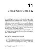
of the upper airway (true for the first decade of life) (Fig. 1-1).
Precise endotracheal tube sizing is critical to avoid cricoid edema
and postintubation croup. A pressure leak should be no greater than
18-20 em H20. Newer high-volume-low-pressure cuffed endotracheal
tubes for infants avoid repeated laryngoscopies to determine the most
appropriate endotracheal tube size.
Given that resistance to airflow is inversely proportional to radius
to the fourth power, a 1-mm reduction in airway diameter increases
resistance to airflow by 16-fold in the infant airway.
The tonsils and adenoids are small in the neonate but reach maximal
size in the first 4-5 years of age. Use of continuous positive pressure
and/or an oral airway will commonly overcome this obstruction.
1
2
CHAPTER1
Introduction
{a) Adult
{b) Infant
FIGURE 1·1 Schematic of an adult (a) and infant (b) airway. A, Anterior; P, Posterior.
[Reprinted from Cote CJ, Todres lD. The pediatric airway. In: Ryan JF, Todres ID, Cote
CJ, et al, eds. A Practice of Anesthesiafor Infants and Children. Philadelphia, PA: WB
Saunders; 1986:35-58, with permission from Elsevier.]
7. The occiput is large. When the infant is placed on a flat surface,
extreme neck flexion will cause airway obstruction. A small roll
placed behind the baby's shoulders will reduce neck flexion and aid in
maintaining the airway.
PEDIATRIC RESPIRATORY PHYSIOLOGY
LOWER AIRWAY
The alveolar bed is incompletely developed at birth; mature alveoli are
seen at 5 weeks of age, with alveolar multiplication with adult morphology
being reached by 8 years of life (Table 1-1 ). Infant lung compliance is
li,):j!IIM
RESPIRATORY SYSTEM DEVELOPMENT
Age
24 weeks gestation
Gas eu:hanging surface forms
Surfactant production begins
Newborn
Decreased reserve because of:
• Increased oxygen consumption
• Decreased FRC
60 weeks postconception
Increased riJk of postoperative apnea in
premature infants until this age
8 years
Number of alveoli reach adult values
lOyears
Fully muscular pulmonary arteries are seen at
the alveolar duct level
19years
Fully muscular pulmonary arteries are seen at
the level of the alveoli
Introduction
CHAPTER1
3
extremely high due to the absence ofelastic fibers; it resembles the emphysematous lung. It is prone to airway collapse and premature airway
closure secondary to low elastic recoil
The cartilaginous rib cage and poorly developed intercostal muscles result in a highly compliant chest wall, leading to inefficient ventilation. The circular configuration of the rib cage (which is ellipsoid
in adults) and the horizontally attached diaphragm (which is oblique
in adults) lead to poor respiratory mechanics. The chest wall begins
to stiffen at 6 months of age, improving the outward recoil of the
chest wall.
The diaphragm has fewer Type I muscle fibers (sustained twitch, highly
oxidative, and fatigue resistant) and is susceptible to fatigue. The adult
diaphragm contains 55%, the neonate 25%, and the preterm only
10% Type I fibers.
LUNG VOLUMES
Functional residual capacity (FRC) in the spontaneously breathing
infant is dynamically maintained at 40% of total lung capacity (similar to
adults). See Table 1-2. The following mechanisms play a role in dynamically maintaining FRC in the awake infant:
• Termination of the expiratory phase before the lung volume reaches
FRC, "auto-PEEP"
• Glottic closure during the expiratory phase (grunting), maintaining
lung volumes
• Diaphragmatic braking: diminished diaphragmatic activity extending
to the expiratory phase
• Tonic activity ofthe diaphragmatic and intercostal muscles, stiffening
the chest wall and maintaining higher lung volumes
Dynamic control of FRC is abolished in the anesthetized child. Under
apneic conditions, the FRC has been estimated to be reduced to 10% of
total lung capacity. The reduced FRC results in reduced intrapulmonary
oxygen reserve and rapid hypoxemia in the infant.
li,1:UIIW
AGE-DEPENDENT RESPIRATORYVALUES
Neooate
Infmt
CbildlAdult
Tidal volume (mL!kg)
6-8
6-8
7-8
Respiratory frequency (bpm)
30-50
20-30
12-16
Minute ventilation (mL/kg/min)
200-260
175-185
80-100
Functional residual capacity (mLJkg)
22-25
25-30
30-45
Total lung capacity (mLikg)
60
70
80
Metabolic rate (mL/kglmin)
6-8
3-4
4
CHAPTER1
Introduction
NEONATAL APNEA
Apnea is defined as cessation ofbreat:hing fur 10-15 seconds and can be associated with bradycardia and loss ofmuscle tone. Apnea is common in premature infants (defined as gestational age <38 weeks) and is related to immature
respiratnry control mechanisms. This phenomenon is rare in full-term
infants. Both theophylline and caffeine have effectively reduced apneic episodes in these infants. Exposure to respiratnry depressants, such as inhaled
agents, opioids, and benzodiazepines, all induce apnea in this population.
Premature infants less than 58-60 weeks postconceptual age have been
shown to be at greater risk of postanesthetic apnea. Apneic episodes have
been described up to 12 hours postoperatively.
Use of a regional anesthetic technique, ie, spinal anesthesia, has been
advocated in this population, although it has not been shown to reduce
the incidence of apnea. Therefore, the need for observation in the perioperative period is not dependent on the anesthetic technique.
NEONATAL HYPOXEMIA
Respiratory control is poorly developed in neonates and preterm infants.
•
•
•
•
•
•
Increased metabolic demand.
Prone to upper airway obstruction.
Immature respiratory control and irregular breathing.
Hypoxia transiently increases then depresses ventilation.
Hypoxia depresses hypercapneic ventilatory response.
Anesthetics abolish mechanisms to maintain FRC.
NEONATAL RENAL FUNCTION
Renal components are incompletely developed at birth, although the formation ofnephrons is complete at 36 weeks gestation. Rapid maturation
occurs during the first month oflife, then these components continue to
fully mature over the first year oflife:
• Reduced glomerular filtration rate (GFR)-25% of adult
• Inadequate tubular function (adult values reached after 2 years of age)
Neonates have difficulty with both volume loading and volume depletion. Volume depletion, though, has more serious implications. Sodium
balance is directly related to intake. The administration of sodium-free
solutions may lead rapidly to hyponatremia.
BODY COMPOSITION
Water constitutes 75% of the weight of a neonate as compared with 65%
of that of a 12-month-old infant and 55% of that of an adult The reduction in total body water is accompanied by a shift in the distribution of
Introduction
li,1:j!iiM
CHAPTER1
5
ASSESSMENT OF HYDRATION/EXTENT OF DEHYDRATION
Slgnt/Symptollli
Dehydration(%)
Thirsty, restless
Fluid De:lidt (miJkg)
5
5(}
Poor tissue turgor, sunken
fontanelle
10
100
Orthostatic, oliguric. comatose
15
150
fluid from extracellular to intracellular. Fat represents 16% of the body
weight of a neonate and increases to 23% by 12 months of age.
Increased fluid requirements occur with:
• Increased metabolic rate
• Increased insensible fluid loss
• Increased obligatory fluid loss
See Table 1-3 for a summary of hydration assessment.
INFANT FLUID REPLACEMENT
Typically 50% of the deficit is replaced over the first hour, with the
remaining deficit being replaced over the next 2 hours. Maintenance fluids can be calculated using the 4/2/1 rule.
Surgical procedures involving only mild tissue trauma may entail
third space losses of3-4 mL/kg/h. This ranges up to 10 mUkg/h in very
large abdominal procedures.
VITAL SIGNS
Changes in heart rate, respiratory rate, and blood pressure as the child
ages are summarized in Table 1-4.
li,1:j!iiM
TYPICAL VITAL SIGNS
Age
Heart Rate
DiutolicBP
Respiratory Rate
Preterm, first day
120
50
35
60
Full term, first
120
65
45
50
1month
160
95
55
40
3months
140
95
60
30
1 year
125
95
60
24
SydolicBP
day
3 years
100
100
65
24
8years
80
105
70
22
12 years
75
115
75
18
6
CHAPTER1
Introduction
NEONATAL HYPOGLYCEMIA
Hypoglycemia in the first 3 days of life is defined in the preterm infant as
BS <20 mg!dL and in the full-term infant as BS <30 mg!dL. After 3 days
of age, blood glucose levels should be >40 mg!dL. Neonates have limited
hepatic glycogen stores, leading to deficient gluconeogenesis. When
these stores are rapidly depleted during increases in metabolic demand.
hypoglycemia ensues.
In addition to limited gluconeogenesis, other causes ofhypoglycemia
include:
•
•
•
•
Increased insulin secretion (Beckwith!Wiedemann)
Perinatal hypoxemia
Sepsis
Toxemia ofpregnancy
Children at greatest risk for hypoglycemic episodes include:
•
•
•
•
•
Preterm neonates
Term infants
Small for gestation infants
Infants of diabetic mothers
Infants receiving total parenteral nutrition (TPN)
Signs and symptoms of hypoglycemia include:
•
•
•
•
•
•
Jitteriness
Cyanosis
Apnea
Lethargy
Hypotonia
Seizures
Treatment is critical to prevent neurologic impairment. Use slow IV
administration of a 250-500-mg/kg glucose bolus followed by an infusion of D1 OW in 0.45NS at 65-85 mL/kg for 24 hours, monitoring glucose level to avoid rebound hypoglycemia.
Monitor blood glucose levels to avoid hyperglycemia and ahyperosmolar
state, intraventricular hemorrhage, osmotic diuresis and dehydration, and
further release of insulin.
INFANT TEMPERATURE REGULATION
The newborn is a homeotherm-compensatory mechanisms exist, but
they regulate only within a limited temperature range (Table 1-5). The
newborn is easily overwhelmed by decreases in environmental temperature. This is compounded by small size, large surface area to volume ratio
(especially the head, which is 20% of the surface area compared with 9%
CHAPTER1
Introduction
li,1:j!iiW
INFANT TEMPERATURE REGULATION
Neutral Temperatme"
Critical Temperaturet
Pretenn infant
34"C
28"C
Term infant
32"C
23"C
Adult
28"C
l"C
~Neutral
7
temperature: the ambient temperature that results in minimal oxygen
consumption.
tCritical temperature: the temperature below which the unanesthetized patient
cannot maintain normal core temperature.
in the adult), thin skin, and limited fat stores. Thermal conductance,
which is heat loss through skin, is inevitable.
The main mechanism for temperature regulation in the newborn
period is nonshivering thermogenesis, also referred to as metabolism of
brown fat. Brown fat differentiates in the fetus at between 26 and 30 weeks
and makes up 2-6% of infant total body weight. These cells have an abundant vascular supply and receive innervation from the beta-adrenergic
system. With exposure to a cold environment, the baby responds with
increased norepinephrine production; brown fat metabolism ensues,
with the production ofheat.
Stores ofbrown fat decline during the first 6 months oflife with a transition to a more adult response to alterations in temperature: shivering.
One problem with the release of norepinephrine is the end organ
effect Norepinephrine produces increased oxygen metabolism and both
pulmonary and peripheral vasoconstriction, with a predisposition to
right-to-left shunting and hypoxemia. The peripheral vasoconstriction
produces mottling. It is therefore incumbent on the anesthesiologist to
maintain the infant's temperature as outlined below.
•
•
•
•
•
•
Transport the infant in a heated "isolette."
Elevate room temperature to 26.6°C or 80°F fur neonates.
Use heating lamps and forced air warming.
Warm fluids and blood products.
Maintain low fresh gas flows and heat-moisture exchanger.
Use protective wrap for extremities and head
INHALATIONAL ANESTHESIA IN PEDIATRIC
PATIENTS
Induction using inhalational agents is more rapid in infants as compared
to adults.
There are four explanations for this:
• Increased alveolar ventilation to FRC ratio (infant 5:1 vs adult 1.4:1 ).
• Increased distribution of cardiac output to highly perfused, vesselrich organs such as the brain and the heart.
8
CHAPTER1
Introduction
• Increased brain mass and reduced muscle mass.
• Potent agents have reduced solubility in infants. The influence of
hematocrit, hemoglobin type, and plasma protein on blood-gas
solubility coefficients is not clear.
INDUCTION WITH INTRACARDIAC SHUNT
Intracardiac shunts, or ventricular septal defects (VSD), alter uptake of
the inhalational agents. 'Ihls is especially true for the more insoluble
agents like Np.
Right~ Left shunts slow uptake and prolong induction
Note: If cardiac function is depressed, it may be equally difficult to
clear an anesthetic and resuscitate the heart and the patient.
Left~ Right shunts are dependent on the size of the shunt
A large shunt (>80%) increases the rate of transfer of anesthetic to
blood and therefore speeds induction.
A small shunt (<50%) has a negligible effect on induction.
MINIMUM ALVEOLAR CONCENTRATION CHANGES
WITH AGE
The minimum alveolar concentration (MAC) increases progressively
through the first month of life, followed by a gradual decline after
6monthsoflife (Fig.l-2).
2.0
1.8
G)'
c
1.6
E
:::)
~
.1!!
£
~ 1.4
::i:
1.2
1.0
0.5
1.0
5
10
Postconceptual age (years)
50
100
FIGURE 1-2 Age and the MAC ofisoflurane from premature infants to adults. [From
LeDez KM, Lennan J, The minimum alveolar concentration (MAC) of isoflurane in
preterm neonates. Aru~sthesiology. 1987;67:301-307.]
Introduction
CHAPTER1
9
SUCCINYLCHOLINE IN CHILDREN
The structure and function of the neuromuscular system is incompletely
developed at birth. The margin of safety for neurotransmission is
reduced in neonates.
• Conduction velocity increases with nerve fiber myelination.
• Slow-contracting muscles are progressively converted to fast
contracting muscles.
• Synaptic transmission is slow.
• During repetitive stimulation, fade occurs because of a limited rate of
acetylcholine release.
Succinylcholine, a depolarizing agonist, given intravenously or
intramuscularly, is useful for rapid tracheal intubation and for
treatment of laryngospasm. Features in infants and children
include:
• Dose requirement is increased based on weight.
• Duration of action is unaffected despite reduced pseudocholinesterase activity.
• Both increased dose and limited duration appear to be due to rapid
redistribution into a larger ECF volume.
• There is no phase II block on first dose.
PEDIATRIC CONTRAINDICATION$ OF SUCCINYLCHOLINE
Contraindications are similar to those in adults with one notable
exception: the myopathic child. The FDA attempted to limit the use of
succinylcholine because of a number of hyperkalemic cardiac arrests in
children with unrecognized myopathies.
Side effects of succinylcholine are as follows:
•
•
•
•
•
•
Cardiac arrhythmias: bradycardia, asystole, and ventricular fibrillation
Hyperkalemia
Postanesthetic myalgias
Pulmonary edema
Increased gastric, intraocular, and intracranial pressure
Associated masseter stiffness, spasm, and malignant hyperthermia
ROCURONIUM
Rocuronium, a nondepolarizing antagonist, is considered a long-acting
relaxant in infants, especially in neonates. A larger volume of distribution and slower clearance results in a prolonged neuromuscular block in
infants (56 minutes vs 26 minutes in children). Onset time is slightly
faster in infants. The duration of action is markedly prolonged when
repeated doses are administered
10
CHAPTER1
Introduction
Neuromuscular function must be evaluated carefully to avoid
hypoventilation-related acidosis and potentiation of relaxant. Observe
the infant prior to induction (muscle tone, depth of respiration, and
vigor of cry) and aim for return ofthis function postoperatively. Useful
clinical signs include the ability to flex arms and lift legs, inspiratory
force less than -25 em Hp, and crying vital capacity greater than
15 mL/kg. The neostigmine requirement is less in children. The onset
of edrophonium is 2-3 minutes faster than that of neostigmine.
ORAL PREMEDICATION IN PEDIATRICS
Benzodiazepine derivatives are widely used for premedicating children. They are given to calm patients, allay anxiety. and diminish the
recall of perianesthetic events. At low doses, minimal drowsiness and
cardiovascular or respiratory depression are produced. Nausea or
vomiting is rare.
Midazolam is a short-acting, water-soluble molecule with a half-life of
2 hours. It is currently the most widely used premedication because of its
rapid uptake and elimination. After oral administration, there is incomplete absorption and extensive first-pass hepatic extraction, explaining
the need for administration ofhigh oral doses.
Other features include:
•
•
•
•
•
•
Peak plasma concentrations in 53 minutes
No increase in gastric pH or residual volume
A calmer child
Acceptable taste for most children
Fewer behavioral changes than in an unpremedicated child
Does not affect the time to recovery
Fentanyl Oralet is most effective when absorbed via the oral mucosa,
not swallowed, since the first-pass metabolism through the liver is high.
The effect is dose-dependent. with signs of sedation in 10 minutes after
receiving 10-15 fig/kg. Desaturation and preoperative nausea are minimized ifthe child is brought to the OR within 10 minutes of completion
of the Oralet. In doses greater than 15 flglkg, there is an increased incidence ofnausea, vomiting, pruritis, and desaturation.
REGIONAL ANESTHESIA
Advantages
•
•
•
•
Faster awakening; reduced anesthetic requirement
Autonomic nervous system suppression
Limb immobilization perioperatively
Reduced stress response