Ebook Critical care nursing made incredibly easy (3rd edition): Part 2
Bạn đang xem bản rút gọn của tài liệu. Xem và tải ngay bản đầy đủ của tài liệu tại đây (28.79 MB, 427 trang )
304
CardiovasCUlar system
Look for a return
• Place the patient in an upright position to relieve dyspnea and
of ST segments to
chest pain. Auscultate lung sounds at least every 2 hours. Adminisbaseline levels with
T-waves flattening
ter supplemental oxygen as needed based on oxygen saturation or
by the end of the
mixed venous oxygen saturation levels.
week, Joy.
• Administer analgesics to relieve pain and nonsteroidal antiinflammatory drugs (NSAIDs), as ordered, to reduce inflammation. Administer steroids if the patient fails to respond
to NSAIDs.
Thanks, and now
on to other news…
• If your patient has a PA catheter, monitor hemodynamic
status. Assess the patient’s cardiovascular status frequently,
watching for signs of cardiac tamponade.
• Administer antibiotics on time to maintain consistent
drug levels in the blood.
• Institute continuous cardiac monitoring to evaluate for
changes in ECG. Look for the return of ST segments to baseline with T-wave flattening by the end of the first 7 days.
• Keep a pericardiocentesis set available if pericardial effusion is suspected, and prepare the patient for pericardiocentesis as indicated.
• Provide appropriate postoperative care, similar to
that given after cardiothoracic surgery.
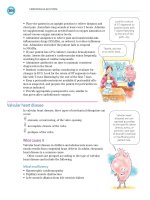
valvular heart disease
In valvular heart disease, three types of mechanical disruption can
occur:
stenosis, or narrowing, of the valve opening
incomplete closure of the valve
prolapse of the valve.
What causes it
Valvular heart disease in children and adolescents most commonly results from congenital heart defects. In adults, rheumatic
heart disease is a common cause.
Other causes are grouped according to the type of valvular
heart disease and include the following:
mitral insufficiency
• Hypertrophic cardiomyopathy
• Papillary muscle dysfunction
• Left ventricle dilation from left ventricle failure
Valvular heart
diseases are categorized according
to the specific valves
(mitral, aortic, or
pulmonic) and type
of disorder (stenosis
or insufficiency) the
patient has.
CardiovasUlar system disorders
305
mitral stenosis
• Endocarditis
• Left atrium tumors
• Miral annulus calcification
aortic insufficiency
• Calcification
• Endocarditis
• Hypertension
• Drugs, especially appetite suppressants
aortic stenosis
• Calcification
Pulmonic stenosis
• Carcinoid syndrome
How it happens
Valvular heart disease may result from numerous conditions,
which vary and are different for each type of valve disorder.
Pathophysiology of valvular heart disease varies according to
the valve and the disorder.
mitral insufficiency
In mitral insufficiency, blood from the left ventricle flows back
into the left atrium during systole, causing the atrium to enlarge
to accommodate the backflow. As a result, the left ventricle also
dilates to accommodate the increased volume of blood from the
atrium and to compensate for diminishing cardiac output.
Ventricular hypertrophy and increased end-diastolic pressure result in increased PAP, eventually leading to left-sided and
right-sided heart failure.
mitral stenosis
In mitral stenosis, the valve narrows as a result of valvular abnormalities, fibrosis, or calcification. This obstructs blood flow from
the left atrium to the left ventricle. Consequently, left atrial volume and pressure increase and the chamber dilates.
Greater resistance to blood flow causes pulmonary hypertension, right ventricular hypertrophy, and right-sided heart failure.
Also, inadequate filling of the left ventricle produces low cardiac
output.
Although the
pathophysiology
varies with the type
of valve and specific
disorder, the end
result seems to be
the same—some
form of heart failure
and pulmonary
involvement.
306
CardiovasCUlar system
aortic insufficiency
In aortic insufficiency, blood flows back into the left ventricle during diastole, causing fluid overload in the ventricle which, in turn,
dilates and hypertrophies. The excess volume causes fluid overload in the left atrium and, finally, the pulmonary system. Leftsided heart failure and pulmonary edema eventually result.
aortic stenosis
In aortic stenosis, elevated left ventricular pressure tries to overcome the resistance of the narrowed valvular opening. The added
workload increases the demand for oxygen, and diminished cardiac output causes poor coronary artery perfusion, ischemia of
the left ventricle, and left-sided heart failure.
Pulmonic stenosis
In pulmonic stenosis, obstructed right ventricular outflow causes
right ventricular hypertrophy in an attempt to overcome resistance to the narrow valvular opening. The ultimate result is rightsided heart failure.
What to look for
The history and physical examination findings vary according to
the type of valvular defects.
mitral insufficiency
Signs and symptoms of mitral insufficiency include:
• orthopnea
• dyspnea
• fatigue
• angina (rare)
• palpitations
• right-sided heart failure (jugular vein distention, peripheral
edema, hepatomegaly)
• systolic murmur
• split S2, S3, and S4 heart sounds.
mitral stenosis
Signs and symptoms of mitral stenosis include:
• dyspnea on exertion, paroxysmal nocturnal dyspnea, orthopnea
• fatigue, weakness
• right-sided heart failure
• crackles on auscultation
• palpitations
• loud S1 and S2
• middiastolic murmur.
CardiovasUlar system disorders
307
aortic insufficiency
Signs and symptoms of aortic insufficiency include:
• dyspnea
• cough
• left-sided heart failure
• pulsus biferiens (rapidly rising and collapsing pulses)
• blowing diastolic murmur or S3
• chest pain with exertion
• crackles on auscultation.
aortic stenosis
Signs and symptoms of aortic stenosis include:
• dyspnea and paroxysmal nocturnal dyspnea
• fatigue
• syncope
• angina
• palpitations and cardiac arrhythmias
• left-sided heart failure
• systolic murmur at the base of the carotids
• chest pain with exertion
• split S1 and S2.
Pulmonic stenosis
Although a patient with pulmonic stenosis may be asymptomatic, possible signs and symptoms include:
• dyspnea on exertion
• right-sided heart failure
• systolic murmur.
What tests tell you
The diagnosis of valvular heart disease can be based on the
results of:
• cardiac catheterization
• chest X-rays
• echocardiography
• ECG.
How it’s treated
Treatments for patients with valvular heart disease commonly
include:
• digoxin, a low-sodium diet, diuretics, vasodilators, and especially ACE inhibitors to correct left-sided heart failure
• oxygen administration in acute situations, to increase oxygenation
Be aware that a
patient with pulmonic
stenosis may have no
symptoms at all.
308
CardiovasCUlar system
• anticoagulants to prevent thrombus formation around diseased
or replaced valves
• prophylactic antibiotics before and after surgery or dental care
to prevent endocarditis
• nitroglycerin to relieve angina in conditions such as aortic stenosis
• beta-adrenergic blockers or digoxin to slow the ventricular rate
in atrial fibrillation or atrial flutter
• cardioversion to convert atrial fibrillation to sinus rhythm
• open or closed commissurotomy to separate thick or adherent mitral valve leaflets
• balloon valvuloplasty to enlarge the orifice of a stenotic mitral, aortic, or pulmonic valve
• annuloplasty or valvuloplasty to reconstruct or repair
the valve in mitral insufficiency
• valve replacement with a prosthetic valve for mitral and
aortic valve disease.
Treatment for
valvular heart disease
typically includes
giving various
combinations of
medications and, in
some cases, valve
repair or replacement.
What to do
• Assess the patient’s vital signs, ABG values, pulse oximetry,
intake and output, daily weights, blood chemistry studies, chest
X-rays, and ECG.
• Place the patient in an upright position to relieve dyspnea if
needed. Administer oxygen to prevent tissue hypoxia as needed
and indicated by ABGs and pulse oximetry.
• Institute continuous cardiac monitoring to evaluate for arrhythmias; if any occur, administer appropriate therapy according to
facility policy and the practitioner’s order.
• For a patient with aortic insufficiency, observe the ECG for arrhythmias, which can increase the risk of pulmonary edema, and
for fever and infection.
• If the patient has mitral stenosis, watch closely for signs of pulmonary dysfunction caused by pulmonary hypertension, tissue
ischemia caused by emboli, and adverse reactions to drug therapy.
• For a patient with mitral insufficiency, observe for signs and
symptoms of left-sided heart failure, pulmonary edema, and
adverse reactions to drug therapy.
Watch those
valves. If the patient
has mitral stenosis,
observe closely for
signs and symptoms
of pulmonary
dysfunction,
emboli, and adverse
reactions to drug
therapy.
QUiCK QUiZ
Quick quiz
1.
Which sign is characteristic of cardiac tamponade?
A. Shortness of breath
B. Beck’s triad
C. Holosystolic murmur
D. Bounding peripheral pulse
Answer: B. Beck’s triad comprises the three classic signs of
cardiac tamponade: elevated CVP with jugular vein distention,
muffled heart sounds, and a drop in systolic blood pressure.
2.
Identify the arrhythmia in the rhythm strip below.
A.
B.
C.
D.
Atrial flutter
Sinus tachycardia
AV junctional rhythm
Atrial fibrillation
Answer: D. The rhythm strip reveals atrial fibrillation. No P
waves are identifiable; ventricular rate is varied; QRS complexes
are uniform in shape but occur at irregular intervals.
3.
Which drug is effective in managing mild to moderate hypotension?
A. Phenylephrine (Neo-Synephrine)
B. Amiodarone (Cordarone)
C. Ibutilide (Corvert)
D. Milrinone
Answer: A. Phenylephrine is indicated for mild to moderate
hypotension.
4.
Which parameter is elevated in right-sided heart failure?
A. CVP
B. Left-ventricular end-diastolic pressure
C. PAWP
D. Cardiac output
Answer: A. CVP is elevated in right-sided heart failure.
309
CardiovasCUlar system
310
5.
ACE inhibitors correct heart failure by:
A. increasing preload.
B. causing vasoconstriction.
C. increasing afterload.
D. reducing afterload.
Answer: D. ACE inhibitors reduce afterload through vasodilation, thereby reducing heart failure.
PPP
PP
P
Scoring
If you answered all five questions correctly, you’re all heart!
(You’d have to be to make it through this cardiovascular
workout!)
If you answered four questions correctly, take heart. You have all
the blood and gumption you need to succeed.
If you answered fewer than four questions correctly, have yourself a heart-to-heart, then try again. You’ll do better next
time.
Good job! Take a
breather and then
move on to the
respiratory system.
LibraryPirate
Respiratory system
Just the facts
In this chapter, you’ll learn:
structure and function of the respiratory system
assessment of the respiratory system
diagnostic tests and procedures for the respiratory
system
respiratory disorders and treatments.
Understanding the respiratory system
The respiratory system delivers oxygen to the bloodstream and
removes excess carbon dioxide from the body.
What a system the
body has going! The
upper airways warm,
filter, and humidify air
before sending it to
the lower airways.
Respiratory system structures
The structures of the respiratory system include the airways and
lungs, bony thorax, and respiratory muscles. (See A close look at
the respiratory system, page 312.)
Airways and lungs
The airways of the respiratory system consist of two parts: the
upper and lower airways. The two lungs are parts of the lower
airway and share space in the thoracic cavity with the heart and
great vessels, trachea, esophagus, and bronchi.
Upper airway
The upper airway warms, filters, and humidifies inhaled air and
then sends it to the lower airway. It also contains the structures
that enable a person to make sounds. Upper airway structures
include the nasopharynx (nose), oropharynx (mouth), laryngopharynx, and larynx.
Critical Care Nursing_Chap05.indd 311
6/29/2011 2:52:04 AM
RespiRAtoRy system
312
A close look at the respiratory system
Get to know the basic structures and functions of the respiratory system so you can
perform a comprehensive respiratory assessment and identify abnormalities. The major
structures of the upper and lower airways are illustrated below. An alveolus, or acinus,
is shown in the inset.
Nasal cavity
Nasopharynx
Oral cavity
Oropharynx
Laryngopharynx
Larynx
Trachea
Right superior
lobar bronchus
Apex of lung
Left main bronchus
Carina
Right main
bronchus
Alveoli
Smooth muscle
Respiratory bronchiole
Alveolar duct
Alveolar sac
Alveolar pore
Terminal bronchiole
Pulmonary vein
Pulmonary artery
Alveoli
Capillary bed
UndeRstAnding the RespiRAtoRy system
313
In the zone
The larynx, which is located at the top of the trachea, houses the
vocal cords. It’s the transition point between the upper and lower
airways.
The larynx is composed of nine cartilage segments. The largest
is the shieldshaped thyroid cartilage. The cricoid cartilage, which
is the only complete ring at the lower end of the larynx, attaches
to the first cartilaginous ring of the trachea.
To flap and protect
The epiglottis is a flap of tissue that closes over the top of the
larynx when the patient swallows. This protects the patient from
aspirating food or fluid into the lower airways.
Lower airway
The lower airway includes the:
• trachea
• bronchi
• lungs.
Lowdown on lower airway
The lower airway begins with the trachea, which divides at the
carina to form the right and left mainstem bronchi of the lungs.
The right mainstem bronchus is shorter, wider, and more vertical
than the left.
The mainstem bronchi branch out in the lungs, forming the:
• lobar bronchi
• tertiary bronchi
• terminal bronchioles
• respiratory bronchioles
• alveolar ducts
• alveoli.
Lungs and lobes
The right lung is larger and has three lobes: upper, middle,
and lower. The left lung is smaller and has only two lobes:
upper and lower.
Plenty of pleura
Each lung is wrapped in a lining called the visceral pleura
and all areas of the thoracic cavity that come in contact with
the lungs are lined with parietal pleura.
A small amount of pleural fluid fills the area between the two
layers of the pleura. This allows the layers to slide smoothly over
each other as the chest expands and contracts. The parietal pleura
also contain nerve endings that transmit pain signals when inflam
mation occurs.
The mainstem
bronchi branch
out in the lungs
to form smaller
airways.
314
RespiRAtoRy system
All about alveoli
The alveoli are the gasexchange units of the lungs. The lungs in a
typical adult contain about 300 million alveoli.
Alveoli consist of type I and type II epithelial cells:
• Type I cells form the alveolar walls, through which gas
exchange occurs.
• Type II cells produce surfactant, a lipidtype substance that
coats the alveoli. During inspiration, the alveolar surfactant allows
the alveoli to expand uniformly. During expiration, the surfactant
prevents alveolar collapse.
Hundreds of
millions of tiny
alveoli conduct
gas exchange in
the lungs.
In circulation
Oxygendepleted blood enters the lungs from the pulmonary
artery of the right ventricle, then flows through the main pulmo
nary arteries into the smaller vessels of the pleural cavities and
the main bronchi, through the arterioles and, eventually, to the
capillary networks in the alveoli.
Trading gases
Gas exchange (oxygen and carbon dioxide diffusion) takes place
in the alveoli. After passing through the pulmonary capillaries,
oxygenated blood flows through progressively larger vessels,
enters the main pulmonary veins and, finally, flows into the left
atrium. (See Tracking pulmonary circulation.)
tracking pulmonary circulation
The right and left pulmonary arteries
carry deoxygenated blood from the
right side of the heart to the lungs.
These arteries divide to form distal
branches called arterioles, which
terminate as a concentrated capillary
network in the alveoli and alveolar sac,
where gas exchange occurs.
Venules—the end branches of the
pulmonary veins—collect oxygenated blood from the capillaries and
transport it to larger vessels, which
carry it to the pulmonary veins. The
pulmonary veins enter the left side of
the heart, where oxygenated blood is
distributed throughout the body.
Trachea
Pulmonary
arterioles
Superior
vena cava
Aorta
Bronchus
Pulmonary
artery
Pulmonary
vein
Pulmonary
trunk
Right atrium
Left atrium
Bronchiole
Left
ventricle
Pulmonary
venules
Alveoli
Inferior vena
cava
Right
ventricle
Diaphragm
UndeRstAnding the RespiRAtoRy system
315
Bony thorax
The bony thorax is composed of:
• clavicles
• sternum
• scapula
• 12 sets of ribs
• 12 thoracic vertebrae.
Imagine that!
Parts of the thorax and some imaginary vertical lines on the chest
are used to describe the locations of pulmonary assessment find
ings. (See Respiratory assessment landmarks, page 316.)
Can you take a ribbing?
Ribs are made of bone and cartilage and allow the chest to
expand and contract during each breath. All ribs are attached to
vertebrae. The first seven ribs also are attached directly to the
sternum. The eighth, ninth, and tenth ribs are attached to the ribs
above them. The eleventh and twelfth ribs are called floating ribs
because they aren’t attached to any other bones in the front.
Respiratory muscles
The primary muscles used in breathing are the diaphragm and
the external intercostal muscles. These muscles contract when
the patient inhales and relax when the patient exhales.
Brain-breath connection
The respiratory center in the medulla initiates each breath by
sending messages over the phrenic nerve to the primary respira
tory muscles. Impulses from the phrenic nerve regulate the rate
and depth of breathing, depending on the carbon dioxide and pH
levels in the cerebrospinal fluid.
Accessory inspiratory muscles
Here’s how other muscles assist in breathing:
In on inspiration
Accessory inspiratory muscles (the trapezius, sternocleidomas
toid, and scalenes) elevate the scapula, clavicle, sternum, and
upper ribs. This expands the fronttoback diameter of the chest
when use of the diaphragm and intercostal muscles isn’t effective.
Out on expiration
Expiration occurs when the diaphragm and external intercostal mus
cles relax. If the patient has an airway obstruction, he may also use
the abdominal muscles and internal intercostal muscles to exhale.
(See Understanding the mechanics of breathing, page 317.)
Ho-hum. The
diaphragm and the
external
intercostal muscles
contract on
inhalation and relax
on exhalation.
RespiRAtoRy system
316
Respiratory assessment landmarks
Use these figures to find the common landmarks used in respiratory assessment.
Anterior view
Suprasternal notch
Manubrium
Angle of Louis
Right upper lobe
Right middle lobe
Right lower lobe
Xiphoid process
Clavicle
First rib
Left upper lobe
Body of the sternum
Left lower lobe
Midsternal line
Left midclavicular line
Left anterior axillary line
Posterior view
Spinous process of C7
First rib
Left upper lobe
Right upper lobe
Scapula
Right middle lobe
Left lower lobe
Right lower lobe
Vertebral line
Left scapular line
UndeRstAnding the RespiRAtoRy system
317
Understanding the mechanics of breathing
Mechanical forces, such as movement of the diaphragm and intercostal muscles, drive the breathing process. In these
depictions, a plus sign (+) indicates positive pressure and a minus sign (–) indicates negative pressure.
At rest
Inhalation
Exhalation
-
-
-
-
-
-
• Inspiratory muscles relax.
• Atmospheric pressure is
maintained in the
tracheobronchial tree.
• No air movement occurs.
-
-
+
- -
-
• Inspiratory muscles contract.
• The diaphragm descends.
• Negative alveolar pressure is
maintained.
• Air moves into the lungs.
+
-
• Inspiratory muscles relax, causing
the lungs to recoil to their resting size
and position.
• The diaphragm ascends.
• Positive alveolar pressure is
maintained.
• Air moves out of the lungs.
Respiration
Effective respiration requires gas exchange in the lungs (external
respiration) and in the tissues (internal respiration).
O2 to lungs
Three external respiration processes are needed to maintain
adequate oxygenation and acidbase balance:
Ventilation (gas distribution into and out of the pulmonary
airways)
Pulmonary perfusion (blood flow from the right side of the
heart, through the pulmonary circulation, and into the left side of
the heart)
Diffusion (gas movement from an area of greater to lesser con
centration through a semipermeable membrane).
RespiRAtoRy system
318
O2 to tissues
Internal respiration occurs only through diffusion, when the red
blood cells (RBCs) release oxygen and absorb carbon dioxide.
Ventilation and perfusion
Gravity affects oxygen and carbon dioxide transport in a positive
way by causing more unoxygenated blood to travel to the lower
and middle lung lobes than to the upper lobes. That’s
why ventilation and perfusion differ in various
parts of the lungs.
Match game
Areas where perfusion and
ventilation are similar have a
ventilationperfusion (V) match; gas
exchange is most efficient in such
areas.
For example, in normal lung
function, the alveoli receive air at a
rate of about 4 L per minute while the capil
laries supply blood to the alveoli at a rate of about 5 L per minute,
creating a V ratio of 4:5, or 0.8. (See Understanding ventilation
and perfusion.)
Mismatch mayhem
A V mismatch, resulting from ventilation–perfusion dysfunc
tion or altered lung mechanics, causes most of the impaired gas
exchange in respiratory disorders.
Ineffective gas exchange between the alveoli and pulmonary
capillaries can affect all body systems by changing the amount of
oxygen delivered to living cells. Ineffective gas exchange causes
three outcomes:
• Shunting (reduced ventilation to a lung unit) causes unoxygen
ated blood to move from the right side of the heart to the left side
of the heart and into systemic circulation. Shunting may result
from a physical defect that allows unoxygenated blood to bypass
fully functioning alveoli. It may also result when airway obstruc
tion prevents oxygen from reaching an adequately perfused area
of the lung. Common causes of shunting include acute respiratory
distress syndrome (ARDS), atelectasis, pneumonia, and pulmo
nary edema.
• Dead-space ventilation (reduced perfusion to a lung unit)
occurs when alveoli don’t have adequate blood supply for gas
exchange to occur, such as with pulmonary emboli and pulmonary
infarction.
Gas exchange
is most efficient
where perfusion
and ventilation
match.
UndeRstAnding the RespiRAtoRy system
319
Understanding ventilation and perfusion
Effective gas exchange depends on the relationship between ventilation and perfusion, or the V ratio. The diagrams
below show what happens when the V ratio is normal and abnormal.
Normal ventilation and perfusion
When ventilation and perfusion are matched, unoxygenated blood from the venous system returns to the right side
of the heart and through the pulmonary artery to the lungs,
carrying carbon dioxide (CO2). The arteries branch into
the alveolar capillaries. Gas exchange takes place in the
alveolar capillaries.
From pulmonary artery
To pulmonary vein
Inadequate perfusion (dead-space ventilation)
When the V ratio is high, as shown here, ventilation is
normal but alveolar perfusion is reduced or absent. Note
the narrowed capillary, indicating poor perfusion. This
commonly results from a perfusion defect, such as pulmonary embolism or a disorder that decreases cardiac
output.
From pulmonary artery
Perfusion blockage
Alveolus
Alveolus
Normal capillary
Narrowed capillary
Inadequate ventilation (shunt)
When the V ratio is low, pulmonary circulation is adequate but not enough oxygen (O2) is available to the alveoli
for normal diffusion. A portion of the blood flowing through
the pulmonary vessels doesn’t become oxygenated.
From pulmonary artery
Ventilation blockage
To pulmonary vein
Inadequate ventilation and perfusion (silent unit)
A silent unit indicates an absence of ventilation and perfusion to the lung area. A silent unit may help compensate
for a V balance by delivering blood flow to better ventilated lung areas.
From pulmonary artery
Perfusion blockage
Alveolus
KEY
To pulmonary vein
Blood with CO2
Ventilation blockage
To pulmonary vein
Alveolus
Blood with O2
• A silent unit (a combination of shunting and deadspace venti
lation) occurs when little or no ventilation and perfusion are pres
ent, such as in cases of pneumothorax and severe ARDS.
Blood with CO2 and O2
RespiRAtoRy system
320
oxygen transport
Most oxygen collected in the lungs binds with hemoglobin to form
oxyhemoglobin; however, a small portion of it dissolves in the
plasma. The portion of oxygen that dissolves in the plasma can
be measured as the partial pressure of arterial oxygen (Pao2) in
blood.
Riding the RBC express
After oxygen binds to hemoglobin, RBCs carry it by way of the
circulatory system to tissues throughout the body. Internal res
piration occurs by cellular diffusion when RBCs release oxygen
and absorb the carbon dioxide produced by cellular metabolism.
The RBCs then transport the carbon dioxide back to the lungs for
removal during expiration.
Acid-base balance
Because carbon dioxide is 20 times more soluble than oxygen,
it dissolves in the blood, where most of it forms bicarbonate (a
base) and smaller amounts form carbonic acid.
Acid-base controller
The lungs control bicarbonate levels by converting bicarbonate
to carbon dioxide and water for excretion. In response to signals
from the medulla, the lungs can change the rate and depth of ven
tilation. This controls acidbase balance by adjusting the amount
of carbon dioxide that’s lost.
In metabolic alkalosis, which results from excess bicarbonate
retention, the rate and depth of ventilation decrease so that car
bon dioxide is retained. This increases carbonic acid levels.
In metabolic acidosis (resulting from excess acid retention or
excess bicarbonate loss), the lungs increase the rate and depth
of ventilation to exhale excess carbon dioxide, thereby reducing
carbonic acid levels.
Off balance
Inadequately functioning lungs can produce
acidbase imbalances. For example, hypoventilation (reduced rate and depth of ventilation)
results in carbon dioxide retention, causing
respiratory acidosis. Conversely, hyperventilation (increased rate and depth of ventilation)
leads to increased exhalation of carbon dioxide
and causes respiratory alkalosis.
Poorly functioning
lungs can produce
acid-base imbalances.
RespiRAtoRy Assessment
321
Respiratory assessment
Respiratory assessment is a critical nursing responsibility. Con
duct a thorough assessment to detect both obvious and subtle
respiratory changes.
history
Build your patient’s health history by asking short, openended
questions. Conduct the interview in several short sessions if you
have to, depending on the severity of your patient’s condition. Ask
his family to provide information if your patient can’t.
Respiratory disorders may be caused or exacerbated by obe
sity, smoking, and workplace conditions so be sure to ask about
these conditions.
Current health status
Begin by asking why your patient is seeking care. Because many
respiratory disorders are chronic, ask how the patient’s latest
acute episode compares with previous episodes and what relief
measures are helpful and unhelpful.
Chronic complaint department
Patients with respiratory disorders commonly report such com
plaints as:
• shortness of breath
• cough
• sputum production
• wheezing
• chest pain
• sleep disturbance.
shortness of breath
Assess your patient’s shortness of breath by asking him to rate
his usual level of dyspnea on a scale of 0 to 10, in which 0 means
no dyspnea and 10 means the worst he has experienced. Then
ask him to rate his current level of dyspnea. Other scales grade
dyspnea as it relates to activity, such as climbing a set of stairs or
walking a city block. (See Grading dyspnea, page 322.)
In addition to using a severity scale, ask these questions: What
do you do to relieve the shortness of breath? How well does it
usually work?
Respiratory
disorders may be
caused or worsened
by obesity, smoking,
and workplace
conditions.
RespiRAtoRy system
322
grading dyspnea
To assess dyspnea as objectively as possible, ask your patient to briefly
describe how various activities affect his breathing. Then, document his
response using this grading system:
• Grade 0: not troubled by breathlessness except with strenuous
exercise
• Grade 1: troubled by shortness of breath when hurrying on a level
path or walking up a slight hill
• Grade 2: walks more slowly on a level path (because of breathlessness) than people of the same age or has to stop to breathe when
walking on a level path at his own pace
• Grade 3: stops to breathe after walking about 100 yards (91 m) on a
level path
• Grade 4: too breathless to leave the house or breathless when dressing or undressing.
Pillow talk
A patient with orthopnea (shortness of breath when lying down)
tends to sleep with his upper body elevated. Ask this patient how
many pillows he uses. The answer reflects the severity of the
orthopnea. For instance, a patient who uses three pillows can be
said to have “threepillow orthopnea.”
Cough
Ask the patient with a cough these questions: At what
time of day do you cough most often? Is the cough
productive? Has it changed recently (if chronic)? If
so, how? What makes the cough better? What makes
it worse?
sputum
If a patient produces sputum, ask him to estimate the
amount produced in teaspoons or some other common mea
surement. Also ask these questions: What’s the color and consis
tency of the sputum? Has it changed recently (if chronic)? If so,
how? Do you cough up blood? If so, how much and how often?
Wheezing
If a patient wheezes, ask these questions: When does wheezing
occur? What makes you wheeze? Do you wheeze loudly enough
for others to hear it? What helps stop your wheezing?
The number of
pillows you need
to sleep indicates
the severity
of your orthopnea.
RespiRAtoRy Assessment
Chest pain
If the patient has chest pain, ask these questions: Where is the
pain? What does it feel like? Is it sharp, stabbing, burning, or ach
ing? Does it move to another area? How long does it last? What
causes it? What makes it better?
Pain provocations
Chest pain due to a respiratory problem is usually the result of
pleural inflammation, inflammation of the costochondral junc
tions, or soreness of chest muscles because of coughing.
It may also be the result of indigestion. Less common
causes of pain include rib or vertebral fractures caused
by coughing or osteoporosis.
323
I guess your
secret is finally
out…you really
are a pain in
the chest!
Well, gee,
only
sometimes!
sleep disturbance
Sleep disturbances may be related to obstructive sleep
apnea or another sleep disorder requiring additional
evaluation.
Daytime drowsiness
If the patient complains of being drowsy or irritable in
the daytime, ask these questions: How many hours of continuous
sleep do you get at night? Do you wake up often during the night?
Does your family complain about your snoring or restlessness?
previous health status
Look at the patient’s health history, being especially watchful for:
• a smoking habit
• exposure to secondhand smoke
• allergies
• previous surgeries
• respiratory diseases, such as pneumonia and tuberculosis (TB).
Ask about current immunizations, such as a flu shot or pneu
mococcal vaccine. Also determine if the patient uses any respira
tory equipment, such as oxygen or nebulizers, at home.
Family history
Ask the patient if he has a family history of cancer, sickle
cell anemia, heart disease, or chronic illness, such as
asthma or emphysema. Determine whether the patient
lives with anyone who has an infectious disease, such as
TB or influenza.
Remember, ladies,
snoring is a symptom
of a respiratory
disorder…it isn’t a
conspiracy to keep us
from getting to sleep.
RespiRAtoRy system
324
Lifestyle patterns
Ask about the patient’s workplace because some jobs, such as
coal mining and construction work, expose workers to substances
that can cause lung disease.
Also ask about the patient’s home, community, and other
environmental factors that may influence how he deals with his
respiratory problems. For example, you may ask questions about
interpersonal relationships, stress management, and coping meth
ods. Ask about the patient’s sex habits and drug use, which may
be connected with acquired immunodeficiency syndrome–related
pulmonary disorders.
physical examination
In most cases, you should begin the physical examination after
you take the patient’s history. However, you may not be able to
take a complete history if the patient develops an ominous sign
such as acute respiratory distress. If your patient is in respira
tory distress, establish the priorities of your nursing assessment,
progressing from the most critical factors (airway, breathing, and
circulation [the ABCs]) to less critical factors. (See Emergency
respiratory assessment.)
Four steps
Use a systematic approach to detect subtle and obvious respira
tory changes. The four steps for conducting a physical examina
tion of the respiratory system are:
• inspection
• palpation
• percussion
• auscultation.
Back, then front
Examine the back first, using inspection, palpation, percus
sion, and auscultation. Always compare one side with the
other. Then examine the front of the chest using the same
sequence. The patient can lie back when you examine the
front of the chest if that’s more comfortable for him.
Making introductions
Before you begin the physical examination, make sure the
room is well lit and warm. Introduce yourself to the patient
and explain why you’re there.
Examine the back
first, and always
compare one side
with the other,
following a
systematic sequence
of inspection,
palpation, percussion,
and auscultation.
RespiRAtoRy Assessment
325
Advice from the experts
emergency respiratory assessment
If your patient is in acute respiratory
distress, immediately assess the ABCs—
airway, breathing, and circulation. If these
are absent, call for help and start cardiopulmonary resuscitation.
Next, quickly check for signs of
impending crisis by asking yourself these
questions:
• Is the patient having trouble breathing?
• Is the patient using accessory muscles
to breathe? If chest excursion is less than
the normal 11/89 to 23/89 (3 to 6 cm), look for
evidence that the patient is using accessory muscles when he breathes, including
shoulder elevation, intercostal muscle
retraction, and use of scalene and sternocleidomastoid muscles.
• Has the patient’s level of consciousness
diminished?
• Is he confused, anxious, or agitated?
• Does he change his body position to
ease breathing?
• Does his skin look pale, diaphoretic, or
cyanotic?
Setting priorities
If your patient is in respiratory distress,
establish priorities for your nursing assessment. Don’t assume the obvious. Note
positive and negative factors, starting with
the most critical factors (the ABCs) and
progressing to less critical factors.
If you don’t have time to go through
each step of the nursing process, make
sure you gather enough data to answer
vital questions. A single sign or symptom
has many possible meanings, so gather
a group of findings to assess the patient
and develop interventions.
inspection
Make a few observations about the patient as soon as you enter
the room and include these observations in your assessment. Note
the patient’s position in the bed. Does he appear comfortable? Is
he sitting up or lying quietly or shifting about? Does he appear
anxious? Is he having trouble breathing? Does he require oxygen?
Is he on a ventilator?
Chest inspection
Help the patient into an upright position, if possible. Ideally, the
patient should be undressed from the waist up or clothed in a
hospital gown. Inspect the patient’s chest configuration, tracheal
position, chest symmetry, skin condition, and nostrils (for flaring),
and look for accessory muscle use.
Beauty in symmetry
Look for chest wall symmetry. Both sides of the chest should
appear equal at rest and expand equally as the patient inhales. The
Your first
observations of the
patient are
important parts of
the assessment.
RespiRAtoRy system
326
diameter of the chest, from front to back, should be about one
half of the width of the chest.
A new angle
Also, look at the angle between the ribs and the sternum at the
point immediately above the xiphoid process. This angle, the
costal angle, should be less than 90 degrees in an adult. The angle
is larger if the chest wall is chronically expanded because of
an enlargement of the intercostal muscles, as can happen with
chronic obstructive pulmonary disease (COPD).
Muscles in motion
When the patient inhales, his diaphragm should descend and the
intercostal muscles should contract. This dual motion causes the
abdomen to push out and the lower ribs to expand laterally. (See
Types of breathing.)
When the patient exhales, his abdomen and ribs return to their
resting positions. The upper chest shouldn’t move much. Accesso
ry muscles may hypertrophy, indicating frequent use. This may be
normal in some athletes, but for most patients it indicates
a respiratory problem, especially when the patient purses
his lips and flares his nostrils when breathing.
Chest wall abnormalities
Inspect for chest wall abnormalities, keeping in mind that
a patient with a deformity of the chest wall might have
completely normal lungs that are cramped in the chest.
The patient might have a smallerthannormal lung capac
ity and limited exercise tolerance.
Barrels, pigeons, and curves
Common abnormalities include:
• Barrel chest—A barrel chest looks like the name implies; it’s
abnormally round and bulging. Barrel chest may be normal in
infants and elderly patients. In other patients, barrel chest occurs
as a result of COPD due to lungs that have lost their elasticity.
The patient typically uses accessory muscles to breathe and easily
becomes breathless. Also note kyphosis of the thoracic spine.
• Pigeon chest—A patient with pigeon chest, or pectus carinatum,
has a chest with a sternum that protrudes beyond the front of
the abdomen. The displaced sternum increases the fronttoback
diameter of the chest but is a minor deformity that doesn’t require
treatment.
• Funnel chest—A patient with funnel chest, or pectus exca
vatum, has a funnelshaped depression on all of or part of the
sternum. This may cause disruptions in respiratory or cardiac
types of
breathing
Men, children, and infants usually use abdominal, or diaphragmatic,
breathing. Athletes and
singers do as well. Most
women, however, usually use chest, or intercostal, breathing.
Hey, I'm pretty
cramped in here!
RespiRAtoRy Assessment
function. Compression of the heart and great vessels may cause
murmurs.
• Thoracic kyphoscoliosis—The patient’s spine curves to one side
and the vertebrae are rotated. Because the rotation distorts lung
tissues, it may be more difficult to assess respiratory status.
Raising a red flag
327
The rate, rhythm,
and quality of
respirations are key
indicators of
respiratory function.
Watch for paradoxical, or uneven, movement of the
patient’s chest wall. Paradoxical movement may
appear as an abnormal collapse of part of the chest
wall when the patient inhales or an abnormal expan
sion when the patient exhales. In either case, such
uneven movement indicates a loss of normal chest wall
function.
Breathing rate and pattern
Assess your patient’s respiratory function by determin
ing the rate, rhythm, and quality of respirations.
Count on it
Adults normally breathe at a rate of 12 to 20 breaths per minute.
To determine the patient’s respiratory rate, count for a full minute,
or longer if you note abnormalities. Don’t tell the patient what
you’re doing or he might alter his natural breathing pattern.
The respiratory pattern should be even, coordinated and regu
lar, with occasional sighs. The normal ratio of inspiration to expi
ration (I:E ratio) is about 1:2.
Abnormal respiratory patterns
Identifying abnormal respiratory patterns can be a great help in
understanding the patient’s respiratory status and overall condi
tion.
tachypnea
Tachypnea is a respiratory rate greater than 20 breaths per
minute; the depth may be normal or shallow. It’s commonly
seen in patients with restrictive lung disease, pain, sepsis, obe
sity, anxiety, and respiratory distress. Fever is another possible
cause. The respiratory rate may increase by 4 breaths per min
ute for every 1° F (0.6° C) increase in body temperature.
Bradypnea
Bradypnea is a respiratory rate below 10 breaths per
minute. It’s commonly noted just before a period of apnea
or full respiratory arrest.
As your patient’s
body temperature
increases with fever,
respiratory rate also
increases.
328
RespiRAtoRy system
Depressed CNS
Patients with bradypnea might have central nervous system
(CNS) depression as a result of excessive sedation, tissue damage,
diabetic coma, or any situation in which the brain’s respiratory
center is depressed. Increased intracranial pressure and metabolic
alkalosis may also cause bradypnea. Note that the respiratory rate
is usually slower during sleep.
Apnea
Apnea is the absence of breathing. Periods of apnea may be short
and occur sporadically, such as in CheyneStokes respirations or
other abnormal respiratory patterns. This condition may be life
threatening if periods of apnea last long enough, and should be
addressed immediately.
hyperpnea
Hyperpnea is characterized by deep breathing with either a nor
mal or increased rate. It occurs during exercise or due to fever,
hypoxia, or acidbase imbalances.
Kussmaul’s respirations
Kussmaul’s respirations are rapid and deep, with sighing breaths.
This type of breathing occurs in patients with metabolic acidosis,
especially when associated with diabetic ketoacidosis, as the
respiratory system tries to lower the carbon dioxide level in the
blood and restore it to normal pH.
Cheyne-stokes respirations
CheyneStokes respirations have a regular cycle of change in the
rate and depth of breathing. Respirations are initially shallow but
gradually become deeper and deeper before becoming shallow
again followed by a period of apnea, lasting 20 to 60 seconds, and
the cycle starts again. This respiratory pattern is seen in patients
with heart failure, kidney failure, or CNS damage. CheyneStokes
respirations can be a normal breathing pattern during sleep in
elderly patients.
Biot’s respirations
Biot’s respirations involve rapid deep breaths that alternate with
abrupt periods of apnea. They’re an ominous sign of severe CNS
damage.
inspecting related structures
Inspect the patient’s skin for pallor, cyanosis, and diaphoresis.
Don’t be blue
Skin color varies considerably among patients, but a patient with
a bluish tint to his skin, nail beds, and mucous membranes is
Address long
periods of apnea
immediately!
They may be
life-threatening.