Ebook Surgical recall (7th edition): Part 2
Bạn đang xem bản rút gọn của tài liệu. Xem và tải ngay bản đầy đủ của tài liệu tại đây (18.7 MB, 338 trang )
Chapter 66 / Vascular Surgery 515
Axillofemoral bypass gra —gra not
in a normal vascular path; usually,
the gra goes from the axillary artery
to the femoral artery and then from one
femoral artery to the other (fem-fem
bypass)
What is an endovascular
repair?
Placement of a stent proximal and distal
to an AAA through a distant percutaneous
access (usually through the groin); less
invasive; long-term results as good as open
2
0'
fr
h
What is an extra-anatomic
bypass gra ?
CLASSIC INTRAOP QUESTIONS DURING AAA REPAIR
Which vein crosses the neck of
the AAA proximally?
Renal vein (le )
What part of the small bowel
crosses in front of the AAA?
Duodenum
Which large vein runs to the
le of the AAA?
IMV
Which artery comes o the
middle of the AAA and runs
to the le ?
IMA
Which vein runs behind the
RIGHT common iliac artery?
LEFT common iliac vein
Which renal vein is longer?
Le
WhiteKnightLove
516 Section II / General Surgery
MESENTERIC ISCHEMIA
Chronic Mesenteric Ischemia
What is it?
Chronic intestinal ischemia from
long-term occlusion of the intestinal
arteries; most commonly results from
atherosclerosis; usually in two or
more arteries because of the extensive
collaterals
What are the symptoms?
Weight loss, postprandial abdominal
pain, anxiety/fear of food because of
postprandial pain, heme occult,
diarrhea/vomiting
What is “intestinal angina”?
Postprandial pain from gut ischemia
What are the signs?
Abdominal bruit is commonly heard
How is the diagnosis made?
A-gram, duplex, MRA
What supplies blood to the gut?
1. Celiac axis vessels
2. SMA
3. IMA
What is the classic nding on
A-gram?
wo of the three mesenteric arteries are
occluded, and there is atherosclerotic
narrowing of the third patent artery
What are the treatment
options?
Bypass, endarterectomy, angioplasty,
stenting
Acute Mesenteric Ischemia
What is it?
Acute onset of intestinal ischemia
What are the causes?
1. Emboli to a mesenteric vessel from
the heart
2. Acute thrombosis of long-standing
atherosclerosis of mesenteric artery
What are the causes of emboli
from the heart?
AFib, MI, cardiomyopathy, valve
disease/endocarditis, mechanical
heart valve
WhiteKnightLove
Chapter 66 / Vascular Surgery 517
What drug has been associated
with acute intestinal ischemia?
Digitalis
To which intestinal artery do
emboli preferentially go?
Superior Mesenteric Artery (SMA)
What are the signs/symptoms
of acute mesenteric ischemia?
Severe pain—classically “pain out of
proportion to physical exam,” no
peritoneal signs until necrosis, vomiting/
diarrhea/hyperdefecation, heme stools
What is the classic triad of
acute mesenteric ischemia?
1. Acute onset of pain
2. Vomiting, diarrhea, or both
3. History of AFib or heart disease
What is the gold standard
diagnostic test?
Mesenteric A-gram
What is the treatment of a
mesenteric embolus?
Perform Fogarty catheter embolectomy,
resect obviously necrotic intestine, and
leave marginal looking bowel until a
“second look” laparotomy is performed
24 to 72 hours postoperatively
What is the treatment of acute
thrombosis?
Papaverine vasodilator via A-gram
catheter until patient is in the OR;
then, most surgeons would perform a
supraceliac aorta gra to the involved
intestinal artery or endarterectomy;
intestinal resection/second look as needed
MEDIAN ARCUATE LIGAMENT SYNDROME
What is it?
Mesenteric ischemia resulting from
narrowing of the celiac axis vessels by
extrinsic compression by the median
arcuate ligament
What is the median arcuate
ligament comprised of?
Diaphragm hiatus bers
What are the symptoms?
Postprandial pain, weight loss
What are the signs?
Abdominal bruit in almost all patients
WhiteKnightLove
518 Section II / General Surgery
How is the diagnosis made?
A-gram
What is the treatment?
Release arcuate ligament surgically
CAROTID VASCULAR DISEASE
Anatomy
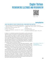
Identify the following structures:
1. Internal carotid artery
2. External carotid artery
3. Carotid “bulb”
4. Superior thyroid artery
5. Common carotid artery
(Shaded area: common site of plaque
formation)
What are the signs/symptoms?
Amaurosis fugax, IA, RIND, CVA
De ne the following terms:
Amaurosis fugax
emporary monocular blindness (“curtain
coming down”): seen with microemboli to
retina; example of IA
TIA
Transient Ischemic Attack: focal
neurologic de cit with resolution of all
symptoms within 24 hours
RIND
Reversible Ischemic Neurologic De cit:
transient neurologic impairment (without
any lasting sequelae) lasting 24 to 72 hours
CVA
CerebroVascular Accident (stroke):
neurologic de cit with permanent brain
damage
What is the risk of a CVA in
patients with TIA?
10% a year
WhiteKnightLove
Chapter 66 / Vascular Surgery 519
What is the noninvasive method
of evaluating carotid disease?
Carotid ultrasound/Doppler: gives
general location and degree of stenosis
What is the gold standard
invasive method of evaluating
carotid disease?
A-gram
What is the surgical treatment
of carotid stenosis?
Carotid EndArterectomy (CEA): the
removal of the diseased intima and media
of the carotid artery, o en performed with
a shunt in place
What are the indications for
CEA in the ASYMPTOMATIC
patient?
Carotid artery stenosis 60% (greatest
bene t is probably in patients with 80%
stenosis)
What are the indications for
CEA in the SYMPTOMATIC
(CVA, TIA, RIND) patient?
Carotid stenosis
Before performing a CEA in
the symptomatic patient, what
study other than the A-gram
should be performed?
Head C
In bilateral high-grade carotid
stenosis, on which side should
the CEA be performed in the
asymptomatic, right-handed
patient?
Le CEA rst, to protect the dominant
hemisphere and speech center
What is the dreaded
complication a er a CEA?
Stroke (CVA)
What are the possible
postoperative complications
a er a CEA?
CVA, MI, hematoma, wound infection,
hemorrhage, hypotension/hypertension,
thrombosis, vagus nerve injury (change
in voice), hypoglossal nerve injury
(tongue deviation toward side of injury—
“wheelbarrow” e ect), intracranial
hemorrhage
What is the mortality rate a er
CEA?
1%
WhiteKnightLove
50%
520 Section II / General Surgery
What is the perioperative
stroke rate a er CEA?
Between 1% (asymptomatic patient) and
5% (symptomatic patient)
What is the postoperative
medication?
Aspirin (inhibits platelets by inhibiting
cyclo-oxygenase)
What is the most common
cause of death during the early
postoperative period a er a CEA?
MI
De ne “Hollenhorst plaque”?
Microemboli to retinal arterioles seen as
bright defects
CLASSIC CEA INTRAOP QUESTIONS
What thin muscle is cut right
under the skin in the neck?
Platysma muscle
What are the extracranial
branches of the internal carotid
artery?
None
Which vein crosses the carotid
bifurcation?
Facial vein
What is the rst branch of the
external carotid?
Superior thyroidal artery
Which muscle crosses the
common carotid proximally?
Omohyoid muscle
Which muscle crosses the
carotid artery distally?
Digastric muscle (T ink: Digastric
Distal)
Which nerve crosses
approximately 1 cm distal to
the carotid bifurcation?
Hypoglossal nerve; cut it and the tongue
will deviate toward the side of the injury
(the “wheelbarrow e ect”)
Inte rnal
c aro tid arte ry
Hypo g lo s s al
ne rve
Exte rnal
c aro tid
arte ry
Co mmo n
c aro tid
arte ry
WhiteKnightLove
Chapter 66 / Vascular Surgery 521
Which nerve crosses the internal
carotid near the ear?
Facial nerve (marginal branch)
What is in the carotid sheath?
1. Carotid artery
2. Internal jugular vein
3. Vagus nerve (lies posteriorly in 98% of
patients and anteriorly in 2%)
4. Deep cervical lymph nodes
SUBCLAVIAN STEAL SYNDROME
What is it?
Arm fatigue and vertebrobasilar
insu ciency from obstruction of the le
subclavian artery or innominate proximal to
the vertebral artery branch point; ipsilateral
arm movement causes increased blood ow
demand, which is met by retrograde ow
from the vertebral artery, thereby “stealing”
from the vertebrobasilar arteries
Which artery is most
commonly occluded?
Le subclavian
WhiteKnightLove
522 Section II / General Surgery
What are the symptoms?
Upper extremity claudication, syncopal
attacks, vertigo, confusion, dysarthria,
blindness, ataxia
What are the signs?
Upper extremity blood pressure
discrepancy, bruit (above the clavicle),
vertebrobasilar insu ciency
What is the treatment?
Surgical bypass or endovascular stent
RENAL ARTERY STENOSIS
What is it?
Stenosis of renal artery, resulting in
decreased perfusion of the juxtaglomerular
apparatus and subsequent activation of the
renin-angiotensin-aldosterone system (i.e.,
hypertension from renal artery stenosis)
S te no s is
What is the incidence?
10% to 15% of the U.S. population have
H N; of these, 4% have potentially
correctable renovascular H N
Also note that 30% of malignant H N
have a renovascular etiology
What is the etiology of the
stenosis?
66% result from atherosclerosis
(men women), 33% result from
bromuscular dysplasia (women
men, average age 40 years, and 50%
with bilateral disease)
Note: Another rare cause is hypoplasia of
the renal artery
WhiteKnightLove
Chapter 66 / Vascular Surgery 523
What is the classic pro le of
a patient with renal artery
stenosis from bromuscular
dysplasia?
Young woman with hypertension
What are the associated risks/
clues?
Family history, early onset of H N, H N
refractory to medical treatment
What are the signs/symptoms?
Most patients are asymptomatic but may
have headache, diastolic H N, ank bruits
(present in 50%), and decreased renal
function
What are the diagnostic tests?
A-gram
Maps artery and extent of stenosis (gold
standard)
IVP
80% of patients have delayed nephrogram
phase (i.e., delayed lling of contrast)
Renal vein renin ratio
(RVRR)
If sampling of renal vein renin levels shows
ratio between the two kidneys 1.5, then
diagnostic for a unilateral stenosis
Captopril provocation test
Will show a drop in BP
Are renin levels in serum
ALWAYS elevated?
No: Systemic renin levels may also
be measured but are only increased
in malignant H N, as the increased
intravascular volume dilutes the elevated
renin level in most patients
What is the invasive
nonsurgical treatment?
Percutaneous Renal Transluminal
Angioplasty (PRTA)/stenting:
With FM dysplasia: use PR A
With atherosclerosis: use PR A/stent
What is the surgical treatment?
Resection, bypass, vein/gra interposition,
or endarterectomy
What antihypertensive
medication is
CONTRAINDICATED in
patients with hypertension
from renovascular stenosis?
ACE inhibitors (result in renal
insu ciency)
WhiteKnightLove
524 Section II / General Surgery
SPLENIC ARTERY ANEURYSM
What are the causes?
Women—medial dysplasia
Men—atherosclerosis
How is the diagnosis made?
Usually by abdominal pain S U/S or
C scan, in the O.R. a er rupture, or
incidentally by eggshell calci cations
seen on AXR
What is the risk factor for
rupture?
Pregnancy
What are the indications
for splenic artery aneurysm
removal?
Pregnancy, 2 cm in diameter, symptoms,
and in women of childbearing age
What is the treatment for
splenic aneurysm?
Resection or percutaneous catheter
embolization in high-risk (e.g., portal
hypertension) patients
POPLITEAL ARTERY ANEURYSM
What is it?
Aneurysm of the popliteal artery caused
by atherosclerosis and, rarely, bacterial
infection
Po plite al
arte ry
Kne e
How is the diagnosis made?
Ane urys m
Usually by physical exam S A-gram, U/S
WhiteKnightLove
Chapter 66 / Vascular Surgery 525
Why examine the contralateral
popliteal artery?
50% of all patients with a popliteal artery
aneurysm have a popliteal artery aneurysm
in the contralateral popliteal artery
What are the indications for
elective surgical repair of a
popliteal aneurysm?
1. 2 cm in diameter
2. Intraluminal thrombus
3. Artery deformation
Why examine the rest of the
arterial tree (especially the
abdominal aorta)?
75%of all patients with popliteal
aneurysms have additional aneurysms
elsewhere; 50% of these are located in
the abdominal aorta/iliacs
What size of the following
aneurysms are usually
considered indications for
surgical repair:
oracic aorta?
6.5 cm
Abdominal aorta?
5.5 cm
Iliac artery?
4 cm
Femoral artery?
2.5 cm
Popliteal artery?
2 cm
MISCELLANEOUS
De ne the following terms:
“Milk leg”
A.k.a. phlegmasia alba dolens
(alba white): o en seen in pregnant
women with occlusion of iliac vein
resulting from extrinsic compression by
the uterus (thus, the leg is “white” because
of subcutaneous edema)
Phlegmasia cerulea dolens
In comparison, phlegmasia cerulea dolens
is secondary to severe venous out ow
obstruction and results in a cyanotic leg;
the extensive venous thrombosis results in
arterial in ow impairment
WhiteKnightLove
526 Section II / General Surgery
Raynaud’s phenomenon
Vasospasm of digital arteries with color
changes of the digits; usually initiated
by cold/emotion
White (spasm), then blue (cyanosis), then
red (hyperemia)
Takayasu’s arteritis
Arteritis of the aorta and aortic branches,
resulting in stenosis/occlusion/
aneurysms
Seen mostly in women
Buerger’s disease
A.k.a. thromboangiitis obliterans:
occlusion of the small vessels of the hands
and feet; seen in young men
who smoke; o en results in digital
gangrene S amputations
What is the treatment for
Buerger’s disease?
Smoking cessation,
What is blue toe syndrome?
Microembolization from proximal
atherosclerotic disease of the aorta
resulting in blue, painful, ischemic toes
What is a “paradoxical
embolus”?
Venous embolus gains access to the le
heart a er going through an intracardiac
defect, most commonly a patent foramen
ovale, and then lodges in a peripheral
artery
What size iliac aneurysm
should be repaired?
What is Behçet’s disease?
sympathectomy
4 cm diameter
Genetic disease with aneurysms from loss
of vaso vasorum; seen with oral, ocular, and
genital ulcers/in ammation (c incidence in
Japan, Mediterranean)
WhiteKnightLove
Se
i
iii
SubspecialtySurgery
Chapter 67 PediatricSurgery
What is the motto o pediatric
surgery?
“Children are NOT little adults!”
What is a simple way to
distract a pediatric patient
when examining the abdomen
or tenderness?
Listen to the abdomen with the
stethoscope and then push down on the
abdomen with the stethoscope to check for
tenderness
PeDiA Ri iV FLUiDS A D U Ri i
What is the maintenance IV
f uid or children?
D5 1/4 NS
Why 1/4 NS?
Children (especially those younger than
4 years of age) cannot concentrate their
urine and cannot clear excess sodium
How are maintenance f uid
rates calculated in children?
4, 2, 1 per hour:
4 cc/ kg or the rst 10 kg o body weight
2 cc/kg or the second 10 kg o body
weight
1 cc/ kg or every kilogram over the
rst 20 (e.g., the rate for a child
weighing 25 kg is 4 10 40 plus
2 10 20 plus 1 5 5, for an
IVF rate of 65 cc/hr)
What is the minimal urine
output or children?
From 1 to 2 mL/kg/hr
What is the best way to present
urine output measurements on
rounds?
Urine output total per shi , THEN cc/kg/hr
What is the major di erence
between adult and pediatric
nutritional needs?
Premature infants/infants/children need
more calories and protein/kg/day
WhiteKnightLove
20 mEq KCl
527
528 Section III / Subspecialty Surgery
What are the caloric
requirements by age or the
ollowing patients:
Premature in ants?
80 Kcal/kg/day and then go up
Children younger than
1 year?
100 Kcal/kg/day (90–120)
Children ages 1 to 7?
85 Kcal/kg/day (75–90)
Children ages 7 to 12?
70 Kcal/kg/day (60–75)
Youths ages 12 to 18
40 Kcal/kg/day (30–60)
What are the protein
requirements by age or the
ollowing patients:
Children younger than
1 year?
3 g/kg/day (2–3.5)
Children ages 1 to 7?
2 g/kg/day (2–2.5)
Children ages 7 to 12?
2 g/kg/day
Youths ages 12 to 18?
1.5 grams/kg/day
How many calories are in
breast milk?
PeDiA Ri BL
20 Kcal/30 cc (same as most formulas)
D V LUMeS
Give blood volume per
kilogram:
Newborn in ant?
85 cc
In ant 1–3 months o age?
75 cc
Child?
70 cc
Fe AL iR ULA i
What is the number o
umbilical veins?
1 (usually)
What is the number o
umbilical arteries?
2
WhiteKnightLove
Chapter 67 / Pediatric Surgery 529
Which umbilical vessel
carries oxygenated blood?
Umbilical vein
T e oxygenated blood travels
through the liver to the IVC
through which structure?
Ductus venosus
Oxygenated blood passes
rom the right atrium to the
le atrium through which
structure?
Foramen ovale
Unsaturated blood goes rom
the right ventricle to the
descending aorta through
which structure?
Ductus arteriosum
De ne the overall etal
circulation
Caro tid arte rie s
To arm
To arm
Duc tus arte rio s is
Lung
Lung
Fo rame n
ovale
Live r
Kidney
Plac e nta
Gut
Fe mo ral arte ry
Fe mo ral arte ry
What are the ADUL
structures o the ollowing
etal structures:
Ductus venosus?
Ligamentum venosum
Umbilical vein?
Ligamentum teres
Umbilical artery?
Medial umbilical ligament
WhiteKnightLove
530 Section III / Subspecialty Surgery
Ductus arteriosus?
Ligamentum arteriosum
Urachus?
Median umbilical ligament
ongue remnant o thyroid’s
descent?
Foramen cecum
Persistent remnant o
vitelline duct?
Meckel’s diverticulum
S to mac h
Duo de num
S upe rio r
me s e nte ric
arte ry
Vite lline duc t
Umbilic us
e M
What is ECMO?
ExtraCorporeal Membrane Oxygenation:
chronic cardiopulmonary bypass—for
complete respiratory support
What are the types o ECMO?
Venovenous: Blood from vein S
oxygenated S back to vein
Venoarterial: Blood from vein (IJ) S
oxygenated S back to artery (carotid)
What are the indications?
Severe hypoxia, usually from congenital
diaphragmatic hernia, meconium
aspiration, persistent pulmonary
hypertension, sepsis
What are the
contraindications?
Weight 2 kg, IVH (IntraVentricular
Hemorrhage in brain contraindicated
because of heparin in line)
WhiteKnightLove
Chapter 67 / Pediatric Surgery 531
e k
What is the major di erential
diagnosis o a pediatric neck
mass?
yroglossal duct cyst (midline), branchial
cle cyst (lateral), lymphadenopathy,
abscess, cystic hygroma, hemangioma,
teratoma/dermoid cyst, thyroid nodule,
lymphoma/leukemia (also parathyroid
tumors, neuroblastoma, histiocytosis
X, rhabdomyosarcoma, salivary gland
tumors, neuro broma)
hyroglossal Duct yst
What is it?
Remnant of the diverticulum formed
by migration of thyroid tissue; normal
development involves migration of thyroid
tissue from the foramen cecum at the base
of the tongue through the hyoid bone to its
nal position around the tracheal cartilage
What is the average age at
diagnosis?
Usually presents around 5 years of age
How is the diagnosis made?
Ultrasound
What are the complications?
Enlargement, infection, and stula
formation between oropharynx or salivary
gland; aberrant thyroid tissue may
masquerade as thyroglossal duct cyst, and
if it is not cystic, deserves a thyroid scan,
MALignancy
WhiteKnightLove
532 Section III / Subspecialty Surgery
What is the anatomic location?
Almost always in the midline
How can one remember the
position o the thyroglossal
duct cyst?
ink: thyroGLOSSAL
midline sticking out
What is the treatment?
Antibiotics if infection is present,
then excision, which must include
the midportion of the hyoid bone and
entire tract to foramen cecum (Sistrunk
procedure)
ONGUE
Branch al l ft Anomal s
What is it?
Remnant of the primitive branchial cle s
in which epithelium forms a sinus tract
between the pharynx (second cle ), or the
external auditory canal ( rst cle ), and the
skin of the anterior neck; if the sinus ends
blindly, a cyst may form
What is the common
presentation?
Infection because of communication
between pharynx and external ear canal
What is the anatomic position?
Second cle anomaly—lateral to the
midline along anterior border of the
sternocleidomastoid, anywhere from
angle of jaw to clavicle
First cle anomaly—less common than
second cle anomalies; tend to be
located higher under the mandible
WhiteKnightLove
Chapter 67 / Pediatric Surgery 533
What is the most common cle
remnant?
Second; thus, these are found most o en
laterally versus thyroglossal cysts, which are
found centrally ( ink: Second Superior)
What is the treatment?
Antibiotics if infection is present, then
surgical excision of cyst and tract once
in ammation is resolved
What is the major anatomic
di erence between thyroglossal
cyst and branchial cle cyst?
yroglossal cyst midline
Branchial cle cyst lateral
( ink: brAnchial lAteral)
Str dor
What is stridor?
Harsh, high-pitched sound heard on
breathing caused by obstruction of the
trachea or larynx
What are the signs/
symptoms?
Dyspnea, cyanosis, di culty with feedings
What is the di erential
diagnosis?
Laryngomalacia—leading cause of stridor
in infants; results from inadequate
development of supporting laryngeal
structures; usually self-limited and
treatment is expectant unless
respiratory compromise is present
Tracheobronchomalacia—similar to
laryngomalacia, but involves the entire
trachea
Vascular rings and slings—abnormal
development or placement of thoracic
large vessels resulting in obstruction of
trachea/bronchus
Acute Allergic Reaction
What are the symptoms o
vascular rings?
Stridor, dyspnea on exertion, or dysphagia
How is the diagnosis o
vascular rings made?
Barium swallow revealing typical
con guration of esophageal compression
Echo/arteriogram
What is the treatment o
vascular rings?
Surgical division of the ring, if the patient
is symptomatic
WhiteKnightLove
534 Section III / Subspecialty Surgery
yst c Hygroma
What is it?
Congenital abnormality of lymph sac
resulting in lymphangioma
What is the anatomic location?
Occurs in sites of primitive lymphatic
lakes and can occur virtually anywhere
in the body, most commonly in the oor
of mouth, under the jaw, or in the neck,
axilla, or thorax
What is the treatment?
Early total surgical removal because they
tend to enlarge; sclerosis may be needed if
the lesion is unresectable
What are the possible
complications?
Enlargement in critical regions, such as the
oor of the mouth or paratracheal region,
may cause airway obstruction; also, they
tend to insinuate onto major structures
(although not malignant), making excision
di cult and hazardous
ASPiRA eD F Rei
B D (FB)
Which bronchus do FBs go into
more commonly (le or right)?
Younger than age 4—50/50
Age 4 and older—most go into right
bronchus because it develops into a
straight shot (less of an angle)
What is the most commonly
aspirated object?
Peanut
What is the associated risk with
peanut aspiration?
Lipoid pneumonia
How can an FB result in “air
trapping and hyperinf ation”?
By forming a “ball valve” (i.e., air in, no
air out) as seen on CXR as a hyperin ated
lung on expiratory lm
How can you tell on
A-P CXR i a coin is in the
esophagus or the trachea?
Coin in esophagus results in the coin lying
“en face” with face of the coin viewed as a
round object because of compression by
anterior and posterior structures
If coin is in the trachea, it is viewed as a
side projection due to the U-shaped
cartilage with membrane posteriorly
WhiteKnightLove
Chapter 67 / Pediatric Surgery 535
What is the treatment o
tracheal or esophageal FB?
Remove FB with bronchoscope or
esophagoscope
HeS
What is the di erential
diagnosis o a lung mass?
Bronchial adenoma (carcinoid is most
common), pulmonary sequestration,
pulmonary blastoma, rhabdomyosarcoma,
chondroma, hamartoma, leiomyoma,
mucus gland adenoma, metastasis
What is the di erential
diagnosis o mediastinal
tumor/mass?
1. Neurogenic tumor (ganglioneuromas,
neuro bromas)
2. Teratoma
3. Lymphoma
4. ymoma
(Classic “ our ’s”: eratoma, errible
lymphoma, hymoma, hyroid tumor)
Rare: pheochromocytoma, hemangioma,
rhabdomyosarcoma, osteochondroma
P ctus D form ty
What heart abnormality
is associated with pectus
abnormality?
Mitral valve prolapse (many patients
receive preoperative echocardiogram)
P ctus excavatum
What is it?
Chest wall deformity with sternum caving
inward ( ink: exCAVatum CAVE)
Pe c tus
e xc avatu m
WhiteKnightLove
536 Section III / Subspecialty Surgery
What is the cause?
Abnormal, unequal overgrowth of rib
cartilage
What are the signs/symptoms?
O en asymptomatic; mental distress,
dyspnea on exertion, chest pain
What is the treatment?
Open perichondrium, remove abnormal
cartilage, place substernal strut; new
cartilage grows back in the perichondrium
in normal position; remove strut 6 months
later
What is the NUSS procedure?
Placement of metal strut to elevate
sternum without removing cartilage
P ctus ar natum
What is it?
Chest wall deformity with sternum outward
(pectus chest, carinatum pigeon); much
less common than pectus excavatum
Pe c tus
c arinatum
What is the cause?
Abnormal, unequal overgrowth of rib
cartilage
What is the treatment?
Open perichondrium and remove
abnormal cartilage
Place substernal strut
New cartilage grows into normal position
Remove strut 6 months later
WhiteKnightLove
Chapter 67 / Pediatric Surgery 537
esophag al Atr s a w thout rach o sophag al ( e) F stula
What is it?
Blind-ending esophagus from atresia
What are the signs?
Excessive oral secretions and inability to
keep food down
How is the diagnosis made?
Inability to pass NG tube; plain x-ray
shows tube coiled in upper esophagus and
no gas in abdomen
What is the primary treatment?
Suction blind pouch, IVFs, (gastrostomy
to drain stomach if prolonged preoperative
esophageal stretching is planned)
What is the de nitive
treatment?
Surgical with 1 anastomosis, o en
with preoperative stretching of blind
pouch (other options include colonic or
jejunal interposition gra or gastric tube
formation if esophageal gap is long)
esophag al Atr s a W th rach o sophag al ( e) F stula
What is it?
Esophageal atresia occurring with a stula
to the trachea; occurs in 90% of cases of
esophageal atresia
What is the incidence?
One in 1500 to 3000 births
De ne the ollowing types
o stulas/atresias:
ype A
Esophageal atresia without TE
stula (8%)
WhiteKnightLove
538 Section III / Subspecialty Surgery
ype B
Proximal esophageal atresia with proximal
TE stula (1%)
ype C
Proximal esophageal atresia with distal TE
stula (85%); most common type
ype D
Proximal esophageal atresia with both
proximal and distal TE stulas (2%)
( ink: D Double connection to
trachea)
WhiteKnightLove
Chapter 67 / Pediatric Surgery 539
ype E
“H-type” TE stula without esophageal
atresia (4%)
How do you remember which
type is most common?
Simple: Most Common type is type C
What are the symptoms?
Excessive secretions caused by an
accumulation of saliva (may not occur
with type E)
What are the signs?
Obvious respiratory compromise,
aspiration pneumonia, postprandial
regurgitation, gastric distention as air
enters the stomach directly from the
trachea
How is the diagnosis made?
Failure to pass an NG tube (although this
will not be seen with type E); plain lm
demonstrates tube coiled in the upper
esophagus; “pouchogram” (contrast
in esophageal pouch); gas on AXR
(tracheoesophageal stula)
What is the initial treatment?
Directed toward minimizing complications
from aspiration:
1. Suction blind pouch (NPO/TPN)
2. Upright position of child
3. Prophylactic antibiotics (Amp/gent)
What is the de nitive
treatment?
Surgical correction via a thoracotomy,
usually through the right chest with
division of stula and end-to-end
esophageal anastomosis, if possible
WhiteKnightLove