Ebook Textbook of anatomy (5/E): Part 1
Bạn đang xem bản rút gọn của tài liệu. Xem và tải ngay bản đầy đủ của tài liệu tại đây (6.33 MB, 495 trang )
VOLUME ONE
Textbook of ANATOMY
Fifth Edition
JAYPEE BROTHERS MEDICAL PUBLISHERS (P) LTD
New Delhi • Panama City • London
Published by
Jaypee Brothers Medical Publishers (P) Ltd
Corporate Office
4838/24 Ansari Road, Daryaganj, New Delhi - 110002, India
Phone: +91-11-43574357, Fax: +91-11-43574314
Website: www.jaypeebrothers.com
Offices in India
• Ahmedabad, e-mail:
• Bengaluru, e-mail:
• Chennai, e-mail:
• Delhi, e-mail:
• Hyderabad, e-mail:
• Kochi, e-mail:
• Kolkata, e-mail:
• Lucknow, e-mail:
• Mumbai, e-mail:
• Nagpur, e-mail:
Overseas Offices
• Central America Office, Panama City, Panama, Ph: 001-507-317-0160
e-mail: , Website: www.jphmedical.com
• Europe Office, UK, Ph: +44 (0) 2031708910, e-mail:
Textbook of Anatomy (Vol. 1)
© 2011, Jaypee Brothers Medical Publishers
All rights reserved. No part of this publication should be reproduced, stored in a retrieval system, or transmitted in any form or by
any means: electronic, mechanical, photocopying, recording, or otherwise, without the prior written permission of the author and
the publisher.
This book has been published in good faith that the material provided by author is original. Every effort is made to ensure
accuracy of material, but the publisher, printer and author will not be held responsible for any inadvertent error (s). In case of
any dispute, all legal matters are to be settled under Delhi jurisdiction only.
First Edition:
Second Edition:
Third Edition:
Fourth Edition:
Fifth Edition:
1996
1999
2003
2007
2011
ISBN 978-93-5025-381-6
Typeset at JPBMP typesetting unit
Printed in India
Preface to the Fifth Edition
Over the years, the considerations taken into account during preparation of the first edition of this book remain
unchanged even as I begin to write this new edition of the “Textbook of Anatomy”. In the present fifth edition, all
chapters have been thoroughly updated and revised. The general format of the book has been changed and now
it holds a completely fresh and new look with the addition of clinical correlations on most of the anatomical structures at the end of each topic, instead of listing them in the form of a single chapter as done in the previous edition.
Clinical matter has been arranged in the form of separate light-green coloured boxes titled “Clinical Correlation”.
Almost every chapter is liberally illustrated with 4-coloured, easy-to-understand illustrations, which the students
can easily draw during their examinations. Most of the text on important topics has been tabulated in an easy-to
-grasp and readily comprehensible format, which would make it pretty interesting for the undergraduate students
to understand the concepts of anatomy.
This textbook is divided into well-elucidated three volumes, with volume one on upper and lower extremities,
volume two on thorax, abdomen and pelvis, and volume three on head, neck and central nervous system. The book
is mainly meant for undergraduate students and may also be of help to students at the postgraduate level.
Through this book, I try to take the young reader through a journey of discovering human anatomy that is as
interesting as it is informative. It contains numerous high quality, hand-drawn simple illustrations, which would
be self-explanatory for undergraduates as well as postgraduates and can be easily drawn at the time of examination.
The information given is graded into different levels for undergraduates and postgraduate level students with
the information meant for students pursuing postgraduation and bright students being arranged in light-pink
coloured boxes titled as “Want to Know More”. This further helps simplify the text.
Printing technology continues to make rapid advances and taking advantage of these, this edition has been made
much more attractive and beautiful. A majority of illustrations have been improved and errors corrected. Overall,
the book has been presented with its original virtues of accuracy and clarity along with the new style and comprehensiveness.
Last but not least, I would like to express deep gratitude to Shri Jitendar P Vij, Chairman and Managing Director,
Jaypee Brothers Medical Publishers for his constant encouragement and support in helping me write a new edition
at 82 years of age.
INDERBIR SINGH
Rohtak, 2011
Preface to the First Edition
Textbooks of anatomy (like the subject itself) have the unenviable reputation of being dull and boring. This book
makes an attempt to (hopefully) change this image. The emphasis throughout the book is on a picture memory
rather than a verbal one; and on understanding of facts rather than their cramming. The author tries to take his
young reader (figuratively) by the hand; and lead him, or her, through a journey of discovery that is as interesting
as it is informative.
It is with this objective that this book incorporates a colour atlas. The atlas is realistic to the extent that normal
contours and relationships are maintained in the illustrations; but it is schematic in that some structures present
in the field of dissection are omitted, or are delineated more clearly than is possible to see in actual dissections. In
describing any part of the body, the region is first reviewed using the atlas figures as a guide. This is followed by
detailed consideration of individual structures.
For the medical student, the study of anatomy is not an end in itself. It is a necessary beginning to the study of
physiology, pathology, and the signs and symptoms of disease. The subject acquires interest if the student is made
aware of the clinical importance of what he studies in the anatomy classroom. This is why there has always been
emphasis on what has been called ‘applied anatomy’. However, many surgeons and physicians feel that much of
what goes under the name of traditional applied anatomy is obsolete, and has to be unlearnt. In this book, therefore,
the emphasis is on providing students some examples of clinical correlations of anatomical structures. Instead of
spreading out this information in small bits throughout the book a separate chapter is devoted to clinical correlations
at the end of each major part.
I shall be grateful to students and teachers who point out errors, typographical or factual, and shall welcome
suggestions for improvement.
I am grateful to the many students and colleagues who have encouraged me in my book writing endeavours,
and this book might never have been written but for their good wishes and encouragement.
INDERBIR SINGH
Rohtak, January 1995
Contents of Volume One
PART 1: UPPER EXTREMITY
1.
Some Essential Terms
The Subject of Anatomy
Main Subdivisions of the Human Body
Some Commonly Used Descriptive Terms
Structures Constituting the Human Body
How Muscles are Named
Some Features of Joints
3
3
3
3
6
7
8
2. Bones of Upper Extremity
Skeleton of the Upper Limb
The Skeleton of the Hand
13
13
34
3.
44
44
44
45
48
49
53
53
54
56
64
Pectoral Region, Axilla and Breast
The Pectoral Region
Cutaneous Nerves of the Pectoral Region
Muscles of the Pectoral Region
The Axilla
The Axillary Artery
The Axillary Vein
Lymph Nodes and Lymphatic Drainage
Lymph Nodes of Upper Limb
The Brachial Plexus and its Branches
The Mammary Glands (Breasts)
4. The Back and Scapular Region
The Back
Muscles of the Back
Nerves of the Back
The Scapular Region
Nerves of Scapular Region
Arteries of Scapular Region
68
68
69
69
73
79
80
5.
84
84
86
88
91
94
96
97
Cutaneous Nerves and Veins of the Free Upper Limb
Cutaneous Nerves of the Free Upper Limb
Veins of the Upper Limb
Anterior Compartment of the Arm
The Brachial Artery
Nerves of the Front of the Arm
Cubital Fossa
Posterior Compartment of the Arm
6. The Forearm and Hand
Front of Forearm and Hand
Muscles of the Front of the Forearm
100
100
100
Important Fascia in the Wrist and Hand
Muscles in the Palm
Nerves of the Forearm and Hand
Arteries of the Forearm
Back of the Forearm and Hand
Muscles of the Back of the Forearm
Nerves and Arteries on the Back of the Forearm and Hand
104
106
109
118
128
128
133
7. General Features of Joints and Joints of the Upper Limb
Classification of Joints
Classification of Joints on the Basis of Structure
Classification of Joints on the Basis of Movements
Joints of the Upper Limb
Joints Connecting the Scapula and Clavicle
The Sternoclavicular Joint
The Shoulder Joint
The Elbow Joint
The Radioulnar Joints
The Wrist Joint
Other Joints of the Upper Limb
135
135
135
139
141
141
141
143
149
151
153
154
8. Surface Marking and Radiological Anatomy of Upper Limb
Surface Marking
Radiological Anatomy
157
157
162
PART 2: LOWER EXTREMITY
9.
Bones of Lower Extremity
The Hip Bone
Pelvis as a Whole
The Femur
The Patella
The Tibia
The Fibula
The Skeleton of the Foot
167
167
177
180
186
188
193
197
10. Cutaneous Nerves, Veins and Lymphatic Drainage: Front and Medial Side of Thigh
Cutaneous Innervation of the Lower Limb
Veins of the Lower Limb
Lymph Nodes and Lymphatic Drainage of the Lower Limb
General Review of the Front and Medial Side of Thigh
Muscles of Front of Thigh
Muscles of Medial Side of Thigh
The Femoral Artery
Femoral Vein
Nerves on Front and Medial Side of Thigh
209
209
214
216
218
224
229
232
235
238
11. Gluteal Region, Back of Thigh and Popliteal Fossa
Gluteal Region
Muscles of the Gluteal Region
245
245
245
Arteries of Gluteal Region
Muscles of the Back of the Thigh
Popliteal Fossa
Popliteal Vessels
Nerves in the Gluteal Region and Back of Thigh
Sacral Ventral Rami and Sacral Plexus
The Sciatic Nerve
The Tibial Nerve
The Common Peroneal Nerve
248
250
253
254
256
256
258
259
260
12. Front and Lateral Side of Leg and the Dorsum of Foot
Compartments of the Leg
Muscles of Anterior Compartment of Leg
Extensor and Peroneal Retinacula
Muscles of Lateral Compartment of Leg
Blood Vessels of the Region
The Tibial Nerve (in Popliteal Fossa)
The Common Peroneal Nerve
The Deep Peroneal (Fibular) Nerve
The Superficial Peroneal (Fibular) Nerve
263
263
263
265
268
270
273
274
274
276
13.
278
279
286
288
294
297
298
300
Back of Leg and Sole
Muscles of the Back of the Leg
Arteries of the Back of the Leg
Muscles and Related Structures in the Sole
Arteries of the Sole
The Tibial Nerve
Medial Plantar Nerve
The Lateral Plantar Nerve
14. Joints of the Lower Limb
Joints and Ligaments of the Pelvis
The Hip Joint
The Knee Joint
The Ankle Joint
Intertarsal Joints
Other Joints of the Lower Limb
Arches of the Foot
303
303
304
306
313
315
316
316
15. Surface Marking and Radiological Anatomy of the Lower Limb
Surface Marking
Radiological Anatomy
319
319
323
Index
Part 1
Upper Extremity
1
Some Essential Terms
CHAPTER
THE SUBJECT OF ANATOMY
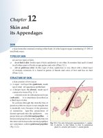
Anatomy is the science that deals with the structure of the human body. Different aspects of the subject are as
follows:
1. Gross anatomy or morphological anatomy is the study of structure that can be seen by naked eye.
2. Microscopic anatomy or histology is the study of structure that can be observed only under a microscope.
3. Cytology is the study of details of the structure of cells.
4. Histochemistry is the study of chemical processes going on in cells and tissues.
5. Ultrastructure is the study of tissues using an electron microscope. It provides very high magnification.
6. Embryology is the study of the development of tissues and organs before birth.
7. Applied anatomy or clinical anatomy is the study of aspects of anatomy that are useful in diagnosis and
treatment of disease.
MAIN SUBDIVISIONS OF THE HUMAN BODY
For convenience of description the human body is divided into a number of major parts.
1. The uppermost part of the body is the head. The face is part of the head.
2. Below the head, there is the neck.
3. Below the neck, there is the region that we call the chest. In anatomical terminology the chest is referred to as
the thorax. The thorax is in the form of a bony cage within which the heart and lungs lie.
4. Below the thorax, there is the region we commonly refer to as ‘stomach’ or ‘belly’.
a. Its proper name is abdomen. The abdomen contains several organs of vital importance to the body.
b. Traced downwards, the abdomen extends to the hips. A part of the abdomen present in the region of the
hips is called the pelvis.
5. The thorax, the abdomen, the neck, and the head together form the trunk.
6. Attached to the trunk, there are the upper and lower limbs, or the upper and lower extremities.
SOME COMMONLY USED DESCRIPTIVE TERMS
1. The study of anatomy is like the learning of a new language. The learning of anatomical terms is the basic
foundation on which all subsequent studies in various subjects of the medical curriculum depend.
2. Of all the terms to be learnt the most fundamental are those used for precise descriptions of the mutual relationships of various structures within the body.
3. In describing such relationships, we usually use terms like ‘in front’, ‘behind’, ‘above’, ‘below’, etc. However,
in a study of anatomy, such terms are found to be inadequate; and the student’s first task is to become familiar
with the specialised terms used.
4
Part 1 ♦ Upper Extremity
Anatomical Position
1. In describing relationships within the body, we presume that the person is standing upright, looking directly
forward, with the arms held by the sides of the body, and with the palms facing forwards.
2. This posture is referred to as the anatomical position. We will now consider some descriptive terms one by
one.
a. When structure A lies nearer the front of the body as compared to structure B, A is said to be anterior to B
(1.1).
The opposite of anterior is posterior. In the above example, it follows that B is posterior to A.
b. When structure C lies nearer the upper end of the body as compared to structure D, C is said to be superior
to D (1.1). The opposite of superior is inferior. In the above example D is inferior to C.
c. The body can be divided into two equal halves, right and left, by a plane passing vertically through it. The
plane separating the two halves is called the median plane (1.2).
i. When a structure lies in the median plane it is said to be median in position (e.g., G in 1.2).
ii. When structure E lies nearer the median plane than structure F, E is said to be medial to F.
iii. The opposite of medial is lateral. In the above example F is lateral to E.
d. In the anatomical position the palm faces forwards and the thumb lies along the outer side of the hand.
Starting from the side of the thumb (or first digit) the fingers are named:
i. Index finger (second digit)
ii. Middle finger (third digit)
iii. Ring finger (fourth digit)
iv. Little finger (fifth digit).
e. Various combinations of the descriptive terms mentioned above are frequently used.
i. For example, each eye is anterior to the corresponding ear; and is also medial to it. Therefore, the eye can
be said to be anteromedial to the ear.
ii. The tip of the nose is inferior and medial to each eye: we can say the nose is inferomedial to the eye.
f. We must now consider terms that are sometimes used as equivalent to some of the terms introduced above.
i. The anterior aspect of the body corresponds to the ventral aspect of the body of four-footed animals.
Hence, the term ventral is often used as equivalent to anterior. (However, we shall see later that the two
terms are not always equivalent e.g., in the thigh).
ii. In the hand, the palm is on the anterior or ventral aspect. This aspect of the hand is often called the
palmar aspect.
iii. The opposite of ventral is dorsal. The back of the hand is the dorsal aspect, or simply the dorsum, of the
hand.
iv. In the case of the foot, the surface towards the sole is ventral: it is called the plantar aspect. The upper
side of the foot is the dorsum of the foot.
v. While referring to structures in the trunk the term cranial (= towards the head) is sometimes used
instead of superior; and caudal (= towards the tail) in place of inferior.
1.1: Scheme to explain the terms anterior, posterior,
superior, and inferior
1.2: Scheme to explain the terms medial, lateral and
median
Chapter 1 ♦ Some Essential Terms
5
vi. In the limbs, the term superior is sometimes replaced by proximal (= nearer) and inferior by distal
(= more distant). Using this convention the phalanges of the hands are designated proximal, middle and
distal.
vii. In the case of the forearm (or hand), the medial side is often referred to as the ulnar side, and the lateral
side as the radial side.
viii. Similarly, in the leg (or foot) we can speak of the tibial (= medial) or fibular (= lateral) sides.
In addition to the terms described above there are some terms that are used to define planes passing through
the body.
1. We have already seen that a plane passing vertically through the midline of the body, so as to divide the
body into right and left halves, is called the median plane. It is also called the mid-sagittal plane.
2. Vertical planes to the right or left of the median plane, and parallel to the latter, are called paramedian or
sagittal planes (1.3).
3. A vertical plane placed at right angles to the median plane (dividing the body into anterior and posterior
parts) is called a coronal plane or a frontal plane (1.4).
4. Planes passing horizontally across the body (i.e., at right angles to both the sagittal and coronal planes) and
dividing it into upper and lower parts, are called transverse or horizontal planes (1.5).
5. There are innumerable oblique planes intermediate between those described above.
6. Sections through any part of the body in any of the planes mentioned above are given corresponding names.
Thus, we speak of:
a. Median sections
b. Sagittal sections
1.3: Scheme showing median and paramedian planes
1.4: Scheme showing a frontal or coronal plane
1.5: Scheme showing a horizontal or transverse plane
6
Part 1 ♦ Upper Extremity
c. Coronal or frontal sections
d. Transverse sections
e. Oblique sections.
STRUCTURES CONSTITUTING THE HUMAN BODY
When we dissect up any part of the body we encounter various elements.
1. The basic framework of the body is provided by a large number of bones that collectively form the skeleton. As
bones are hard they not only maintain their own shape, but also provide shape to the part of the body within
which they lie.
2. In some situations (e.g., the nose or the ear) part of the skeleton is made up, not of bone but of, a firm but flexible
tissue called cartilage.
3. Bones meet each other at joints, many of which allow movements to be performed. At joints, bones are united
to each other by fibrous bands called ligaments.
4. Overlying (and usually attached to) bones we see muscles.
a. Muscles are what the layman refers to as flesh. In the limbs, muscles form the main bulk.
b. Muscle tissue has the property of being able to shorten in length. In other words muscles can contract, and
by contraction they provide power for movements.
c. A typical muscle has two ends, one (traditionally) called the origin, and the other called the insertion.
d. Both ends are attached, typically, to bones.
5. The attachment of a muscle to bone may be a direct one, but quite often the muscle fibres end in cord like structures called tendons which convey the pull of the muscle to bone. Tendons are very strong structures.
6. Sometimes a muscle may end in a flat fibrous membrane. Such a membrane is called an aponeurosis.
7. When we dissect a limb we find that the muscles within it are separated from skin, and from each other, by a
tissue in which fibres are prominent. Such tissue is referred to as fascia.
a. Immediately beneath the skin the fibres of the fascia are arranged loosely and this loose tissue is called superficial fascia.
b. Over some parts of the body the superficial fascia may contain considerable amounts of fat.
c. Deep to the superficial fascia the muscles are covered by a much better formed and stronger membrane.
This membrane is the deep fascia.
d. In the limbs, and in the neck, the deep fascia encloses deeper structures like a tight sleeve.
8. Membranes similar to deep fascia may also intervene between adjacent muscles forming intermuscular septa.
Such septa often give attachment to muscle fibres.
9. Running through the intervals between muscles (usually in relation to fascial septa) there are blood vessels,
lymphatic vessels, and nerves.
a. Blood vessels are tubular structures through which blood circulates.
b. The vessels that carry blood from the heart to various tissues are called arteries.
c. Those vessels that return this blood to the heart are called veins.
d. Within tissues, arteries and veins are connected by plexuses of microscopic vessels called capillaries.
10. Lymphatic vessels are delicate, thin walled tubes. They are difficult to see. They often run alongside veins.
11. Along the course of these lymphatic vessels small bean-shaped structures are present in certain situations.
These are lymph nodes.
12. Lymphatic vessels and lymph nodes are part of a system that plays a prominent role in protecting the body in
various ways that you will study later.
13. Running through tissues, often in the company of blood vessels, we have solid cord like structures called nerves.
a. Each nerve is a bundle of a large number of nerve fibres.
b. Each nerve fibre is a process arising from a nerve cell (or neuron).
c. Most nerve cells are located in the brain and in the spinal cord.
d. Nerves transmit impulses from the brain and spinal cord to various tissues. They also carry information
from tissues to the brain.
Chapter 1 ♦ Some Essential Terms
7
e. Impulses passing through nerves are responsible for contraction of muscle, and for secretions by glands.
Sensations like touch, pain, sight and hearing are all dependent on nerve impulses travelling through nerve
fibres.
14. Bones, muscles, blood vessels, nerves etc., which we have spoken of in the previous paragraphs are to be seen
in all parts of the body. In addition to these many parts of the body have specialized organs, also commonly
called viscera.
15. Some of the viscera are solid (e.g., the liver, or the kidney), while others are tubular (e.g., the intestines) or sac
like (e.g., the stomach).
16. The viscera are grouped together in accordance with function to form various organ systems.
a. Some examples of organ systems are the respiratory system responsible for providing the body with
oxygen.
b. The alimentary or digestive system responsible for the digestion and absorption of food.
c. The urinary system responsible for removal of waste products from the body through urine; and the genital
system which contains organs concerned with reproduction.
17. From the discussions in the previous paragraphs, it will be clear that in the study of the anatomy of any part of
the body we have to consider the following:
a. The skeletal basis of the part including bones and joints.
b. The muscles and fasciae.
c. The blood vessels and nerves.
d. The lymph nodes and their areas of drainage.
e. Viscera present in the region.
HOW MUSCLES ARE NAMED
1. The human body contains a very large number of muscles, and each has a name. The student spends a great
deal of time learning these names.
2. A muscle may be named on the basis of its action, its shape and size, and the region in which it lies.
3. The name of a given muscle usually consists of two or more words based on these characteristics.
4. How muscles are named will be clear from the following examples.
Some Names Based on Region
1. The region on the front of the chest is called the pectoral region. There are two muscles in this region. The larger
of the two is called the pectoralis major. The smaller one is called the pectoralis minor.
2. The region of the buttock is called the gluteal region. It contains three large muscles that are given the names
gluteus maximus (largest), gluteus medius (intermediate in size) and gluteus minimus (smallest).
3. In each of the above examples note that the first word in the name refers to the region concerned, and the
second to relative size.
Some Names Based on Shape
1. Muscles that are straight are given the name rectus (compare with ‘erect’). One such muscle present in the wall
of the abdomen is called the rectus abdominis. Another in the thigh is called the rectus femoris. (Femoris =
thigh—that is why the bone of the thigh is the femur).
2. Over the shoulder there is a strong triangular muscle called the deltoid (after the Greek letter delta, which is
shaped like a triangle).
3. A quadrilateral muscle present in the lumbar region is called the quadratus lumborum.
4. Most muscles have a fusiform shape. The central thicker part is muscular and is called the belly. The ends are
usually tendinous.
5. Some muscles have two (or more) bellies each with a distinct origin (or head). A muscle having two heads is
given the name biceps.
8
Part 1 ♦ Upper Extremity
6. There is one such muscle in the arm and another in the thigh. The one in the arm is the biceps brachii (brachium
= arm) and that in the thigh is the biceps femoris.
7. On the back of the arm, there is a muscle that arises by three heads. It is called the triceps.
8. On the front of the thigh, there is a muscle that has four heads. It is called the quadriceps femoris. (Distinguish
carefully between quadriceps and quadratus).
Some Names Based on Action
1. Muscles that produce flexion may be named flexors; and those that cause extension may be called extensors.
2. Similarly, a muscle may be an abductor, an adductor, a supinator or a pronator.
3. In each case, the word indicating action is followed by another word indicating the part on which the action is
produced. For example, on the back of the forearm there is a muscle that is an extensor of the digits: it is called
the extensor digitorum.
4. Sometimes, we can have more than one muscle that qualifies for such a name. In that case we add a third word
indicative of position.
a. On the front of forearm there are two muscles that produce flexion at the wrist (or carpus).
b. One of them, which lies towards the medial (or ulnar) side is called the flexor carpi ulnaris (= ulnar flexor
of the carpus).
c. The second muscle lies towards the lateral (or radial) side and is called the flexor carpi radialis.
5. Sometimes, it is necessary to add a fourth word to the name. On the back of the forearm there are two radial
extensors of the wrist: we call the longer one the extensor carpi radialis longus and the shorter one is named
the extensor carpi radialis brevis.
6. On the medial side of the thigh, there are three muscles that adduct it. Because of variations in size they are
called the adductor longus, the adductor brevis, and the adductor magnus (magnus = largest).
7. Appreciation of these principles, used in naming muscles, can go a long way in easing the burden of remembering
the names of muscles and their actions.
SOME FEATURES OF JOINTS
1. Joints are formed where two (or more) bones meet.
2. Some joints are merely bonds of union between different bones and do not allow movement. Joints of the skull
(sutures) belong to this category.
3. Some joints allow slight movement, while some (like the shoulder joint) allow great freedom of movement.
4. In describing movements, we use certain terms that the student must understand clearly.
a. Movements at any joint can take place in various planes.
i. Movements taking place in a sagittal plane are referred to as flexion (= bending), and extension
(= straightening).
ii. For example, when we bend the upper limb at the elbow joint so that the front of the forearm tends to
approach the front of the arm this movement is called flexion (1.6).
iii. Straightening the limb at elbow is called extension.
iv. Bending the neck forwards is flexion of the neck, and straightening it, is extension. Similarly, when
we bow, the vertebral column is being flexed, and when the body is made upright the spine is being
extended.
b. Movements in the coronal plane are referred to as abduction (= taking away) or adduction (= bringing near).
i. When a limb is moved laterally so that it moves away from the trunk it is said to undergo abduction
(1.7).
ii. For example, such a movement takes place at the shoulder joint when the upper limb is raised
sidewards.
iii. A similar movement takes place at the hip joint. Adduction and abduction can also take place at the wrist
and at some other joints.
Chapter 1 ♦ Some Essential Terms
1.6: Diagram to explain the movement of flexion of the forearm
9
1.7: Diagram to explain the movement
of abduction of the arm
c. Some joints allow rotatory movements.
i. When the forearm is rotated so that the palm comes to face forwards, the movement is called supination.
ii. The opposite movement is called pronation.
iii. Side-to-side movements of the neck are also rotatory movements.
iv. The movement of the arm performed by a cricketer in bowling is a rotatory movement at the shoulder.
v. Note that during this movement the hand moves in a circle. This movement is, therefore, called circumduction.
d. When the foot is turned so that the sole looks somewhat inwards, the movement is called inversion. The
opposite movement is called eversion.
CLINICAL CORRELATION
Introduction
1. A medical student in India spends the first 12 months in the study of what are called the basic sciences of
anatomy, physiology and biochemistry.
2. This study is an essential preparation for the understanding and treatment of disease.
3. The purpose of this section is to give students a preliminary glimpse of some facets of clinical practice that
make the study of anatomy meaningful and relevant.
4. A patient may come to a doctor with various kinds of problems that may pertain to any part of the body. Some
of these are as follows.
Injury (trauma)
1. Any part of the body may be affected by injury.
2. The mechanical force inflicting injury may be direct or indirect.
3. It may be produced by a sharp object, or may be a blunt injury.
4. The effects of injury will depend on the tissues injured, and on the severity of injury.
Fractures
1. Injury to a bone can break it—breaking of a bone is fracture.
a. The line along which a bone fractures may be transverse, oblique, or spiral.
b. A fracture in which a bone breaks into several small pieces is called a comminuted fracture.
c. Sometimes a bone, made up mainly of cancellous bone, (e.g., body of a vertebra) may be compressed
(compression fracture).
d. In young children, with soft bones, fractures are often incomplete (i.e., the two parts of a fractured bone
may remain together). These are referred to as green-stick fractures.
10
Part 1 ♦ Upper Extremity
2. The two fragments of a fractured long bone may sometimes retain their normal relative position, but quite
commonly there is displacement. Displacement is produced by the actions of muscles on the two fragments.
3. In treating a fracture, the surgeon tries to bring the fragments back to their normal relative position. This is
called reduction of the fracture.
4. Thereafter, measures are taken to prevent the fragments from being displaced again (immobilisation).
a. Immobilisation can be done by applying a suitable plaster cast round the limb, or by operation in which
the two fragments are united using metal appliances of various types (internal fixation and external fixation).
b Immobilisation aids the process of healing, and relieves pain.
Fracture Healing
1. Immediately after a fracture, there is bleeding from vessels within the bone. This collection of blood (haematoma) surrounds the site of the fracture.
2. The bone contains cells that help in repair.
a. These cells proliferate and invade the haematoma. Cells growing out of the two bone ends meet to form a
single mass of cells. New bone is formed within the mass.
b. This bone forms a covering for the two bone ends and unites them. This covering is called the callus.
3. Immature bone of the callus is gradually replaced by mature bone. In this way the bone becomes one again,
but the region of the fracture is thick and may be irregular.
4. As the newly formed bone becomes strong, excess bone around the fracture site is gradually removed. This is
called remodelling.
5. Following remodelling in the bones of children, no trace of the fracture site may remain. However, in adults,
the fracture site usually shows a recognisable irregularity.
Injuries to Joints and Ligaments
1. Severe injury can result in separation of the two bones taking part in a joint. This is called dislocation.
a. Dislocation is more likely to occur in joints that allow free movement e.g., the shoulder joint.
b. Dislocation usually involves damage to the capsule and to some ligaments.
2. In some cases, the two articular surfaces are displaced from their normal position but retain some contact with
each other. This condition is called subluxation.
3. When dislocation at a joint is combined with fracture of one of the bones within the joint the condition is
called fracture-dislocation.
4. A force that strongly stretches a ligament can cause its rupture. This usually leads to displacement of the joint
surfaces.
5. However, injury to a ligament short of rupture can be a cause of serious pain at a joint, specially during movements that tend to stretch the ligament. Such a condition is referred to as sprain, or strain.
6. Ligaments can also be damaged by prolonged mild stress and some authorities use the term strain only for
such injury.
Injuries to Blood Vessels
1. Injury to an artery is dangerous because loss of blood can, if unchecked, lead to death.
a. Bleeding from an artery can be stopped by applying pressure over a suitable point.
b. Knowledge of points where major arteries can be palpated and pressure applied on them is therefore of
importance.
2. Injuries to large veins can also be serious.
a. In some veins the pressure can be lower than atmospheric pressure and air can be sucked into them.
b. This air travels into the heart and lungs and can block small vessels and capillaries there (air embolism).
3. Injured vessels have to be ligated (tied up). In the case of large vessels repair of the vessel may be possible.
4. Ligation of an artery carries the risk of necrosis (death) of the part supplied if its blood supply through alternative channels (collateral circulation) is not adequate.
Chapter 1 ♦ Some Essential Terms
11
5. For this reason, a knowledge of anastomoses established by various arteries becomes of importance.
6. Anastomoses are most abundant in regions where the main artery is subjected to compression because of
movements e.g., around joints.
Injuries to Nerves
1. Injuries to nerves, if complete, can lead to paralysis (loss of the power of movement) of all muscles supplied,
and anaesthesia (loss of sensations) over the area of sensory supply.
2. When a nerve is injured all structures supplied by branches arising distal to the point of injury are affected.
However, injury may be partial and only some of the structures are then affected.
3. The extent of sensory loss is usually less than the area supplied by the nerve, because of overlap in the territories supplied by adjoining cutaneous nerves.
Injuries to Other Tissues
1. A muscle may be injured by any kind of direct violence.
a. It may also be injured during rigorous exercise (as in athletes).
b. In persons following sedentary occupations, and in old age, even mild unaccustomed movement can lead
to strain within a muscle leading to pain and discomfort.
c. However, the most serious effects on muscles are seen following injury to the nerves supplying them.
d. Muscles can also be paralysed as a result of injury to the brain, to the spinal cord, or to nerve roots.
2. Tendons can be injured as a result of injury.
a. A sharp injury can cut right through a tendon.
b. A tendon can be damaged by a fractured bone.
c. A tendon weakened by degenerative changes may rupture with relatively mild force.
3. The skin is the tissue most commonly affected by injury.
a. However, because of great regenerative capacity superficial injuries are easily repaired.
b. When large areas of skin are lost these areas can be covered with skin taken from other parts of the body.
This process is called skin grafting.
c. Injury to skin may also be caused by extreme heat (burns), or by extreme cold; by chemicals (e.g., strong
acids or alkalis); electrical currents; and by various kinds of radiations.
d. Large areas of skin can be lost as a result of burns. In such cases death can occur because of loss of large
amounts of water from the body, or because of infection.
4. Injuries to internal organs are usually serious and require urgent surgery.
a. An injured organ may bleed into a body cavity.
b. Such an internal haemorrhage can lead to death if it is not recognised and treated in time.
5. Injury to the brain is always very serious and often a cause of death. Damaged nerve cells cannot regenerate
and if the patient survives some effects of injury may persist.
INFLAMMATION AND INFECTIONS
1. Any tissue or organ in the body can be infected with bacteria or viruses.
a. Infection may be acute or chronic.
b. In acute infection the tissue usually shows signs of inflammation.
c. The part becomes red in colour and warm because of greater blood flow.
d. Accumulation of fluid causes swelling; and pressure on nerves in the area causes pain.
2. Infection can lead to pus formation. If the pus is in an enclosed space (as on the tip of a finger) it can cause
considerable pain.
3. Infection often spreads along fascial planes. Its spread can be limited by fascial septa (as in the palm). In the
treatment of infections knowledge of the anatomy of the part is, therefore, important.
4. Many of the terms used to describe infections are made up of the name of the part followed by the suffix ‘itis’.
a. Infection of the tonsil is tonsillitis.
b. Infection of the vermiform appendix is called appendicitis.
12
Part 1 ♦ Upper Extremity
5. Such terms are often used to describe inflammation of the organ that may be caused by agents other than
infection.
a. For example, inflammation of the mucosa of the stomach (gastritis) can be caused by any substance that
irritates it (e.g. alcohol, or a drug).
b. A common cold (rhinitis) can result from a virus infection, but it can also be caused by allergy (undue
sensitivity of the tissue to some foreign substance).
c. Inflammation can also be caused by physical agents like heat, cold, mechanical trauma and radiations.
Some infections are caused by protozoa.
d. Infection by amoeba histolytica can cause colitis (inflammation of the colon) or hepatitis (inflammation
in the liver).
e. Another important infection caused by protozoa is malaria.
f. When there is infection in any part of the body, lymph nodes draining the area may enlarge and become
painful. This condition is called lymphadenitis. Lymph vessels may also get inflamed (lymphangitis) and
may be seen as red streaks over the skin.
NEOPLASIA
1. In our body, cells in various tissues are constantly multiplying to replace dead cells.
2. The rate of multiplication varies from tissue to tissue and is strictly controlled.
3. Under certain circumstances the mechanisms that control cell proliferation do not work. As a result, there can
be uncontrolled multiplication of cells leading to the formation of a neoplasm or tumour.
4. Some tumours remain confined to their original site. Such tumours are said to be benign, and their surgical
removal leads to complete cure.
5. In the case of other tumours, some cells that get detached from the main tumour spread to distant sites
(through lymphatic vessels, or sometimes through veins), and start multiplying forming new tumours. Such
tumours are said to be malignant.
6. The original tumour is the primary tumour, while the ones formed by spread from it are called secondaries.
7. The spread of malignant tumours greatly adds to the difficulty of treating them, and once secondaries form
complete eradication of the tumour may become impossible.
8. A surgeon examining a malignant tumour examines the lymph nodes of the region very carefully, as such
examination can provide vital clues about the degree to which the growth has spread.
9. Because of the spread of malignant growths and of infections through lymphatics a knowledge of the lymphatic drainage of various parts of the body is of great importance.
10. Malignant growths fall into two major categories.
a. A malignant growth arising from epithelial cells is called a carcinoma (cancer). Carcinoma can arise in the
skin, in any tube or cavity lined with epithelium, and from epithelia of glands.
b. A malignant tumour arising from non-epithelial tissue is usually referred to as sarcoma.
c. Such tumours can arise from connective tissue (fibrosarcoma), from muscle (myosarcoma), from bone
(osteosarcoma) etc.
OTHER CAUSES OF DISEASE
Apart from trauma, inflammation due to various causes, and neoplasms, some other causes that can lead to disease are as follows:
1. An individual may be born with physical defects that may affect the exterior or interior of the body.
a. Such defects are called congenital malformations.
b Many diseases can be traced to genetic causes.
c. Genetic defects can result in biochemical alterations that can lead to various disorders.
2. Diseases can also be produced as a result of malnutrition, and can be a feature of normal ageing processes.
3. With increasing age there is narrowing of the arteries. Lack of adequate blood supply to the heart or to the
brain can lead to serious consequences.
4. Wear and tear in joints is a common cause of joint pains in old persons.
5. Abnormal outgrowths from bone ends can reduce mobility at joints and can also cause pain (osteoarthritis).
2
Bones of Upper Extremity
CHAPTER
Skeleton of the Upper Limb
The bones of the upper limb are shown in 2.1 and 2.2.
The bones join each other at a number of joints.
1. The upper end of the humerus is joined to the scapula at the shoulder joint (2.2).
2. The lower end of the humerus is joined to the upper ends of the radius and ulna to form the elbow joint.
3. The wrist joint is formed where the lower ends of the radius and ulna meet the carpal bones.
4. The upper and lower ends of the radius and ulna are united to one another at the superior and inferior radioulnar
joints.
5. There are numerous small joints in the hand:
a. The intercarpal joints between the carpal bones themselves.
b. The carpometacarpal between the carpal and metacarpal bones.
c. The metacarpo-phalangeal between each metacarpal bone and the proximal phalanx.
d. The interphalangeal joints between the phalanges themselves.
During embryonic development most bones of the body are first seen in the form of cartilage. The replacement
of these cartilages by bone is called ossification.
1. In most bones, ossification begins during intrauterine life at an area called the primary centre of ossification.
2. The part of the bone formed by extension of bone formation from the primary centre is called the diaphysis.
3. However, the ends of long bones are still cartilaginous at birth. These are ossified from secondary centres that
(as a rule) appear after birth.
4. Each part ossified from a secondary centre is called an epiphysis.
2.1: Bones of upper extremity
BONES ATTACHED
TO TRUNK
FORMING
PECTORAL GIRDLE
BONES OF FREE LIMB
Name
Location
Clavicle
Rod-like bone lying in front of upper part of thorax
Scapula
Triangular plate of bone behind upper part of thorax behind upper
part of thorax
Humerus
Arm
Radius
Forearm (laterally)
Ulna
Forearm(medially)
Carpal bones (8)
Wrist (arranged in 2 rows)
Metacarpal bones (5)
Palm
Phalanges
Fingers or digits (3 in each finger and 2 in the thumb)
14
Part 1 ♦ Upper Extremity
5. For many years after birth, the bone of the epiphysis and
diaphysis is separated by a plate of cartilage called the
epiphyseal plate.
6. This plate is a site of active bone growth. Growth in length of a
bone is possible only as long as the plate exists.
7. When a bone has attained its full length the epiphyseal plate
disappears and the diaphysis and epiphysis fuse with each
other. This is referred to as fusion of the epiphysis.
8. Knowledge of the ages at which various centres of ossification
appear, and the ages at which epiphyses fuse to the diaphysis
is of practical importance in determining the age of a person.
Determination of the Side to which a Limb Bone Belongs
1. The first step in the study of any bone is to orientate it as it lies
in the body.
2. To do this, we have to distinguish the anterior aspect from the
posterior; the upper end from the lower; and the medial side
from the lateral.
3. Once, we have this information we can find out whether the
bone belongs to the right limb or the left one.
The Clavicle
Determination of Side
The clavicle is a long bone having a shaft, and two ends (2.3 and 2.4).
1. The medial end is much thicker than the shaft and is easily
distinguished from the lateral end that is flattened.
2. The anterior and posterior aspects of the bone can be distinguished
by the fact that the shaft (which has a gentle S-shaped curve) is
convex forwards in the medial two-thirds, and concave forwards
in its lateral one-third.
3. The inferior aspect of the bone is distinguished by the presence
of a shallow groove on the shaft, and by the presence of a rough
area near its medial end.
For purposes of description it is convenient to divide the clavicle
into the lateral one-third that is flattened, and the medial two-thirds
that are cylindrical.
1. The lateral one-third has two surfaces i.e., superior and inferior.
a. These surfaces are separated by two borders: anterior and
posterior.
2.2: Drawing showing bones and joints of upper
b. The anterior border is concave and shows a small thickened
extremity
area called the deltoid tubercle.
c. The lower surface (of the lateral one-third) shows a prominent thickening near the posterior border; this is the conoid tubercle.
d. Lateral to the tubercle, there is a rough ridge that runs obliquely up to the lateral end of the bone, and is
called the trapezoid line.
2. The medial two-thirds of the shaft has four surfaces: anterior, posterior, superior and inferior, that are not
clearly marked off from each other.
a. The large rough area present on the inferior aspect of the bone near the medial end forms part of the inferior
surface.
Chapter 2 ♦ Bones of Upper Extremity
15
2.3: Right clavicle seen from above
2.4: Right clavicle seen from below
b. The middle-third of the inferior aspect shows a longitudinal groove, the depth of which varies considerably
from bone to bone.
3. The lateral or acromial end of the clavicle bears a smooth facet which articulates with the acromion of the
scapula to form the acromioclavicular joint.
4. The medial or sternal end of the clavicle articulates with the manubrium sterni, and also with the first costal
cartilage.
a. The articular area is smooth and extends on to the inferior surface of the bone for a short distance.
b. The uppermost part of the sternal surface is rough for ligamentous attachments.
5. The clavicle can be easily felt in the living person as it lies just deep to the skin in its entire extent. The sternal
end of the bone forms a prominent bulge that extends above the upper border of the manubrium sterni.
want to know more?
Attachments on the Clavicle
The muscles attached to the clavicle are as follows:
1.
2.
3.
4.
5.
6.
The pectoralis major (clavicular head) arises from the anterior surface of the medial half of the shaft.
The deltoid arises from the anterior border of the lateral one-third of the shaft.
The sternocleidomastoid (clavicular head) arises from the medial part of the upper surface.
The sternohyoid (lateral part) arises from the lower part of the posterior surface just near the sternal end.
The trapezius is inserted into the posterior border of the lateral one-third of the shaft.
The subclavius is inserted into the groove on the inferior surface of the shaft.
See 2.5 and 2.6.
16
Part 1 ♦ Upper Extremity
Ossification of the Clavicle
1.
2.
3.
4.
5.
The clavicle is the first bone in the body to start ossifying.
The greater part of the clavicle is formed by intramembranous ossification.
The sternal and acromial ends are preformed in cartilage.
Two primary centres appear in the shaft during the 6th week of fetal life and soon fuse with each other.
The sternal end ossifies from a secondary centre that appears between 15 and 20 years of age, and fuses with
the shaft by the age of 25 years.
6. An additional centre may appear in the acromion.
Clinical correlation
Fractures of the Clavicle
Most fractures of the clavicle are caused by indirect violence.
1. The bone is most commonly fractured at the junction of its middle and outer one-thirds (2.12), this being the
weakest point of the bone.
a. In this fracture, the outer fragment is pulled downwards by the weight of the upper limb. It is pulled
medially by the pectoralis major.
b. The inner segment is pulled upwards by the sternocleidomastoid.
2. Less commonly, the clavicle is fractured near its lateral end.
2.5: Right clavicle showing attachments, seen from above
2.6: Right clavicle showing attachments, seen from below