Ebook Pocket atlas of oral diseases: Part 2
Bạn đang xem bản rút gọn của tài liệu. Xem và tải ngay bản đầy đủ của tài liệu tại đây (9.13 MB, 172 trang )
199
6 Papillary Lesions
Papillary lesions of the oral mucosa are a small group, appearing clinically as exophytic growths with a verrucous or cauliflower-like surface.
Reactive lesions, benign tumors, malignancies, and systemic diseases are
included in this group. Etiologically, traumatic, viral, and neoplastic
factors may cause these lesions. The diagnosis is based on clinical and
histopathological criteria.
O Focal epithelial hyperplasia
O Papilloma
O Epulis fissuratum
O Condyloma acuminatum
O Crohn disease
O Verruca vulgaris
O Acanthosis nigricans, maligO Verruciform xanthoma
nant
O Verrucous carcinoma
O Familial acanthosis nigricans
O Squamous-cell carcinoma
O Darier disease
O Verrucous leukoplakia
Laskaris, Pocket Atlas of Oral Diseases © 2006 Thieme
All rights reserved. Usage subject to terms and conditions of license
200
Papillary Lesions
Papilloma
Papilloma is a common benign proliferation, originating from the stratified squamous epithelium (see also p. 34). Clinically, papilloma presents
as a painless, exophytic, well-circumscribed and usually pedunculated
lesion. Typically, it consists of numerous fingerlike projections, which
give the lesion a “cauliflower” appearance (Fig. 197). The tumor has a
white or grayish color, and is usually between 0.5 cm and 1 cm in size.
The tongue, gingiva, and soft palate are the sites of predilection
(Figs.198, 199). The differential diagnosis includes verruca vulgaris, condyloma acuminatum, early verrucous carcinoma, and verruciform xanthoma.
Fig. 197
Papilloma of the gingiva.
Laskaris, Pocket Atlas of Oral Diseases © 2006 Thieme
All rights reserved. Usage subject to terms and conditions of license
Papillary Lesions
Fig. 198
Papilloma of the palate.
Fig. 199
Papilloma of the tongue.
Laskaris, Pocket Atlas of Oral Diseases © 2006 Thieme
All rights reserved. Usage subject to terms and conditions of license
201
202
Papillary Lesions
Condyloma Acuminatum
Definition Condyloma acuminatum is a sexually transmitted benign
lesion, mainly occurring in the anogenital region, and rarely in the
mouth.
Etiology
Human papillomavirus, types 6 and 11.
Clinical features Oral lesions appear as single, or more often multiple,
small, sessile, well-demarcated, exophytic masses with a cauliflower-like
surface (Fig. 200). The lesions have a whitish or normal color, and usually
recur; the average size is 0.5–1 cm. The labial mucosa, tongue, gingiva,
buccal mucosa, and soft palate are the sites most frequently affected.
Oral condyloma acuminatum occurs more frequently in HIV-infected
patients (Fig. 201). The anogenital lesions present as discrete or multiple,
sessile or pedunculated, exophytic, small nodules with cauliflower-like
appearance. The lesions may have whitish or brownish color and size
that varies from 1–5 mm to several centimeters in diameter.
Laboratory tests Histopathological examination, in-situ hybridization,
polymerase chain reaction (PCR).
Differential diagnosis Papilloma, verruca vulgaris, focal epithelial hyperplasia, verruciform xanthoma, sialadenoma papilliferum, focal dermal hypoplasia syndrome, early verrucous carcinoma, molluscum contagiosum.
Treatment Surgical excision, cryosurgery, CO2 laser, electrocautery,
topical podophyllin.
Laskaris, Pocket Atlas of Oral Diseases © 2006 Thieme
All rights reserved. Usage subject to terms and conditions of license
Papillary Lesions
Fig. 200
Multiple condylomata acuminata on the lower lip mucosa.
Fig. 201
Multiple condylomata acuminata of the gingiva.
Laskaris, Pocket Atlas of Oral Diseases © 2006 Thieme
All rights reserved. Usage subject to terms and conditions of license
203
204
Papillary Lesions
Verruca Vulgaris
Definition Verruca vulgaris, or common wart, is a benign, mainly
cutaneous lesion that may rarely appear in the oral mucosa.
Etiology
Human papillomavirus (HPV-2, 4, and 40).
Clinical features Verruca vulgaris frequently develops on the hands of
children. From the skin lesions, the virus can be autoinoculated into the
oral mucosa, usually on the vermilion border and the lip mucosa, commissures, and tongue. Clinically, it appears as a painless, small, sessile,
and well-defined exophytic growth with a cauliflower surface and whitish color (Figs. 202, 203, 204). The lesions may be single or multiple.
Laboratory tests Histopathological examination.
Differential diagnosis Papilloma, condyloma acuminatum, verruciform xanthoma, focal epithelial hyperplasia.
Treatment
Fig. 202
Surgical excision, electrosurgery.
Verruca vulgaris, multiple lesions on the buccal mucosa.
Laskaris, Pocket Atlas of Oral Diseases © 2006 Thieme
All rights reserved. Usage subject to terms and conditions of license
Papillary Lesions
Fig. 203
Verruca vulgaris: multiple lesions on the lip mucosa.
Fig. 204
Verruca vulgaris: solitary lesion on the lip mucosa.
Laskaris, Pocket Atlas of Oral Diseases © 2006 Thieme
All rights reserved. Usage subject to terms and conditions of license
205
206
Papillary Lesions
Verruciform Xanthoma
Definition Verruciform xanthoma is a rare hyperplastic disorder of the
oral mucosa.
Etiology
trauma.
Unknown. Presumably, it may represent a reaction to local
Clinical features The lesion is more common in women in the 50–70
year age group. Typically, it appears as a well-demarcated, painless,
sessile, slightly elevated lesion. It has a cauliflower-like surface with a
reddish-yellowish or normal color (Fig. 205). The size ranges from 0.5 cm
to 2 cm, and the gingiva and alveolar ridge, tongue, and palate are the
most common locations.
Laboratory tests Histopathological examination.
Differential diagnosis Papilloma, verruca vulgaris, condyloma acuminatum, sialadenoma papilliferum, verrucous carcinoma.
Treatment
Surgical excision.
Verrucous Carcinoma
Verrucous carcinoma (see also p. 36) is a low-grade variant of squamouscell carcinoma. Typically, it presents as an exophytic, whitish mass with a
papillary or verruciform surface (Fig. 206). Along with the clinical features, biopsy and histopathological examination should be performed to
rule out other papillary growths. Verrucous carcinoma is well-differentiated, slow-growing, rarely metastasizes, and has a good prognosis.
Laskaris, Pocket Atlas of Oral Diseases © 2006 Thieme
All rights reserved. Usage subject to terms and conditions of license
Papillary Lesions
Fig. 205
Verruciform xanthoma of the tongue.
Fig. 206
Extensive verrucous carcinoma of the tongue.
Laskaris, Pocket Atlas of Oral Diseases © 2006 Thieme
All rights reserved. Usage subject to terms and conditions of license
207
208
Papillary Lesions
Squamous-Cell Carcinoma
Squamous-cell carcinoma has a wide range of clinical presentations (see
also pp. 172, 272). A common clinical feature is an exophytic mass. It has
a papillary or verruciform surface and a red, whitish, or normal color
(Fig. 207). The surface is usually ulcerated, and the base of the lesion is
indurated on palpation. The buccal mucosa, tongue, floor of the mouth,
and gingiva are the most common regions affected by this clinical form
of carcinoma.
Verrucous Leukoplakia
Verrucous leukoplakia is a rare clinical form of leukoplakia with a greater
risk of malignant transformation (see also p. 2). Clinically, it presents as
an irregular, white, exophytic plaque with a papillary surface (Figs. 208,
209) and a relative tendency to spread. Verrucous leukoplakia occurs
more frequently in women (the female to male ratio is about 4 : 1).
Biopsy and histopathological examination must always be performed.
The treatment of choice is surgical excision. The lesions tend to recur.
Fig. 207
Late squamous-cell carcinoma of the floor of the mouth.
Laskaris, Pocket Atlas of Oral Diseases © 2006 Thieme
All rights reserved. Usage subject to terms and conditions of license
Papillary Lesions
209
Fig. 208
tongue.
Generalized proliferative verrucous leukoplakia on the dorsum of the
Fig. 209
Proliferative verrucous leukoplakia of the buccal mucosa.
Laskaris, Pocket Atlas of Oral Diseases © 2006 Thieme
All rights reserved. Usage subject to terms and conditions of license
210
Papillary Lesions
Focal Epithelial Hyperplasia
Definition Focal epithelial hyperplasia, or Heck disease, is a benign
hyperplastic lesion of the oral squamous epithelium.
Etiology Human papillomavirus (HPV-13 and 32). A genetic factor may
also be involved.
Clinical features The disease frequently occurs among the Eskimos,
North American Indians, South Africans, and, rarely, in other ethnic
groups. Children are more often affected. The condition is characterized
clinically by multiple painless, sessile, slightly elevated, soft nodules or
plaques 1–10 mm in diameter (Figs. 210, 211, 212). The lesions may
occasionally have a slightly papillary surface, and they have a whitish
or normal color. The buccal mucosa, lips, tongue, and gingiva are the sites
more frequently involved.
Laboratory tests Histopathological examination, in-situ hybridization,
polymerase chain reaction (PCR).
Differential diagnosis Multiple condylomata acuminata and verruca
vulgaris, multiple papillomas, focal dermal hypoplasia syndrome, Cowden disease.
Treatment Conservative surgical excision only for aesthetic purposes.
Spontaneous regression may occur.
Epulis Fissuratum
Definition Epulis fissuratum, or denture fibrous hyperplasia, is a relatively common hyperplasia of the fibrous connective tissue.
Etiology
Poorly fitting partial or complete denture.
Laskaris, Pocket Atlas of Oral Diseases © 2006 Thieme
All rights reserved. Usage subject to terms and conditions of license
Papillary Lesions
Fig. 210
Focal epithelial hyperplasia: multiple lesions on the upper lip.
Fig. 211
Focal epithelial hyperplasia: multiple lesions on the buccal mucosa.
Laskaris, Pocket Atlas of Oral Diseases © 2006 Thieme
All rights reserved. Usage subject to terms and conditions of license
211
212
Papillary Lesions
Clinical features The lesion presents as multiple or single inflamed
and elongated papillary folds, usually in the mucolabial or mucobuccal
grooves (Fig. 213). The lesions are mobile, and usually ulcerated at the
base of the folds. The diagnosis is usually made at the clinical level.
Laboratory test Histopathological examination.
Differential diagnosis Neurofibromatosis, fibroma, fibroepithelial
polyp, squamous-cell carcinoma.
Treatment
Surgical excision and construction of a new denture.
Laskaris, Pocket Atlas of Oral Diseases © 2006 Thieme
All rights reserved. Usage subject to terms and conditions of license
Papillary Lesions
Fig. 212
Focal epithelial hyperplasia: multiple lesions on the buccal mucosa.
Fig. 213
Epulis fissuratum.
Laskaris, Pocket Atlas of Oral Diseases © 2006 Thieme
All rights reserved. Usage subject to terms and conditions of license
213
214
Papillary Lesions
Crohn Disease
Definition Crohn disease or regional ileitis is a chronic inflammatory
disease that primarily affects the ileum and other parts of the gastrointestinal tract.
Etiology
Unknown; probably immunologically mediated.
Clinical features The disease usually affects young individuals, and
presents clinically with abdominal pain, nausea, diarrhea, weight loss,
low-grade fever, and rectal bleeding. Extra-abdominal involvement includes arthritis, spondylitis, uveitis, and oral manifestations. Oral lesions
occur in 10–20% of patients and are characterized by nodular swelling,
which may be ulcerated. Diffuse raised nodules resulting in a cobblestone appearance of the mucosa or mucosal tag lesions may occur
(Fig. 214). Granulomatous lip swelling, angular cheilitis, gingival swelling, and atypical ulcerations may be seen.
Laboratory tests Histopathological examination.
Differential diagnosis Orofacial granulomatosis, epulis fissuratum,
pyogenic granuloma.
Treatment
Topical steroids, systemic steroids, sulfasalazine.
Laskaris, Pocket Atlas of Oral Diseases © 2006 Thieme
All rights reserved. Usage subject to terms and conditions of license
Papillary Lesions
Fig. 214
Crohn disease: cobblestone appearance of the buccal mucosa.
Laskaris, Pocket Atlas of Oral Diseases © 2006 Thieme
All rights reserved. Usage subject to terms and conditions of license
215
216
Papillary Lesions
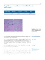
Acanthosis Nigricans, Malignant
Definition Acanthosis nigricans is a rare disorder involving the skin
and mucosae, characterized by papillary lesions and brownish alteration
of the skin.
Etiology
Unknown.
Clinical features The disorder is classified into two major types: the
benign form (genetic or acquired) and the malignant form, which is
associated with an internal malignancy, particularly adenocarcinoma.
Oral manifestations are more common in the malignant form and are
characterized by papillomatous growths that most often involve the lips,
tongue, and gingiva (Fig. 215). Hypertrophy and elongation of the filiform papillae may result in a shaggy appearance of the tongue. The skin
manifestations present as small, velvety papillary lesions, tags, and dark
pigmentation (Fig. 216). The axillae, the genitofemoral area, the neck,
and, less commonly, the palms of the hand and soles of the foot are the
sites of predilection.
Laboratory tests Histopathological examination.
Differential diagnosis Benign acanthosis nigricans, pyostomatitis vegetans, focal epithelial hyperplasia, multiple papillomas, lipoid proteinosis, multiple verruca vulgaris, pemphigus vegetans.
Treatment Symptomatic. Treatment of the underlying malignancy
may resolve the oral and skin lesions in the malignant form of the
disease.
Laskaris, Pocket Atlas of Oral Diseases © 2006 Thieme
All rights reserved. Usage subject to terms and conditions of license
Papillary Lesions
Fig. 215
lips.
217
Malignant acanthosis nigricans: verrucous and papillomatous lesions of the
Fig. 216 Malignant acanthosis nigricans: marked pigmentation and papillary hyperplasia of the skin.
Laskaris, Pocket Atlas of Oral Diseases © 2006 Thieme
All rights reserved. Usage subject to terms and conditions of license
218
Papillary Lesions
Familial Acanthosis Nigricans
Definition. Familial or genetic acanthosis nigricans is a rare benign
mucocutaneous disorder, characterized by papillary lesions and skin
discoloration.
Etiology. Genetic. It is inherited as an autosomal dominant trait.
Clinical features. The cutaneous lesions appear as multiple, painless
small papillary growths (skin tags) and dark discoloration (Fig. 217).
The axillae, groin, neck, umbilicus, genitalia, and perianal area are more
frequently affected. Oral lesions occur in 10–25% of the cases and present
as multiple, small, painless, papillomatous growths with normal color
(Fig. 218). Hypertrophy and elongation of the filiform papillae result in a
shaggy appearance of the tongue. The tongue, lips, gingiva, and palate
are more frequently affected. The disorder usually begins during childhood or at puberty. The diagnosis is mainly based on the history and the
clinical features. Biopsy and histopathological examination may also be
helpful.
Differential diagnosis. Endocrine-related acanthosis nigricans, malignant acanthosis nigricans, Darier disease, Cowden disease.
Treatment. Good oral hygiene, electrosurgery, cryosurgery.
Laskaris, Pocket Atlas of Oral Diseases © 2006 Thieme
All rights reserved. Usage subject to terms and conditions of license
Papillary Lesions
Fig. 217
Benign acanthosis nigricans, multiple skin tags.
Fig. 218
Familial acanthosis nigricans
Laskaris, Pocket Atlas of Oral Diseases © 2006 Thieme
All rights reserved. Usage subject to terms and conditions of license
219
220
Papillary Lesions
Darier Disease
Definition. Darier disease, or dyskeratosis follicularis, is a relatively rare
mucocutaneous disease.
Etiology. Genetic. It is inherited as an autosomal dominant trait.
Clinical features. The disease affects mainly the skin and nails, but the
mucosae may also be involved (oral mucosa, pharynx, genitalia, rectum).
The skin lesions appear as multiple, painless, brownish-red papules that
usually coalesce into plaques (Fig. 219). The forehead, ears, scalp, chest,
and back are more frequently affected. The nails exhibit subungual
keratosis and longitudinal ridges and lines. Oral lesions occur in
20–40% of cases and appear as small multiple whitish confluent papules,
which may become hypertrophic, assuming a cobblestone or papillary
pattern (Fig. 220). The palate, gingiva, buccal mucosa, and tongue are
more frequently affected. The oral lesions develop after the cutaneous
ones. The clinical diagnosis should be confirmed by a biopsy and histopathological examination.
Differential diagnosis. Familial acanthosis nigricans, familial benign
pemphigus, papillary hyperplasia of the palate, Cowden disease.
Treatment. Good oral hygiene, systemic aromatic retinoids.
Laskaris, Pocket Atlas of Oral Diseases © 2006 Thieme
All rights reserved. Usage subject to terms and conditions of license
Papillary Lesions
Fig. 219
221
Darier disease, multiple skin papules.
Fig. 220 Darier disease, multiple whitish confluent papules on the gingiva and
alveolar mucosa.
Laskaris, Pocket Atlas of Oral Diseases © 2006 Thieme
All rights reserved. Usage subject to terms and conditions of license
Laskaris, Pocket Atlas of Oral Diseases © 2006 Thieme
All rights reserved. Usage subject to terms and conditions of license
223
7 Gingival Enlargement
A common characteristic of this group of lesions is that they are located
on the gingiva and present as a submucosal enlargement covered by
normal epithelium. The lesions can be either generalized or localized.
Local diseases, drug-induced lesions, systemic diseases, and tumors are
included in this particular group of disorders.
I Generalized
II Localized
O Hyperplastic gingivitis
O Pyogenic granuloma
O Mouth-breathing gingivitis
O Peripheral giant-cell granuloO Drug-induced gingival overma
growth
O Peripheral ossifying fibroma
O Gingival overgrowth in pregO Granular-cell tumor of the
nancy
newborn
O Gingival overgrowth due to
O Periodontal abscess
leukemia
O Parulis
O Hereditary gingival fibromaO Multiple exostoses
tosis
O Gingival cyst
O Scurvy
O Eruption cyst
O Wegener granulomatosis
O Acanthosis nigricans
Laskaris, Pocket Atlas of Oral Diseases © 2006 Thieme
All rights reserved. Usage subject to terms and conditions of license