Ebook Essentials of anatomy and physiology (7th edition): Part 2
Bạn đang xem bản rút gọn của tài liệu. Xem và tải ngay bản đầy đủ của tài liệu tại đây (21.67 MB, 390 trang )
3957_Ch11_282-305 06/10/14 10:50 AM Page 282
CHAPTER
Blood
3957_Ch11_282-305 06/10/14 10:50 AM Page 283
STUDENT OBJECTIVES
■
Describe the composition and explain the functions of blood plasma.
CHAPTER OUTLINE
■
Name the primary hemopoietic tissue and the kinds of blood cells
produced.
Plasma
■
State the function of red blood cells, including the protein and the
mineral involved.
■
Name the nutrients necessary for red blood cell production, and state the
function of each.
■
Explain how hypoxia may change the rate of red blood cell production.
■
Describe what happens to red blood cells that have reached the end of
their life span; what happens to the hemoglobin?
■
Explain the ABO and Rh blood types.
■
Name the five kinds of white blood cells and describe the function of each.
Classification
■
State what platelets are, and explain how they are involved in hemostasis.
Functions
■
Describe the three stages of chemical blood clotting.
■
Explain how abnormal clotting is prevented in the vascular system.
Function
■
State the normal values in a complete blood count.
Prevention of Abnormal
Clotting
NEW TERMINOLOGY
ABO group (A-B-O GROOP)
Albumin (al-BYOO-min)
Bilirubin (BILL-ee-roo-bin)
Chemical clotting (KEM-i-kuhl
KLAH-ting)
Embolism (EM-boh-lizm)
Erythrocyte (e-RITH-roh-sight)
Hemoglobin (HEE-moh-GLOW-bin)
Hemostasis (HEE-moh-STAY-sis)
Heparin (HEP-ar-in)
Immunity (im-MYOO-ni-tee)
Leukocyte (LOO-koh-sight)
Macrophage (MAK-roh-fahj)
Normoblast (NOR-moh-blast)
Reticulocyte (re-TIK-yoo-loh-sight)
Rh factor (R-H FAK-ter)
Thrombocyte (THROM-boh-sight)
Thrombus (THROM-bus)
Characteristics of Blood
Blood Cells
Red Blood Cells
Function
Production and Maturation
Life Span
Blood Types
White Blood Cells
RELATED CLINICAL
TERMINOLOGY
Anemia (uh-NEE-mee-yah)
Differential count (DIFF-er-EN-shul
KOWNT)
Erythroblastosis fetalis (e-RITHroh-blass-TOH-sis fee-TAL-is)
Hematocrit (hee-MAT-oh-krit)
Hemophilia (HEE-moh-FILL-ee-ah)
Jaundice (JAWN-diss)
Leukemia (loo-KEE-mee-ah)
Leukocytosis (LOO-koh-sigh-TOHsis)
RhoGAM (ROH-gam)
Tissue typing (TISH-yoo-TIGHping)
Typing and cross-matching (TIGHping and KROSS-match-ing)
Terms that appear in bold type in the chapter text are defined in the glossary,
which begins on page 603.
Platelets
BOX 11–1
Anemia
BOX 11–2
Jaundice
BOX 11–3
Rh Disease of the
Newborn
BOX 11–4
Leukemia
BOX 11–5
White Blood Cell Types:
HLA
BOX 11–6
Hemophilia
BOX 11–7
Dissolving and
Preventing Clots
283
3957_Ch11_282-305 06/10/14 10:50 AM Page 284
284 Blood
O
ne of the simplest and most familiar life-saving
medical procedures is a blood transfusion. As you
know, however, the blood of one individual is not
always compatible with that of another person. The ABO
blood types were discovered in the early 1900s by Karl
Landsteiner, an Austrian American. He also contributed
to the discovery of the Rh factor in 1940. In the early 1940s,
Charles Drew, an African American, developed techniques
for processing and storing blood plasma, which could then
be used in transfusions for people with any blood type.
When we donate blood today, our blood may be given to a
recipient as whole blood, or it may be separated into its
component parts, and recipients will then receive only
those parts they need, such as red cells, plasma, Factor 8,
or platelets. Each of these parts has a specific function, and
all of the functions of blood are essential to our survival.
The general functions of blood are transportation, regulation, and protection. Materials transported by the
blood include nutrients, waste products, gases, and hormones. The blood contributes to the regulation of fluid–
electrolyte balance, acid–base balance, and the body
temperature. Protection against pathogens is provided by
white blood cells, and the blood clotting mechanism prevents excessive loss of blood after injuries. Each of these
functions is covered in more detail in this chapter.
CHARACTERISTICS OF BLOOD
Blood has distinctive physical characteristics:
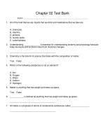
Amount—a person has 4 to 6 liters of blood, depending
on his or her size. Of the total blood volume in the
human body, 38% to 48% is composed of the various
blood cells, also called formed elements. The remaining
52% to 62% of the blood volume is plasma, the liquid
portion of blood (Fig. 11–1).
Color—you’re probably saying to yourself, “Of course, it’s
red!” Mention is made of this obvious fact, however,
because the color does vary. Arterial blood is bright red
because it contains high levels of oxygen. Venous blood
has given up much of its oxygen in tissues, and has a
darker, dull red color. This may be important in the
assessment of the source of bleeding. If blood is bright
red, it is probably from a severed artery, and dark red
blood is probably venous blood.
pH—the normal pH range of blood is 7.35 to 7.45, which
is slightly alkaline. Venous blood normally has a slightly
lower pH than does arterial blood because of the presence of more carbon dioxide. Recall from Chapter 2 that
blood contains buffer systems, pairs of chemicals (such
as carbonic acid and sodium bicarbonate) that will react
in less than a second to change a strong acid or base to
molecules that will not bring about a drastic change in
the pH of the blood.
Viscosity—this means thickness or resistance to flow.
Blood is about three to five times thicker than water.
Viscosity is increased by the presence of blood cells and
the plasma proteins, and this thickness contributes to
normal blood pressure.
PLASMA
Plasma is the liquid part of blood and is approximately
91% water. The solvent ability of water enables the plasma
to transport many types of substances. Nutrients absorbed
in the digestive tract, such as glucose, amino acids, vitamins, and minerals, are circulated to all body tissues.
Waste products of the tissues, such as urea and creatinine,
circulate through the kidneys and are excreted in urine.
Hormones produced by endocrine glands are carried in
the plasma to their target organs, and the antibodies produced by lymphocytes are also transported in plasma.
Most of the carbon dioxide produced by cells is carried in
the plasma in the form of bicarbonate ions (HCO3–).
When the blood reaches the lungs, the CO2 is re-formed,
diffuses into the alveoli, and is exhaled.
Also in the plasma are the plasma proteins. The clotting
factors prothrombin, fibrinogen, and others are synthesized by the liver and circulate until activated to form a clot
in a ruptured or damaged blood vessel. Albumin is the most
abundant plasma protein. It, too, is synthesized by the liver.
Albumin contributes to the colloid osmotic pressure of
blood, which pulls tissue fluid into capillaries. This is important to maintain normal blood volume and blood pressure. Other plasma proteins are called globulins. Alpha and
beta globulins are synthesized by the liver and act as carriers
for molecules such as fats. The gamma globulins (also called
immunoglobulins) are the antibodies produced by lymphocytes. Antibodies are labels that initiate the destruction of
pathogens and provide us with immunity.
Plasma also carries body heat. Heat is one of the byproducts of cell respiration (the production of ATP in cells).
Blood becomes warmer as it flows through active organs
such as the liver and muscles (blood flows slowly in capillaries, so there is time for warming). This heat is distributed
to cooler parts of the body as blood continues to circulate.
BLOOD CELLS
There are three kinds of blood cells: red blood cells,
white blood cells, and platelets. Blood cells are produced
from stem cells in hemopoietic tissue. After birth this
3957_Ch11_282-305 06/10/14 10:50 AM Page 285
Blood
Other body tissues and fluids 92%
Blood plasma 52–62%
Water 91.5%
Blood
8%
Blood cells 38–48%
Total body weight
Blood volume
Erythrocytes 4.5–6.0 million
Thrombocytes 150,000 – 300,000
Other substances
1.5%
Nutrients
Proteins
Leukocytes 5,000–10,000
7%
Fibrinogen 7%
Basophils 0.5–1.0%
Eosinophils 1–3%
Monocytes 3–8%
Hormones
Globulins
38%
Lymphocytes
20–35%
Nitrogenous
wastes
Respiratory
gases
Albumins
55%
Neutrophils
55–70%
Electrolytes
Other substances
Proteins
Leukocytes
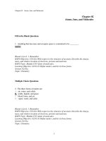
Figure 11–1 Components of blood and the relationship of blood to other body tissues.
QUESTION: Blood plasma is mostly what substance? Which blood cells are the most numerous?
Blood cells
(per microliter)
285
3957_Ch11_282-305 06/10/14 10:50 AM Page 286
286 Blood
is primarily the red bone marrow, found in flat and
irregular bones such as the sternum, hip bone, and vertebrae. Lymphocytes mature and divide in lymphatic
tissue, found in the spleen, lymph nodes, and thymus
gland. The thymus contains stem cells that produce
T lymphocytes, and the stem cells in other lymphatic
tissue also produce lymphocytes.
RED BLOOD CELLS
Also called erythrocytes, red blood cells (RBCs) are biconcave discs, which means their centers are thinner than
their edges. You may recall from Chapter 3 that red blood
cells are the only human cells without nuclei. Their nuclei
disintegrate as the red blood cells mature and are not
needed for normal functioning.
A normal RBC count ranges from 4.5 to 6.0 million
cells per microliter (μL) of blood (1 microliter = 1 mm3 =
one millionth of a liter, a very small volume). RBC counts
for men are often toward the high end of this range; those
for women are often toward the low end. Another way to
measure the amount of RBCs is the hematocrit. This test
involves drawing blood into a thin glass tube called a capillary tube and centrifuging the tube to force all the cells
to one end. The percentages of cells and plasma can then
be determined. Because RBCs are by far the most abundant of the blood cells, a normal hematocrit range is just
like that of the total blood cells: 38% to 48%. Both RBC
count and hematocrit (Hct) are part of a complete blood
count (CBC).
Function
Red blood cells contain the protein hemoglobin (Hb),
which gives them the ability to carry oxygen. Each red
blood cell contains approximately 300 million hemoglobin
molecules, each of which can bond to four oxygen molecules (see Box Fig. 3–B in Box 3–2 of Chapter 3 for the
structure of hemoglobin). In the pulmonary capillaries,
RBCs pick up oxygen and oxyhemoglobin is formed. This
blood circulates from the lungs back to the heart and is
then sent off to the body. In the systemic capillaries,
hemoglobin gives up much of its oxygen and becomes
reduced hemoglobin.
A determination of hemoglobin level is also part of a
CBC; the normal range is 12 to 18 grams per 100 mL of
blood. Essential to the formation of hemoglobin is the
mineral iron; there are four atoms of iron in each molecule
of hemoglobin. It is the iron that actually bonds to the oxygen and also makes RBCs red.
Hemoglobin is also able to bond to carbon dioxide
(CO2) and does transport some CO2 from the tissues to
the lungs. But hemoglobin accounts for only about 10%
of total CO2 transport (most is carried in the plasma as
bicarbonate ions).
Production and Maturation
During embryonic and fetal development, the production
of RBCs can be likened to a relay race, with the “baton” of
production passed from one organ or tissue to another. In
the embryo (the first 8 weeks after fertilization) RBCs are
first produced by an external membrane called the yolk
sac (see Fig. 21–3 in Chapter 21). The fetal liver then takes
over for a while, and the fetal spleen also makes a contribution to RBC manufacture later in gestation. The red
bone marrow becomes active during the fifth month of
gestation, becomes ever more important, and shortly after
birth is the only site of RBC formation.
In older children and adults, red blood cells are formed
in the red bone marrow (RBM) in flat and irregular bones.
Within red bone marrow are precursor cells called stem
cells. Recall from Chapter 3 that stem cells are unspecialized cells that may develop, or differentiate, in several
ways. The stem cells of the red bone marrow may also
be called hemocytoblasts (hemo = “blood,” cyto = “cell,”
blast = “producer”), and they constantly undergo mitosis
to produce new stem cells and all the kinds of blood cells,
many of which are RBCs (Figs. 11–2 and 11–3). The rate
of production is very rapid (estimated at several million
new RBCs every second), and a major regulating factor is
oxygen. If the body is in a state of hypoxia, or lack of oxygen, the kidneys produce a hormone called erythropoietin, which stimulates the red bone marrow to increase
the rate of RBC production (that is, the rate of stem cell
mitosis). This will occur following hemorrhage or if a person stays for a time at a higher altitude. As a result of the
action of erythropoietin, more RBCs will be available to
carry oxygen and correct the hypoxic state.
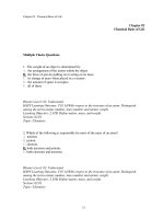
The stem cells that will become RBCs go through a number of developmental stages, only the last two of which we
will mention: normoblasts and reticulocytes (see Fig. 11–2).
The normoblast is the last stage with a nucleus, which then
disintegrates. Hemoglobin has been produced, and the chromosomes with the DNA code for hemoglobin are no longer
needed. The reticulocyte has fragments of the endoplasmic
reticulum (also no longer needed), which are visible as purple stippling when blood smears are stained for microscopic
evaluation. These immature cells are usually found in the
red bone marrow, although a small number of reticulocytes
in the peripheral circulation is considered normal (up to
1.5% of the total RBCs). Large numbers of reticulocytes or
normoblasts in the circulating blood mean that the number
3957_Ch11_282-305 06/10/14 10:50 AM Page 287
Blood
Erythrocytes
Reticulocytes
Neutrophil
Basophil
Normoblasts
Band cell
Megakaryocyte
Eosinophil
Stem cell
Lymphoblast
Thrombocytes
(platelets)
Natural
killer
cell
B cell
Monocyte
T cell
Plasma cell
Macrophage
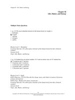
Figure 11–2 Production of blood cells. Stem cells are found primarily in red bone marrow and
are the precursor cells for all the types of blood cells.
QUESTION: Where are normoblasts and reticulocytes usually found, and why?
287
3957_Ch11_282-305 06/10/14 10:50 AM Page 288
288 Blood
A
C
E
B
D
F
of mature RBCs is not sufficient to carry the oxygen needed
by the body. Such situations include hemorrhage, or when
mature RBCs have been destroyed, as in Rh disease of the
newborn, and malaria.
The maturation of red blood cells requires many nutrients. Protein and iron are necessary for the synthesis of
hemoglobin and become part of hemoglobin molecules.
Copper is part of some of the enzymes involved in hemoglobin synthesis, though it does not become part of hemoglobin itself (if it did, it would make our blood blue, like
that of horseshoe crabs). The vitamins folic acid and B12
are required for DNA synthesis in the stem cells of the red
bone marrow. As these cells undergo mitosis, they must
continually produce new sets of chromosomes. Vitamin B12
contains the mineral cobalt and is also called the extrinsic factor because its source is external, our food. Parietal cells of the stomach lining produce the intrinsic
factor, a chemical that combines with the vitamin B12 in
food to prevent its digestion and promote its absorption
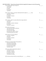
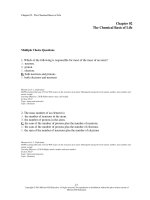
Figure 11–3 Blood cells.
(A) Red blood cells, platelets,
and a basophil. (B) Lymphocyte
(left) and neutrophil (right).
(C) Eosinophil. (D) Monocytes.
(E) Megakaryocyte with
platelets. (A–E ×600) (F) Normal
bone marrow (×200). (From
Harmening, DM: Clinical
Hematology and Fundamentals
of Hemostasis, ed. 3. FA Davis,
Philadelphia, 1997, pp 14, 17,
19, 26, 48, with permission.)
QUESTION: Look at the RBCs in
picture B. Why do they have pale
centers?
in the small intestine. A deficiency of either vitamin B12
or the intrinsic factor results in pernicious anemia (see
Box 11–1: Anemia).
Life Span
Red blood cells live for approximately 120 days. As they
reach this age they become fragile; their membranes
begin to disintegrate. These damaged cells are removed
from circulation by cells of the tissue macrophage
system (formerly called the reticuloendothelial or RE
system). The organs that contain macrophages (literally,
“big eaters”) are the liver, spleen, and red bone marrow.
Look at Fig. 11–4 as you read the following. The old
RBCs are phagocytized and digested by macrophages,
and the iron they contained is put into the blood to be
returned to the red bone marrow to be used for the synthesis of new hemoglobin. If not needed immediately
for this purpose, excess iron is stored in the liver. The
iron of RBCs is actually recycled over and over again.
3957_Ch11_282-305 06/10/14 10:50 AM Page 289
Blood
Box 11–1
289
| ANEMIA
Anemia is a deficiency of red blood cells, or insufficient hemoglobin within the red blood cells.
There are many different types of anemia.
Iron-deficiency anemia is caused by a lack of
dietary iron, when there is not enough of this mineral to form sufficient hemoglobin. A person with
this type of anemia may have a normal RBC count
and a normal hematocrit, but the hemoglobin
level will always be below normal.
A deficiency of vitamin B12, which is found
only in animal foods, leads to pernicious anemia, in which the RBCs are large, misshapen,
and fragile. Another cause of this form of anemia
is lack of the intrinsic factor due to autoimmune
destruction of the parietal cells of the stomach
lining.
Sickle-cell anemia has already been discussed in Chapter 3. It is a genetic disorder of
A
B
C
D
hemoglobin, which causes RBCs to sickle, clog
capillaries, and rupture.
Aplastic anemia is suppression of the red bone
marrow, with decreased production of RBCs,
WBCs, and platelets. This is a very serious disorder that may be caused by exposure to radiation,
certain chemicals such as benzene, or some medications. There are several antibiotics that must
be used with caution because they may have this
potentially fatal side effect.
Hemolytic anemia is any disorder that causes
rupture of RBCs before the end of their normal life
span. Sickle-cell anemia and Rh disease of the newborn are examples. Another example is malaria,
in which a protozoan parasite reproduces in RBCs
and destroys them. Hemolytic anemias are often
characterized by jaundice because of the increased
production of bilirubin.
Box Figure 11–A Anemia. (A) Iron-deficiency anemia; notice the pale, oval RBCs (×400).
(B) Pernicious anemia, with large, misshapen RBCs (×400). (C) Sickle-cell anemia (×400).
(D) Aplastic anemia, bone marrow (×200). (A, B, and C from Listen, Look, and Learn, Vol 3;
Coagulation, Hematology. The American Society of Clinical Pathologists Press, Chicago,
1973, with permission. D from Harmening, DM: Clinical Hematology and Fundamentals of
Hemostasis, ed 3. FA Davis, Philadelphia, 1997, p 49, with permission.)
3957_Ch11_282-305 06/10/14 10:50 AM Page 290
290 Blood
Circulate
120 days
RBCs
Macrophages in
liver, spleen, and
red bone marrow
phagocytize old RBCs
New RBCs
formed in
red bone marrow
Protein synthesis
Amino acids
Used to make
new RBCs
Iron
Heme
Globin
Stored in
liver
Kidney
Small
intestine
Large intestine
Bilirubin
Bilirubin
Urobilin
Colon
bacteria
Urine
Urobilin
Figure 11–4
Life cycle of red blood cells. See text for description.
QUESTION: Which components of old RBCs are recycled? Which is excreted? (Go to the macrophage and
follow the arrows.)
The globin or protein portion of the hemoglobin molecule is also recycled. It is digested to its amino acids,
which may then be used for the synthesis of new proteins.
Another part of the hemoglobin molecule is the heme
portion, which cannot be recycled and is a waste product.
The heme is converted to bilirubin by macrophages. The
liver removes bilirubin from circulation and excretes it into
bile; bilirubin is a bile pigment. Bile is secreted by the liver
into the duodenum and passes through the small intestine
and colon, so bilirubin is eliminated in feces and gives
feces their characteristic brown color. In the colon some
bilirubin is changed to urobilinogen by the colon bacteria.
3957_Ch11_282-305 06/10/14 10:50 AM Page 291
Blood
Some urobilinogen may be absorbed into the blood, but it
is changed to urobilin and excreted by the kidneys in urine.
If bilirubin is not excreted properly, perhaps because of
liver disease such as hepatitis, it remains in the blood. This
may cause jaundice, a condition in which the whites of the
eyes appear yellow. This yellow color may also be seen in
the skin of light-skinned people (see Box 11–2: Jaundice).
Blood Types
Our blood types are genetic; that is, we inherit genes from
our parents that determine our own types. There are many
red blood cell factors or types; we will discuss the two most
important ones: the ABO group and the Rh factor. (The
genetics of blood types is discussed in Chapter 21.)
Box 11–2
291
The ABO group contains four blood types: A, B, AB,
and O. The letters A and B represent antigens (proteinoligosaccharides) on the red blood cell membrane. A person with type A blood has the A antigen on the RBCs, and
someone with type B blood has the B antigen. Type AB
means that both A and B antigens are present, and type O
means that neither the A nor the B antigen is present.
Circulating in the plasma of each person are natural
antibodies for those antigens not present on the RBCs.
Therefore, a type A person has anti-B antibodies in the
plasma; a type B person has anti-A antibodies; a type AB
person has neither anti-A nor anti-B antibodies; and a
type O person has both anti-A and anti-B antibodies (see
Table 11–1 and Fig. 11–5).
| JAUNDICE
Jaundice is not a disease, but rather a sign caused
by excessive accumulation of bilirubin in the
blood. Because one of the liver’s many functions
is the excretion of bilirubin, jaundice may be a sign
of liver disease such as hepatitis or cirrhosis. This
may be called hepatic jaundice because the problem is with the liver.
Other types of jaundice are prehepatic jaundice
and posthepatic jaundice: The name of each tells
us where the problem is. Recall that bilirubin is
the waste product formed from the heme portion
of the hemoglobin of old RBCs. Prehepatic jaundice means that the problem is “before” the liver;
that is, hemolysis of RBCs is taking place at a
more rapid rate. Rapid hemolysis is characteristic
of sickle-cell anemia, malaria, and Rh disease of
the newborn; these are hemolytic anemias. As excessive numbers of RBCs are destroyed, bilirubin
is formed at a faster rate than the liver can excrete
it. The bilirubin that the liver cannot excrete remains in the blood and causes jaundice. Another
name for this type is hemolytic jaundice.
Posthepatic jaundice means that the problem
is “after” the liver. The liver excretes bilirubin into
bile, which is stored in the gallbladder and then
moved to the small intestine. If the bile ducts are
obstructed, perhaps by gallstones or inflammation of the gallbladder, bile cannot pass to the
small intestine and backs up in the liver. Bilirubin
may then be reabsorbed back into the blood and
cause jaundice. Another name for this type is
obstructive jaundice.
| ABO BLOOD TYPES
Table 11–1
PERCENTAGE IN U.S. POPULATION*
TYPE
ANTIGENS PRESENT
ON RBCs
ANTIBODIES PRESENT
IN PLASMA
A
A
B
WHITE
BLACK
ASIAN
anti-B
40
27
31
B
anti-A
11
20
26
AB
both A and B
neither anti-A nor anti-B
4
4
8
O
neither A nor B
both anti-A and anti-B
45
49
35
*Average.
3957_Ch11_282-305 06/10/14 10:50 AM Page 292
A
ABO blood types
Red blood cells
Plasma
Type A
Type B
A antigens
B
Typing and cross-matching
Anti-A serum
Anti-B serum
B antibodies
Type A
B antigens
A antibodies
Type B
Type AB
A and B antigens
Type O
Neither A
nor B antigens
Neither A nor B antibodies
Type AB
A and B antibodies
Type O
Type
O
Universal donor
C
Type
A
Type
O
Type
B
Type
A
Type
AB
Type
B
Type
AB
Universal recipient
Figure 11–5 (A) The ABO blood types. Schematic representation of antigens on the RBCs and
antibodies in the plasma. (B) Typing and cross-matching. The A or B antiserum causes agglutination of RBCs with the matching antigen. (C) Acceptable transfusions are diagrammed and
presuppose compatible Rh factors.
QUESTION: In part C, find your blood type. To whom (that is, to which blood types) can you donate blood?
3957_Ch11_282-305 06/10/14 10:50 AM Page 293
Blood
Why we have these natural antibodies is not known
(they begin to be formed several months after birth), but
we do know that they are of great importance for transfusions. If possible, a person should receive blood of his or
her own type; only if this type is not available should type
O negative blood be given. For example, let us say that a
type A person needs a transfusion to replace blood lost in
hemorrhage. If this person were to receive type B blood,
what would happen? The type A recipient has anti-B antibodies that would bind to the type B antigens of the
RBCs of the donated blood. The type B RBCs would first
clump (agglutination) then rupture (hemolysis), thus defeating the purpose of the transfusion. An even more serious consequence is that the hemoglobin of the ruptured
RBCs, now called free hemoglobin, may clog the capillaries of the kidneys and lead to renal damage or renal failure.
You can see why typing and cross-matching of donor and
recipient blood in the hospital laboratory is so important
before any transfusion is given (see Fig. 11–5). This procedure helps ensure that donated blood will not bring
about a hemolytic transfusion reaction in the recipient.
You may have heard of the concept that a person with
type O blood is a “universal donor.” Usually, a unit of type
O negative blood may be given to people with any other
blood type. This is so because type O RBCs have neither
the A nor the B antigens and will not react with whatever
Box 11–3
293
antibodies the recipient may have. If only one unit (1 pint)
of blood is given, the anti-A and anti-B antibodies in the
type O blood plasma will be diluted in the recipient’s blood
plasma and will not have a harmful effect on the recipient’s
RBCs. The term negative, in O negative, the universal
donor, refers to the Rh factor, which we will now consider.
The Rh factor is another antigen (often called D) that
may be present on RBCs. People whose RBCs have the
Rh antigen are Rh positive; those without the antigen are
Rh negative. Rh-negative people do not have natural antibodies to the Rh antigen, and for them this antigen is foreign. If an Rh-negative person receives Rh-positive blood
by mistake, antibodies will be formed just as they would
be to bacteria or viruses. A first mistaken transfusion often
does not cause problems because antibody production is
slow upon the first exposure to Rh-positive RBCs, and
those RBCs have a relatively short lifespan. A second
transfusion, however, when anti-Rh antibodies are already
present will bring about a transfusion reaction, with hemolysis and possible kidney damage (see also Box 11–3:
Rh Disease of the Newborn).
WHITE BLOOD CELLS
White blood cells (WBCs) are also called leukocytes.
There are five kinds of WBCs; all are larger than RBCs and
have nuclei when mature. The nucleus may be in one piece
| Rh DISEASE OF THE NEWBORN
Rh disease of the newborn may also be called
erythroblastosis fetalis and is the result of an Rh
incompatibility between mother and fetus. During
a normal pregnancy, maternal blood and fetal
blood do not mix in the placenta. However, during
delivery of the placenta (the “afterbirth” that follows the birth of the baby), some fetal blood may
enter maternal circulation.
If the woman is Rh negative and her baby is
Rh positive, this exposes the woman to Rhpositive RBCs. In response, her immune system
will now produce anti-Rh antibodies following
this first delivery. In a subsequent pregnancy,
these maternal antibodies will cross the placenta and enter fetal circulation. If this next
fetus is also Rh positive, the maternal antibodies will cause destruction (hemolysis) of the
fetal RBCs. In severe cases this may result in the
death of the fetus. In less severe cases, the baby
will be born anemic and jaundiced from the loss
of RBCs. Such an infant may require a gradual
exchange transfusion to remove the blood with
the maternal antibodies and replace it with Rhnegative blood. The baby will continue to produce its own Rh-positive RBCs, which will not
be destroyed once the maternal antibodies have
been removed.
Much better than treatment, however, is prevention. If an Rh-negative woman delivers an
Rh-positive baby, she should be given RhoGAM
within 72 hours after delivery. RhoGAM is an antiRh antibody that will destroy any fetal RBCs that
have entered the mother’s circulation before her
immune system can respond and produce antibodies. The RhoGAM antibodies themselves break
down within a few months. The woman’s next
pregnancy will be like the first, as if she had never
been exposed to Rh-positive RBCs.
3957_Ch11_282-305 06/10/14 10:50 AM Page 294
294 Blood
or appear as several lobes or segments. Special staining for
microscopic examination gives each kind of WBC a distinctive appearance (see Figs. 11–2 and 11–3).
A normal WBC count (part of a CBC) is 5,000 to
10,000 per μL. Notice that this number is quite small compared with a normal RBC count. Many of our WBCs are
not circulating within blood vessels but are carrying out
their functions in tissue fluid or in lymphatic tissue.
Classification
The five kinds of white blood cells, all produced in the
red bone marrow (and some lymphocytes in lymphatic
tissue), may be classified in two groups: granular and
agranular. The granular leukocytes are the neutrophils,
eosinophils, and basophils, which usually have nuclei in
Table 11–2
two or more lobes or segments and have distinctly colored granules when stained. Neutrophils have light blue
granules, eosinophils have red granules, and basophils
have dark blue granules. The agranular leukocytes are
lymphocytes and monocytes, which have nuclei in one
piece. Monocytes are usually quite a bit larger than lymphocytes. A differential WBC count (part of a CBC) is
the percentage of each kind of leukocyte. Normal ranges
are listed in Table 11–2, along with other normal values
of a CBC.
Functions
White blood cells all contribute to the same general function, which is to protect the body from infectious disease
and to provide immunity to certain diseases. Each kind
| COMPLETE BLOOD COUNT
MEASUREMENT
NORMAL RANGE*
VARIATIONS
Red blood cells
4.5–6.0 million/L
Decrease: anemia
Increase: polycythemia
Hemoglobin
12–18 grams/100 mL
Decrease: iron deficiency, other anemias
Increase: polycythemia
Hematocrit
38%–48%
Decrease: anemia
Increase: polycythemia, heavy smoking
Reticulocytes
0%–1.5%
Decrease: RBM suppression
Increase: insufficiency of mature RBCs
White blood cells
(total)
5000–10,000/L
Decrease: leucopenia
Increase: leukocytosis
Neutrophils
55%–70%
Decrease: radiation, chemotherapy for cancer
Increase: infection, inflammation
Eosinophils
1%–3%
Decrease: corticosteroid excess
Increase: allergies, parasitic infections
Basophils
0.5%–1%
Decrease: cancer
Increase: allergies
Lymphocytes
20%–35%
Decrease: HIV/AIDS, severe burns, cancer, radiation
Increase: many viral diseases
Monocytes
3%–8%
Decrease: corticosteroid excess
Increase: many viral diseases, chronic inflammation
Platelets
150,000–300,000/L
Decrease: thrombocytopenia that may be idiopathic or
accompany aplastic anemia
Increase: not considered a clinical condition, but may
follow removal of the spleen
*The values on hospital lab slips may vary somewhat but will be very similar to the normal ranges given here.
3957_Ch11_282-305 06/10/14 10:50 AM Page 295
Blood
of leukocyte makes a contribution to this very important
aspect of homeostasis.
Neutrophils and monocytes are capable of the
phagocytosis of pathogens. Neutrophils are the more
abundant phagocytes, but the monocytes are the more
efficient phagocytes, because they differentiate into
macrophages, which also phagocytize dead or damaged
tissue at the site of any injury, helping to make tissue repair possible. Monocytes also contribute to tissue repair.
During an infection, neutrophils are produced more
rapidly, and the immature forms, called band cells (see
Fig. 11–2), may appear in greater numbers in peripheral
circulation (band cells are usually less than 10% of the
total neutrophils). The term “band” refers to the nucleus
that has not yet become segmented and may look somewhat like a dumbbell.
Eosinophils are believed to detoxify foreign proteins
and will phagocytize anything labeled with antibodies.
Eosinophils become more abundant during allergic reactions and parasitic infections such as trichinosis (caused
by a worm parasite). Basophils contain granules of heparin
and histamine. Heparin is an anticoagulant that helps prevent abnormal clotting within blood vessels. Histamine,
you may recall, is released as part of the inflammation
process, and it makes capillaries more permeable, allowing
tissue fluid, proteins, and white blood cells to accumulate
in the damaged area.
There are two major kinds of lymphocytes, T cells and
B cells, and a less numerous third kind called natural killer
cells. For now we will say that T cells (or T lymphocytes)
help recognize foreign antigens and may directly destroy
some foreign antigens. B cells (or B lymphocytes) become
plasma cells that produce antibodies to foreign antigens.
Both T cells and B cells provide memory for pathogens.
The memory T cells and B cells are the reason vaccines or
recovery from a disease can provide immunity to future
cases of that disease. Natural killer cells (NK cells) destroy
foreign cells by chemically rupturing their membranes.
These functions of lymphocytes are discussed in the context of the mechanisms of immunity in Chapter 14.
As mentioned earlier, leukocytes function in tissue fluid
and blood. Many WBCs are capable of self-locomotion
(ameboid movement) and are able to squeeze between
the cells of capillary walls and out into tissue spaces.
Macrophages provide a good example of the dual locations of leukocytes. Some macrophages are “fixed,” that
is, stationary in organs such as the liver, spleen, and
red bone marrow (part of the tissue macrophage or RE
system—the same macrophages that phagocytize old
RBCs) and in the lymph nodes. They phagocytize
295
pathogens that circulate in blood or lymph through
these organs. Other “wandering” macrophages move
about in tissue fluid, especially in the areolar connective
tissue of mucous membranes and below the skin.
Pathogens that gain entry into the body through natural
openings or through breaks in the skin are usually destroyed by the macrophages and other leukocytes in
connective tissue before they can cause serious disease.
The alveoli of the lungs, for example, have macrophages
that very efficiently destroy pathogens that enter with
inhaled air.
A high WBC count, called leukocytosis, is often an indication of infection. Leukopenia is a low WBC count,
which may be present in the early stages of diseases such
as tuberculosis. Exposure to radiation or to chemicals such
as benzene may destroy WBCs and lower the total count.
Such a person is then very susceptible to infection.
Leukemia, or malignancy of leukocyte-forming tissues, is
discussed in Box 11–4: Leukemia.
The white blood cell types (analogous to RBC types
such as the ABO group) are called human leukocyte antigens (HLAs). These cell types are created by cell membrane proteins that are a genetic characteristic. The genes
for these self-antigens are collectively called the major
histocompatibility complex (MHC) and are on chromosome number 6. The purpose of the antigens is discussed
in Box 11–5: White Blood Cell Types: HLA.
PLATELETS
The more formal name for platelets is thrombocytes,
which are not whole cells but rather fragments or pieces
of cells. Some of the stem cells in the red bone marrow
differentiate into large cells called megakaryocytes (see
Figs. 11–2 and 11–3), which break up into small pieces
that enter circulation. These small, oval, circulating
pieces are platelets, which may last for 5 to 9 days, if not
utilized before that. Thrombopoietin is a hormone
produced by the liver that increases the rate of platelet
production.
A normal platelet count (part of a CBC) is 150,000 to
300,000/μL (the high end of the range may be extended to
500,000). Thrombocytopenia is the term for a low platelet
count.
Function
Platelets are necessary for hemostasis, which means prevention of blood loss. With respect to intact blood vessels,
platelets help maintain the junctions between adjacent
epithelial cells that form capillaries and line larger vessels (the endothelium). Without platelets, zipper-like
3957_Ch11_282-305 06/10/14 10:50 AM Page 296
296 Blood
Box 11–4
| LEUKEMIA
Leukemia is the term for malignancy of a bloodforming tissue. There are many types of leukemia,
which are classified as acute or chronic, by the
types of abnormal cells produced, and by either
childhood or adult onset.
In general, leukemia is characterized by an overproduction of immature white blood cells. These
immature cells cannot perform their normal functions, and the person becomes very susceptible to
infection. As a greater proportion of the body’s nutrients are used by malignant cells, the production
of other blood cells decreases. Severe anemia is a
consequence of decreased red blood cell production, and the tendency to bruise easily, then hemorrhage, is the result of decreased platelets.
Chemotherapy may bring about cure or remission for some forms of leukemia, but other forms
remain resistant to treatment and may be fatal
within a few months of diagnosis. In such cases,
the cause of death is often pneumonia or some
Box 11–5
other serious infection because the abnormal
white blood cells cannot prevent the growth and
spread of pathogens within the body.
Box Figure 11–B Leukemia. Notice the many darkly staining WBCs (×300); compare with normal blood in Fig. 11–3 A
and B. (From Sacher, RA, and McPherson, RA: Widmann’s
Clinical Interpretation of Laboratory Tests, ed 11. FA Davis,
Philadelphia, 2000, with permission.)
| WHITE BLOOD CELL TYPES: HLA
Human leukocyte antigens (HLAs) are antigens
on WBCs that are representative of the antigens
present on all the cells of an individual. These are
our “self” antigens that identify cells that belong
in the body.
Recall that in the ABO blood group of RBCs,
there are only two antigens, A and B, and four
possible types: A, B, AB, and O. HLA antigens are
also given letter names. HLA A, B, and C are
called class I proteins, with from 100 to more than
400 possibilities for the specific protein each can
be. The several class II proteins are given various
D designations and, again, there are many possibilities for each. Each person has two genes for
each HLA type because these types are inherited,
just as RBC types are inherited. Members of the
same family may have some of the same HLA
types, and identical twins have exactly the same
HLA types.
The purpose of the HLA types is to provide a
“self” comparison for the immune system to use
when pathogens enter the body. The T lymphocytes compare the “self” antigens on macrophages
to the antigens on bacteria and viruses. Because
these antigens do not match ours, they are recognized as foreign; this is the first step in the destruction of a pathogen.
The surgical transplantation of organs has also
focused on the HLA. The most serious problem for
the recipient of a transplanted heart or kidney is rejection of the organ and its destruction by the immune system. You may be familiar with the term
tissue typing. This process involves determining
the HLA types of a donated organ to see if one or
several will match the HLA types of the potential recipient. If even one HLA type matches, the chance
of rejection is lessened. Although all transplant recipients (except corneal) must receive immunosuppressive medications to prevent rejection, such
medications make them more susceptible to infection. The closer the HLA match of the donated
organ, the lower the dosage of such medications,
3957_Ch11_282-305 06/10/14 10:50 AM Page 297
Blood
Box 11–5
297
| WHITE BLOOD CELL TYPES: HLA (Continued)
and the less chance of serious infections. (The
chance of finding a perfect HLA match in the general population is estimated at 1 in 20,000.)
There is yet another aspect of the importance
of HLA: People with certain HLA types seem to be
more likely to develop certain autoimmune diseases. For example, type 1 (insulin-dependent)
diabetes mellitus is often found in people with
HLA DR3 or DR4, and an arthritis of the spine
called ankylosing spondylitis is often found in
those with HLA B27. These are not genes for
these diseases but may be predisposing factors.
glycoproteins called cadherins tend to come apart, the
epithelial cells separate, and RBCs and excess plasma leak
out. Should a blood vessel rupture or be cut, three mechanisms help minimize blood loss, and platelets are involved in each. Two of these mechanisms are shown in
Fig. 11–6.
1. Vascular spasm—when a large vessel such as an artery
or vein is severed, the smooth muscle in its wall contracts in response to the damage (called the myogenic
response). Platelets in the area of the rupture release
serotonin, which also brings about vasoconstriction.
The diameter of the vessel is thereby made smaller, and
the smaller opening may then be blocked by a blood
clot. If the vessel did not constrict first, the clot that
formed would quickly be washed out by the force of the
blood pressure.
2. Platelet plugs—when capillaries rupture, the damage
is too slight to initiate the formation of a blood clot.
The rough surface, however, causes platelets to change
shape (become spiky) and become sticky. These activated platelets stick to the edges of the break and to
each other. The platelets form a mechanical barrier
or wall to close off the break in the capillary. Capillary
ruptures are quite frequent, and platelet plugs, although
small, are all that is needed to seal them.
Would platelet plugs be effective for breaks in larger
vessels? No, they are too small, and though they do
form, they are washed away (until a clot begins to
form that can contain them). Would vascular spasm
be effective for capillaries? Again, the answer is no,
because capillaries have no smooth muscle and cannot constrict at all.
What may happen is this: A virus enters the body
and stimulates the immune system to produce
antibodies. The virus is destroyed, but one of the
person’s own antigens is so similar to the viral
antigen that the immune system continues its activity and begins to destroy this similar part of the
body. Another possibility is that a virus damages
a self-antigen to the extent that it is now so different that it will be perceived as foreign. These
are two theories of how autoimmune diseases
are triggered, a topic that is the focus of much research in the field of immunology.
3. Chemical clotting—The stimulus for clotting is a
rough surface within a vessel, or a break in the vessel,
which also creates a rough surface. The more damage
there is, the faster clotting begins, usually within 15 to
120 seconds.
The clotting mechanism is a series of reactions involving chemicals that normally circulate in the blood and
others that are released when a vessel is damaged.
The chemicals involved in clotting include platelet factors, chemicals released by damaged tissues, calcium ions,
and the plasma proteins prothrombin, fibrinogen, Factor 8,
and others synthesized by the liver. (These clotting factors
are also designated by Roman numerals; Factor 8 would
be Factor VIII.) Vitamin K is necessary for the liver to
synthesize prothrombin and several other clotting factors
(Factors 7, 9, and 10). Most of our vitamin K is produced
by the intestinal microbiota, the bacteria that live in the
colon; the vitamin is absorbed as the colon absorbs water
and may be stored in the liver.
Chemical clotting is usually described in three stages,
which are listed in Table 11–3 and illustrated in Fig. 11–7.
Stage 1 begins when a vessel is cut or damaged internally
and includes all of the factors shown. As you follow the
pathway, notice that the product of stage 1 is prothrombin activator, which may also be called prothrombinase.
Each name tells us something. The first name suggests
that this chemical activates prothrombin, and that is true.
The second name ends in “ase,” which indicates that this
is an enzyme. The traditional names for enzymes use the
substrate of the enzyme as the first part of the name, and
add “ase.” So this chemical must be an enzyme whose
substrate is prothrombin, and that is also true. The stages
of clotting may be called a cascade, where one leads to the
3957_Ch11_282-305 06/10/14 10:50 AM Page 298
298 Blood
Skin is cut and
blood escapes from a
capillary and an
arteriole.
Capillary
Arteriole
Platelets
Figure 11–6 Hemostasis.
Platelet plug formation in a capillary and chemical clotting and
clot retraction in an arteriole.
Fibrin
In the capillary, platelets
stick to the ruptured wall
and form a platelet plug.
Table 11–3
In the arteriole, chemical
clotting forms a fibrin clot.
| CHEMICAL CLOTTING
CLOTTING STAGE
FACTORS NEEDED
Stage 1
■
■
■
■
Stage 2
■
■
■
Stage 3
Clot retraction pulls the
edges of the break together.
QUESTION: Look at the diameter of the arteriole (compared
with that of the capillary) and
explain why platelet plugs
would not be sufficient to stop
the bleeding.
■
■
■
■
REACTION
Platelet factors
Chemicals from damaged tissue
(tissue thromboplastin)
Factors 5, 7, 8, 9, 10, 11, 12
Calcium ions
Platelet factors + tissue
thromboplastin + other
clotting factors + calcium ions
form prothrombin activator
(prothrombinase)
Prothrombin activator from stage 1
Prothrombin
Calcium ions
Prothrombin activator converts
prothrombin to thrombin
Thrombin from stage 2
Fibrinogen
Calcium ions
Factor 13 (fibrin stabilizing factor)
Thrombin converts fibrinogen to fibrin
3957_Ch11_282-305 06/10/14 10:50 AM Page 299
Blood
Stage 1
Liver
Factors 5, 7, 8, 9,
10, 11, 12
Platelet factors
Chemicals from
damaged tissue
Calcium
ions
Prothrombin
activator
Stage 2
Prothrombin
Calcium
ions
Thrombin
Fibrinogen
Stage 3
Calcium ions
Factor 13
Fibrin
Figure 11–7
Stages of chemical blood clotting.
QUESTION: Based only on this picture, explain why the liver is a vital organ.
299
3957_Ch11_282-305 06/10/14 10:50 AM Page 300
300 Blood
next, as inevitable as water flowing downhill. Prothrombin activator, the product of stage 1, brings about the
stage 2 reaction: converting prothrombin to thrombin.
The product of stage 2, thrombin, brings about the stage
3 reaction: converting fibrinogen to fibrin (see Box 11–6:
Hemophilia).
The clot itself is made of fibrin, the product of stage 3.
Fibrin is a threadlike protein. Many strands of fibrin form
a mesh that traps RBCs and platelets and creates a wall
across the break in the vessel.
Once the clot has formed and bleeding has stopped,
clot retraction and fibrinolysis occur. Clot retraction
requires platelets, ATP, and Factor 13 and involves folding of the fibrin threads to pull the edges of the rupture
in the vessel wall closer together. This will make the area
to be repaired smaller. The platelets contribute in yet
another way because as they disintegrate they release
platelet-derived growth factor (PDGF), which stimulates
mitosis for the repair of blood vessels. As repair begins,
the clot is dissolved, a process called fibrinolysis. It is important that the clot be dissolved because it is a rough
surface, and if it were inside a vessel it would stimulate
more and unnecessary clotting, which might eventually
obstruct blood flow.
Prevention of Abnormal Clotting
Clotting should take place to stop bleeding, but too
much clotting would obstruct vessels and interfere with
Box 11–6
normal circulation of blood. Clots do not usually form
in intact vessels because the endothelium (simple squamous epithelial lining) is very smooth and repels the
platelets and clotting factors. If the lining becomes
roughened, as happens with the lipid deposits of atherosclerosis, a clot will form.
Heparin, produced by basophils, is a natural anticoagulant that inhibits the clotting process (although heparin
is called a “blood thinner,” it does not “thin” or dilute the
blood in any way; rather it prevents a chemical reaction
from taking place). The liver produces a globulin called antithrombin, which combines with and inactivates excess
thrombin. Excess thrombin would exert a positive feedback
effect on the clotting cascade and result in the splitting
of more prothrombin to thrombin, more clotting, more
thrombin formed, and so on. Antithrombin helps to prevent this, as does the fibrin of the clot, which adsorbs excess
thrombin and renders it inactive. All of these factors are
the external brake for this positive feedback mechanism.
Together they usually limit the fibrin formed to what is
needed to create a useful clot but not an obstructive one.
Thrombosis refers to clotting in an intact vessel; the
clot itself is called a thrombus. Coronary thrombosis, for
example, is abnormal clotting in a coronary artery, which
will decrease the blood (oxygen) supply to part of the heart
muscle. An embolism is a clot or other tissue transported
from elsewhere that lodges in and obstructs a vessel (see
Box 11–7: Dissolving and Preventing Clots).
| HEMOPHILIA
There are several forms of hemophilia; all are genetic and are characterized by the inability of the
blood to clot properly. Hemophilia A is the most
common form and involves a deficiency of clotting Factor 8 (VIII). The gene for hemophilia A is
located on the X chromosome, so this is a sexlinked trait, with the same pattern of inheritance
as red-green color blindness and Duchenne’s
muscular dystrophy.
Without Factor 8, the first stage of chemical
clotting cannot be completed, and prothrombin
activator is not formed. Without treatment, a hemophiliac experiences prolonged bleeding after
even minor injuries and extensive internal bleeding, especially in joints subjected to the stresses
of weight bearing. Treatment, but not cure, is
possible with Factor 8 obtained from blood donors.
The Factor 8 is extracted from the plasma of donated blood and administered in concentrated
form to hemophiliacs, enabling them to live normal lives.
In what is perhaps the most tragic irony of
medical progress, many hemophiliacs were inadvertently infected with HIV, the virus that causes
AIDS. Before 1985, there was no test to detect HIV
in donated blood, and the virus was passed to
hemophiliacs in the very blood product that was
meant to control their disease and prolong their
lives. Today, all donated blood and blood products are tested for HIV, and the risk of AIDS transmission to hemophiliacs, or anyone receiving
donated blood, is now very small.
3957_Ch11_282-305 06/10/14 10:50 AM Page 301
Blood
Box 11–7
301
| DISSOLVING AND PREVENTING CLOTS
Abnormal clots may cause serious problems in
coronary arteries, pulmonary arteries, cerebral
vessels, and even veins in the legs. However, if
these clots can be dissolved before they cause
death of tissue, normal circulation and tissue
functioning may be restored.
One of the first substances used to dissolve
clots in coronary arteries was streptokinase,
which is actually a bacterial toxin produced by
some members of the genus Streptococcus.
Streptokinase did indeed dissolve clots, but its
use created the possibility of clot destruction
throughout the body, with serious hemorrhage a
potential consequence.
Safer thrombolytic chemicals are now used
(thrombo = “clot” and lytic = “to lyse” or “split”). In
a case of a coronary thrombosis, if a thrombolytic
SUMMARY
All of the functions of blood described in this chapter—
transport, regulation, and protection—contribute to the
homeostasis of the body as a whole. However, these
can be directed into the affected vessel within a
few hours, the clot may be dissolved and permanent heart damage prevented. The same procedure is also used to prevent permanent brain
damage after strokes (CVAs) caused by blood
clots.
Some people, such as those with atrial fibrillation or a tendency to form clots in veins of the
legs, require clot prevention. You have probably
heard of warfarin, which has been a standard clotpreventing drug for many years. Several new
medications are available (and can be taken
orally) that inhibit the action of thrombin or other
clotting factors. Slower clotting and excessive
bleeding are possible side effects, but episodes of
major bleeding have been less likely with the
newer medications than with warfarin.
functions could not be carried out if the blood did not
circulate properly. The circulation of blood throughout
the blood vessels depends on the proper functioning of
the heart, the pump of the circulatory system, which is
the subject of our next chapter.
STUDY OUTLINE
The general functions of blood are transportation, regulation, and protection.
Characteristics of Blood
1. Amount—4 to 6 liters; 38% to 48% is cells; 52%
to 62% is plasma (Fig. 11–1).
2. Color—arterial blood has a high oxygen content
and is bright red; venous blood has less oxygen
and is dark red.
3. pH—7.35 to 7.45; venous blood has more CO2
and a lower pH than arterial blood; buffer systems help maintain normal pH.
4. Viscosity—thickness or resistance to flow; due
to the presence of cells and plasma proteins;
contributes to normal blood pressure.
Plasma—the liquid portion of blood
1. 91% water.
2. Plasma transports nutrients, wastes, hormones,
heat, antibodies, and CO2 as HCO3–.
3. Plasma proteins: many are produced by the
liver.
a. Clotting factors (prothrombin, fibrinogen,
and others) are synthesized by the liver.
b. Albumin is synthesized by the liver and provides colloid osmotic pressure that pulls tissue fluid into capillaries to maintain normal
blood volume and blood pressure.
3957_Ch11_282-305 06/10/14 10:50 AM Page 302
302 Blood
c. Alpha and beta globulins are synthesized by
the liver and are carriers for fats and other
substances in the blood.
d. Gamma globulins (immunoglobulins) are
antibodies produced by lymphocytes.
Blood Cells
1. Formed elements are RBCs, WBCs, and platelets
(Figs. 11–2 and 11–3).
2. After birth the primary hemopoietic tissue is the
red bone marrow, which contains stem cells.
Lymphocytes mature and divide in the lymphatic
tissue of the spleen, lymph nodes, and thymus,
which also have stem cells for lymphocytes.
Red Blood Cells—erythrocytes (see Table 11–2
for normal values)
1. Biconcave discs; no nuclei when mature.
2. RBCs carry O2 bonded to the iron in hemoglobin; oxyhemoglobin is formed in pulmonary
capillaries; the oxygen is dropped off in systemic capillaries.
3. Before birth, RBCs are formed by the embryonic yolk sac and then by the fetal liver, spleen,
and RBM.
4. After birth, RBCs are formed in the RBM from
hemocytoblasts (stem cells, the precursor cells).
5. Hypoxia stimulates the kidneys to produce the
hormone erythropoietin, which increases the
rate of RBC production (mitosis of stem cells) in
the RBM.
6. Immature RBCs: normoblasts (have nuclei) and
reticulocytes; large numbers in peripheral circulation indicate a need for more RBCs to carry
oxygen.
7. Vitamin B12 contains cobalt and is called the extrinsic factor, needed for DNA synthesis (mitosis) in stem cells in the RBM. Intrinsic factor is
produced by the parietal cells of the stomach
lining; it combines with B12 to prevent its digestion and promote its absorption in the small
intestine.
8. RBCs live for 120 days and are then phagocytized by macrophages in the liver, spleen, and
RBM (see Fig. 11–4).
a. The iron is returned to the RBM or stored in
the liver.
b. The heme of the hemoglobin is converted
to bilirubin, which the liver excretes into bile
to be eliminated in feces.
c. Colon bacteria change bilirubin to urobilinogen.
d. Any urobilinogen absorbed is converted
to urobilin and excreted by the kidneys in
urine.
e. Jaundice is the accumulation of bilirubin
in the blood, perhaps the result of liver
disease.
9. ABO blood types are hereditary.
a. The type indicates the antigen(s) on the
RBCs (see Table 11–1 and Fig. 11–5).
b. Antibodies in plasma are for those antigens
not present on the RBCs and are an important consideration for transfusions.
10. The Rh blood type (D antigen) is also hereditary.
a. Rh positive means that the D antigen is
present on the RBCs.
b. Rh negative means that the D antigen is not
present on the RBCs.
c. Rh-negative people do not have natural
antibodies but will produce them if given
Rh-positive blood.
3957_Ch11_282-305 06/10/14 10:50 AM Page 303
Blood
White Blood Cells—leukocytes (see Table 11–2
for normal values)
1. Larger than RBCs; have nuclei when mature;
produced in the red bone marrow, except some
lymphocytes produced in the thymus or other
lymphatic tissue (Figs. 11–2 and 11–3).
2. Granular WBCs are the neutrophils, eosinophils,
and basophils.
3. Agranular WBCs are the lymphocytes and
monocytes.
4. Neutrophils and monocytes phagocytize
pathogens; monocytes become macrophages,
which also phagocytize dead tissue.
5. Eosinophils detoxify foreign proteins during allergic reactions and parasitic infections; they
phagocytize anything labeled with antibodies.
6. Basophils contain the anticoagulant heparin and
histamine, which makes capillaries more permeable during inflammation.
7. Lymphocytes: T cells, B cells, and natural killer
cells.
a. T cells recognize foreign antigens and destroy them and also provide memory for
pathogens, in turn providing immunity.
b. B cells become plasma cells, which produce
antibodies to foreign antigens, and also provide memory.
c. NK cells destroy the cell membranes of foreign
cells.
8. WBCs carry out their functions in tissue fluid and
lymphatic tissue, as well as in the blood.
Platelets—thrombocytes (see Table 11–2 for
normal values)
1. Platelets are formed in the RBM and are fragments
of megakaryocytes; the hormone thrombopoietin
from the liver increases platelet production.
303
2. Platelets help maintain the endothelium of blood
vessels and are involved in all mechanisms of
hemostasis (prevention of blood loss) (Fig. 11–6).
3. Vascular spasm—large vessels constrict when
damaged, the myogenic response. Platelets release serotonin, which also causes vasoconstriction. The break in the vessel is made smaller and
may be closed with a blood clot.
4. Platelet plugs—rupture of a capillary creates a
rough surface to which platelets stick and form
a barrier over the break.
5. Chemical clotting involves platelet factors, chemicals from damaged tissue, prothrombin, fibrinogen and other clotting factors synthesized by the
liver, and calcium ions. Vitamin K from the intestinal microbiota is required for synthesis of some
clotting factors. See Table 11–3 and Fig. 11–7 for
the three stages of chemical clotting.
a. Stage 1: Prothrombin activator is formed.
b. Stage 2: Prothrombin activator converts
prothrombin to thrombin.
c. Stage 3: Thrombin splits fibrinogen to fibrin.
The clot is formed of fibrin threads that form
a mesh over the break in the vessel.
6. Clot retraction is the folding of the fibrin
threads to pull the cut edges of the vessel
closer together to facilitate repair. Fibrinolysis
is the dissolving of the clot once it has served
its purpose.
7. Abnormal clotting (thrombosis) is prevented by
the very smooth endothelium (simple squamous
epithelium) that lines blood vessels; heparin,
which inhibits the clotting process; and antithrombin (synthesized by the liver), which inactivates excess thrombin.
3957_Ch11_282-305 06/10/14 10:50 AM Page 304
304 Blood
REVIEW QUESTIONS
1. Name four different kinds of substances transported in blood plasma. (p. 284)
9. Explain how and why platelet plugs form in
ruptured capillaries. (p. 297)
2. Name the precursor cell of all blood cells.
Name the primary hemopoietic tissue and state
its locations. (p. 286)
10. Describe what happens in vascular spasm, and
explain how it prevents excessive blood loss
when a large vessel is severed. (p. 297)
3. State the normal values (CBC) for RBCs, WBCs,
platelets, hemoglobin, and hematocrit. (p. 294)
11. With respect to the chemical blood clotting
mechanism: (pp. 297, 300)
4. State the function of RBCs; include the protein
and mineral needed. (p. 286)
5. Explain why iron, protein, folic acid, vitamin B12,
and the intrinsic factor are needed for RBC production. (p. 288)
6. Explain how bilirubin is formed and excreted.
(pp. 290–291)
7. Explain what will happen if a person with type O
positive blood receives a transfusion of type A
negative blood. (p. 293)
8. Name the WBC with each of the following functions: (p. 295)
a. Become macrophages and phagocytize dead
tissue
b. Produce antibodies
c. Detoxify foreign proteins
d. Phagocytize pathogens
e. Contain the anticoagulant heparin
f. Recognize antigens as foreign
g. Secrete histamine during inflammation
a. Name the mineral necessary
b. Name the organ that produces many of the
clotting factors
c. Name the vitamin necessary for prothrombin synthesis
d. State what the clot itself is made of
12. Explain what is meant by clot retraction and fibrinolysis and why they are important. (p. 300)
13. State two ways abnormal clotting is prevented
in the vascular system. (p. 300)
14. Explain what is meant by blood viscosity, the
factors that contribute, and why viscosity is
important. (p. 284)
15. State the normal pH range of blood. What gas
has an effect on blood pH? (p. 284)
16. Define anemia, leukocytosis, and thrombocytopenia. (pp. 289, 295)
3957_Ch11_282-305 06/10/14 10:50 AM Page 305
Blood
305
FOR FURTHER THOUGHT
1. Explain why type AB+ blood may be called the
“universal recipient” for blood transfusions.
Explain why this would not be true if the
transfusion required 6 units (about 3 liters) of
blood.
7. An artificial blood may someday be available;
many are being tested. What specific function of
blood will it definitely have? Are there any advantages to an artificial blood compared with
blood from a human donor?
2. The liver has many functions that are directly related to the composition and functions of blood.
Name as many as you can.
8. Disseminated intravascular coagulation (DIC) is
a serious condition that may follow certain kinds
of infections or traumas. First, explain what the
name means. This is best done one word at a
time. In DIC, clotting becomes a vicious cycle,
and the blood is depleted of clotting factors.
What do you think will be the consequence for
the affected person?
4. Anthony moved from New Jersey to a mountain cabin in Colorado, 8000 feet above sea
level. When he first arrived, his hematocrit was
44%. After 6 months in his new home, what
would you expect his hematocrit to be? Explain
your answer and what brought about the
change.
5. The lab results for a particular patient show these
CBC values:
RBCs—4.2 million/μL
Hct—40%
Hb—13 g/100 mL
WBCs—8,500/μL
Platelets—30,000/μL
Is this patient healthy or would you expect any
symptoms of a disorder? Explain your answer.
6. Using the model in Question 5, make a list of possible CBC values for a patient with iron-deficiency
anemia. Then make a list of possible CBC values
for a person with aplastic anemia.
9. Look at Question Figure 11–A: Red blood cell
production before birth. The graph shows the
contributions made by the liver, spleen, yolk sac,
and red bone marrow to RBC formation during
the 9 months of gestation. Label each line with
the proper organ or tissue.
100%
% RBCs Produced
3. Constructing a brick wall requires bricks and
bricklayers. List all the nutrients that are needed
for RBC production, and indicate which are
bricks and which are bricklayers.
75%
50%
25%
0
1
2
3 4 5 6 7
Prenatal Months
8
9
1 2 3 4 5
Postnatal Months
Birth
QUESTION FIGURE 11–A: Red blood cell production before
birth.
3957_Ch12_306-325 06/10/14 10:48 AM Page 306
CHAPTER
The Heart