Ebook Paediatric intensive care: Part 2
Bạn đang xem bản rút gọn của tài liệu. Xem và tải ngay bản đầy đủ của tài liệu tại đây (4.27 MB, 573 trang )
Part 00 3
Section
Specifi
Part
title
c
specialties
20
21
22
23
241
251
26
27
28
29
30
31
32
33
34
35
36
37
38
39
Cardiac disorders and postoperative care
Respiratory disease
Neurocritical care
Trauma and burns
kdl;fjh;lgfk;ghjkl'hkg;l
Infection
control policies and PICU
kdl;fjh;lgfk;ghjkl'hkg;l
Immunity
and infection
Sepsis and multiple organ failure
Laboratory investigations for infectious disease
Antimicrobial use on the PICU
Neonatology
Gastroenterology and hepatology
Nephrology
Diabetes and endocrinology
Metabolic disorders
Haematology
Brain death, organ donation, and
transplantation
Poisoning
Technology-dependent children
Genetic syndromes
Paediatric intensive care medicine in
the developing world
347
407
451
493
24
531
24
535
569
585
589
603
625
645
669
693
709
723
741
755
761
779
This page intentionally left blank
Chapter 20
Cardiac disorders and
postoperative care
Applied cardiovascular anatomy 348
Applied cardiovascular physiology 350
Bedside monitoring of the cardiovascular
system/circulation 350
Cardiac arrhythmias 350
Congestive heart failure 356
Pathophysiology of congenital heart disease 357
Pulmonary hypertension syndromes 361
Systemic hypertension 363
Dilated cardiomyopathy and myocarditis 364
Infective endocarditis 366
Pericarditis and cardiac tamponade 367
Postoperative care 369
Immediate postoperative care 369
Early postoperative problems 373
Late postoperative problems 382
Staged palliation of a univentricular heart 384
Common surgical procedures (A to Z) 385
347
348
CHAPTER 20
Cardiac disorders
Applied cardiovascular anatomy
This section describes salient features in a normal heart.
Cardiac anatomy (see Fig. 20.1)
Right heart
• Deoxygenated blood from the systemic circulation returns to the right
atrium (RA) through the superior and inferior caval veins (SVC and IVC)
• Cardiac venous blood enters the heart through the coronary sinus and
directly through the thebesian veins
• During diastole, blood flows from the RA to the right ventricle (RV)
through the tricuspid valve; this valve has 3 leaflets (anterosuperior,
septal, and inferior leaflets)
• The RV is triangular shaped, and much thinner than the left
ventricle (LV). It is heavily trabeculated, and it has a muscular sleeve
(infundibulum) separating the tricuspid valve from the pulmonary valve
• The main pulmonary trunk arises to the left and anterior relative to the
aorta
• It courses posteriorly before branching into the left and right pulmonary
arteries.
Left heart
• Oxygenated blood from the lungs returns to the left atrium (LA)
through the right- and left-sided pulmonary veins
• During diastole, blood enters the LV through the mitral valve, which is a
bicuspid valve (posterior/mural leaflet and anterior leaflet)
• Each leaflet is secured at the base to the mitral annulus, and the free
end is linked to the papillary muscles via thin tendinous structures
(chordae tendineae)
• During systole, the papillary muscles contract to increase tension on
the chordal apparatus and thus maintain valvar competency
• The aortic valve is in fibrous continuity with the mitral valve, and is a
trileaflet structure
• 2 of its cusps (left and right) support the origin of the appropriate
coronary arteries, the 3rd leaflet being termed non-coronary
• The left ventricular wall is 3 times thicker than the RV
• Its fibres are oriented in 3 layers; the inner (subendocardial) layer is the
most important in children, and young adults
• The outermost oblique layer, along with the subendocardial layer,
have their fibres running longitudinally from the apex to the base, while
the middle layer is made up of a radial arrangement of fibres
• Systole involves ventricular contraction which shortens, thickens, and
twists towards the apex
• The aorta ascends as a central structure from the heart, and usually
arches to the left curving over the heart to descend posteriorly to the
left of the spine.
APPLIED CARDIOVASCULAR ANATOMY
75%
100/60
95%
30/10
3
75%
8
95%
75%
100/6
30/3
95%
75%
(a)
(b)
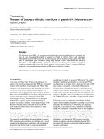
Fig. 20.1 a) Normal heart structures; b) normal O2 sat and pressure measurements.
Sequential segmental analysis
To evaluate patients with suspected congenital heart disease, it is imperative to analyse the heart in a segmental pattern based on:
• Position of the heart, and other organs (thoracic and abdominal):
• Visceral sidedness (situs solitus or inversus)
• Cardiac position (location and orientation)
• Connections between the different regions (veins, atria, ventricles, and
arteries)
• Description of a cardiac region based on its morphological
characteristics rather than its position, or relation to other structures.
It is also important to understand that:
• Connection is an anatomic term showing a direct link between 2
structures; drainage a haemodynamic one, referring to flow of blood
• Single refers to an absence of a corresponding contralateral
structure (single valve in tricuspid atresia); common refers to bilateral
components with an absent division (e.g. common AV valve).
The endocardium is the inner layer of the heart, which is metabolically
active in contributing to cardiovascular function.
The pericardium, a fibroserous sac consisting of visceral and parietal
layers, is a dynamic and adaptive structure which:
• Protects the heart by acting as a barrier
• Reduces friction due to cardiac motion.
Conduction system
Contraction is triggered by electrical impulses which are generated and
conducted through a system of specialized cells—the conduction system.
The sinoatrial node (SA node) generates the electrical impulse which
spreads through the atrial chambers.
• SA node is situated at the SVC/right atrium junction.
There is a single point of electrical connectivity between the atria and the
ventricles; the atrioventricular node (AV node)
• AV node is situated in the triangle of Koch (near the coronary sinus).
The conduction system then proceeds as the bundle of His before dividing
into the left and right bundles and then into various fascicles.
349
350
CHAPTER 20
Cardiac disorders
Circulation (see also b Chapter 11)
• There are 2 vascular beds in the circulation—the pulmonary and
systemic through which the blood is driven by the appropriate
ventricles (see Fig. 20.1):
• The pressure in the pulmonary circulation is significantly lower than
that in the systemic circulation
• The vessels become smaller and thinner as they get farther from the
great arteries, becoming arterioles, and finally capillaries which are the
units where gas and metabolic exchange takes place:
• Arterioles are small arteries with relatively thick muscle and
constitute the majority of the resistance to the relevant vascular
bed; they regulate blood flow
Applied cardiovascular physiology
(See b Chapter 7)
Bedside monitoring of the
cardiovascular system/circulation
(See b Chapter 7)
Cardiac arrhythmias
Cardiac arrhythmias can be due to (Box 20.1):
• Disturbances of rhythm (tachyarrhythmia):
• Supraventricular tachycardia (SVT)
• Ventricular tachycardia (VT)
• Disturbances of conduction (bradyarrhythmia):
• Sinus node dysfunction
• AV dissociations (1st, 2nd, or 3rd degree).
Box 20.1 Cardiac arrhythmias in children can be due to:
• Structural heart disease:
• Native
• Postoperative
• Abnormal pathway
• Cardiomyopathy/myocarditis
• Heart failure
• Miscellaneous:
• Electrolyte imbalance
• Drugs
• Systemic disturbance.
CARDIAC ARRHYTHMIAS
Substrates for the genesis of the arrhythmias are:
• Re-entrant mechanisms: these require the presence of 2 electrical
pathways separated by an electrically inert tissue, having different
properties, setting up an electrical circuit
• Automatic mechanisms: this is due to an abnormally active electrical
focus either inherent (atrial ectopic tachycardia) or due to a secondary
cause (imbalance, strain)
• Triggered mechanisms.
Presentation
Palpitations
Funny turns
Dizziness
Syncope
Cardiac compromise
• deffort tolerance
• Failure to thrive
• Breathless, difficulty in feeding
• Ventricular dysfunction (if prolonged)
• Incidental finding.
•
•
•
•
•
Supraventricular arrhythmias
These are conditions that involve structures above the bifurcation of the
bundle of His.
Re-entrant tachycardias are the commonest mechanism for arrhythmias
seen in children with normal hearts.
• Usually paroxysmal
• Narrow QRS complex
• Regular (constant R–R interval)
• Initiated or terminated by a premature event.
Classically, these rhythms can be terminated with cardioversion. Examples
are:
• AV re-entry tachycardia (AVRT)
• AV nodal re-entry tachycardia (AVNRT)
• Wolff–Parkinson–White (WPW) syndrome
• Atrial flutter.
Automatic mechanisms are much less frequently seen.
• Usually incessant
• Can present with cardiomyopathy or cardiac compromise
• Usually have a narrow QRS complex but have varying R–R interval
(irregular)
• Do not respond to cardioversion.
Examples are:
• Atrial ectopic tachycardia (AET)
• Junctional ectopic tachycardia (JET)
• Atrial fibrillation.
351
352
CHAPTER 20
Cardiac disorders
Diagnosis
• Detailed history
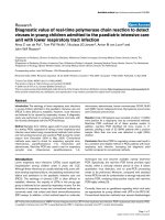
• 12-lead ECG, preferably during an episode (Fig. 20.2)
• Intracardiac electrophysiological studies (‘EP study’): an identification of
the pathway (or focus) can be made invasively; usually combined with
definitive management (ablation) in the same procedure.
P
Atrial ECG from
pacing wires
P waves are conducted
in a retrograde fashion
represented as a spike
following the QRS
P waves on corresponding
surface ECG not identified
Fig. 20.2 Atrial (V1) and surface (V2) ECGs in nodal rhythm. Reproduced from
Mackay J and Arrow Smith, J (eds) (2004) Core topics in cardiac anaesthesia, with
permission from Cambridge University Press.
Treatment
Based on presentation and identification of the mechanism. Acute management will usually involve IV adenosine or synchronized cardioversion.
Specific treatment depends on the mechanism:
• Vagal manoeuvres: Valsalva, diving reflex
• Drug therapy:
• Re-entry tachycardias will respond to adenosine (200mcg kg -1) given
as a fast IV bolus
• B-blockers (sotalol, propranolol) or digoxin (contraindicated in
WPW syndrome)
• Flecanide
• Amiodarone reserved for resistant tachycardias
• Definitive therapy:
• Radio-frequency ablation of the pathway (or focus) by intracardiac
mapping has become the mainline of treatment even in children.
Ventricular arrhythmias
VT is defined as at least 3 consecutive beats of ventricular origin with a
rate >120beats/min.
• Extremely rare in paediatric practice
CARDIAC ARRHYTHMIAS
• A group of heterogeneous conditions with variable substrates and
mechanisms
• As in SVTs the mechanisms involved are re-entrant or triggered
automaticity (more common in VT)
• Ventricular fibrillation (VF) is a series of uncoordinated ventricular
depolarizations associated with an absence of cardiac output.
Important clues on ECG to help identification (Fig. 20.3):
• QRS axis
• QRS morphology
• Propensity to remain same (monomorphic) or vary (polymorphic).
Box 20.2 Causes
• Primary (idiopathic):
• RV outflow tract tachycardia
• Arrhythmogenic RV dysplasia
• Catecholamine sensitive polymorphic VT
• Familial (Brugada syndrome, congenital long QT syndrome)
• Myocardial:
• Cardiomyopathy
— hypertrophic cardiomyopathy
— dilated cardiomyopathy
• Myocarditis
• Myocardial ischaemia
• Conduction abnormality (heart block)
• Miscellaneous:
• Structural heart disease (native or palliated)
• Metabolic derangements
• Drugs
• Trauma with myocardial injury.
Fig. 20.3 Ventricular tachycardia.
Diagnosis
• A detailed history, with emphasis on family history along with a 12-lead
ECG, and identification for a cause should be the primary aim
• ECG monitoring (Holter, loop recorders)
• Specific investigations: genetic testing (long QT syndrome,
cardiomyopathy) or cardiac MRI (arrhythmogenic RV dysplasia)
• Invasive EP study will help to map the focus, and ablate it.
353
354
CHAPTER 20
Cardiac disorders
Treatment
Acute management of VT depends upon hemodynamic status:
• If the patient is stable, amiodarone can be considered
• If the subject has any evidence of cardiac compromise cardioversion
should be used:
• Synchronized cardioversion for VT
• Non-synchronized cardioversion for VF.
Specific management of ventricular arrhythmias includes:
• Individuals at risk (previous history, family history):
• Surveillance
• Prophylactic measures (ß-blockers, implantable defibrillators (ICDs)
• Pharmacotherapy:
• Class IA (procainamide), IB (mexiletine) IC (flecanide), B-blockers,
Amiodarone
• Definitive management:
• Ablation of focus (pathway)
• Implantation of ICD 9 pacemaker.
Bradyarrhythmias
These are due to abnormalities in the generation of an electrical impulse
or conduction defects. They can be seen in children with structurally
normal hearts (complete congenital heart block) or with structural heart
disease (ventricular inversion, post surgical).
They can be classified as:
• AV block:
• 1st degree: prolonged PR interval
• 2nd degree:
— Type I (Wenckebach)
— Type II
• 3rd degree: complete AV block
• Sinus node dysfunction: bradycardia
Chronotropic incompetence
2nd-degree AV blocks with Wenkebach phenomenon (Type I) is a progressive prolongation of PR interval leading to a blocked impulse. Type II
is an abrupt block of an impulse without prolongation of PR interval.
Presentation and coexisting conditions
In children, overt symptoms due to bradycardia are relatively uncommon.
Neonates and infants
• Heart failure (fetal hydrops)
• Apnoea
• Hypoxia
• Gastro-oesophageal reflux with laryngospasm
• Breath holding.
Older children and adolescents
• Heart failure
• Syncope
• deffort tolerance
• Easy fatigability
• Sudden death.
CARDIAC ARRHYTHMIAS
Diagnosis
• A detailed history, with emphasis on family history along with a 12-lead
ECG, and identification for a cause should be the primary aim
• ECG monitoring (Holter, loop recorders) may identify a long pause,
especially at night
• Specific investigations; antibodies (anti-Ro, anti-La) for SLE-related
maternal or fetal condition.
Treatment
Fetal management
• Consider the need for early delivery
• Sympathomimetic agents (ritodrine) have been used with limited effect,
but are poorly tolerated by mothers.
Acute management of a compromised child
• Chronotropic agents (isoprenaline)
• Atropine
• Temporary pacing.
Pacemakers
• Transvenous (via subclavian vein) or epicardial (surgically implanted)
• In infants a single chamber system (ventricular—VVI) is used, and
upgraded to a dual chambered system (DDD) to maintain AV
synchrony in older children.
355
356
CHAPTER 20
Cardiac disorders
Congestive heart failure
Congestive cardiac failure develops when systemic oxygen supply is inadequate for oxygen demands, or is maintained at the expense of higher
atrial filling pressures. In paediatric practice, the cause is frequently a large
L-to-R shunt (LlR) (large VSD) with ‘preserved myocardial function’ as
opposed to ‘pump failure’ as commonly seen in adults.
A range of compensatory mechanisms, initially beneficial, contribute to
the pathophysiology. These include:
• Salt and water retention:
• Aldosternone stimulation (sodium retention)
• Arginine vasopressin (water and sodium retention)
• Natriuretic peptides
• Neuro-hormonal changes:
• Sympathetic stimulation:
— sympathetic cholinergic fibres (sweating)
— A-adrenoreceptors (vasoconstriction)
— B-adrenoreceptors (tachycardia)
• Renin–angiotensin activation (vasoconstriction)
• ired cell mass
• Hypertrophy of cardiomyocytes.
Pulmonary oedema occurs due to a combination of:
• Fluid retention
• ifilling pressures (left atrium)
• ipulmonary blood flow (in LlR shunts)
• Lower oncotic pressures (low albumin concentrations).
Causes
• Volume overload:
• Intracardiac shunt
• Extracardiac shunt, e.g. AV malformation, aneurysm of great vein of
Galen
• Valvar regurgitation
• Pressure overload: obstruction—cardiac (aortic stenosis) or arterial
(coarctation)
• Intrinsic myocardial contractile dysfunction
• Myocarditis
• Cardiomyopathy
• Rhythm disorders:
• Persistent tachycardia/bradycardia
• Lack of AV synchrony (heart blocks)
• icardiac output (‘high output’ states):
• Sepsis (‘warm shock’)
• Severe anaemia
• Hyperthyroidism
• Liver failure.
Symptoms and signs
• iadrenergic tone:
• Clammy, pale, vasoconstriction, oliguria
• Tachycardia
PATHOPHYSIOLOGY OF CONGENITAL HEART DISEASE
• Impaired myocardial contractility:
• Poor perfusion, weak pulses
• Altered sensorium, irritability
• deffort tolerance, chest pain
• Failure to thrive, breathless on feeding
• Salt and water retention:
• Cardiomegaly
• Hepatomegaly
• Pulmonary congestion
• Tachypnoea, respiratory distress, frequent ‘chest infections’.
Treatment
Box 20.3 Treatment of congestive heart failure
• Specific management of treatable causes, e.g. structural heart disease,
myocarditis
• General interventions:
• Optimize nutrition, haemoglobin
• Optimize respiratory function
— oxygen
— respiratory support: CPAP, ventilation
• Impaired myocardial contractility:
• Inotropes (sympathomimetics, PDEIs)
• Vasodilators
• Mechanical support (ventricular assist device, ECLS)
• Compensatory mechanisms:
• Salt and water retention—diuretics
• Renin–angiotensin–aldosterone axis—captopril, losartan,
spironolactone
• Minimize risk from cardiac impairment:
• Rhythm abnormalities
• Thromboembolic phenomena—heparin prophylaxis for severely
impaired ventricular function.
Pathophysiology of congenital
heart disease
Congenital heart disease lesions can be classified as:
• LlR shunts
• Hypoxaemic lesions
• Obstructive and regurgitant lesions of left and right heart.
Left-to-right shunts
Lesions
• Ventricular septal defect (VSD
• Persistence of arterial duct (PDA)
• Atrial septal defect (ASD)
• AV septal defect (AVSD)
• Aortopulmonary window (AP window).
357
358
CHAPTER 20
Cardiac disorders
Clinical manifestations are related to:
• Size of the defect
• Postnatal changes in vascular resistance of the pulmonary and systemic
beds.
Following birth, there is a rapid reduction in the pulmonary vascular resistance. This usually takes place over 2-6 weeks following birth; however in
the presence of large defects this may be delayed by 1-3 months, and in
some cases, there is no significant reduction in the resistance across the
pulmonary bed.
Consequences of LlR shunting:
• ipulmonary blood flow
• Left atrial dilatation and left ventricular volume overload
• Pulmonary tree:
• ivolume and pressure of pulmonary vasculature
• Large shunts can result in pulmonary vascular disease if not
corrected in the 1st year of life
• Airway obstruction with hyperinflation
• Stretching of oval foramen liatrial shunting (LlR).
Hypoxaemic lesions
Cyanosis is defined as the presence of >5g.L–1 of reduced haemoglobin.
The cardiac causes of hypoxaemia can be classified broadly into:
• Obstruction to pulmonary blood flow
• Transposition physiology
• Common mixing.
Common lesions with obstruction to pulmonary blood flow
• Tetralogy of Fallot
• Pulmonary atresia (with or without VSD)
• Double outlet ventricle with pulmonary stenosis.
Clinical manifestations
The degree of hypoxaemia is determined by the severity of the pulmonary
obstruction, and the patency of the PDA. If the obstruction is progressive,
there will be a continuing decline in O2 saturation.
• Adaptive mechanisms:
• Hyperpnoea
• Increase in red cell mass—polycythaemia may develop
• i2,3 DPG levels
• Dilated coronary vessels.
Hypercyanotic spells in tetralogy of Fallot
Characterized by a pronounced fall in O2 saturation often associated with
a manoeuvre causing an increase in intrathoracic pressure, whilst dropping
the SVR. Treatment consists of:
• Calm the child
• Oxygen
• iSVR:
• Hip and knee flexion (‘squatting’)
• A-receptor sympathetic agent if profound
PATHOPHYSIOLOGY OF CONGENITAL HEART DISEASE
• Medication:
• Propranolol (or esmolol in intensive care environment)
• Morphine.
Initial palliation of any hypoxaemic lesion is to create a stable systemicpulmonary shunt to replace the PDA.
Transposition physiology
Severity of hypoxaemia is linked to the degree of mixing between the 2
parallel circulations at an intracardiac level.
• Associated lesions:
• VSD
• Pulmonary stenosis—quite often complex
• In absence of a significant VSD, neonates are dependent on the size of
the atrial communication to maintain adequate oxygen levels.
Immediate management consists of a prostaglandin E infusion and a balloon
atrial septostomy
Common mixing
Lesions
• Common arterial trunk (truncus arteriosus)
• Common atrium
• Single ventricle (HLHS)
• Spectrum of hypoplastic RV (tricuspid atresia)
• Anomalies of pulmonary venous return (TAPVD).
Pathophysiology
• Characterized by mixing of systemic and pulmonary blood at some
level
• Systemic arterial oxygenation is dependent on the magnitude of the
pulmonary venous return relative to systemic venous return
• Manipulation of PVR and SVR can be useful in manipulating the Qp:Qs
which will influence O2 saturation.
Obstruction to systemic output
Can be broadly divided into obstruction to the LV outflow and inflow.
LV outflow tract obstruction
Lesions include:
• Subvalvar, valvar, and supravalvar aortic stenosis
• Aortic arch hypoplasia
• Interrupted aortic arch
• Coarctation.
During fetal development systemic perfusion is not compromised, due
to ductal patency, but LV hypertrophy and compromise to LV development can occur to an extent that it is not able to maintain adequate independent systemic circulation (hypoplastic left heart syndrome).
In the postnatal period, ductal patency is essential to maintain systemic
flow. The systemic perfusion may be dependent entirely on the ductal
flow and right ventricular function (hypoplastic left heart syndrome/ aortic
Atresia), or partially (aortic coarctation) where reasonable systemic perfusion can be maintained as long as the aortic end of the ductal patency
is maintained.
359
360
CHAPTER 20
Cardiac disorders
LV inflow obstruction
Lesions include:
• Mitral stenosis
• Cor triatriatum
• Pulmonary venous obstruction.
The hemodynamic changes in this group cause derangements due to ‘back
pressure’ changes in addition to compromising forward flow. These are:
• Compromised LV output (reduced preload)
• ‘Back pressure’ changes related to elevated LA pressure:
• ipulmonary venous pressures
• Pulmonary and RV hypertension
• Systemic venous congestion (if RV dysfunction).
Regurgitant lesions
Valvar regurgitation is usually associated with other cardiac abnormalities.
It can be congenital or acquired—due to an infection or secondary to
ventricular dilatation. Symptoms are related to the duration, and severity
of the lesions; chronic lesions are better tolerated.
Mitral valve regurgitation (MR) can be due to:
• Isolated (rare)
• Mitral cleft
• AV junction abnormalities (AVSD)
• Papillary muscle infarction (ALCAPA).
Haemodynamic derangements cause:
• Left atrial and ventricular volume overload
• i filling pressures
• Back pressure changes:
• Pulmonary venous congestion
• Right heart dilatation and hypertension
• Atrial thrombi
• Atrial dysrhythmias.
Once there is progressive LA dilation, the mitral annulus stretches leading
to further i in mitral regurgitation (progressive)
Tricuspid valve regurgitation (TR) can be due to:
• Dysplastic tricuspid valve
• Ebstein’s anomaly of the tricuspid valve.
Haemodynamic derangements can be similar to discussed earlier, the
major differences are:
• Significant instability in neonatal period due to iPVR. Haemodynamics
improve as resistance d
• iRA pressures
• RlL shunts l hypoxaemia
• Potential for lung hypoplasia l lung function compromise.
Aortic valve regurgitation (AR) is rarely an isolated anomaly. Haemodynamic
derangements include:
• Volume loading of the LV
• LV hypertrophy
PULMONARY HYPERTENSION SYNDROMES
• i filling pressures
• Large AR l diastolic runoff l dcoronary blood flow.
Pulmonary valve regurgitation (PR) can be due to:
• Absent pulmonary valve syndrome
• Following repair/reconstruction of RV outflow tract, e.g. tetralogy of
Fallot
Haemodynamic effects:
• RV volume overload
• RV hypertrophy
• Compromised lung perfusion
• LV dysfunction:
• dpreload
• Ventricular interaction.
Pulmonary hypertension syndromes
Pulmonary hypertension (PH) is defined as a mean pulmonary artery pressures of Ĕ25mmHg.
It can be classified according to aetiology (Box 20.4).
Box 20.4 Aetiology of PH syndromes
• Pulmonary arterial hypertension:
• Primary PH—unknown cause:
— familial
— sporadic
• Collagen vascular disease
• Congenital heart disease with systemic-to-pulmonary shunt
• Miscellaneous:
— persistent PH of the newborn (PPHN)
— drugs
— HIV
— portal hypertension
• Pulmonary venous abnormalities: left-sided heart disease
(mitral stenosis)
• Pulmonary veno-occlusive disease
• Associated with respiratory disease and hypoxemia
• Chronic thrombo-embolic disease: sickle cell disease
• Other: pulmonary vasculature (e.g. sarcoidosis).
Presentation
• Dependent upon the primary pathology
• Primary PH predominantly affects young people, and has a very
aggressive progression
• Diagnosis often delayed in absence of an intracardiac shunt
• Symptoms:
• Dyspnoea (often diagnosed as ‘asthma’)
• Frequent respiratory exacerbations (repeated ‘chest infections’)
361
362
CHAPTER 20
•
•
•
•
•
•
•
Cardiac disorders
Failure to thrive
Decreasing effort tolerance
Palpitations, chest pain
Cyanosis with or without exercise
Headaches
Pedal oedema
Syncope/near syncope.
Diagnosis
History
Chest radiograph
ECG (right-sided changes in 70–80%)
Echocardiography:
• Exclude structural heart disease
• Non-invasive estimation of pulmonary pressure if TR jet is present
• Other investigations:
• Cardiac catheterization and angiogram
• Lung perfusion
• CT scan
• Lung biopsy.
•
•
•
•
Treatment
Management of pulmonary hypertensive crises is described on b p.379.
• Treatment of the causative pathology
• Pulmonary vasodilator therapy:
• Selective type V phosphodiesterase inhibitor (sildenafil)
• Non-selective endothelin receptor blocker (bosentan)
• Calcium channel blockers
• Prostacyclin infusion
• Inhaled NO
• Home oxygen
• Anticoagulation (aspirin, warfarin)
• Other:
• Blade atrial septostomy—to allow RlL shunt and preserve cardiac
output at the expense of cyanosis
• Lung transplantation
• Newer therapeutic agents:
• Vasoactive mediators
• Potassium channel blockers
• Serine elastase inhibitors.
Outcome
• Bimodal presentation: aggressive course in infants and adolescents
• Survival:
• 37% at 1 year following diagnosis
• 12.5% at 2.5 years
• Lung transplant outcomes (survival):
• 1 year: 73% (90% for congenital heart disease)
• 10 years: 30–40%.
SYSTEMIC HYPERTENSION
Systemic hypertension
Hypertension is uncommon in childhood, but often goes unrecognized for
a long time. It is defined as systolic and/or diastolic BP being >95 percentile for age on Ĕ3 occasions.
Cardiac sequelae of childhood hypertension are uncommon, but acute,
severe forms (malignant hypertension) can result in ventricular dysfunction and congestive heart failure. Long standing hypertension can
result in:
• Diastolic dysfunction with ilate filling (prominent ‘A’ contribution
relative to ‘E’)
• LV hypertrophy
• ifilling pressures.
Causes
Box 20.5 Causes of hypertension
• Essential (idiopathic)
• Cardiovascular:
• Coarctation
• Defects with diastolic runoff (result in systolic hypertension):
— arteriovascular malformation
— PDA
— severe AR/MR
• Reno-vascular:
• Parenchymal renal disease
• Polycystic kidneys
• Renal artery stenosis
• Tumours (Wilms’)
• Endocrine:
• Phaeochromocytoma
• Congenital adrenal hyperplasia
• Cushing’s disease
• Drugs: steroid therapy.
Presentation
•
•
•
•
•
May be asymptomatic and be detected coincidentally
Symptoms of primary pathology
Headaches, visual disturbance
Encephalopathy (if severe)
Epistaxis.
Treatment
Hypertensive crisis: after initial resuscitation, aim should be to reduce
BP but to avoid a precipitous drop to maintain organ perfusion. Rule of
thumb is to reduce BP by no more than 25% in first 12–24H. A quicker
reduction is safe if the BP rise is of very recent onset.
363
364
CHAPTER 20
Cardiac disorders
Drugs include:
• Nifedepine (oral)
• Labetelol or esmolol infusion
• Sodium nitroprusside infusion
• Hydralazine.
Treatment of primary pathology
• Management of BP:
• Non-pharmacological
• Weight management
• Dietary modification (sodium reduction)
• Pharmacological
• ACE inhibitors (captopril, enalapril)
• Angiotensin receptor blockers (ARBs) (losartan)
• B-adrenergic receptor blockers (propranolol, atenolol)
• Diuretics (frusemide, thiazide)
• Calcium-channel blockers (nifedepine)
• Miscellaneous, e.g. minoxidil
Dilated cardiomyopathy and
myocarditis
Dilated cardiomyopathy is a group of heterogeneous aetiologies uniformly
characterized by ventricular dilatation and impairment of contractility. LV
function is usually more affected than RV. Causes are multifactorial, see
Box 20.6.
Box 20.6 Causes
•
•
•
•
•
•
Idiopathic (>50%)
Myocarditis (10–15%)
Familial/genetic (20–35%, autosomal dominance is most frequent)
Autoimmune
Drug induced (anthracycline)
Miscellaneous:
• Persistent arrhythmias
• Structural heart disease
• Inborn errors of metabolism
• Coronary arterial disease (ALCAPA)
• Neuromuscular disorders.
Myocarditis is an acute process characterized by inflammatory infiltration
of the myocardium along with cellular necrosis, and is caused by infectious
agents (e.g. enterovirus, coxsacchie), or can be an autoimmune process
(e.g. lupus).
Incidence and prevalence are low in children; however there is some evidence of an increasing trend. It is likely that a number of less severe cases
of myocarditis associated with a viral infection go undetected.
DILATED CARDIOMYOPATHY AND MYOCARDITIS
Presentation
There is a variable period during which the child is asymptomatic as the
heart undergoes dilatation and hypertrophy to maintain cardiac output.
Cases with a family history may be picked up at this stage on screening.
With progression congestive heart failure ensues:
• Infants: tachypnoea, feeding difficulties, failure to thrive, sweating
• Older children: decreasing effort tolerance with exertional dyspnoea,
palpitations, arrhythmias, or syncope (13%).
Diagnosis
• Detailed history including relevant past and family history
• CXR, ECG, and Echo to confirm the diagnosis
• Cardiac catheterization and myocardial biopsy are not routinely
performed due to associated high risk
• Cardiomyopathy screen to identify cause—details are available from
any paediatric cardiology unit.
Treatment
Acute treatment consists of stabilization and may include inotropes,
cautious use of diuretics, and mechanical ventilation if the child has
decompensated.
Induction of anaesthesia is high risk if ventricular function is severely
compromised:
• Summon expert assistance
• Start inotropic therapy ahead of induction, have epinephrine available
• Use small doses of induction drugs that are unlikely to acutely drop
cardiac output or SVR (ketamine, fentanyl, etomidate rather than
thiopentone, propofol).
Once stable, management should be aimed towards:
• Identification and treatment of cause
• Decreasing cardiac afterload (ACE inhibitors)
• Diuretics (frusemide)
• Prevention of arrhythmias (digoxin)
• Prevention of thromboembolic phenomena (aspirin or heparin)
• Newer therapies (stable patients in chronic heart failure):
• B-blockers—carvedilol
• ARBs—losartan
• Cardiac resynchronization therapy.
Specific issues
Myocarditis therapy: unproven but used in some centres.
• Immunomodulatory therapy—IV immunoglobulin (IVIG)
• Immunosuppressive therapy—steroids, cyclosporine, azathioprine.
Continuing cardiac instability
• Mechanical support—LV assist device (LVAD), e.g. Berlin Heart Excor®
as a bridge to heart transplant, ECLS for myocarditis.
Outcome
• Complete resolution (25–35%)
• Residual cardiac dysfunction (30–35%)
• Deterioration and death/transplant (25–35%).
365
366
CHAPTER 20
Cardiac disorders
Poor prognostic factors
• Idiopathic
• Age at diagnosis: <2 years
• First 2 years after presentation.
Prognosis for acute myocarditis in newborns is very poor (up to 75%
mortality; highest within 1st week of presentation). Older infants and
children bear a better prognosis with 10–25 % mortality, and complete
recovery in >50%.
Infective endocarditis
Infection of the endocardium, heart valves, or related structures is known
as infective endocarditis. Risk factors include:
• Structural heart disease
• Neonates with invasive procedures or lines
• Prosthetic material in the heart or great vessels
Pathophysiology
The genesis is multifactorial, and difficult to confirm; there is turbulent
blood flow leading to endothelial damage. Aggregation of platelets,
and fibrin deposition (non-infective thrombotic vegetation) follows.
Colonization can occur and is more likely with bacteria producing dextran,
in the presence of fibronectin at the local site (Box 20.7).
Infection can damage cardiac structures, vegetations may obstruct blood
flow, and occasionally will embolize to other areas, most commonly the
lungs (or brain).
Immunological mechanisms are central to pathogenesis, and sequelae of
this process, and involves cell-mediated and humoral mediated pathways.
• Hypergammaglobulinaemia:
• Polyclonal and antigen specific B-cell activation
• Rheumatoid factors
• ilevels of:
• Circulating immune complexes
• Mixed-type cryoglobulinaemia
• Renal involvement (due to immune complex deposition).
Box 20.7 Commonest organisms include:
•
•
•
•
•
Streptococcus viridians (~40%; commonest)
Other streptococci
Staphylococcus (most common in postoperative period)
Gram-negative organisms
Fungi.
Presentation
• Fever
• Anorexia/weight loss
• Malaise
PERICARDITIS AND CARDIAC TAMPONADE
Arthralgia
Chest pain
Congestive heart failure
Specific skin lesions less common in children:
• Petechiae (1/3 cases)
• Osler’s nodes/Janeway lesion/Roth spots/splinter haemorrhage
(<10%)
• Splenomegaly
• New or changing murmur
• Embolization of infective foci to other organs (e.g. cerebral infarcts).
•
•
•
•
Investigation (Box 20.8)
• Blood cultures: at least 3–5 samples in the first 24h of presentation
(negative in 10–15%)
• FBC: neutrophilia
• Inflammatory markers: very high ESR, raised CRP
• Echocardiography: TTE has 44% sensitivity to detect vegetations
• ECG: ectopic beats, blocks, ST/T changes
• Circulating immunologic complexes (in difficult cases).
Box 20.8 Learning point
Infective endocarditis is a clinical diagnosis confirmed by presence of
positive blood cultures. Presence of an echogenic focus (vegetation)
on echocardiography supports the diagnosis; however a negative result
does not rule out the diagnosis.
Treatment
• Antibiotics for 4–6 weeks (guidance from microbiologists):
• Usually 2 antibiotics
• Initial parenteral therapy
• Central venous access only after sterilization of blood and resolution of
symptoms: prophylaxis is mandatory for prevention/relapse.
Prognosis
• 20–30% mortality even in the modern era of antimicrobial, therapy
• Always at higher risk for subsequent infection.
Pericarditis and cardiac tamponade
Pericarditis can occur due to:
• Infections:
• Viral infection:
— coxsackie, varicella, influenza, infectious mononucleosis, Echo,
mumps
• Purulent infection:
— S. aureus (1/3 cases; 3/4 of those who die)
— Haemophilus influenza, type b
— Streptococcus
367
368
CHAPTER 20
•
•
•
•
Cardiac disorders
— M. pneumonia
— Candida, Aspergillus (immunocompromised host)
• TB
• HIV
Auto-immune disorders
Connective tissue disorders
Malignancy
Others: drugs, therapeutic procedures.
Presentation
•
•
•
•
Clinical manifestations of the primary disorder
Chest pain
Pericardial rub
More severe forms tend to show manifestations of abnormal perfusion:
• Tachycardia
• Low volume pulses
• Pulsus paradoxus
• Tamponade.
Diagnosis
Specific investigation for underlying aetiology
Acute inflammatory markers: CRP/ESR
Cardiac enzymes (may be elevated)
ECG:
• Low voltage complexes
• Widespread T-wave/ST changes
• Chest radiograph (enlarged heart)
• Echo:
• Pericardial effusion
• Echogenic objects (fibrin, clots)
• Atrial collapse
• Ventricular function
• Other: cardiac MRI, CT scan, cardiac catheterization.
•
•
•
•
Treatment
• Treatment of primary cause—antibiotics
• Treatment of haemodynamic alterations:
• Stabilization of haemodynamics (fluid, inotropes, ventilation if
required)
• Arrhythmias
• Decompression of pericardial fluid.
Cardiac tamponade
Tamponade occurs when there is sufficient fluid in the pericardial cavity
to cause compromise to cardiac filling or contractility, and is not necessarily related to the amount of fluid in the cavity (or size of cardiac silhouette on CXR).
It can be seen as a result of acute pericarditis or in the postoperative
period following cardiac (thoracic) surgery.
IMMEDIATE POSTOPERATIVE CARE
Presentation
• Dyspnoea
• Tachycardia, small volume pulses
• Narrow arterial pulse, hypotension
• Elevation of systemic venous pressures (CVP, LAP if line present)
• Pulsus paradoxus (decreasing/absent pulses in inspiration).
Treatment
• ABC stabilization—volume, inotropes, ventilation
• Pericardial drainage is the definitive acute management:
• Needle aspiration can be done as an emergency procedure at the
bedside
• Pericardial tap and insertion of a drain can be done under US or
angiographic guidance
• Surgical drainage, advised if:
— fluid is posterior
— adhesions
— purulent fluid
— pericardial thickening.
Postoperative care
The practice of cardiac intensive care has evolved considerably over the
past 10 years with:
• Greater use of interventional procedures in the catheter lab with
device closures of ASD, VSD, PDA, and even percutaneous valve
replacements
• Earlier surgical intervention so that postoperative pulmonary
hypertension is much less common
• Changes in inotrope and vasodilator therapy
• Greater use of ultrafiltration following bypass and ECLS for children in
a low cardiac output state
• Fast-tracking of suitable patients to achieve short PICU and hospital
stays.
Cardiac surgical patients epitomize the importance of a multidisciplinary
approach in a specialized paediatric or cardiac ICU.
Immediate postoperative care
The major goal is to establish adequate cardiac output to ensure tissue
oxygen delivery and end-organ function. This is best addressed with a systematic approach.
Cardiovascular
• Anaesthetic and surgical handovers provide vital information from the
operating room whilst the patient was on and off CPB.
• It is important that both occur as they will provide different
information
• Note CPB, aortic cross clamp, and circulatory arrest durations
369