Ebook Color atlas of cytology, histology and microscopic anatomy: Part 2
Bạn đang xem bản rút gọn của tài liệu. Xem và tải ngay bản đầy đủ của tài liệu tại đây (25.95 MB, 281 trang )
Endocrine Glands
344
Hypophysis—Pituitary Gland
The human hypophysis (hypophysis cerebri) weighs about 600–900 mg. It is
enveloped by a thin connective tissue capsule 1 . Distinguished by their
modes of development and detail structure, there are the large glandular
lobe, the adenohypophysis 2 , and the smaller cerebral lobe, the neurohypophysis 3 . The adenohypophysis (anterior lobe, pars distalis) continues toward the cranium as tubular part of the hypophysis cerebri (pars infundibularis) 4 . The infundibulum 5 connects the neurohypophysis (posterior lobe,
pars nervosa) to the diencephalon. The intermediary lobe 6 between anterior
and posterior lobes is part of the adenohypophysis. This sagittal section distinctly shows these different parts of the hypophysis.
1 Capsule
2 Anterior lobe, pars distalis
3 Posterior lobe, pars nervosa
4 Infundibular lobe, pars infundibularis
5 Infundibulum with eminentia mediana
6 Intermediary lobe with colloid cysts (adenohypophysis)
Stain: azan; magnification: × 7
345
Adenohypophysis—Anterior Lobe
The anterior lobe of the adenohypophysis (pars distalis) consists of cords and
nests of different types of epithelial cells. These are surrounded by reticular
fibers and wide blood sinuses. The cells are grouped according to their affinities to dyes as acidophilic 1 , basophilic 2 or chromophobic 3 cells. The
three cell types can be distinguished in this figure without much effort. Acidophilic cells 1 are round and contain a dense (acidophilic) population of
granules. There are somatotropic and mammotropic acidophilic cells. The granules in somatotropic acidophilic cells have diameters of about 300 nm,
those in mammotropic acidophilic cells have diameters of 600–900 nm. Basophilic cells 2 come in various sizes. They contain granules. There are gonadotropic basophilic cells (granule size: 300–400 nm), thyrotropic basophilic
cells (granule size: 60–160 nm), adrenotropic basophilic cells (granule size:
200–500 nm), lipotropic basophilic cells (granule size: 200–500 nm) and melanotropic basophilic cells (granule size: 200–400 nm). According to current
opinion, the chromophobic cells 3 do not participate in the biosynthesis of
hormones. They are very likely precursors of hormone-producing cells.
1 Acidophilic cells
3 Chromophobic cells
2 Basophilic cells
4 Capillary
Stain: hematoxylin (Carazzi)-eosin; magnification: × 320
346
Adenohypophysis—Anterior Lobe
Section of the anterior lobe (pars distalis) from the adenohypophysis. Compare this micrograph with Fig. 345. The legend includes both commonly used
terminologies.
254
1 α-cells (acidophilic)
2 β-cells (basophilic)
3 δ-cells
Stain: azan; magnification: × 400
4 γ-cells (chromophobic)
5 e-cells
Kuehnel, Color Atlas of Cytology, Histology, and Microscopic Anatomy © 2003 Thieme
All rights reserved. Usage subject to terms and conditions of license.
344
1
4
5
3
2
6
1
345
2
1
2
1
1
3
4
Endocrine Glands
4
3
346
1
2
2
4
1
5
3
2
255
Kuehnel, Color Atlas of Cytology, Histology, and Microscopic Anatomy © 2003 Thieme
All rights reserved. Usage subject to terms and conditions of license.
347
Adenohypophysis—Intermediary Lobe
Because of its developmental origin, the intermediary lobe (pars intermedia)
between anterior and posterior lobes 1 of the hypophysis belongs to the
adenohypophysis (cf. Fig. 344). The intermediary lobe constitutes about 3% of
the adenohypophysis. The details of its structure are very complex. Groups of
the anterior lobe cells 3 are captured in the lower part of the figure. Basophilic cells may enter the dorsal hypophysis (basophil invasion). Colloidfilled cysts (colloid cysts) 2 are conspicuous elements in the intermediary
lobe. They derive from the hypophyseal pouch (remnant of the Rathke pouch).
The cysts may be lined by a single-layered epithelium or sometimes by a
multilayered stratified epithelium at different levels of differentiation. The
cells of the intermediary lobe synthesize melanotropin (melanocyte stimulating hormone, MSH).
1 Posterior lobe (pars nervosa)
2 Colloid cysts
Stain: azan; magnification: × 80
Endocrine Glands
348
3 Cells of the adenohypophysis
Neurohypophysis
The neurohypophysis consists of the posterior lobe (pars nervosa) and the infundibulum, including the eminentia mediana (see Fig. 344).
The constituent cells of the neurohypophysis are neuroglia cells (pituicytes,
protoplasmic glial cells), numerous unmyelinated nerve fibers, which stem
from neurosecretory neurons of the hypothalamus, connective tissue and
vessels. Routine staining procedures reveal a dense matted layer of fibers (a)
or a woven meshwork of fine, unmyelinated nerve fascicles 1 (cross-sectioned or cut longitudinally). The meshwork of the nerve fascicles contains
pituicytes and wide capillaries. Figure (b) shows numerous basophilic cells
2 , which have invaded from the intermediary lobe (basophil invasion) into
the posterior lobe. Two main neurosecretory systems exist; their morphological and biochemical attributes are different.
1 Bundles of unmyelinated nerve fibers
2 Basophilic cells, invaded from the intermediary lobe
3 Vein
Stain: alum hematoxylin-eosin; magnifications: a) × 40; b) × 100
349
Pineal Gland—Epiphysis Cerebri
The pineal gland has a conical shape. Its surface is covered by the pia mater.
Connective tissue septa 1 originate at the pia mater and subdivide the parenchyme into incompletely separated lobes of different sizes. The pineal gland
consists of pinealocytes (modified photoreceptor cells), interstitial cells and
glial cells (astrocytes). The interstitial cells often form structures in the same
way that epithelial cells do. Degeneration of the pineal gland proceeds
throughout human life. It leads to the formation of a gritty substance (acervulus), brain sand. In this figure, the gritty substance 2 takes the form of
stacked lamellae (stained red or blue), which consist of pebbles of organic
substances as well as calcium and magnesium salts.
256
1 Connective tissue septa
2 Gritty substance of the pineal gland (acervulus)
Stain: azan; magnification: × 150
Kuehnel, Color Atlas of Cytology, Histology, and Microscopic Anatomy © 2003 Thieme
All rights reserved. Usage subject to terms and conditions of license.
347
1
2
2
3
2
348
2
1
Endocrine Glands
3
1
2
a
b
349
1
2
257
Kuehnel, Color Atlas of Cytology, Histology, and Microscopic Anatomy © 2003 Thieme
All rights reserved. Usage subject to terms and conditions of license.
350
Pineal Gland—Epiphysis Cerebri
The specific cells of the pineal gland are the pinealocytes 1 . They usually have
a bizarre geometry, although some may appear rounded or polygonal. Their
nuclei have little chromatin. Pinealocytes have numerous branched cell processes with synaptic ribbons in close proximity to blood vessels. As is the
case in the neurohypophysis, there are nerve-blood contact zones. In addition, the epiphysis contains contact neurons in the pineal recess. The pinealocytes 1 in this figure are embedded in a meshwork of glial cells 2 .
1 Pineal cells
2 Meshwork of glial cells
Stain: azan; magnification: × 400
Endocrine Glands
351
Adrenal Gland—Glandula Suprarenalis
The furrowed surface of the adrenal gland (suprarenal gland) is encased in a
vascularized connective tissue capsule 1 . The adrenal gland consists of the
cortex 2 and the medulla 3 . The zona reticularis 4 of the cortex is emphasized as a heavily stained dark band. This band makes the border between cortex and medulla particularly obvious in this preparation. A large muscular
vein 3 in the medulla is cut (cf. Fig. 357).
Cortex and medulla are of different phylogenetic origin and have distinct
functions within the organ.
1 Organ capsule
2 Cortex
3 Medulla with vein
4 Zona reticularis
5 Adipose tissue
Stain: alum hematoxylin-eosin; magnification: × 4
352
Adrenal Gland—Glandula Suprarenalis
The adrenal gland is enveloped by a fiber-rich connective tissue capsule 1 .
Above it, there is a layer of vascularized loose connective tissue with many
adipocytes. The zona glomerulosa 2 is the outermost narrow layer of the cortex. The epithelial cells in this zone have formed round aggregates or nests.
The underlying cells are part of the wide zona fasciculata 3 . The cells are arranged in lightly stained strands and columns. The zona reticularis 4 follows
without demarcation. It is a loose web-like tissue (cf. Fig. 353c), which borders on the medulla. Part of the ectodermal medulla 5 is visible on the right
of this figure.
1 Capsule
2 Zona glomerulosa
3 Zona fasciculata
4 Zona reticularis
5 Medulla
6 Medullary vein
Stain: azan; magnification: × 25
258
Kuehnel, Color Atlas of Cytology, Histology, and Microscopic Anatomy © 2003 Thieme
All rights reserved. Usage subject to terms and conditions of license.
350
2
1
2
1
5
2
4
1
3
Endocrine Glands
351
1
352
2
5
3
4
6
1
5
259
Kuehnel, Color Atlas of Cytology, Histology, and Microscopic Anatomy © 2003 Thieme
All rights reserved. Usage subject to terms and conditions of license.
353
Adrenal Gland—Glandula Suprarenalis
a) Zona glomerulosa, zona multiformis.
The fiber-rich organ capsule can be found at the left edge of the microphotograph. The round or oval groups of cells and cell nests in the following cortical layer are the glomeruli. The right part of the figure shows part of the zona
fasciculata.
b) Zona fasciculata.
The top half of the figure shows the large columnar cells, which are only
lightly stained. The cells are evenly filled with small lipid droplets (honeycomb structure, spongiocytes). The cell cords in the lower part of the image
are more loosely arranged. The cells are smaller and contain pigments. The
pigments make the cells appear darker.
c) Zona reticularis.
The sizes of cells in this layer may vary. Its cell cords are arranged in a loose
web-like structure. Sinusoid blood vessels and strong connective tissue
trabeculae are found between cells.
Figures b and c are rotated 90° in comparison with Figure a.
Stain: azan; magnification: × 100
Endocrine Glands
354
Adrenal Gland—Glandula Suprarenalis
The connective tissue capsule 1 of the adrenal gland contains nerves and
blood vessels. The small rounded groups of cells of the zona glomerulus are
located underneath it (cf. 353a). The large round nuclei of the glomerular
cells are heavily stained. The lipid droplets in the cells have been removed
during preparation. This has left empty, vacuole-like spaces in the place of fat
droplets in the acidophilic cytoplasm (cf. Fig. 63). Strands of delicate connective tissue start at the organ capsule and continue between the glomerular
cells. They contain wide sinusoid blood vessels 2 . Note the button-shaped
endothelial cells, which push into the lumen.
Cells in the zona glomerulosa mostly biosynthesize mineral corticoids,
which predominantly regulate the potassium and sodium balances. The
most important mineral corticoids are aldosterone and deoxycorticosterone.
1 Connective tissue capsule
2 Sinusoids
Semi-thin section; stain: methylene blue-azure II; magnification: × 800
355
Medulla of the Adrenal Gland
The medulla of the suprarenal gland is derived from sympathochromaffin
tissue. It is therefore a sympathetic paraganglion. The polygonal cells 1 are
arranged in cords and show a fine granulation. Due to their affinity to chromium salts, the cells are often called chromaffin or pheochrome cells. Their
secretory granules contain adrenaline or noradrenaline. The medullary cell
nests (cell clusters) 1 are pervaded by large, strong muscular veins 2 (cf.
Fig. 357). All cells of the medulla have immediate access to capillaries and venules. The medulla contains autonomous multipolar sympathetic ganglion
cells 3 , either singly or as clusters. Unmyelinated nerve fibers are seen at the
left edge of the figure.
1 Cords of chromaffin medullary cells
2 Medullary veins
260
3 Multipolar ganglion cells
4 Cushion of smooth muscle cells,
smooth muscle prominence
Stain: alum hematoxylin-eosin; magnification: × 40
Kuehnel, Color Atlas of Cytology, Histology, and Microscopic Anatomy © 2003 Thieme
All rights reserved. Usage subject to terms and conditions of license.
353
a
b
c
354
1
2
2
Endocrine Glands
2
355
1
2
2
4
3
1
261
Kuehnel, Color Atlas of Cytology, Histology, and Microscopic Anatomy © 2003 Thieme
All rights reserved. Usage subject to terms and conditions of license.
356
Medulla of the Adrenal Gland
Most of the medullary parenchyme of the adrenal gland (suprarenal gland)
consists of chromaffin or pheochrome cells. There are also multipolar sympathetic ganglion cells 1 with long cell processes and satellite cells. Ganglia
cells occur singly or in small clusters.
Stain: azan; magnification: × 400
Endocrine Glands
357
Medulla of the Adrenal Gland
The adrenal gland is a richly vascularized organ. There are connections between the vascular networks of the cortex and medulla. Having passed the
medullary sinuses, the venous blood accumulates in the throttling veins,
which are characterized by irregularly arranged subendothelial longitudinal
muscle strands (muscle cushions). In some places, very strong underlying
muscles may cause the venous walls to bulge into the lumen. The contraction
of muscles underneath the tunica intima presumably throttles the blood
flow in the capillaries, which supply the surrounding tissue.
The figure shows a throttling vein in the adrenal medulla and its powerful
muscle bulges 1 . Note the cross-sectioned smooth muscle cells. The muscle
cells run parallel to the longitudinal axis (cf. Figs. 284, 285).
Stain: hematoxylin-eosin; magnification: × 80
358
Thyroid Gland
In contrast to other endocrine glands, the thyroid gland stores large amounts
of its hormone-containing secretory product in the extracellular space. Consequently, the ultrastructure with its irregular lobes and follicles looks more
like that of an exocrine gland.
The follicles of the thyroid gland are round, ovoid or tube-like hollow organs
with a diameter between 0.1–0.8 mm. Their walls consist of a single-layered
epithelium (see Fig. 362). The height of this epithelium depends on the functional status of the follicle. The epithelium is isoprismatic (cuboidal) in the
inactive thyroid gland and the spaces are filled with secretory product (colloid) (a). The secretory product contains the thyroid hormones T4 and T3 in an
inactive, glycoprotein-bound form. The height of the epithelium increases
with increasing secretory activity (b). The follicles here are free of colloid.
The epithelial cells of thyroid follicles have a polar structure. The apical surface is covered with microvilli of various lengths (see Figs. 361, 362). This is
the site where secretory vesicles are released or retrieved by the cell. Note
that the thyroid follicles are surrounded by vascularized connective tissue
(stained blue) (cf. Figs. 160, 362). Parafollicular cells (C-cells) (see Fig. 361)
are found in the epithelium. They have no contact to the colloid bodies.
Stain: azan; magnification: × 300
262
Kuehnel, Color Atlas of Cytology, Histology, and Microscopic Anatomy © 2003 Thieme
All rights reserved. Usage subject to terms and conditions of license.
356
1
1
1
Endocrine Glands
357
1
358
a
b
Kuehnel, Color Atlas of Cytology, Histology, and Microscopic Anatomy © 2003 Thieme
All rights reserved. Usage subject to terms and conditions of license.
263
359
Thyroid Gland
Thyroid follicles are lined by a single-layered isoprismatic (cuboidal) epithelium with round nuclei. Their lumina contain homogeneous colloid material
(cf. Fig. 358a). The clearly defined cells are lightly stained. A capsule with an
outer and inner lamina encloses the thyroid gland, as seen in other organs.
Strands of connective tissue start at the inner lamina and pervade the gland.
This creates a lobular structure. A basal membrane and a dense meshwork of
fenestrated capillaries and sympathetic nerve fibers encircle the thyroid follicles.
Stain: iron hematoxylin; magnification: × 200
Endocrine Glands
360
Thyroid Gland
Thyroid follicles are mostly round or ovoid. They are encased by a singlelayered epithelium 1 of various heights (cf. Figs. 358–362). First, a liquid secretion of low viscosity is released into the follicular lumen. This secretory
product is called colloid (mostly thyroglobulin) 2 . It is the carrier for thyroid
hormones. Colloid secretion leads to the storage of thyroid hormones inside
the follicle (thyroid hormone is entrapped in a thyroglobulin scaffolding). In
the process, the heights of the epithelial cells decrease (see Fig. 358, 359). The
basal lamina underneath the follicular epithelium can only be seen on the
level of electron microscopy. Dense nets of blood and lymph capillaries surround the follicles. The epithelial cells around the capillaries are fenestrated
3 , as is the case in other endocrine organs. The connective tissue 4 (see
Fig. 160) between the thyroid follicles contains nerve fibers. C-cells 6 , also
known as parafollicular cells, occur dispersed in the epithelium (see
Fig. 361).
1 Follicular epithelium
4 Connective tissue
2 Colloid
5 Connective tissue cells
3 Fenestrated capillaries
6 C-cells
Electron microscopy; magnification: × 2300
361
Thyroid Gland
This figure shows a section from the wall of a thyroidal follicle. Four follicles
1 are cut. Their lumina are filled with colloid 2 (cf. Figs. 359, 360). Microvilli
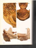
extend from the epithelial surfaces into the follicle lumina. The cytoplasm
contains regions with elaborate ergastoplasm 3 , Golgi complexes 4 and secretory granules. The cell on the right displays the entire nucleus. There is a
web of terminal bars (terminal complexes) at the apicolateral surfaces of epithelial cells, which border at the follicular lumen. A large C-cell (parafollicular cell) 5 is seen in the lower part of the figure. C-cells originate with the ultimobranchial body and have developed as part of the thyroid gland. Their
important attribute is the presence of secretory granules. The granules have
diameters of 100 to 180 nm and contain the hormone calcitonin. Calcitonin
consists of 32 amino acids.
264
1 Follicular epithelium
4 Golgi apparatus (complexes)
2 Colloid
5 C-cell
3 Ergastoplasm
6 Connective tissue
Electron microscopy; magnification: × 5520
Kuehnel, Color Atlas of Cytology, Histology, and Microscopic Anatomy © 2003 Thieme
All rights reserved. Usage subject to terms and conditions of license.
359
360
1
Endocrine Glands
3
2
6
4
5
3
6
3
361
2
1
4
3
4
1
5
6
Kuehnel, Color Atlas of Cytology, Histology, and Microscopic Anatomy © 2003 Thieme
All rights reserved. Usage subject to terms and conditions of license.
265
362
Thyroid Gland
This view into three sectioned thyroid follicles shows the polygonal follicular
epithelial cells. They are covered with short microvilli. There are residual secretory products.
1 Cut surface of the follicular epithelium
2 Vascularized connective tissue lining of the follicle (cf. Fig. 160)
Scanning electron microscopy; magnification: × 1800
363
Thyroid Gland—Capillary Network
An extraordinarily rich vascularization is the common characteristic of all
endocrine organs. A capillary network encircles the thyroid follicles (see
Fig. 360). This figure provides a vivid image of the capillary network at the
surface of feline thyroid follicles. An interlobular artery is seen in the lower
part of the figure. It continues in interfollicular vessels.
In this preparation, the vessel system of the thyroid gland was only partially
filled with resin. The capillary network is in reality much more elaborate.
Endocrine Glands
Corrosion preparation (cf. Fig. 303, 304); scanning electron microscopy; magnification: ×
250
364
Parathyroid Gland
The parathyroid glands (glandulae parathyroideae) consist of epithelial cell
clusters that are about as big as a wheat kernel. They are richly vascularized.
Fat cells and an occasional colloid-containing follicle interrupt the parathyroid tissue organization. Based on their affinity to dyes, three cell types can
be distinguished in light microscopy: 1, clear chief cells (lightly stained cells);
2, dark chief cells; and 3, oxyphilic cells (chromophilic cells, Welsh cells).
The parathyroid glands biosynthesize parathormone (PTH), which regulates
the levels of calcium and phosphate ions, including the blood calcium level.
1 Oxyphilic cell
2 Main light cell
3 Connective tissue
Stain: azan; magnification: × 400
266
Kuehnel, Color Atlas of Cytology, Histology, and Microscopic Anatomy © 2003 Thieme
All rights reserved. Usage subject to terms and conditions of license.
362
2
1
1
2
Endocrine Glands
363
364
2
1
3
2
3
267
Kuehnel, Color Atlas of Cytology, Histology, and Microscopic Anatomy © 2003 Thieme
All rights reserved. Usage subject to terms and conditions of license.
365
Pancreatic Islets of Langerhans
Clusters of endocrine cells are found in a sea of pancreatic exocrine cells.
They are clusters of vascularized epithelium, called the islets of Langerhans
(Paul Langerhans, 1847–1888), or simple islet cells. Exocrine pancreatic cells
and islet cells have different structures and stain differently (see Fig. 455, 457,
459). There are three types of islet cells, α-, β- and δ-cells (A-, B- and D-cells.
In figure (a), the α-cells 2 are stained red (their secretory product is glucagon) and the β-cells 3 are stained blue (they secrete insulin). About 60–80%
of the islet cells are β-cells, i.e., the insulin-producing cells predominate.
Figure (b) shows necrotic islet cells after parenteral administration of Alloxan.
There are still α-cells 2 at the periphery. The lighter stained regions represent necrotic β-cells 4 with almost completely degenerated nuclei. There are
acini 1 of the exocrine pancreas adjacent to the necrotic islet (cf. Fig. 366).
Endocrine Glands
1 Serous acinar cells, exocrine pancreas
2 α-cells (A-cells)
3 β-cells (B-cells)
4 Necrotic β-cells
5 Fat cells
Stain: a) Ivic; magnification: × 64; b) alum hematoxylin-eosin; magnification: × 100
366
Pancreatic Islets of Langerhans
Section of a human pancreas with an islet of Langerhans (cf. Fig. 18, 365). Islet
cells do not contain zymogen granules. Therefore, they always appear lighter
after staining than the exocrine cells. The acinar cells of the eccrine gland 1
contain secretory granules in their apical region. The acidophilic granules are
stained blue. There is loose connective tissue around the eccrine gland cells.
The connective tissue contains larger blood vessels.
1 Acini of the exocrine pancreas
2 Capillaries within the islet organ
3 Vessels of the exocrine pancreas
Semi-thin section; stain: methylene blue-azure II; magnification: × 400
367
Pancreatic Islets of Langerhans
The entire endocrine portion of the pancreas consists of Langerhans islets
(insulae pancreaticae). The islets consist of cords of cells, which form an irregular network. This network is extensively vascularized so that virtually
every islet cell is connected to the bloodstream. Five cell types can be defined
for the cell cords: α-, β-, δ-, PP- and D1-cells. α-, β- and δ-cells produce the following polypeptide hormones: α-cells secrete glucagon, β-cells secrete insulin and δ-cells secrete somatostatin. About 5–7% of the cells are δ-cells.
This figure shows the α-cells of a Langerhans islet using fluorescence-labeled
antibody to glucagon. The yellow fluorescence indicates that the α-cells
mostly reside at the islet periphery. There are also fluorescent α-cells in the
duct epithelium. None of the other islet cells are stained and neither is the
exocrine portion of the pancreas. The PP-cells biosynthesize the pancreatic
polypeptide.
268
Fluorescence microscopy, × 150
Kuehnel, Color Atlas of Cytology, Histology, and Microscopic Anatomy © 2003 Thieme
All rights reserved. Usage subject to terms and conditions of license.
365
1
2
1
3
4
2
2
5
1
a
b
1
366
2
1
2
3
Endocrine Glands
3
367
269
Kuehnel, Color Atlas of Cytology, Histology, and Microscopic Anatomy © 2003 Thieme
All rights reserved. Usage subject to terms and conditions of license.
368
Pancreatic Islets of Langerhans
There are at least five different cell types in the islets of Langerhans. Each
type biosynthesizes a different hormone (see Fig. 365, 367). This section
shows three cell types: α-cells, β-cells and δ-cells. The α-cells biosynthesize
glucagon. The average diameter of the hormone-containing granules (α-granules) is 300 nm. In electron microscopy, they show an electron-dense
center, which is surrounded by a narrow, less electron-dense halo. About 80%
of all islet cells are insulin producing β-cells 2 . Their secretory granules (βgranules) are surrounded by a membrane. The sizes of β-granules vary, and
they come in different geometrical forms (polygonal crystalloid). The granules always show a lighter halo. δ-Cells 3 occur mostly in the center of the
islet. They biosynthesize the hormone somatostatin. Their secretory granules
measure about 320 nm. Somatostatin containing secretory granules are not
as electron-dense as β-granules. Note the close proximity of islet cells and capillaries 4 .
Endocrine Glands
1 α-cells
2 β-cells
3 δ-cells
4 Capillaries
Electron microscopy; magnification: × 3100
369
Pancreatic Islets of Langerhans
δ-Cell (somatostatin cell) 1 from the Langerhans islet of a mouse. About 5% of
all islet cells are δ-cells. They are usually located at the end of an islet cell
cord. δ-Cell granules have diameters of about 320 nm. They are not as electron-dense as the granules from α- or β-cells, and do not have the characteristic light halo of β-granules (cf. Fig. 368). The content of the δ-cell granules is
either homogeneous or shows a very fine granulation.
Somatostatin inhibits the secretion of insulin and glucagon. Glucagon stimulates the release of pancreatic somatostatin. In contrast, insulin inhibits somatostatin release.
1 δ-cell
2 δ-cell granule
3 β-cells
4 Nucleus
Electron microscopy; magnification: × 10 000
270
Kuehnel, Color Atlas of Cytology, Histology, and Microscopic Anatomy © 2003 Thieme
All rights reserved. Usage subject to terms and conditions of license.
368
2
1
4
3
Endocrine Glands
1
2
369
2
3
1
4
2
271
Kuehnel, Color Atlas of Cytology, Histology, and Microscopic Anatomy © 2003 Thieme
All rights reserved. Usage subject to terms and conditions of license.
Digestive System
370
Oral Cavity—Nasal Cavity
Frontal section through the head of a human embryo. The crown–rump
length is 80 mm. The palate is closed.
1 Telencephalon
6 Middle nasal concha
2 Eye
7 Inferior nasal concha
3 Nasal septum
8 Palate
4 Nasal cavity
9 Tongue
5 Superior nasal concha
10 Oral cavity
11 Tooth germ—mandible (cf. Figs. 388–391)
Stain: Masson-Goldner trichrome; magnification: × 10
371
Lips—Labia
The outer lips are covered by skin 1 (pars cutanea, multilayered stratified
nonkeratinizing squamous epithelium), hair, sebaceous glands and sweat
glands. The mucous membrane 2 of the vestibulum oris (pars mucosa, multilayered stratified nonkeratinizing squamous epithelium, seromucous salivary
glands) covers the inner part of the lips. The transition from the outer to the
inner epithelium is made in the vermilion border 3 (pars intermedia). This
sagittal section of a human adult lip shows the characteristic epithelial
covering: the outer skin is on top, mucous membranes are in the lower part.
The vermilion border is on the right. A plate of connective tissue and striated
muscle fibers 4 ( orbicularis oris muscle) forms the middle part of the pecten.
The orbicular ring muscle abruptly turns outward in the region of the vermilion border (pars marginalis of the ring muscles) 4 . The fibers of the orbicular
ring muscle are cut vertical to their long axis. Note the clearly defined cell
groups of the seromucous labial glands 5 . The keratinization and pigmentation of the epithelium is marginal in the vermilion border. Therefore, the
color of blood shines through the epithelium. Note the thick multilayered
nonkeratinizing epithelium on the mucous membrane side of the lips. Hair
shafts and sebaceous glands are seen on the outer (skin) side 1 .
1 Skin, pars cutanea
4 Marginal part of orbicularis oris muscle
2 Mucous membrane, pars mucosa
5 Labial gland
3 Vermilion border
Stain: hematoxylin-eosin; magnification: × 15
372
Tongue—Lingua
This frontal section of the tongue from a human newborn shows the arrangement of the musculature (cf. 233). The dorsal tongue (dorsum linguae) is in
the top part of the figure. The larger vessels are filled with Indian ink. The
three-dimensional grid formed by the inner musculature can easily be discerned in the center of the figure. Distinct muscle fibers run in longitudinal
2 , transverse and vertical direction (M. longitudinalis, verticalis and transversus linguae; cf. Fig. 373).
272
1 Lingual artery
5 Geniohyoid muscle
2 Inferior longitudinal muscle of
6 Mucous membrane of the dorsal tongue
tongue
with lingual papilla
3 Genioglossus muscle
7 Aponeurosis of the tongue
4 Submandibular duct
Indian ink injection; stain: van Gieson picric acid; magnification: × 12
Kuehnel, Color Atlas of Cytology, Histology, and Microscopic Anatomy © 2003 Thieme
All rights reserved. Usage subject to terms and conditions of license.
370
1
1
2
2
4
3
5
6
7
8
9
10
11
11
371
1
3
4
5
Digestive System
4
2
372
6
7
1
2
3
4
Kuehnel, Color Atlas of Cytology, Histology, and Microscopic Anatomy © 2003 Thieme
All rights reserved. Usage subject to terms and conditions of license.
1
2
3
4
5
273
373
Lingual Papillae—Filiform Papillae
The mucous membranes everywhere on the tongue are a multilayered stratified squamous epithelium and may have many forms. The dorsal tongue displays raised mucous membrane epithelium called lingual papillae. These
come in all kinds of forms and structures. The base of the papillae is formed
by the lamina propria mucosae. This figure shows that the papillae subdivide
into secondary papillae, which appear toward the throat as arcuate, sometimes fimbriated lappets. The keratinized epithelium lappets of filiform
(thread-like) papillae 1 are constantly scuffed off and replaced. They give the
tongue the velvety appearance. Scuffed-off epithelial cells, along with the
mycelium of the fungus Leptothrix buccalis, form the tongue coating. The
lower part of the figure shows the regular pattern of the inner tongue musculature. The strong lingual aponeurosis 2 is located between mucous membrane and tongue muscles. The lingual aponeurosis forms a rigid (kinetically
stable) connection with the mucous membrane (cf. Fig. 372).
Feline tongue.
Digestive System
1 Filiform papilla
2 Lingual aponeurosis
3 Vertical muscle of the tongue
4 Transverse muscle of the tongue
Stain: alum hematoxylin-eosin; magnification: × 10
374
Lingual Papillae—Filiform and Vallate Papillae
Toward the throat oriented filiform papillae 1 of a feline tongue. The keratinized points are intact. Filiform papillae are well developed in many animals. They give their tongues a grating quality (see Fig. 373).
The vallate papillae 2 on the posterior third of the feline tongue are less
evenly distributed than on a human tongue. They are found between the
mostly parallel rows of filiform papillae, which will occasionally tower over
them. A moat-like groove surrounds the vallate papillae.
1 Filiform papillae (thread-like)
2 Vallate papillae (round, knob-like)
3 Circular wall
4 Moat-like groove, moat or furrow
Scanning electron microscopy; magnification: × 4 .5
375
Lingual Papillae—Vallate Papillae
The 6–12 vallate papillae at the border to the root of the tongue and before
the sulcus terminalis (groove) (papillae vallatae) barely rise over the level of
the tongue’s mucous membrane 1 . The wall-enclosed papillae are separated
from the wall by a narrow moat. The secretory ducts of the serous gustatory
glands 3 end at the bottom of the groove (moat). Their lobular structure is
shown in this figure. The ducts connect with the groove (moat) on both sides
of the papilla base 4 . The multilayered stratified squamous epithelium of the
grooves shows lighter spots. These correspond to taste buds (see Fig. 662).
274
1 Groove, moat
3 Ebner’s glands
2 Secretory ducts
4 Papilla base
Stain: alum hematoxylin-eosin; magnification: × 10
Kuehnel, Color Atlas of Cytology, Histology, and Microscopic Anatomy © 2003 Thieme
All rights reserved. Usage subject to terms and conditions of license.
373
1
2
3
4
374
2
3
4
2
1
Digestive System
1
375
1
4
2
3
1
2
3
275
Kuehnel, Color Atlas of Cytology, Histology, and Microscopic Anatomy © 2003 Thieme
All rights reserved. Usage subject to terms and conditions of license.
376
Lingual Papillae—Foliate Papillae
Human foliate papillae are well developed only in children. However, rabbits
possess dense fields of foliate papillae at the posterior lateral edges of the
tongue. This preparation shows rabbit foliate (leaf-like) papillae. The surface
folds are separated from each other by deep grooves (cf. Fig. 377). The foliate
papillae are supported by several parallel connective tissue cords 1 . The
multilayered stratified epithelium contains taste buds 2 (cf. Fig. 662). The
ducts of serous gustatory glands 3 end at the bottom of the deep grooves.
Two small groups of glands 3 are present in the subepithelial connective
tissue 4 . They are located in close proximity to the striated skeletal muscle
fibers 5 of the tongue.
1 Connective tissue cords
2 Taste buds
3 Serous gustatory glands
4 Lamina propria
5 Striated lingual musculature
Stain: van Gieson iron hematoxylin-picrofuchsin; magnification: × 80
Digestive System
377
Lingual Papillae—Foliate Papillae
The lingual vessels initially follow the patterns of the inner lingual musculature. Then, having traversed the tightly structured aponeurosis, they spread
into the mucous membrane. The vessel terminals form a densely woven network, which ends in a capillary web area. Each secondary papilla has a capillary loop 3 (cf. Fig. 376).
The lower part of the figure shows the striated lingual musculature 2 . Serous
glands have not been sectioned.
1 Multilayered stratified squamous epithelium
2 Lingual musculature
3 Capillary loop (ansa)
Stain: alum hematoxylin-eosin after injection with Indian ink; magnification: × 15
378
Lingual Glands—Posterior Lingual Glands
Groups of mucous glands (glandulae radices linguae) in considerable numbers exist in the area of the lingual root, radix lingua, under the lingual tonsil
(see Figs. 338, 339). The clustered mucous glands are not restricted to the
tunica mucosa but extend deeper between the muscle fiber bundles. The
glandular ducts end in the canals of the lingual follicles. Connective tissue
septa (trabeculae) (stained blue) and sectioned striated muscle fibers 1 are
present between the glandular lobes. The nuclei (stained red) in the basal cell
region and the cytoplasm with its honeycomb structure (cf. Fig. 130) are characteristic of mucous glands. The secretory ducts have wide lumina.
The paired seromucous anterior lingual gland (Nuhn’s gland) is located close
to the tip of the tongue.
1 Striated skeletal musculature of the tongue
2 Artery
Stain: azan; magnification: × 100
276
Kuehnel, Color Atlas of Cytology, Histology, and Microscopic Anatomy © 2003 Thieme
All rights reserved. Usage subject to terms and conditions of license.
376
2
1
4
2
1
3
5
3
377
3
3
2
Digestive System
1
378
1
2
277
Kuehnel, Color Atlas of Cytology, Histology, and Microscopic Anatomy © 2003 Thieme
All rights reserved. Usage subject to terms and conditions of license.
379
Parotid Gland
The parotid gland (glandula parotidea, or short parotis) is the largest human
salivary gland. It is a purely serous gland with long, branched secretory ducts.
The entire length of the secretory duct is always contained in the glandular
lobe. Groups of fat cells 2 occur between the wide serous acini 1 . The nuclei
of serous acinar cells are round and located in the basal cell region. Their cytoplasm is finely granulated (see Fig. 129). A long intercalated duct 3
traverses the center of the image from top left to bottom right. There are also
cross-sectioned intercalated ducts 4 . The ducts are lined by an isoprismatic
(cuboid) epithelium (see Figs. 380, 381).
1 Serous acini
2 Fat cells
3 Intercalated duct, cut longitudinally
4 Intercalated duct, cross-sectioned
Stain: alum hematoxylin-eosin; magnification: × 200
Digestive System
380
Parotid Gland
When paraffin sections are stained using the Masson-Goldner method, they
will clearly render the cone or pyramid-shaped cell groups of the acini 1 and
their narrow lumina. The acinar cell cytoplasm contains slightly acidophilic
granules (cf. Figs. 379, 381). A sectioned salivary or striated duct 2 is visible
in the lower right half of the image. The oval nuclei of the pseudostratified
columnar epithelial cells are located in the central or basal cell regions. A
basal striation is visible underneath the nuclei (see Fig. 90). It is caused by involutions of the basal plasmalemma and densely packed mitochondria between the membrane pleats (cf. Fig. 91). A longitudinally section of an intercalated duct 3 is visible in the right part of the figure, another duct is crosssectioned 4 (cf. Figs. 379, 381). Note the sparse connective tissue (stained
green) and the numerous adipocytes 5 .
1 Serous acini
2 Salivary duct, cross-sectioned
3 Intercalated duct, cut longitudinally
4 Intercalated duct, cross-sectioned
5 Adipocytes
Stain: Masson-Goldner trichrome; magnification: × 200
381
Parotid Gland
The epithelium of serous acini 1 consists of nearly pyramid-shaped cells
with central or basal cell nuclei. The supranuclear or apical cell region of the
serous acinar cells is filled with secretory granules (stained blue). The number of granules varies with the secretory activity. A cross-sectioned intercalated duct 2 is shown in the center of the figure. Spindle-shaped, flat
myoepithelial cells (cf. Figs. 379, 380) surround it. There are fibrocytes and
capillaries in the loose connective tissue between acini. Compare with Figs.
127 and 129.
278
1 Serous acinus
2 Intercalated duct with myoepithelial cells
Semi-thin section; stain: methylene blue-azure II; magnification: × 200
Kuehnel, Color Atlas of Cytology, Histology, and Microscopic Anatomy © 2003 Thieme
All rights reserved. Usage subject to terms and conditions of license.