Bài giảng Rối loạn nhịp thất trong nhồi máu cơ tim cấp - ThS. Hoàng Việt Anh
Bạn đang xem bản rút gọn của tài liệu. Xem và tải ngay bản đầy đủ của tài liệu tại đây (9.24 MB, 44 trang )
Hà Nội 26/11/2016
RỐI LOẠN NHỊP THẤT
trong nhồi máu cơ tim cấp
ThS.BS Hoàng Việt Anh
Trưởng phòng Q3B - Viện Tim mạch quốc gia Việt Nam
Nhồi máu cơ tim
• Là bệnh lý tim mạch có tỷ lệ mắc ngày càng tăng và tỷ lệ tử vong cao
• Tỷ lệ mới mắc tại Hoa Kỳ: 735.000 người/năm
• Yếu tố nguy cơ: Tăng huyết áp, ĐTĐ2, Rối loạn lipid máu, Béo phì, ít vận
động thể chất, cuộc sống căng thẳng….
Rối loạn nhịp là một biến chứng của NMCT
Reference
to Ventricular
MYOCAItDIAL
to Ventricular
%
No.
Episodes-
16
Dallas,
14
2 INTRODUCTION3
Tachycardia*
mean
E. with
JONES,
Total F.C.C,P,,**
M.D.,
F.Inferior
A. BASHOUR,
Anterior
Tachycardia*
Reference
LESION
10
SGOT
of 213 units per ml,
M.D.f
AND
R. EDMONSON, tachycardia
M.D.
true
ventricular
Texas
per
ml.
In
the
absence
of ventricular
cardia,generally the accepted,
mean becauseSGOTof the sequence
rise
the ligation
of coronary
with of events
acute following
myocardial
infarction
i
Cardiac
Arrhythmias
in Acute
Myocardial
T Infarction
BASHOUR,
M.D.,
F.C.C,P,,** Supraventricular
E.
JONES,
M.D.f
ANDper ml. R.
EDMONSO
cardial
infarction
changed
tachycardia
4
1
5has
16.6
units
The
number
of patie
in with
years;
in the
II. Incidence
of the Common
Arrhythmias
3 remained 10
Complete
AV block
0 the past 303 Special
The
reported
incidence
of
cardiac
arshock
or
heart
failure
on
admission
neighborhood
of 30
cent.
In the maMultifocal
PVC’s
16
14
30
100
jority
of instances,
death
occurred
in
Reference
to Ventricular
Tachycardia*
to
demonstrate
a
close
relationship
Voiume
Si, No. S
Ectopic Dallas,
ventricular
first 48Texas
50 per cent
rhythms:
clinical
features
and
the a
of these patients,
death
is sudden
and un- these
May,
1967
myocardial
infarction.
The development
of
a) True
ventricular
expected.
These
patients
are clinically
well of ectopic
ventricular
rhythms.
Atrial
F. A.
fibrillation
1
HE
MORTALITY
RATE
IN
ACUTE
MYO-
arteries
little
in dogs,
tricular
it
that
arrhythmia
this
group
and
fibrillation.
dies
of ven-
per
rhythmias
the
hours.
Approximately
the
F. A.
olume
ay,
ine
No.
51,
INCIDENCE
1967
was
or
S
used
T
atrial
alone
in
arrhythmia.
Cardiac
wo groups
myocardial
one
other
T
arrhythmias
were
OF
BASHOUR,
M.D.,
COMMON
and
patient
one
inferior
into
Texas
myocardial
infarction)
bigeminy
in
the bigeminal
INTRODUCTION
one of these,
R.
JONES,
Dallas,
INTRODUCTION
and ventricular
divided
E.
F.C.C,P,,**
ARRHYTHMIAS
M.D.f
tachycardia AND (rate
than
523 greater
than
100/mm)
b) Slow ventricular
rhythm
(rate
from
70-100/mm)
11 patients.
In
*One
patient accepted,
had
generally
rhythm
lastedventricular
rhythms.
atEDMONSON,
the time of death
struction
*From
HE
M.D. sufficient
without
myocardial
tissue
the 7 terminal 15 event,
to 8 explain
the
Department
at
been
University
Southwestern
Professor
Medical
generally
f5*Present
address
1
Medicine.
of
6in
intern
-
to
widely.
the
20
at
this
In
groupthe run
dies of
one,
byfibrillation.
ventricular
This
frequency
rhythm
for
cardiac
Dilantin
of
the true
patients
continuous
The
fold:
acute
recording
incidence
and
of
the evaluation
patients. arrhythmiaThe
cardiac
the
purpose
are
to
of
reportedthe
describe
the
of the
that abnormal
occur
(Dila
becau
3.
beats
were
ventricular
these
the bi
this
period.
encountered
study
was
in Table
incidence
of
ous
premature
in
is
recording
with
incardiac
the immediatearrhythmias
post-infarction
Stan- group
variation
of
rhythmwas has used
permitted prophylactically
the study
of
consecutive
types
of
the
School.
Medicine
ford
University.
slow
Resident
in and
Medicine. rapid
ectopic
Part
of
this
work
was
presented
at the Texas
Heart
Association
Meeting,
Texarkana
(February 2, 1965)
and
the Midwestern
Section,
American
Federation
for Clinical
Research,
Chicago,
November,
l965.’
both
cardiac
methods
de-
necropsy
50It has
Medicine,
of
due
hydantoin
accepted,
because
of the sequence
the of
ligationevents
of coronary
agent
followingin
of Texas
**Associate
of events
following
HE
MORTALITY
three RATE
hours. IN TrueACUTEventricular
tachycardia
to MORTALITY
the location
of the
RATE
IN MYOACUTE
MYOthat
was present
in 15 patients,little and
the arteries
incianterior
and inferior infarction
ventricular in dogs,
tachycardia.
cardial
has changed
according
infarction:
of
varied
largely
in
threeseri-
the
cardiac
arrhythmia,
ventricular
presence
of ventricular
to correlate
tachycardia
a preliminary
of the disease,
and
the use of diphenyl-
13
known patients
clinical
three.
of ven-
and
features
report
on
premature
The
the
with
ventricular
tricular little
arrhythmia
andarteries
in dogs, that thi
past 30 years;
it remained
in the
cardialin theassociatedinfarction
has changed
MATERIAL
TheThe reported
incidenceoxaloaceticof cardiac
arneighborhood
of 30 per cent.
In the maserum
glutamic
transwere grouped
tricular
arrhythmia
and
rhythmias
varied
widely.
This
variation
is
(SGOT)
in the pastjority 30of instances,
years; death it occurred
remained
in the
in the
largely
due to the frequency
of recording
first 48 hours.
Approximately
in 50 per cent
Patients incidence
with
The
reported
The neighborhood
commonly
encountered
cardiac of ar- 30
the
cardiac
rhythm
of
patients
with
acute
per
cent.
In
the
maof these patients,
death
is sudden
and unmyocardial
infarction.
The development dence
of
of widely.
an
patients
are clinicallyarrhythmia.
well
for the ventricular
The slow
ibrillation
appeared
in expected.
three
patients These
(or
rhythmias
varied
jority
of
instances,
death
occurred
in
the
methods
for
continuous
recording
of the
at the
time ofre- death
without
sufficient
de10 per cent),
with a rapid
ventricular
cardiac
rhythm
has permitted
the study were
of
struction
of myocardial
tissue
at necropsy
largely
due
to studied.
the frequ
first 48 hours.
Approximately
in
50
per
cent
the
true
incidence
and
the
evaluation
of
the
to explain
the terminal
event,
It has been
1:arrhythmia
Avionics’ that monitoring
system.
The
unit,
of the FIGURE
cardiac
occur
30 portable
episodes
of
the
cardiac
rhythm
ner.
The recorder
is the small
unit
in of
the
of these patients,
death
is sudden pertypes
and
unin minthe immediate
post-infarction
period.
cardiocharter.
Medicine.
(referred
The
purpose
of this myocardial
study
was threewhom
had anterior
and one inferior
myoinfarction. to be
fThese
Stanexpected.
patients
are
clinically
well
cardial
infarction.
fold:pre- to describe
the incidence
of the seritored
for a pe
ous cardiac
arrhythmia, methods
to correlate
the
for
continuous
at the time of death
withoutat
sufficient
depresenceof
of ventricular
tachycardia
with
stances,
m
cardiac
rhythm
hasthe perm
known
clinical
features
of the disease,
and
struction
of myocardial
tissue
at necropsy
a preliminary
report
on the use of diphenylthe true incidence
and
in the anterior
to explain
the terminal
event,
It has been
types
of of
the thecardiacheart arrh
*From
the Department
of
Medicine,
University
Downloaded From: on 11/15/2016
in the immediate
diaphragmatic post-inf
of Texas
Southwestern
Medical
School.
(or posterior).
nvolvement
Arrhythmias
of the
lateral
with
either
dence
with
myocardial
previous
terior
wall
cardia
are reported
in
Table
more
ectopic
ponse
in two
patients.
Wandering
maker
was observed
in 11 patients.
ure
atrial
beats
were
frequent
in
**Associate
Present
observed
pacePremafour pathe tachyDepartment
*From
supraventricular
of Texas four Southwestern
in five patients,
of
Paroxysmal
occurred
Multifocal
ventricular
were seen in all patients.
was
or
1. Atrial
beats
in
three
Professor
address
-
six
and
per
ranged
minute.
ventricular
ventricular
PVC’s
infarction. fibrillation
tachyaminase
one
and
70. The
in 27
between
100 and
blood
In five patients,
two
foci
were
was
(Fig.
followed
4).
rhythm
patients,
five of whom
one inferior
myocardial
ute. In one patient,
both types
intern
in Medicine
at
rhythm
were
present.
Runs
multifocal,
number.
Atrial
cardia
levels
patients.
levels
The
was
peak
correlated
were
determined
rise
of
with
SGOT
the
ap-
were
were
tient
respectively.
was
observed
had
ventricular
quite
r
and
nod
fibrillation
noted
in
two
and
Ventricular
in four
i
t
patients,
ach
two
fibrillation
on
responsible
was
present
in
had
anterior
infarction.
The
attacks
numbered
between
one and
a singleof Medicine,
patient
and University
the
ventricular
ranged Medical
between School.
72 and
90 beats
of
three
an-
of ventricular
between
rate
between
myocardial
of attacks
varied
ventricular
distributed
inferior
number
220
rhythmias
equally
and
The
location
depending
on whether
the anterior or the
nferior
wall
was predominantly
involved.
ients.
cardia
was
though
ten
in
rate
FIGURE
1:
Avionics’
ner.
The recorder
is
monitoring
the small
system.
The
unit,
portable
unit
in the
of ventricular
of three
ford
University.
premature
beats Medicine. mature
ventricular
beats
interrupted
Resident
in
normally
conducted
beats Texas
were
seen in
R/TPart phenomenon
of
this
work
was
presented
the
Meeting,
Texarkana
(Febru(twoHeart had Association
anterior
patients;
12
of these
patients
had
runs
ary 2, 1965)
and
the Midwestern
Section,
American
Federation
for Clinical
Research,
Chicago,
November,
l965.’
on
center
cardiocharter.
520
by
16
Downloaded From: on 11/15/2016
V.W.
(PMH
283525)
1-28-65
the
right
of
the
side
picture.
of
the
On
picture,
the
represents
left
side
the
is
the
scan-
eLectro-
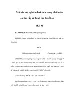
Table 1. Occurrence of arrhythmias in STEMI patients during and
Tỷ lệ mắc các rối loạn nhịp thất trong ACS
immediately after primary PCI.71
Accelerated idioventricular rhythm (50-120 b.p.m.)
15-42%
Sinus bradycardia (<50 b.p.m.)
28%
Non-sustained VT
26%
Sinus tachycardia (>100 b.p.m.)
22%
Atrial fibrillation
High-degree AV block
9%
5-10%
Sustained VT
2-4%
VF
2-5%
AV: atrioventricular; b.p.m.: beats per minute; PCI: percutaneous
coronary intervention; STEMI: ST-elevation myocardial infarction;
VF: ventricular fibrillation; VT: ventricular tachycardia
Tỷ lệ mắc các rối loạn nhịp thất theo vùng NMCT
International Journal of Contemporary Medical Research Volume 3 | Issue 5 | May 2016 | ICV: 50.43 |
Sinh lý bệnh
của rối loạn
nhịp thất trong
NMCT cấp
Piccini et al
Rối loạn nhịp thất sau NSTEMI
Sustaine
EARLY ACS trial
Fi
ta
fib
w
de
Piccini et al Sustained VT/VF After NSTE ACS.
Circulation July 3, 2012
These variables included the same covariates as in the 30-day
Table 1. More patient
Các YTNC của rối loạn nhịp thất bền bỉ trong NMCT cấp
• Sốc tim hay tổn thương cấp động mạch vành lớn (VD:
thân chung ĐMV trái)
• Chậm trễ trong tái thông ĐMV
• Tái thông không được hay không hoàn toàn ĐMV thủ
phạm do vấn đề kỹ thuật hay giải phẫu khó khăn
• Có suy chức năng thất trái hay sẹo cơ tim do NMCT cũ
hay ST do bệnh cơ tim trước đó
• Bệnh cơ tim rối loạn nhịp do di truyền
Willich and Goette. Int J Crit Care Emerg Med 2015, 1:2
Rối loạn nhịp thất sau NMCT cấp
• RL nhịp thất hay gặp hơn trong STEMI so với NSTEMI
(gấp 4 lần)
• STEMI: 90% xảy ra trong 48 giờ đầu
• NSTEMI: 60% xảy ra sau 48 giờ
• Tỷ lệ xuất hiện: NNT không bền bỉ (13%), NNT bền bỉ
(3%) và rung thất (3%)
• Một nghiên cứu khác: 6%
• Tại viện Tim mạch Việt Nam: 13% NNT, 48% NTT thất
trong 24h sau can thiệp STEMI.
Willich and Goette. Int J Crit Care Emerg Med 2015, 1:2
Tiên lượng của NMCT có rối loạn nhịp thất
• Tổng hợp 4 nghiên cứu lớn: GUSTO, PURSUIT,
PARAGON A, PARAGON B
• Tổng cộng 26.416 bệnh nhân NSTEMI
• Có 552 bệnh nhân có rối loạn nhịp thất trong thời gian
nằm viện (nhịp nhanh thất và/hoặc rung thất): 2.1%
• Tỷ lệ tử vong sau 30 ngày và sau 6 tháng đều tăng ở
nhóm có rối loạn nhịp thất.
Sustained Ventricular Arrhythmias Among Patients With Acute Coronary Syndromes With No ST-Segment Elevation – Al Khatib et
al – Circulation July16,2002
tions was unavailable, we could not explore their relationships with outcomes.
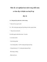
Tiên lượng của NMCT có rối loạn nhịp thất
Sustained Ventricular Arrhythmias Among Patients With Acute Coronary Syndromes With No ST-Segment Elevation – Al Khatib et
al – Circulation July16,2002
Kaplan-Meier curves of mortality by ventricular arrhythmia.
Tiên lượng của NMCT có rối loạn nhịp thất
• Rung thất sớm trong vòng 48 giờ sau NMCT: tử vong
tăng 5 lần so với NMCT không có rung thất
• Trên những bệnh nhân NMCT sau can thiệp ĐMV, nếu
có NNT/RT: tỷ lệ tử vong tăng 4 lần, biến cố tim mạch
tăng 3 lần, thời gian nằm viện tăng 50%.
Sustained Ventricular Arrhythmias Among Patients With Acute Coronary Syndromes With No ST-Segment
Elevation – Al Khatib et al – Circulation July16,2002
The American Journal of Medicine, Vol 121, No 9, September 2008
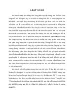
Tử vong tăng khi có rung thất
120
FAST-AMI 2005
A
B 60
60
50
In-hospital mortality, %
50
In-hospital mortality, %
W. Bougouin et al.
40
30
20
40
30
20
10
10
0
0
Patients without VF
Patients with VF
No VF
Early VF
Late VF
Figure 1 In-hospital mortality according to occurrence of ventricular fibrillation.
Table 3 Cause-specific death during hospitalization
European
H
Journal
(2014)
35,
116–122
according
toeart
occurrence
of VF
Table 4 Treatments at hospital discharge among
survivors by occurrence of VF
Các yếu tố dự báo NNT bền bỉ sau NMCT cấp
The American Journal of Medicine 2008 121, 797-804DOI: (10.1016/j.amjmed.2008.04.024)
Copyright © 2008 Elsevier Inc. Terms and Conditions
Tử vong và RLNT tăng theo số yếu tố nguy cơ
The American Journal of Medicine 2008 121, 797-804DOI: (10.1016/j.amjmed.2008.04.024)
Copyright © 2008 Elsevier Inc. Terms and Conditions
Tử vong tăng ở nhóm can thiệp thất bại
The American Journal of Medicine 2008 121, 797-804DOI: (10.1016/j.amjmed.2008.04.024)
Copyright © 2008 Elsevier Inc. Terms and Conditions
Làm thế nào phòng ngừa và điều trị
rối loạn nhịp thất à giảm tử vong
trong NMCT cấp ???
nal of the American College of Cardiology
013 by the American College of Cardiology Foundation, the American Heart Association, Inc.,
the Heart Rhythm Society
ished by Elsevier Inc.
V
ISSN
/>
PRACTICE GUIDELINE
2012 ACCF/AHA/HRS Focused Update Incorporated
Into the ACCF/AHA/HRS 2008 Guidelines for
Device-Based Therapy of Cardiac Rhythm Abnormalities
A Report of the American College of Cardiology Foundation/American Heart Association
Phòng ngừa và điều trị
đột tử trong hội chứng
vành cấp ngoại viện
may reveal disease-specific findings are detailed in Web Table 4.
of these conditions must be avoided. Co-morbidities that may
Phác đồ xử trí RL nhịp thất theo nguyên nhân
Clinical History
• Angina pectoris or shortness of breath
• Family history of premature SCD (age <40 years) or ealy-onset heart disease
• ECG during tachycardia
Acute ischemia
(STEMI, NSTEMI)
ECG
Echocardiogram
History and
Family history a
Urgent angiogram
and
revascularisation
Reverse
transient cause
Evaluate for
cardiovascular
diseases
• ECG
• Echocardiogram / CMR
• History
• Other tests
Evaluate for
complete
reversal of
cause
Secondary
prevention for
SCD (ACEi,
beta-blockers, statin,
antiplatelets)
Re-evaluate
LVEF
6–10 weeks
after event
Consider ICD
according to
secondary
prevention
Structural heart disease
and congenital heart
diseases
suspected (e.g. Stable CAD,
sarcoidosis, aortic valve
disease, DCM)
Sudden death victims
• Autopsy in collaboration with pathologists
• Obtain blood and tissue samples
• Molecular autopsy after autopsy
• Offer family councelling and support
• Refer family for cardiology / SCD workup
No detectable
heart disease
Inherited
arrhythmogenic
disease or
cardiomyopathy
suspected
Further patient assessment, e.g.b
• Stress test, Holter 48 hours,
• Consider coronary angiogram
• Refer patients to experienced centers for risk evaluation,
catheter ablation, drugs and ICD
• Drug challenges, EPS
• CMR, CT, myocardial biopsy
• Signal averaged ECG, TOE based on suspected disease
• Treatment of underlying
heart disease (e.g. valve
repair, medication)
• Assess risk for SCD
Specific treatment
• Genetic testing
• Family screening
• Assess risk for SCD
Consider to
obtain second opinion
on cause of
VT/VF
Downloaded from by guest on November 21, 2016
Other transient
cause e.g.
• Drugs
• Electrolytes
• Chest trauma
ment of sustained VA.
014
e drugs
n of the
nts after
ompared
largely
in ACS.
ong carcacy on
did not
ejection
f dronemortality
treated
interval
none of
eatment
for this
Phác đồ điều trị rối loạn nhịp thất trong ACS
tructure
Recurrent VT/VF and Electrical Storm in ACS
Cardioversion/defibrillation
Overdrive pacing
Attempt complete revascularization
Treat ischaemia
Correct electrolyte imbalance
β-blocker therapy
Deep sedation
Recurrent VT/VF
Electrical Storm
Amiodarone
Lidocaine
Consider catheter ablation
Amiodarone
Consider ICD reprogramming
Consider catheter ablation
Consider LVAD implantation
EuroIntervention
2014;10-online
publish-ahead-of-print
August 2014
Phòng ngừa đột tử
trong hội chứng vành
cấp trong viện:
Tái thông ĐMV
Phòng ngừa đột tử
trong hội chứng
vành cấp trong viện:
Tái thông ĐMV
ment
C
Điều trị rối loạn nhịp thất và mục tiêu
FIGURE 52-29 Cardiac rupture syndromes complicating STEMI. A, Anterior myocardial rupture in an acute infarct. B, Rupture of the ventricular septum. C, Complete
rupture of a necrotic papillary muscle. (From Schoen FJ: The heart. In Kumar V, Abbas AK, Fausto N [eds]: Robbins & Cotran Pathologic Basis of Disease. 7th ed. Philadelphia,
WB Saunders, 2005.)
TABLE 52-12 Cardiac Arrhythmias and Their Management During Acute Myocardial Infarction
CATEGORY
1. Electrical
instability
ARRHYTHMIA
OBJECTIVE OF TREATMENT
THERAPEUTIC OPTIONS
Correction of electrolyte deficits and
increased sympathetic tone
Prophylaxis against ventricular fibrillation,
restoration of hemodynamic stability
Urgent reversion to sinus rhythm
Observation unless hemodynamic function
is compromised
Search for precipitating cause (e.g., digitalis
intoxication); suppress arrhythmia only if
hemodynamic function is compromised
Potassium and magnesium solutions,
beta blocker
Antiarrhythmic agents, beta blocker;
cardioversion/defibrillation
Defibrillation; amiodarone, lidocaine
Increase sinus rate (atropine, atrial pacing);
antiarrhythmic agents
Atrial overdrive pacing; antiarrhythmic agents;
cardioversion relatively contraindicated if
digitalis intoxication present
Sinus tachycardia
Reduce heart rate to diminish myocardial
oxygen demands
Atrial fibrillation and/or
atrial flutter
Paroxysmal
supraventricular
tachycardia
Reduce ventricular rate; restore sinus
rhythm
Reduce ventricular rate; restore sinus
rhythm
Antipyretics; analgesics; consider betablocking agent unless congestive heart
failure present
Verapamil, digitalis glycosides; amiodarone;
treat heart failure; cardioversion
Vagal maneuvers; verapamil, cardiac
glycosides, beta-adrenergic blocking
agents; cardioversion
Sinus bradycardia
Acceleration of the heart rate only if
hemodynamic function is compromised
Acceleration of the sinus rate only if loss of
atrial “kick” causes hemodynamic
compromise
Ventricular premature
beats
Ventricular tachycardia
Ventricular fibrillation
Accelerated idioventricular
rhythm
Nonparoxysmal AV
junctional tachycardia
2. Pump failure/
excessive
sympathetic
stimulation
3. Bradyarrhythmias
and conduction
disturbances
Junctional escape rhythm
AV block and
intraventricular block
Atropine; atrial pacing
Atropine; atrial pacing
Insertion of a pacemaker
Modified from Antman EM, Rutherford JD (eds): Coronary Care Medicine: A Practical Approach. Boston, Martinus Nijhoff, 1986, p 78.
imaging (Fig. 52-30) or by insertion of a pulmonary artery balloon
Rupture of a Papillary Muscle