3D culture and analysis of the expression of cancer stem cell markers from gastric cancer cell line MKN45
Bạn đang xem bản rút gọn của tài liệu. Xem và tải ngay bản đầy đủ của tài liệu tại đây (428.57 KB, 8 trang )
JOURNAL OF MEDICAL RESEARCH
3D CULTURE AND ANALYSIS OF THE EXPRESSION
OF CANCER STEM CELL MARKERS FROM GASTRIC
CANCER CELL LINE MKN45
Ngo Thu Ha¹, Le Thi Thanh Huong¹, La Thi Huyen², Nguyen Phu Hung¹,³
¹Thai Nguyen University of Sciences
²Institute of Biotechnology - Vietnam Academy of Science and Technology
³French National Institute of Health and Medical Research U1235, Nantes, France
Gastric cancer is the fourth leading cause of worldwide cancer mortality, Vietnam belongs to the
specified high-risk group of developing gastric cancer. Gastric cancer stem cells (CSCs) are considered
to be the origin of gastric adenocarcinoma. 3D cell culture is an important step of identification of stem
cell as well as CSC. In this study, a non-adherent cell culture model (3D) was developed with a culture
medium that had growth factors selective for the development of gastric CSCs from gastric cancer
cell line MKN45. The result showed that only 6% single cancer cells developed into tumorspheres
during culture process. Then, flow cytometry and immunofluorescence analysis was carried out
to evaluate the expression of CSC markers CD44 and ALDH in samples taken from tumorspheres
which arose from gastric CSCs. The results showed that gastric CSCs formed tumorspheres in 3D
culture condition with a high expression of 92% and 40,1% for markers CD44 and ALDH, respectively.
Keywords: 3D culture, gastric cancer stem cell, CD44, ALDH, MKN45
I. INTRODUCTION
Cancer is the leading cause of death
globally. Gastric cancer has the fourth highest mortality, accounting for 723,000 deaths
in 2012 according to World Health Organization [1]. Vietnamese are at high risk of
developing gastric cancer. Important risk
factors for development of gastric cancer include smoked foods, salted fish and meat,
Corresponding author: Nguyen Phu Hung,
Thai Nguyen University of Sciences
Email:
Received: 10 July 2017
Accepted: 09 Octorber 2017
JMR 111 E2 (2) - 2018
family history, chronic gastritis, smoking and
Helicobacter pylori (H. pylori) infection [2].
Epidemiological studies show the close relationship between chronic atrophic gastritis
and H. pylori infection [3]. In atrophic gastritis, the reduction of parietal cells and chief
cells lead to decrease HCl secretion which
is a favorable environment for H. pylori development. H. pylori overgrowth increases
genetic mutation in epithelial cells of stomach which can cause gastric cancer [4].
Lauren's gastric cancer classification
system subdivided gastric adenocarcinoma
into two main types: intestinal type accounts
53
JOURNAL OF MEDICAL RESEARCH
for 54% and diffuse type is about 32%. The
remaining 14% belongs to indeterminate
type (mixture of two main types above) [5;
6]. While intestinal type tumors are typically
found in the gastric antrum of older individuals, diffuse type tumors are more common
in the gastric corpus of younger individuals
[7; 8].
Recent research indicated that although
cancer stem cells (CSCs) frequency in tumors is low, CSCs are responsible for proliferation and differentiation of tumor cells.
Gastric stem cells which are located in the
isthmus or progenitor zone of gastric gland
and differentiate into mucous, parietal, chief
and endocrine cells can be considered the
origin of gastric CSCs [9]. In in-vitro experiments, if one cell can form tumorsphere in
non-adherent culture condition (3D) with
medium containing growth factors, it can
qualify as a CSCs.
Cancer cells grown in 3D cell culture
condition were more similar to cells in tumors when compared with cells that were
cultured in adherent (2D) condition [10].
CSC identification and culture are important
parts of cancer research; single cell culture
to form tumorspheres (3D culture) is a significant tool in CSCs culture.
CD44 and ALDH are two popular markers used for evaluation and isolation of
CSCs in many different types of cancers
[11]. In gastric adenocarcinoma, these
markers expression identifies gastric cancer stem cell markers and is related to drug
tolerance of tumor [12]. There are many
different methods to identify expression of
CD44 and ALDH markers, but Flow cytometry and immunofluorescence are common
54
methods in CSC studies [13].
CD44 (Cluster of differentiation 44), a
receptor of hyaluronic acid, was used to
identify gastric CSCs for the first time by
Takaishi et al. in 2009 [14]. ALDH (aldehyde
dehydrogenase) is an important marker of
CSCs in solid tumors and gastric carcinoma
in particular. It is involved in cell detoxification, regulation of cellular proliferation, and
relates to drug tolerance in cancer [15].
Studies on stem cells and CSCs have
not been done previously in Vietnam, and
3D cell culture is an emerging technique in
cancer research in general and gastric cancer in particular. In this study, MKN45 gastric cancer cells were seeded in 3D culture
condition. The existence of gastric CSCs
was evaluated by measuring tumorsphere
formation and the expression of CD44 and
ALDH gastric cancer stem cell markers.
CD44 and ALDH expression was measured
using flow cytometry and immunofluorescence analysis.
II. MATERIALS AND METHODS
1. Materials: Gastric cancer cell line
MKN45
Gastric cancer cell line MKN45 was a
generous gift from Inserm U1053 Laboratory – French National Institute of Health and
Medical Research in Bordeaux. Cells were
maintained in RPMI media supplemented
with 10% Fetal Bovine Serum (FBS), and
1% ampicillin/streptomycin (P/S). The seeding density was 1 x 10⁵ cells/well in 3.8 cm²
wells using adherent cell culture condition
(2D). Cells were incubated at 37oC in the
presence of 5% CO2. After 2 days, culture
JMR 111 E2 (2) - 2018
JOURNAL OF MEDICAL RESEARCH
medium was replaced. After 4 days, cells
were passaged into a new culture well.
2. Methods
3D cell culture method
To create non-adherent culture plates,
12 well culture plate (area of 3,8 cm² for
each well) from BD Bioscience were supplemented with 500 µM Poly-HEMA solution
(Poly(2-hydroxy-ethyl methacrylate from
Sigma) and 10 mg/ml of concentration. Culture plates were then dried surface in clean
benches overnight. Plate were washed
3 times using PBS 1X buffer before commencing cell culture.
3D culture: Trypsinized cells were collected from 2D cell culture plate and diluted in PBS 1X buffer until reaching the
concentration 1000 cell/µl. A 2 µl solution
containing 2000 cells was resuspended in
2 ml of specific stem cell culture medium in
a non-adherent plate (treated by Poly-HEMA). This medium contains basic medium
DMEMF12/Glutamax (from Invitrogen) supplemented with 1% P/S, Epithelial Growth
Factor (EGF) 20 ng/ml, Fibroblast Growth
Factor (FGF) 20 ng/ml, glucose 0.3%, and
insulin 5 µg/ml (all components from Sigma). 1 ml of medium was replaced with new
medium each 2 days. Cells were incubated
at 37oC, 5% CO2. The formation of tumorspheres was evaluated for from the 5th day
to 14th day of the culture process. Tumorspheres were collected for cell analysis.
Immunofluorescence
Collected tumorspheres were centrifuged at 1300 rpm for 3 min and washed
2 times with PBS 1X buffer. Tumorsphere
were stained with anti-human CD44-PE
JMR 111 E2 (2) - 2018
monoclonal antibody 1:50 (from BD Biosciences) and with ALDH substrate 1:100
in 500 µl ALDEFLUOR buffer (ALDEFLUOR kit from Stem cell Technology) at 37oC
for 20 min. Tumorspheres were washed 2
times with ALDEFLUOR buffer by centrifugation. The second wash used ALDEFLUOR buffer contains 10 µg/ml Hoechst 33342
for nuclei staining (from Sigma).
After tumorspheres were incubated with
CD44 monoclonal antibody or stained with
ALDH substrate in ALDEFLUOR kit, images
were taken using NIKON fluorescence microscope at magnification of 200 and 400
times with specialized software.
Flow cytometry analysis
Tumorspheres collected on the 10th day
of culture were trypsinized and separated
into single cells. Single cells were incubated with CD44-APC specific antibody (from
BD Biosciences) and specific ALDH substrate in ALDEFLUOR kit (from Stem cell
technology, Canada) according to manufactory instruction for 20 min at 37oC. Samples
were washed 2 times by ALDEFLUOR buffer (500 µl for each) before being placed in
specialized 4 ml glass tube (from BD Bioscience) and analysed by FACS Canto II flow
cytometry system (BD - Biosciences) at the
compatible wavelength of APC (Allophycocyanin) and FITC (Fluorescein isothiocyanate) for CD44 and ALDH, respectively.
20,000 events recorded in flow cytometry
were analyszed using DIVA 6.1 software.
3. Ethics
All MKN45 cells were provided by Inserm U1053 Laboratory – French National
Institute of Health and Medical Research in
55
JOURNAL OF MEDICAL RESEARCH
Bordeaux. There is no intervention to patients and animals.
III. RESULTS
1. Formation of tumorsphere from
MKN45 single cell in 3D cell culture
condition
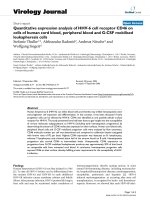
Images of the tumorsphere formation
and percentage of MKN45 single cell that
formed tumorspheres is presented in Figure
1. The result showed that only 6% ± 1,2%
single cancer cells developed into tumorspheres by the 5th days of culture process.
This means that for every 100 single cultured cells, only 6 of them have ability to
form tumorsphere.
Figure 1. Tumorsphere formation from a single cell of gastric cancer cell line MKN45
A) Tumorsphere formation at the 1st, 5th and 12th day of observation
B) Tumorsphere forming percentage (estimated in the 5th day of culture process)
Scale bar (to estimate tumorsphere size): 50µm.
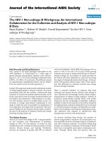
2. The expression of CD44 and ALDH using immunofluorescence method
As shown in Figure 2, the proportion of CD44 marker labeled with red fluorochrome was
high at 90%. Similarly, the proportion of ALDH maker labeled with green fluorochrome was
50%. The immunofluorescence image indicated that CD44 was expressed in the cell membrane, whereas ALDH was located in the cytosol.
Figure 2. Tumorsphere immunofluorescence after 7 days of culture process
CD44 (red), ALDH (green) and Hoechst (blue). Scale bar: 50µm.
56
JMR 111 E2 (2) - 2018
JOURNAL OF MEDICAL RESEARCH
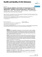
3. The expression of CD44 and ALDH using Flow cytometry analysis
To determine correctly the number of cells expressing CD44 or ALDH and to confirm the
immunofluorescence result above, we used flow cytometry analysis. The result is shown in
Figure 3. The dot blot data in P7 range shows that most MKN45 gastric cancer cell expressed
CD44 at a proportion of 92,0% ± 2,7% (Figure 3A(b)). On the other hand, the expression of
ALDH was 40,1% ± 2,5%, much lower than CD44 (Figure 3B(b)).
Figure 3. Result of Flow cytometry analysis
(A) for CD44 and (B) for ALDH marker. (a) Control (cells only)
(b) Sample (CD44 antibody or ALDEFLUOR™ reagent added)
IV. DISCUSSION
In the 2D cell culture, the connection between cells occurs on one side of the cell (on
the surface of cultured plate). However, in 3D culture condition, cell attachments occur all
around the surface of the cell. This influences the cell proliferation and differentiation [16].
The roles of Epithelial Growth Factor (EGF) and Fibroblast Growth Factor (FGF) in stem cell
self-renewal have been demonstrated [17; 18]. Therefore, the single cancer cells which were
cultured in medium supplied with the above growth factors should have self-renewal capacity
allowing cancer stem cells to form tumorspheres. Our result showed that only 6% ± 1,2%
single cancer cells formed tumorspheres, this is compatible with the report of Jianming et al.
in 2013 about tumorsphere formation of gastric cancer cell line MKN45 [19].
Tumor is comprised of different cells which present distinct phenotypic and functional
profiles. The heterogenous tumorsphere which arose from a single cell in 3D cell culture
conditions indicate that different tumor cells can originate from a single unique stem cell.
JMR 111 E2 (2) - 2018
57
JOURNAL OF MEDICAL RESEARCH
The result of immunofluorescence analysis
showed 20% of the colored cells displayed
the nucleus dyed Hoechst (blue). Hoechst,
also called bis-Benzimide, is a cytotoxic organic compound and tends to bind to double-strained AT-rich regions of DNA [20].
Hoechst has been used to identify side populations - which comprise stem cell-like cells
exhibiting specific markers - in research of
stem cell in multiple mammalian species
and many different tumor types [21; 22].
Cells have the ability to be unstained by this
dye because they are able to actively pump
Hoechst out of the cell. Besides, ALDH is
a significant enzyme in cell detoxification
through oxidation of aldehydes to carboxylic
acids, ALDH participates in ABC (ATP binding cassette) transport system. In addition,
side population occupies only a small proportion of tumor cells, but it contains a large
amount of CSC [23], and ALDH+ cells did
not incorporate the Hoechst dye, whereas
ALDH is a selective gastric cancer stem cell
marker. It claims that tumorsphere comprises a large proportion of gastric cancer stem
cell and these can exclude chemicals out
of the cell through ABC membrane transport proteins. The lower expression level of
ALDH in comparison with CD44 shown in
Flow cytometry analysis result indicate that
ALDH is more selective than CD44 (40,1%
± 2,5% in comparison with 92,0% ± 2,7%),
this also is compatible with previous research [14; 15].
V. CONCLUSION
By culturing MKN45 gastric cancer
cells in 3D condition to form tumorspheres
that have in-vivo tumor characteristics,
58
we demonstrated that gastric cancer cell
populations include a small rate of gastric CSCs. These gastric CSC had the capacity of tumorsphere formation. Besides,
immunofluorescence and flow cytometric
results confirmed tumorspheres contain a
large amount of gastric CSC, as measured
by gastric CSC markers CD44 and ALDH.
These results show 3D cell culture is a good
model for gastric CSC assays. The expression level of CD44 and ALDH marker was
confirmed by using both immunofluorescence and flow cytometry analysis simultaneously. The results from the 2 methods
indicated consistent expression proportions
for each marker. In Vietnam, 3D cell culture has not been widely used in research,
therefore deploying this method will contribute to later studies assessing the effects of
drugs on tumor cells.
Acknowledgements
This work was supported by Research
Grant 55/B2017-TNA-55 from the Ministry
of Science and Technology in Vietnam.
REFERENCES
1. Ferlay J, Soerjomataram I, Ervik M,
et al. (2013). GLOBOCAN 2012 cancer incidence and mortality worldwide: IARC cancerbase No. 11. Lyon, France. International
Agency for Research on Cancer.
2. El-Omar E.M., Carrington M., Chow
W.H., et al (2000). Interleukin-1 polymorphisms associated with increased risk of
gastric cancer. Nature, 404, 398 - 402.
3. Weck M.N., Gao L., Brenner H
(2009). Helicobacter pylori infection and
chronic atrophic gastritis: associations acJMR 111 E2 (2) - 2018
JOURNAL OF MEDICAL RESEARCH
cording to severity of disease. Epidemiol,
Camb, Mass, 20, 569 - 574.
4. Haruma K., Mihara M., Okamoto E.,
et al (1999). Eradication of Helicobacter pylori increases gastric acidity in patients with
atrophic gastritis of the corpus-evaluation of
24-h pH monitoring. Alimentary Pharmacology and Therapeutics, 13, 155 - 162.
5. Kumar V., Abbas A.K., Fausto N., et
al (2010). Pathologic Basic of Disease 8th
edition. Saunders elsevier, Philadelphia,
784 - 786.
6. Polkowski W., van Sandick J.W., Offerhaus G.J., et al (1999). Prognostic value
of Laurén classification and c-erbB-2 oncogene overexpression in adenocarcinoma of
the esophagus and gastroesophageal junction. Ann Surg Oncol, 6, 290 - 296.
7. Henson D.E., Dittus C., Younes M.,
et al (2004). Differential trends in the intestinal and diffuse types of gastric carcinoma in
the United States, 1973 - 2000: increase in
the signet ring cell type. Arch, Pathol, Lab,
Med, 128, 765 - 770.
8. Noda S., Soejima K., Inokuchi K.
(1980). Clinicopathological analysis of the
intestinal type and diffuse type of gastric
carcinoma. Jpn, J. Surg, 10, 277 - 283.
9. Mills J.C., Shivdasani R.A. (2011).
Gastric epithelial stem cells. Gastroenterology, 140, 412 - 424.
10. Vinci M., Gowan S., Boxall F., et
al (2012). Advances in establishment and
analysis of three-dimensional tumor spheroid - based functional assays for target validation and drug evaluation. BMC Biol, 10,
29.
11. Adams A., Warner K., Pearson A.T.,
et al (2015). ALDH/CD44 identifies uniqueJMR 111 E2 (2) - 2018
ly tumorigenic cancer stem cells in salivary
gland mucoepidermoid carcinomas. Oncotarget, 6 (29), 26633.
12. Kang L., Zeng D., Yu-Qiang N.
(2014). Gastric cancer stem cells in gastric
carcinogenesis, progression, prevention
and treatment. World J Gastroenterol, 20
(18), 5420 - 5425.
13. Prince M.E., Sivanandan R.,
Kaczorowski A., et al (2007). Identification
of a subpopulation of cells with cancer stem
cell properties in head and neck squamous
cell carcinoma. Proceedings of the National
Academy of Sciences, 104 (3), 973 - 978.
14. Takaishi S., Okumura T., Tu S., et al
(2009). Identification of gastric cancer stem
cells using the cell surface marker CD44.
Stem cell Dayt, Ohio, 27, 1006 - 1020.
15. Nguyen P.H., Giraud J., Chambonnier L., et al (2017). Characterization of
Biomarkers of Tumorigenic and Chemoresistant Cancer Stem Cells in Human Gastric
Carcinoma. Clin Cancer Res Off J Am Assoc Cancer Res, 23 (6), 1586 - 1682.
16. Singhvi R., Kumar A., Lopez G.P.,
et al (1994). Engineering Cell Shape and
Function. Science, 264 (5159), 696 - 698.
17. Herbst R.S. (2004). Review of epidermal growth factor receptor biology. International Journal of Radiation Oncology,
Biology, Physics, 59 (2 Suppl), 21 - 26.
18. Coutu D.L., and Jacques G. (2011).
Roles of FGF signaling in stem cell self-renewal, senescence and aging. Aging (Albany NY), 3 (10), 920.
19. Jianming L., Lilin M., Junfei X., et
al (2013). Spheroid body-forming cells in
the human gastric cancer cell line MKN-45
process cancer stem cell properties. Int J
59
JOURNAL OF MEDICAL RESEARCH
Oncol, 42 (2), 453 - 459.
20. Portugal J., Waring M. J. (1988).
Assignment of DNA binding sites for 4′, 6-diamidine-2-phenylindole and bisbenzimide
(Hoechst 33258). A comparative footprinting study. Biochimica et Biophysica Acta
(BBA)-Gene Structure and Expression, 949
(2), 158 - 168.
21. Goodell M.A., Brose K., Paradis G.,
et al (1996). Isolation and functional properties of murine hematopoietic stem cells that
are replicating invivo. J. Exp. Med., 183,
60
1797 – 1806.
22. Goodell M.A., Rosenzweig M., Kim
H., et al (1997). Dye efflux studies suggest
that hematopoietic stem cells expressing
low or undetectable levels of CD34 antigen
exist in multiple species. Nat. Med., 3, 1337
– 1345.
23. Brown M.D., et al (2010). Identification of a cancer stem cell enriched side
population using Hoechst 33342 based isolation. Cancer Stem Cells
JMR 111 E2 (2) - 2018