Ebook Feigenbaum’s echocardiography (8/E): Part 2
Bạn đang xem bản rút gọn của tài liệu. Xem và tải ngay bản đầy đủ của tài liệu tại đây (35.77 MB, 1,624 trang )
Chapter 13
Infective Endocarditis
Infective endocarditis remains a challenging and often fatal condition. One
reason for this is the difficulty of establishing an accurate diagnosis,
particularly early in the course of the disease when proper management can
be lifesaving. As therapeutic approaches have become more successful, the
importance of early and accurate diagnosis is self-evident. Unfortunately, no
single test or finding establishes the diagnosis in all cases. Instead, a
constellation of findings that constitutes the diagnostic criteria continues to
evolve.
The central role that echocardiography plays in the diagnosis of
endocarditis began in the early 1970s with the echocardiographic
demonstration of a valvular vegetation by the M-mode technique. With the
advent of two-dimensional and Doppler modalities, echocardiography has
become virtually indispensable in the diagnosis and management of these
patients. Today, echocardiographic findings are a central part of the
diagnostic criteria for infective endocarditis.
CLINICAL PERSPECTIVE
Despite improvements in therapy, infective endocarditis remains a potentially
lethal disease with an incidence of 4 to 8/100,000 patient-years. Although the
overall incidence has not increased appreciably over time, several factors
have contributed to substantial recent changes in the epidemiology of the
disease. For example, Staphylococcus aureus is now the most common cause
of endocarditis in most series, in part due to an aging population and the
increasing prevalence of intracardiac devices, including prosthetic valves,
conduits, pacing wires, and indwelling catheters. This has led to the concept
of “healthcare contact” as a recognized risk factor for the development of
infective endocarditis. Currently, approximately 25% of infective
endocarditis cases in this country are attributable to a previous medical event
or procedure, such as implantation of a prosthetic valve or pacemaker. More
recently, the opioid epidemic that has plagued the United States has been
associated with a striking increase in the incidence of drug-dependence–
associated endocarditis. Such patients appear to have a particularly poor
prognosis, in part due to the likelihood of recurrent infection following valve
replacement therapy.
Infective endocarditis is defined as a localized infection anywhere on the
endocardium, including the chamber walls, vessels, and within congenital
defects. The vast majority of vegetations, however, occur on valve leaflets.
Infection may also develop on any implanted or prosthetic material such as
prosthetic valves, conduits, pacing electrodes, and catheters. The process of
developing endocarditis occurs in the setting of bacteremia or fungemia. The
initiating event usually requires the presence of a high-velocity jet, which
may be due to a congenital anomaly such as a ventricular septal defect, a
regurgitant valve, or a prosthetic valve. It is thought that the jet interferes
with the protective endothelial surface, allowing the blood-borne pathogens
to adhere and coalesce. As the nidus of infection organizes, masses of
microorganisms attract platelets, fibrin, and other materials and become
adherent to the endothelial surface to form a vegetation. The vegetation will
grow in size, either as a sessile clump or as a highly mobile and even
pedunculated mass with the potential for embolization. As the hallmark of
endocarditis, the ability to detect the vegetation is the focal point of
diagnosis. This sequence of events offers a mechanism for development of
endocarditis in patients with underlying heart disease. However, since as
many as 50% of patients who get endocarditis do not have lesions associated
with a high-velocity jet, some other set of conditions must be operational in
these patients to explain the link between bacteremia and cardiac
involvement.
ECHOCARDIOGRAPHIC CHARACTERISTICS OF
VEGETATION
The versatility of echocardiography in the evaluation of endocarditis is
illustrated in Table 13.1. Among its important functions is the identification
of underlying heart disease known to increase a patient’s risk of infection.
Although the absence of underlying disease does not confer protection
against endocarditis, particular conditions, such as congenital heart disease,
bicuspid aortic valve, and a myxomatous mitral valve, are known risk factors.
At the same time, these conditions often confound the diagnosis of
endocarditis by creating abnormalities that mimic or conceal
echocardiographic evidence of infection.
An essential first step in the echocardiographic evaluation is to search for
evidence of acute ongoing infection. Although there are several
manifestations of endocarditis, including abscesses and fistulae, the most
common and direct evidence of infective endocarditis is the vegetation.
Because a vegetation begins as a microscopic focus of infection and
gradually grows into a conspicuous mass, its presence may or may not be
evident on an imaging study. Thus, echocardiography must be sensitive
enough to detect the vegetation and specific enough to distinguish it from
other echocardiographic abnormalities or artifacts. As can be seen in Table
13.2, certain echocardiographic features can be used to either increase or
decrease the probability that a visualized mass is due to endocarditis. A
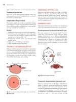
vegetation is typically irregularly shaped, highly mobile, and attached to the
free edge of a valve leaflet. They tend to develop on the upstream side of the
valve, that is, the ventricular side of the aortic valve and the atrial side of the
mitral valve (Fig. 13.1). Vegetations may be sessile or pedunculated but
usually have motion that is independent of the valve itself. Figure 13.2 is an
example of endocarditis involving the tricuspid valve in a patient with a
history of intravenous drug use. The infectious process can be seen encasing
the valve leaflets and chordae. Severe tricuspid regurgitation was present.
Because vegetations often occur in the path of a high-velocity jet, their
motion is frequently described as oscillating or fluttering. The presence of
significant mobility, or oscillating motion, is a classic feature of most
vegetations. In fact, the absence of mobility argues against the diagnosis and
should suggest the possibility of an alternative diagnosis, including a healed
vegetation. The shape and size of vegetations are quite variable and may
either increase (due to progression of disease) or decrease (due to healing or
embolization) over time (Fig. 13.3). Fungal vegetations tend to be larger than
those caused by bacterial infections, and those involving the tricuspid valve
tend to be larger compared with vegetations that affect the aortic or mitral
valve (Fig. 13.4).
Table 13.1
COMPREHENSIVE ROLE OF ECHOCARDIOGRAPHY IN
PATIENTS WITH ENDOCARDITIS
Initial/early role:
Identifies predisposing heart disease
Early assessment in all cases of suspected IE
Detects complications
Assesses hemodynamic consequences
Serial evaluation (assesses efficacy of therapy)
Intraoperative assessment of extent of disease
Prognosis (risk of complications)
Establishes new baseline after therapy
Repeat/follow-up role:
TEE (after positive TTE) in patients at high risk for complications
Repeat TEE (after negative initial TEE) if clinical suspicion persists
Repeat TEE if suboptimal course during therapy (e.g., clinical deterioration, persistently
positive blood cultures, worsening physical examination)
FIGURE 13.1. An example of a large, mobile vegetation on the aortic valve that
fills the left ventricular outflow tract, seen from the parasternal long-axis (A) and
the apical views (B).
Video 13-1a
Video 13-1b
Table 13.2
ECHOCARDIOGRAPHIC CRITERIA FOR DEFINING A
VEGETATION
Positive Feature
Negative Feature
Low reflectance
High echogenicity
Attached to valve, upstream side
Nonvalvular location
Irregular shape, amorphous
Smooth surface or fibrillar
Mobile, oscillating
Nonmobile
Associated tissue changes, valvular regurgitation
Absence of regurgitation
Although typically attached to a valve, vegetations may also attach to
chordae, chamber walls, or any foreign body, such as a pacemaker lead,
indwelling catheter, or prosthetic valve sewing ring. Figure 13.5 is an
example of endocarditis involving a porcine tricuspid valve as well as the
pacing wire that extends through it. The mass itself is typically homogeneous
with echogenicity similar to that of the myocardium. However, vegetations
can occasionally be cystic or appear more dense and calcified. The infectious
process often alters valve structure and function. As a result, some degree of
regurgitation is associated with most cases of acute endocarditis. In Figure
13.6, a patient with an aortic valve vegetation is shown. The involvement is
extensive, and the valve is partially flail. There is severe aortic regurgitation.
A patient with significant mitral regurgitation associated with an extensive
aortic valve vegetation is shown in Figure 13.7. Despite the presence of
severe mitral regurgitation, neither a vegetation nor leaflet perforation were
demonstrated. Figure 13.8 shows a patient with intravenous drug use who
presented early in the course of their endocarditis. Small vegetations were
noted on the aortic, mitral, and tricuspid valves. These were not fully
appreciated on transthoracic imaging.
FIGURE 13.2. Extensive infection involving the right heart from a patient with a
history of intravenous drug use. The vegetation involves the tricuspid valve
(arrow) and chordae (arrowhead).
Video 13-2
FIGURE 13.3. A: An example of disease progression from a patient with positive
blood cultures, mild aortic regurgitation, and a sclerotic aortic valve but no definite
vegetation (day 1). B: Two weeks later (day 14), a small vegetation is seen on the
valve (arrow). C: By day 22, the vegetation has increased in size (arrow) and
there is severe aortic regurgitation, despite antibiotic therapy.
Video 13-3a
Video 13-3b
Video 13-3c
FIGURE 13.4. A very large fungal vegetation in an immunocompromised patient
is shown. Extensive involvement of the mitral valve is demonstrated in the apical
long-axis (A) and four-chamber (C) views. In B, the dimensions of the mass are
recorded.
Video 13-4a
Video 13-4c
FIGURE 13.5. Transesophageal echocardiography shows a large mass (large
arrow) attached to a pacemaker lead, most likely an infected thrombus. There are
also multiple small vegetations (small arrows) attached to the pacemaker wire in
the right atrium.
Video 13-5
If the process results in destruction of underlying tissue leading to a flail
or perforated valve structure, the degree of regurgitation will be severe. For
example, if the infection leads to disruption of the aortic valve, severe aortic
regurgitation will ensue. This is demonstrated in Figure 13.9, taken from a
patient with a severely disrupted aortic valve in the setting of staphylococcal
endocarditis. Figure 13.10 is an example of a small perforation of the
noncoronary cusp of the aortic valve due to infection. Mild aortic
regurgitation was present, but no definite vegetation was identified. Another
example of a perforated valve due to S. aureus infection is shown in Figure
13.11. This patient had a large, highly mobile aortic valve vegetation. A
perforation at the base of the anterior mitral leaflet is also demonstrated,
along with severe mitral regurgitation. Much less often, a large vegetation
will obstruct the valve orifice, leading to a functional form of valve stenosis
(Fig. 13.12).
Although most vegetations involve the valves, in some cases the infection
may extend to other structures, such as the chamber wall. Figure 13.13 shows
an unusual vegetation attached to the posterior wall of the aortic root.
Multiple vegetations involving the aortic valve are also present.
It should be emphasized that there is no single characteristic on the
echocardiogram that will conclusively identify a mass as a vegetation. The
ability to detect a vegetation depends on vegetation size, location, the
presence of underlying heart disease, image quality, and instrument settings.
All available echocardiographic windows should be used, and Doppler flow
mapping should be performed to identify any associated valvular
regurgitation. Often nonstandard imaging planes are needed for detection,
especially for smaller vegetations. Although masses as small as 2 mm have
been reported, in most cases, a vegetation must be at least 3 to 6 mm in size
to be reliably seen. Image quality will also influence our ability to visualize
small structures. As is discussed later, these are areas in which the advantages
of transesophageal echocardiography have been demonstrated.
To avoid false-positive results, vegetations must be differentiated from
other echo-producing abnormalities, such as myxomatous processes,
degenerative changes (including Lambl excrescences and calcification),
tumors, thrombi, and imaging artifacts. Figure 13.14 is taken from a patient
who was asymptomatic. The large mitral valve mass could easily be mistaken
for a vegetation. However, the absence of clinical signs of infection suggests
an alternative diagnosis. In this case, the mass was a blood cyst. Underlying
heart disease both obscures the presence of a vegetation and increases the
likelihood of false-positive findings through misinterpretation. Figure 13.15
is from a patient with mitral valve prolapse who had fever due to a viral
syndrome. The prolapsing scallop was incorrectly interpreted as a vegetation.
The correct diagnosis was established based on absence of signs of infection
and direct comparison of the current echocardiogram to a prior study.
Nonbacterial thrombotic endocarditis (Libman–Sacks) is also easily confused
with infective endocarditis. In Figure 13.16, two examples of nonbacterial
thrombotic endocarditis are shown. In both cases, nodular masses on present
on the free edge of the mitral leaflets that could easily be misinterpreted as
bacterial vegetations. Distinguishing between these two conditions can be
very difficult and must rely on clinical correlation.
Thus, the accuracy of echocardiography is greater in patients without
underlying valve disease. Furthermore, active vegetations must be
differentiated from old or healed vegetations. Some studies have suggested
that vegetations tend to become smaller and more circumscribed and
echogenic over time as part of the healing process (Fig. 13.17). Although this
is generally true, a reduction in vegetation size might also suggest
embolization. Thus, distinguishing active from healed vegetations can never
rely on echocardiography alone but must take into account clinical factors.
FIGURE 13.6. Transesophageal echocardiography demonstrates a vegetation
(arrow) on a partially disrupted aortic valve (A). In B, severe aortic regurgitation is
present (arrow).
Video 13-6a
Video 13-6b
FIGURE 13.7. Panels A and B demonstrate extensive and diffuse involvement of
the aortic valve (arrows). Although a mitral valve vegetation was not seen,
Doppler shows severe mitral regurgitation (arrows, C).
Video 13-7a
Video 13-7b
Video 13-7c
FIGURE 13.8. Multi-valve involvement of Staphylococcus aureus endocarditis is
shown. Transesophageal echocardiography demonstrated small vegetations
(arrows) on the aortic (A), mitral (B), and tricuspid valves (C).
Video 13-8a
Video 13-8b
Video 13-8c
FIGURE 13.9. Another example of Staphylococcus aureus endocarditis is
provided. A shows a prolapsing or disrupted aortic cusp (arrow). In B, a
vegetation on the aortic valve is apparent (arrows). Color (C) and continuouswave Doppler (D) demonstrate severe aortic regurgitation. Note the steep slope of
the spectral Doppler signal (arrows).
FIGURE 13.10. A transesophageal echocardiogram demonstrates a small
perforation of the noncoronary cusp of the aortic valve. A: Focal thickening is
seen but no definite vegetation. B: Color Doppler imaging demonstrates the jet
extending through the cusp (arrows). C: A short-axis view confirms the location of
the perforation (arrow).
Video 13-10
FIGURE 13.11. A large aortic valve vegetation (A, arrow) and a perforated
anterior mitral valve leaflet (B, arrow) from a patient with intravenous drug use. In
C, severe mitral regurgitation is demonstrated with color Doppler (arrows).
Video 13-11a
Video 13-11b
FIGURE 13.12. A: A large vegetation involving the anterior mitral leaflet (arrows).
B: Spectral Doppler imaging recorded a 10 mm Hg mean gradient across the
mitral valve.
FIGURE 13.13. A transesophageal echocardiogram demonstrates an aortic valve
vegetation (arrow) and a second vegetation adherent to the posterior aortic wall
(arrowhead).
Video 13-13
DIAGNOSTIC ACCURACY OF ECHOCARDIOGRAPHY
Numerous clinical studies have tested the accuracy of echocardiography to
detect vegetations and other manifestations of acute endocarditis. A limitation
of all these studies is the difficulty in defining the standard by which the
diagnosis is established. In most series, a clinical standard for diagnosis was
used that incorporated clinical findings, blood culture results, response to
therapy, and outcome measures. Although practical, this approach has
obvious limitations and very likely permitted the inclusion of some patients
who had bacteremia but never had endocarditis. More rigorous diagnostic
standards that required pathologic and/or surgical confirmation must, by
definition, exclude patients who have endocarditis but never come to either
surgery nor autopsy. As a result, only the “sickest of the sick” would be
included in such series. Finally, the recognition over time of the fundamental
involvement of echocardiography in establishing a diagnosis made it
increasingly difficult to “test the test.” That is, it becomes impossible to
establish the accuracy of a test (in this case, echocardiography) that is
fundamentally involved in the definition of disease. For all these reasons, the
exact sensitivity and specificity of the various echocardiographic techniques
must be interpreted in context. Despite these limitations, the overall utility of
echocardiography as an integral part of the diagnostic algorithm is well
established.
FIGURE 13.14. An example of a blood cyst (arrow) is demonstrated within the
mitral valve. Diastolic (A) and systolic (B) frames are shown. Such an appearance
could easily be confused with vegetation.
Video 13-14
FIGURE 13.15. This echocardiogram was recorded from a patient with mitral
valve prolapse and significant mitral regurgitation. The mitral valve was
myxomatous and partially flail. A: The prolapsing valve is indicated by the arrows.
B: Severe mitral regurgitation is demonstrated (arrow). C: A transesophageal
echocardiogram demonstrates the prolapsing scallop (arrows). This could easily
be mistaken for a vegetation.
Video 13-15
FIGURE 13.16. Two examples of nonbacterial thrombotic endocarditis are
shown. In A, from a patient with metastatic cancer, masses on the tips of the
mitral leaflets are noted (arrows). B is from a patient with systemic lupus, and
demonstrates a nodular mass on the free edge of the anterior mitral leaflet
(arrow).