Ebook GENOSYS–exam preparatory manual for undergraduates biochemistry: Part 2
Bạn đang xem bản rút gọn của tài liệu. Xem và tải ngay bản đầy đủ của tài liệu tại đây (9.53 MB, 95 trang )
Chapter
7
Nutrition
Vitamins are essential organic compounds that the animal
organism is not capable of forming itself, although it requires them in small amounts for metabolism. Most vitamins are precursors of coenzymes; in some cases, they are
also precursors of hormones or act as antioxidants.
FAT-SOLUBLE VITAMINS
Transport from Liver to Tissues
The vitamin A from liver is transported to peripheral tissues as trans-retinol by the retinol-binding protein (RBP).
The fat-soluble vitamin A is present only in foods of animal
origin. However, its provitamin carotenoids are found in
plants. All the compounds with vitamin A activity are referred as retinoids. They include retinol, retinal and retinoic acid.
Vitamin A
1. Wald’s visual cycle: Rhodopsin is a conjugated protein present in rods. It contains 11-cis-retinal. The aldehyde group (of retinal) is linked to amino group of
lysine (opsin).
The primary event in visual cycle, on exposure to light, is the isomerization of 11-cis-retinal
to all-trans-retinal (Fig. 7.1). This leads to a conformational change in opsin, which is responsible
for the generation of nerve impulse. The all-transretinal is immediately isomerized by retinal isomerase (retinal epithelium) to 11-cis-retinal. This
combines with opsin to regenerate rhodopsin and
complete the visual cycle. However, the conversion of
Biochemical Role of Vitamin A
Fat-soluble vitamins are vitamin A, vitamin D, vitamin E
and vitamin K. Their general properties include:
1. Their precursors are called provitamins and are found
in plants.
2. They are absorbed from gastrointestinal (GI) lumen in
the presence of lipids and are emulsified with the bile.
3. They are stored in liver and adipose tissue.
4. Large doses for a long duration cause hypervitaminosis.
VITAMINS
1. Beta-carotene is cleaved by a dioxygenase to form retinal. The retinal is reduced to retinol by a nicotinamide
adenine dinucleotide (NADH) or nicotinamide adenine dinucleotide phosphate (NADPH)-dependent
retinal reductase present in the intestinal mucosa. Intestine is the major site of absorption.
2. The absorption is along with other fats and requires
bile salts. In biliary tract, obstruction and steatorrhea,
vitamin A is reduced.
3. Within the mucosal cell, the retinol is re-esterified
with fatty acids, incorporated into chylomicrons and
transported to liver. In the liver stellate cells, vitamin A
is stored as retinol palmitate.
Absorption of Vitamin A
Nutrients are the constituents of food, necessary to sustain
the normal functions of the body. All energy is provided by
three classes of nutrients namely fats, carbohydrates, proteins, and in some diets and ethanol. The intake of these
energy-rich molecules is larger than that of the other dietary nutrients. Therefore, they are called macronutrients.
Those nutrients needed in lesser amounts, vitamins and
minerals are called micronutrients.
/>Cha-7-Nutrition.indd 71
30-01-2015 14:02:57
72
Section 1: Theories
Fig. 7.1: Wald’s visual cycle (GDP, guanosine diphosphate; GMP,
cyclic guanosine monophosphate; GTP, guanosine triphosphate;
pi, inorgamic phosphate).
all-trans-retinal to 11-cis-retinal is incomplete. Therefore, most of the all-trans-retinal is transported to the
liver and converted to all-trans-retinol by alcohol. The
all-trans-retinol undergoes isomerization to 11-cisretinol, which is then oxidized to 11-cis-retinal to participate in the visual cycle.
2.Rods and cones: The retina of the eye possesses two
types of cells, which are called rods and cones. The
rods are in the periphery, while cones are at the center
of retina. Rods are involved in dim light vision whereas
cones are responsible for bright light and color vision.
3.Dark adaptation time: When a person shifts from a
bright light to a dim light, rhodopsin stores are depleted
and vision is impaired. However, within a few minutes,
known as dark adaptation time, rhodopsin is resynthesized and vision is improved. Dark adaptation time is
increased in vitamin A deficient individuals.
4.Color vision:
a. Cones are responsible for vision in bright light as
well as color vision. They contain the photosensitive protein and conopsin.
Cha-7-Nutrition.indd 72
b. There are three types of cones, each is characterized by a different conopsin that is maximally sensitive to blue (cyanopsia), green (iodopsin) or red
(porphyropsin).
c. In cone proteins also, 11-cis-retinal is the chromophore. Reduction in number of cones or the cone
proteins will lead to color blindness.
5.Other biochemical functions:
a. Retinol and retinoic acid function almost like steroid hormones. They regulate the protein synthesis
and thus are involved in the cell growth and differentiation.
b. Vitamin A is essential to maintain healthy epithelial
tissue. This is due to the fact that retinol and retinoic acid are required to prevent keratin synthesis
(responsible for horny surface).
c. Retinyl phosphate synthesized from retinol is necessary for the synthesis of certain glycoprotein,
which is required for growth and mucous secretion.
d. Retinol and retinoic acid are involved in the synthesis of transferring the iron transport protein.
e.Vitamin A is considered to be essential for the
maintenance of proper immune system to fight
against various infections.
f. Cholesterol synthesis requires vitamin A. Mevalonate, an intermediate in the cholesterol biosynthesis
is diverted for the synthesis of coenzyme Q in vitamin A deficiency.
g. Carotenoids (most important beta-carotene) function as antioxidants and reduce the risk of cancers
initiated by free radicals and strong oxidants. Betacarotene is found to be beneficial to prevent heart
attacks. This is also attributed to the antioxidant
property.
Recommended Daily Allowance
The recommended daily allowance (RDA) of vitamin A for:
• Children: 400–600 mg/day
• Men: 750–1,000 mg/day
• Women: 750 mg/day
• Pregnancy: 1,000 mg/day.
One international unit is 0.3 mg of retinol. One retinol
equivalent to 1 mg of retinol or 6 mg of beta-carotene.
Dietary Sources of Vitamin A
Animal sources include milk, butter, cream, cheese, egg
yolk and liver. Fish liver oils (cod liver oil and shark liver oil) are very rich sources of the vitamin A. Vegetable
sources contain the yellow pigments of beta-carotene.
Carrot contains significant quantity of beta-carotene.
Papaya, mango, pumpkins and green leafy vegetables
30-01-2015 14:02:57
Chapter 7: Nutrition
(spinach, amaranth) are other good sources for vitamin A
activity. During cooking, the activity is not destroyed.
Mnemonic
Increased vitamin A makes you ‘HARD’:
• Headache/Hepatomegaly
• Anorexia/Alopecia
• Really painful bones
• Dry skin/Drowsiness
Deficiency Manifestations
Vitamin D (Cholecalciferol)
Vitamin D is fat soluble. It resembles steroids in structure
and function like a hormone.
Fig. 7.2: Bitot’s spots
Biochemical Role of Vitamin D
Calcitriol (1, 25-DHCC) is the biologically active form of vitamin D. It regulates plasma levels of calcium and phosphate.
Calcitriol acts at three different levels (intestine, kidney
and bone) to maintain plasma calcium (normal 9–11 mg/
dL) as follows:
1. Action of calcitriol on the intestine: Calcitriol increases the intestinal absorption of calcium and phosphate.
In the intestinal cells, calcitriol binds with a cytosolic
receptor to form a calcitriol receptor complex. This
complex then approaches the nucleus and interacts
with a specific DNA, leading to synthesis of a specific
calcium-binding protein. This protein increases the
calcium uptake by the intestine. The mechanism of
action in calcitriol on the target tissue (intestine) is
similar to the action of a steroid hormone.
2. Action of calcitriol on the bone: In the osteoblasts
of bone, calcitriol stimulates calcium uptake
for deposition as calcium phosphate. Thus calcitriol is essential for bone formation. The bone
is an important reservoir of calcium and phosphate. Calcitriol, along with parathyroid hormone,
Hypervitaminosis of vitamin A include dermatitis (drying
and redness of skin), enlargement of liver, skeletal decalcification, tenderness of long bones, loss of weight, irritability, loss of hair, joint pains, etc.
Activation of vitamin D:
1. Vitamin D is a prohormone. The cholecalciferol is
first transported to liver, where hydroxylation at 25th
position occurs, to form 25-hydroxy-cholecalciferol
(25-HCC).
2. In plasma, 25-HCC is bound to ‘vitamin D-binding
protein’ (VDBP), an alpha 2-globulin.
In the kidney, it is further hydroxylated at the first
position. It requires cytochrome P450, NADPH and
ferrodoxin (an iron-sulfur protein). Thus 1, 25-dihydroxy cholecalciferol (DHCC) is generated. Since it
contains three hydroxyl groups at 1, 3 and 25 positions,
it is also called calcitriol. The calcitriol thus formed is
the active form of vitamin; it is a hormone.
Hypervitaminosis
Formation of Vitamin D (Fig. 7.3)
Effect on the eyes
1. Night blindness (nyctalopia): It is one of the earliest
symptoms of vitamin A deficiency. The individuals
have difficulty to see in dim light, since the dark adaptation time is increased. Prolonged deficiency irreversibly damages a number of visual cells.
2. Xerophthalmia: Severe deficiency of vitamin A leads
to xerophthalmia. This is characterized by dryness in
conjunctiva and cornea, and keratinization of epithelial cells.
3. In certain areas of conjunctiva, white triangular
plaques are seen, known as Bitot’s spots (Fig. 7.2).
4. Keratomalacia: If xerophthalmia persists for a long
time, corneal ulceration and degeneration occur. This
result in the destruction of cornea, a condition referred to as keratomalacia, causing total blindness.
Effect on reproduction
The reproductive system is adversely affected in vitamin A
deficiency. Degeneration leads to sterility in males.
Effect on skin and epithelial cells
The skin becomes rough and dry. Keratinization of epithelial cells of GI, urinary tract and respiratory tract is noticed.
This leads to increased bacterial infection.
Effect on renal system
Vitamin A deficiency is associated with formation of urinary stones.
73
/>Cha-7-Nutrition.indd 73
30-01-2015 14:02:58
74
Section 1: Theories
Fig. 7.3: Vitamin D synthesis
increases, the mobilization of calcium and phosphate.
This causes elevation in the plasma calcium and phosphate levels.
3.Action of calcitriol on the kidney: Calcitriol is also involved in minimizing the excretion of calcium and phosphate through the kidney by decreasing their excretion
and enhancing reabsorption.
Recommended Daily Allowance
Requirement of vitamin D for:
• Children: 10 mg (400 IU)/day
• Adults: 5 mg (200 IU)/day
• Pregnancy, lactation: 10 mg/day
• Above the age of 60: 600 IU/day.
Dietary Sources of Vitamin D
Clinical features
The classical features of rickets are bone deformities.
Weight bearing bones are bent (Fig. 7.4).
Clinical manifestations
The clinical manifestations of rickets include bow legs,
knock-knee, rickety rosary, bossing of frontal bones and
pigeon chest.
An enlargement of the epiphysis at the lower end of
ribs and costochondral junction leads to beading of ribs
or rickety rosary.
Harrison’s sulcus is a transverse depression passing
outwards from the costal cartilage to axilla. This is due to
the indentation of lower ribs at the site of the attachment
of diaphragm.
Different types of rickets
1.The classical vitamin D deficiency—rickets can be
cured by giving vitamin D in the diet.
2.The hypophosphatemic rickets mainly result from
defective renal tubular reabsorption of phosphate.
Supplementation of vitamin D along with phosphate
is found to be useful.
3.Vitamin D-resistant rickets is found to be associated
with Fanconi syndrome, where the renal tubular reabsorption of bicarbonate, phosphate, glucose and
amino acids are also deficient.
4.Renal rickets: In kidney diseases, even if vitamin D is
available, calcitriol is not synthesized. These cases will
respond to administration of calcitriol.
5.End organ refractoriness to 1, 25-DHCC will also lead
to rickets.
Clinical Features of Osteomalacia
1.The bones are softened due to insufficient mineralization and increased osteoporosis. Patients are more
prone to get fractures.
Exposure to sunlight produces cholecalciferol. Moreover
fish liver oil, fish and egg yolk are good sources of the vitamin D. Milk contains moderate quantity of the vitamin D.
Deficiency Manifestations of Vitamin D
Vitamin D deficiency is relatively less common, since this
vitamin can be synthesized in the body. However, insufficient exposure to sunlight and consumption of diet lacking
vitamin D results in its deficiency.
Rickets
Rickets is seen in children. There is insufficient mineralization of bone. Bones become soft and pliable. The
bone growth is markedly affected. Plasma calcium and
phosphorus are low-normal with alkaline phosphatase
(bone isoenzyme) being markedly elevated.
Cha-7-Nutrition.indd 74
Fig. 7.4: Rickets
30-01-2015 14:02:58
11. It works in association with vitamins A, C and betacarotene, to delay the onset of cataract.
12. Vitamin E has been recommended for the prevention
of chronic diseases such as cancer and heart diseases.
75
2. The abnormalities in biochemical parameters are slightly lower serum calcium and a low serum phosphate.
3. Serum alkaline phosphatase and bone isoenzyme are
markedly increased.
Chapter 7: Nutrition
Hypervitaminosis D
Vitamin E and Selenium
Doses above 1,500 units per day for very long periods may
cause toxicity. Symptoms include weakness, polyuria,
intense thirst, difficulty in speaking, hypertension and
weight loss. Hypercalcemia leads to calcification of soft tissues (metastatic calcification, otherwise called calcinosis,
especially in vascular and renal tissues).
The element selenium is found in the enzyme glutathione
peroxidase that destroys free radicals. Thus, selenium is
also involved in antioxidant functions like vitamin E and
both of them act synergistically. To a certain extent, selenium can spare the requirement of vitamin E and vice versa.
Vitamin E (Tocopherol)
Vitamin E (tocopherol) is a naturally occurring antioxidant. It is essential for normal reproduction in many animals, hence known as antisterility vitamin.
Chemistry
Vitamin E is the name, given to a group of tocopherols and
tocotrienols. About eight tocopherols have been identified—a, b, g, d, etc. Among these, a-tocopherol is the most
active. The tocopherols are derivatives of 6-hydroxychromane (tocol) ring with isoprenoid (3 units) side chain. The
antioxidant property is due to the chromane ring.
Requirement of vitamin E for:
• Man: 10 mg (15 IU)
• Woman: 8 mg (12 IU)
• Vitamin E-supplemented diet is advised for pregnant
and lactating women.
Dietary Sources
Many vegetable oils are rich sources of vitamin E. Wheat
germ oil, cotton seed oil, peanut oil, corn oil and sunflower
oil are the good sources of this vitamin. It is also present in
meat, milk, butter and eggs.
Deficiency Manifestations
In many animals, the deficiency is associated with sterility, degenerative changes in muscle, megaloblastic anemia and changes in central nervous system (CNS). Severe
symptoms of vitamin E deficiency are not seen in humans
except increased fragility of erythrocytes and minor neurological symptoms.
Hypervitaminosis
Among the fat-soluble vitamins (A, D, E and K), vitamin
E is the least toxic. No toxic effect has been reported even
after ingestion of 300 mg/day.
Vitamin K
Vitamin K is the only fat-soluble vitamin with a specific
coenzyme function. It is required for the production of
blood-clotting factors, essential for coagulation.
1. Vitamin E is essential for the membrane structure and
integrity of the cell, hence it is regarded as a membrane antioxidant.
2. It prevents the peroxidation of polyunsaturated fatty
acids in various tissues and membranes. It protects
RBC from hemolysis by oxidizing agents (e.g. H2O2).
3. It is closely associated with reproductive functions
and prevents sterility.
4. It increases the synthesis of heme by enhancing the
activity of enzymes aminolevulinic acid (ALA) synthase and ALA dehydratase.
5. It is required for cellular respiration through electron
transport chain.
6. Vitamin E prevents the oxidation of vitamin A and
carotene.
7. It is required for proper storage of creatine in skeletal
muscle.
8. Vitamin E is needed for optimal absorption of amino
acids from the intestine.
9. It is involved in proper synthesis of nucleic acids.
10. Vitamin E protects liver from being damaged by toxic
compounds such as carbon tetrachloride.
Biochemical Role of Vitamin E
Recommended Daily Allowance of Vitamin E
Biochemical Role of Vitamin K
Vitamin K is necessary for coagulation. Factors dependent
on vitamin K are factor II (prothrombin); factor VII [serum
prothrombin conversion accelerator (SPCA)]; factor IX
(Christmas factor); factor X (Stuart-Prower factor).
/>Cha-7-Nutrition.indd 75
30-01-2015 14:02:58
76
Section 1: Theories
All these factors are synthesized by the liver as inactive
zymogens. They undergo post-translational modification;
gamma carboxylation of glutamic acid (GCG) residues.
These are the binding sites for calcium ions. The GCG synthesis requires vitamin K as a cofactor.
Vitamin K-dependent gamma carboxylation is also
necessary for the functional activity of osteocalcin as well
as structural proteins of kidney, lung and spleen. Osteocalcin is synthesized by osteoblasts and seen only in bone.
It is a small protein (40–50 amino acids length) that binds
tightly to hydroxyapatite crystals of bone. Osteocalcin also
contains hydroxyproline, so it is dependent on both vitamins K and C.
Recommended Daily Allowance
Recommended daily allowance is 50–100 mg/day. This is
usually available in a normal diet.
WATER-SOLUBLE VITAMINS
Water-soluble vitamins include vitamin B complex and vitamin C. Their general properties include:
1.Most of them are converted into coenzymes for various metabolic reactions.
2.Due to their water solubility, they cannot be stored to
any significant extent.
3. Large doses are passed out in urine and they rarely result in toxicity.
Thiamine (Vitamin B1)
Vitamin B1 is also called anti-beriberi factor and antineuritic factor (since it can relieve neuritis).
Chemistry
Dietary Sources of Vitamin K
Green leafy vegetables are good dietary sources. Even if
the diet does not contain the vitamin, intestinal bacterial
synthesis will meet the daily requirements, as long as absorption is normal.
ATP, adenosine triphosphate; AMP, adenosine monophosphate; TPP, thiamine pyrophosphate (TPP).
Deficiency Manifestations
1.Hemorrhagic disease of the newborn is attributed to
vitamin K deficiency. The newborns, especially the
premature infants have relative vitamin K deficiency.
This is due to lack of hepatic stores and absence of intestinal bacterial flora.
2. It is often advised that premature infants be given prophylactic doses of vitamin K (1 mg menadione).
3.In children and adults, vitamin K deficiency may be
manifested as bruising tendency, ecchymotic patches, mucous membrane, hemorrhage, post-traumatic
bleeding and internal bleeding.
4.Prolongation of prothrombin time and delayed clotting time are characteristic of vitamin K deficiency.
5.Warfarin and dicoumarol will competitively inhibit
the gamma carboxylation due to structural similarity
with vitamin K. Hence they are widely used as anticoagulants for therapeutic purposes.
6.Treatment of pregnant women with warfarin can lead
to fetal bone abnormalities (fetal warfarin syndrome).
Hypervitaminosis of Vitamin K
Hemolysis, hyperbilirubinemia, kernicterus and brain
damage are the manifestations of toxicity. Administration
of large quantities of menadione may result in toxicity.
Cha-7-Nutrition.indd 76
Thiamine contains a pyrimidine ring and a thiazole
ring by means of methylene bridge. Alcohol group of thiamine is esterified with 2 moles of phosphate to form its active coenzyme thiamine pyrophosphate.
Biochemical Functions
1.Pyruvate dehydrogenase: The coenzyme form is thiamine pyrophosphate (TPP). It is used in oxidative
decarboxylation of alpha-keto acids, e.g. pyruvate dehydrogenase catalyzes the breakdown of pyruvate to
acetyl-CoA and carbon dioxide.
2.Alpha-ketoglutarate dehydrogenase: An analogous
biochemical reaction that requires TPP is the oxidative decarboxylation of alpha-ketoglutarate to succinyl-CoA and CO2.
3.Transketolase: The second group of enzymes that use
TPP as coenzyme are the transketolases in the hexose
monophosphate shunt pathway of glucose.
30-01-2015 14:02:58
Recommended Daily Allowance
Dietary Sources
LDH
Lactate
Leading to lactic
acidosis
Vitamin B2 (Riboflavin)
Vitamin B2 is also called lactoflavin, ovoflavin, hepatoflavin.
Chemistry
Riboflavin has a dimethyl isoalloxazine ring to which a ribitol is attached.
Coenzyme
• Flavin adenine dinucleotide (FAD)
• Flavin mononucleotide (FMN).
Recommended daily allowance depends on calorie intake:
• Adult: 1–1.5 mg/day (0.5 mg/1,000 calories of energy)
• Children: 0.7–1.2 mg/day
• Pregnancy and lactation: 2 mg/day.
Pyruvate
77
4. Alpha-keto acid decarboxylase: Thiamine pyrophosphate is required for alpha-keto acid decarboxylase to
catalyze oxidative decarboxylation of branched-chain
amino acids (valine, leucine isoleucine).
5. Tryptophan pyrrolase: Thiamine is required in tryptophan metabolism for the activity of tryptophan
pyrrolase.
Thiamine antagonists: As follows:
• Pyrithiamine
• Oxythiamine.
Chapter 7: Nutrition
Aleurone layer of cereals (food grains) is a rich source of
thiamine. Therefore whole wheat flour and unpolished
hand-pound rice have better nutritive value. Yeast is also a
very good source. Thiamine is partially destroyed by heat.
Carbohydrate metabolism
• Pyruvate to acetyl-CoA by pyruvate dehydrogenase
• Alpha-ketoglutarate to succinyl-CoA by alpha-ketoglutarate dehydrogenase
• Succinate to fumarate by succinate dehydrogenase.
Lipid metabolism
• Acyl-CoA to alpha-beta unsaturated acyl-CoA by acylCoA dehydrogenase.
Protein metabolism
• Glycine to glyoxylate and ammonia by glycine oxidase
• D-amino acid to alpha-keto acid and ammonia by Damino acid oxidase.
Purine metabolism
• Xanthine to uric acid by xanthine oxidase.
FMN-dependent enzymes
• L-amino to alpha-keto acid and ammonia by alphaamino acid oxidase.
• NAD+
FMN
CoQ. By NADH dehydrogenase.
Riboflavin antagonists
• Dichloro-riboflavin
• Isoriboflavin.
Biochemical Functions
Beriberi
Deficiency of thiamine leads to beriberi. It is a Sinhalese
word, meaning ‘weakness’. The early symptoms are anorexia, dyspepsia, heaviness and weakness.
Types of beriberi
1. Wet beriberi: Here cardiovascular manifestations are
prominent. Edema of legs, face, trunk and serous cavities
are the main features. Death occurs due to heart failure.
2. Dry beriberi: In this condition, CNS manifestations
are the major features. Edema is not commonly seen.
Muscles become weak. Peripheral neuritis with sensory disturbance leads to complete paralysis.
3. Infantile beriberi: It occurs in infants born to mothers
suffering from thiamine deficiency.
4. Wernicke-korsakoff syndrome: It is also called cerebral
beriberi. Clinical features are those of encephalopathy:
• Ophthalmoplegia
• Nystagmus
• Cerebellar ataxia—loss of muscle coordination
caused by disorders of cerebellum with psychosis.
Polyneuritis
Polyenuritis is common in chronic alcoholics. Alcohol
inhibits intestinal absorption of thiamine, leading to thiamine deficiency. Polyneuritis may also be associated with
pregnancy and old age. Impairment of conversion of acetate to acetyl-CoA.
Deficiency Manifestations
Recommended Daily Allowance
• Adult: 1.5 mg/day
• Pregnancy and lactation: 2–2.5 mg/day.
/>Cha-7-Nutrition.indd 77
30-01-2015 14:02:58
78
Section 1: Theories
Dietary Sources
Rich sources are liver, dried yeast, egg and whole milk.
Good sources are fish, whole cereals, legumes and green
leafy vegetables.
Deficiency Manifestations
Causes
Natural deficiency of riboflavin in man is uncommon, because riboflavin is synthesized by the intestinal flora. Riboflavin deficiency usually accompanies other deficiency
diseases such as beriberi, pellagra and kwashiorkor.
Manifestations
Symptoms are confined to skin and mucous membranes:
• Glossitis
• Magenta-colored tongue
• Cheilosis
• Angular stomatitis (inflammation at the corners of mouth)
• Circumcorneal vascularization
• Proliferation of the bulbar conjunctival capillaries.
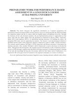
Vitamin B3 (Niacin)
Vitamin B3 is also called pellagra-preventing factor of
Goldberger and nicotinic acid.
Chemistry
Niacin is pyridine-3-carboxylic acid. In tissues, it occurs
principally as amide form.
Coenzyme
• Nicotinamide adenine dinucleotide (NAD+)
• Nicotinamide adenine dinucleotide phosphate (NADP+).
Biochemical Functions
NAD+-dependent enzymes.
Carbohydrate metabolism include:
1.Lactate dehydrogenase (lactate
pyruvate).
2. Glyceraldehyde-3-phosphate dehydrogenase (glyceraldehyde-3-phosphate
1, 3-bisphosphoglycerate).
3.Pyruvate dehydrogenase (pyruvate
acetyl-CoA).
NADPH-dependent enzymes
1.
Ketoacyl-ACP dehydrogenase (beta-ketoacyl-ACP
beta-hydroxyacyl-ACP).
2.a,b-unsaturated acyl-ACP
acyl-ACP.
3.HMG-CoA reductase (HMG-CoA
mevalonate.
4.Folate reductase (folate
dihydrofolate
tetrahydrofolate).
5.Phenylalanine hydroxylase (phenylalanine
tyrosine).
Recommended Daily Allowance
• Adult: 20 mg/day
• Pregnancy and lactation: 25 mg/day.
Dietary Sources
The richest natural sources of niacin are dried yeast, polished
rice, liver, peanut, whole cereals, legumes, meat and fish.
Deficiency Manifestations
Pellagra
Pellagra is characterized by three Ds, which are as follows:
1.Dermatitis: Increased pigmentation around the neck
is known as Casal’s necklace (Fig. 7.5).
2.Dementia: It is frequently seen in chronic cases. Delirium is common in acute pellagra.
3.Diarrhea: The diarrhea may be mild or severe with
blood and mucus.
Vitamin B6 (Pyridoxal Phosphate)
Coenzyme
• Pyridoxine
• Pyridoxal
• Pyridoxamine.
Active form of pyridoxine is pyridoxal phosphate (PLP).
Lipid metabolism
1.Beta hydroxyacyl-CoA dehydrogenase (beta hydroxyacyl-CoA
beta-ketoacyl CoA).
NADP+ dependent enzymes
1.Glucose-6-phosphate dehydrogenase in the hexose
monophosphate shunt pathway (glucose-6 phosphate
6-phosphogluconolactone).
2.Malic enzyme (malate to pyruvate).
Cha-7-Nutrition.indd 78
Fig. 7.5: Pellagra
30-01-2015 14:02:58
Chapter 7: Nutrition
Biochemical Functions
Dermatological manifestations
Deficiency of B6 will also affect tryptophan metabolism.
Since, niacin is produced from tryptophan, B6 deficiency
in turn leads to niacin deficiency, which is manifested as
pellagra.
Hematological manifestations
In adults, hypochromic microcytic anemia may occur due
to the inhibition of heme biosynthesis. The metabolic disorders, which respond to vitamin B6 therapy are xanthurenic aciduria and homocystinuria.
Vitamin B9 (Folic Acid)
decreased formation of GABA. The PLP is involved in the
synthesis of sphingolipids; so B6 deficiency leads to demyelination of nerves and consequent peripheral neuritis.
Vitamin B9 is also called liver lactobacillus casei factor,
Streptococcus lactis resistance (SLR) factor, pteroylglutamic acid (PGA).
1. Transamination: These reactions are catalyzed by
aminotransferases (transaminases), which employ
PLP as coenzyme. For example,
Alanine +
Pyruvate+
Alpha-ketoglutarate
glutamic acid
Alanine transaminase
2. Decarboxylation: All decarboxylation reactions of
amino acids require PLP as coenzymes. For examples,
a. Glutamate
GABA.
GABA is an inhibitory neurotransmitter and hence
in B6 deficiency, especially in children, convulsions may occur.
b. Histidine
histamine.
3. Sulfur-containing amino acids: Pyridoxal phosphate
plays an important role in methionine and cysteine
metabolism.
Homocysteine + Serine
Cystathionine (by
cystathionine synthase).
Cystathionine
Homoserine + Cysteine (by
cystathionase).
4. Heme synthesis: Aminolevulinic acid synthase is a
PLP-dependent enzyme. This is the rate-limiting step
in heme biosynthesis so, in B6 deficiency, anemia may
be seen.
5. Production of niacin from tryptophan require PLP.
6. Glycogenolysis: Phosphorylase enzyme (glycogen to
glucose-1-phosphate) requires PLP. In fact, more than
70% total PLP content of the body is in muscles, where
it is a part of the phosphorylase enzyme.
79
Chemistry
Recommended Daily Allowance
• Adult: 1–2 mg/day
• Pregnancy and lactation: 2.5 mg/day.
Dietary Sources of Vitamin B6
Rich sources are yeast, polished rice, wheat germs, cereals,
legumes (pulses), oil seeds, egg, milk, meat, fish and green
leafy vegetables.
Deficiency Manifestations
Neurological manifestations
In vitamin B6 deficiency, PLP-dependent enzymes function poorly. So, serotonin, epinephrine, noradrenalin
and GABA are not produced properly. Neurological
symptoms are therefore quite common in B6 deficiency.
In children, B6 deficiency leads to convulsions due to
The pteridine group with para-aminobenzoic acid (PABA)
is pteroic acid. It is then attached to glutamic acid to form
pteroylglutamic acid or folic acid.
Coenzyme
Active form is reduced 5, 6, 7, 8-tetrahydrofolic acid (THFA).
The THFA is the carrier of one-carbon groups. One carbon compound is an organic molecule that contains only a
single carbon atom. The following groups are one-carbon
compounds:
• Formyl (—CH=O)
• Formimino (—CH=NH)
• Methenyl (—CH=)
• Methylene (—CH2–)
• Hydroxymethyl (—CH2OH)
• Methyl (—CH3).
Folate antagonists:
• Sulfonamides
• Pyrimethamine
• Aminopterin.
Recommended Daily Allowance
• Adult: 200 mg/day
• Pregnancy: 400 mg/day
• Lactation: 300 mg/day.
Dietary Sources
Rich sources of folate are yeast, green vegetables. Moderate sources are cereals, oil seeds and egg.
/>Cha-7-Nutrition.indd 79
30-01-2015 14:02:58
80
Section 1: Theories
Deficiency Manifestations
Reduced DNA synthesis
In folate deficiency, THFA is reduced and thymidylate
synthase enzyme is inhibited. Hence deoxyuridine monophosphate (dUMP) is not converted to deoxythymidine
monophosphate (dTMP). So deoxythymidine triphosphate (dTTP) is not available for DNA synthesis. Thus cell
division is arrested.
Macrocytic anemia
1.It is the most characteristic feature of folate deficiency. During erythropoiesis, DNA synthesis is delayed,
but protein synthesis is continued. Thus hemoglobin
accumulates in RBC precursors leading to immature
looking nucleus and macrocytic cells.
2.Reticulocytosis is often seen. These abnormal RBCs
are rapidly destroyed. Reduced generation and increased destruction of RBCs result in anemia.
3.Leukopenia and thrombocytopenia are also manifested.
Homocysteinemia
Folic acid deficiency may cause increased homocysteine
levels in blood (above 15 mmol/L) with increased risk of
coronary artery diseases. It is treated by adequate doses of
pyridoxine, vitamins B12 and B9.
Birth defects
Folic acid deficiency during pregnancy causes homocysteinemia and neural tube defects in fetus. Folic acid prevents
birth defects malformations such as spina bifida.
Cancer
Bronchial carcinoma and cervical carcinoma.
linked to a substituted benzimidazole ring. This is then
called cobalamin. The 6th valence of the cobalt is satisfied
by any of the following groups namely cyanide, hydroxyl,
adenosyl or methyl.
When cyanide is added at the (R) position, the molecule is called cyanocobalamin. When cyanide group
is substituted by hydroxyl group, it forms hydroxy, cobalamin.
Absorption of Vitamin B12
Absorption of vitamin B12 requires two binding proteins.
First is the intrinsic factor (IF) of Castle. The second factor
is cobalophilin (Figs 7.6A and B).
Transport and storage
In the blood, methyl B12 form is predominant. Transcobalamin, a glycoprotein is the specific carrier. It is stored
in the liver cells, as ado-B12 form, in combination with
transcorrin.
Biochemical Functions
In B12 deficiency, methylmalonyl-CoA is excreted in urine
(methylmalonic aciduria).
Mnemonic: Folate deficiency causes:
‘A FOLIC DROP’
• Alcoholism
• Folic acid antagonists
• Oral contraceptives
• Low dietary intake
• Infection with Giardia
• Celiac sprue
• Dilantin
• Relative folate deficiency
• Old
• Pregnant
Vitamin B12 (Cobalamin)
Vitamin B12 is called antipernicious anemia factor and extrinsic factor of Castle.
Chemistry
Four pyrrole rings coordinated with a cobalt atom is called
a corrin ring. The 5th valence of the cobalt is covalently
Cha-7-Nutrition.indd 80
Figs 7.6A and B: Absorption of vitamin B12 (R, cobalophilin; Cbl,
cobalamin; IF, intrinsic factor; TC, trans cobalamin)
30-01-2015 14:02:58
1. Homocysteine Methyltransferase (Fig. 7.7).
2. Methyl folate trap and folate deficiency.
The production of methyl THFA is an irreversible step.
Therefore, the only way for generation of free THFA is
step no. 1 in the Figure 7.7. When B12 is deficient, this
reaction cannot take place. This is called the methyl
folate trap, this leads to the associated folic acid scarcity in B12 deficiency.
Chapter 7: Nutrition
Recommended Daily Allowance
• Adult: 1–2 mg/day
• Pregnancy and lactation: 2 mg/day.
Dietary Sources
Richest source is liver. Curd is a good source.
Causes of B12 Deficiency
Nutritional: Nutritional vitamin B12 deficiency is very common in India.
Decrease in absorption: Absorptive surface is reduced by
gastrectomy, resection of ileum and malabsorption syndromes.
Addisonian pernicious anemia: It is manifested usually in
persons over 40 years. It is an autoimmune disease. Antibodies are generated against IF. So, the IF becomes deficient, leading to defective absorption of B12.
Gastric atrophy: Similar atrophy of gastric epithelium leading to deficiency of IF and decreased B12 absorption is
common in India. In chronic iron deficiency anemia, there
is generalized mucosal atrophy.
81
Pregnancy: Increased requirement of vitamin in pregnancy is
another common cause for vitamin B12 deficiency in India.
Fish tapeworm: The fish tapeworm, diphyllobothrium latum has a special affinity to B12 causing reduction in available vitamin.
Deficiency Manifestations
Folate trap: Vitamin B12 deficiency causes simultaneous
folate deficiency due to the folate trap. Therefore all the
manifestations of folate deficiency are also seen.
Megaloblastic anemia: In the peripheral blood, megaloblasts and immature RBCs are observed.
Homocysteinemia: In vitamin B12 deficiency, homocysteine
is accumulated, leading to homocystinuria and myocardial infarction.
Demyelination: In vitamin B12 deficiency, nonavailability
of active methionine leads to inadequate methylation of
phosphatidylethanolamine to phosphatidylcholine. This
leads to deficient formation of myelin sheaths of nerves,
demyelination and neurological lesions.
Subacute combined degeneration: Damage to nervous system
is seen in B12 deficiency. There is demyelination affecting
cerebral cortex as well as dorsal column and pyramidal tract
of spinal cord. Since sensory and motor tracts are affected,
it is named as combined degeneration. Symmetrical paresthesia of extremities, alterations of tendon, and deep senses
and reflexes, loss of position sense, unsteadiness in gait,
positive Romberg’s sign (falling when eyes are closed) and
positive Babinski’s sign (extensor plantar reflex) are seen.
Achlorhydria: Absence of acid in gastric juice is associated
with vitamin B12 deficiency.
Vitamin C (Ascorbic Acid)
Vitamin C is also called antiscorbutic vitamin.
Chemistry
Fig. 7.7: Action of homocysteine methyltransferase (THFA,
tetrahydrofolic acid; 1, –CH3; 2, homocysteine methyltransferase)
Vitamin C is easily destroyed by heat, alkali and storage. In
the process of cooking, 70% of vitamin C is lost.
The structural formula of ascorbic acid closely resembles that of carbohydrates. The strong reducing property
of vitamin C depends on the double-bonded (enediol)
carbons.
/>Cha-7-Nutrition.indd 81
30-01-2015 14:02:59
82
Section 1: Theories
Only L-ascorbic acid and dehydroascorbic acid have
antiscorbutic activity. The D-ascorbic acid has no activity.
Biochemical Functions
Reversible oxidation-reduction
The vitamin can change between ascorbic acid and dehydroascorbic acid. Most of the physiological properties of
the vitamin could be explained by this redox system.
Oxidation
L-ascorbic acid
Dehydroascorbic acid
Reduction
Hydroxylation of proline and lysine
Ascorbic acid is necessary for the post-translational hydroxylation of proline and lysine residue. Hydroxyproline
and hydroxylysine are essential for the formation of cross
links in the collagen, which gives the tensile strength to the
fibers. This process is necessary for the normal production
of osteoid, collagen and intercellular cement substance of
capillaries.
Tryptophan metabolism
Ascorbic acid is necessary for the hydroxylation of tryptophan to 5-hydroxytryptophan. This is required for the formation of serotonin.
Tyrosine metabolism
Vitamin C helps in the oxidation of para-hydroxyphenyl
pyruvate to homogentisic acid.
Iron metabolism
Ascorbic acid enhances the iron absorption from the intestine. Ascorbic acid reduces ferric iron to ferrous state,
which is preferentially absorbed.
Hemoglobin metabolism
Vitamin C is useful for reconversion of methemoglobin to
hemoglobin (Hb) by methemoglobin reductase.
Folic acid metabolism
Ascorbic acid is helping the enzyme folate reductase to reduce the folic acid to tetrahydrofolic acid. Thus it helps in
the maturation of RBC.
Steroid synthesis
Adrenal gland possesses increased level of ascorbic acid,
particularly in periods of stress. Vitamin C is necessary for
hydroxylation reactions for synthesis of corticosteroids.
Vitamin C helps in synthesis of bile acids from cholesterol.
Phagocytosis
Ascorbic acid stimulates phagocytic action of leukocytes
and helps in the formation of antibodies.
Antioxidant property
As an antioxidant, it may prevent cancer formation.
Cataract
Vitamin C is concentrated in the lens of eye. Regular intake
of ascorbic acid reduces the risk of cataract formation.
Recommended Daily Allowance
• Adult: 75 mg/day
• Pregnancy, lactation and in aged people: 100 mg/day.
Dietary Sources
Rich sources are amla (Indian gooseberry), guava, lime,
lemon and green leafy vegetables.
Deficiency Manifestations
Scurvy
Gross deficiency of vitamin C results in scurvy.
Infantile scurvy (Barlow’s disease)
In infants between 6 and 12 months of age, (period in
which weaning from breast milk), the diet should be supplemented with vitamin C sources. Otherwise, deficiency
of vitamin C is seen.
Hemorrhagic tendency
In ascorbic acid deficiency, collagen is abnormal and the
intercellular cement substance is brittle. So capillaries are
fragile, leading to the tendency to bleed even under minor
pressure subcutaneous hemorrhage may be manifested
as petechiae (small red or purple spots on skin caused by
minor hemorrhage due to broken capillaries) in mild deficiency and as ecchymoses (large purple or black and blue
spots produced by extravasation of blood into tissues) or
even hematoma in severe conditions.
Internal hemorrhage
In severe cases, hemorrhage may occur in the conjunctiva and retina resulting in epistaxis, hematuria or melena
(black colored stools due to oxidation of iron in Hb).
Oral cavity
In severe cases of scurvy, the gum becomes painful, swollen and spongy. The pulp is separated from the dentine
and finally teeth are lost. Wound healing may be delayed.
Bones
In the bones, the deficiency results in the failure of the
osteoblasts to form the intercellular substance, osteoid.
Without the normal ground substance, the deposition of
bone is arrested. The resulting scorbutic bone is weak and
Cha-7-Nutrition.indd 82
30-01-2015 14:02:59
Milk is a good source of calcium. Egg, fish and vegetables
are medium sources for calcium. Cereals contain only
small amount of calcium.
1. Calcification of the growing bones and teeth and
maintenance of the mature bones are dependent on
adequate dietary intake of calcium and phosphorus.
2. Calcium is an activator for a number of enzymes, e.g.
adenylate cyclase, ATPases, protein kinases, etc. Calcium, even in very low concentration, activates phosphorylase kinase through its binding to calmodulin
and thus increases the rate of glycogen breakdown.
It activates pyruvate dehydrogenase phosphatase,
which in turn activates pyruvate dehydrogenase complex to produce acetyl-CoA. Calcium also regulates
the enzymes of citric acid cycle at several steps. For
example, Ca2+ activates isocitrate dehydrogenase and
alpha-ketoglutarate dehydrogenase. Thus, Ca2+ stimulates the production of ATP.
3. It is essential for clotting of blood.
4. It is required for the contraction of muscles (excitation-contraction coupling).
5. It regulates the permeability of the capillary walls and
excitability of the nerve fibers.
Biological Function
Adult: 500 mg/day.
Children: 1,200 mg/day.
Pregnancy and lactation: 1,500 mg/day.
Recommended Daily Allowance
Sources of Calcium
Total calcium in the human body is about 1–1.5 kg. About
99% of which is seen in bone and 1% in extracellular fluid.
Mechanism of absorption of calcium is taking place from
the first and second part of duodenum. Calcium is absorbed against a concentration gradient and requires
energy. Absorption requires a carrier protein, helped by
calcium-dependent ATPase.
Factors causing increased absorption
1. Vitamin D: Calcitriol induces the synthesis of the carrier protein (calbindin) in the intestinal epithelial cells
and so facilitates the absorption of calcium.
2. Parathyroid hormone: It increases calcium transport
from the intestinal cells.
3. Acidity: It favors calcium absorption.
4. Amino acids: Lysine and arginine increases calcium
absorption.
Factors causing decreased absorption
1. Phytic acid: Hexaphosphate of inositol is present in
cereals. Fermentation and cooking reduce phytate
content.
2. Oxalates: They are present in some leafy vegetables,
which cause formation of insoluble calcium oxalates.
3. Malabsorption syndromes: Fatty acid is not absorbed,
causing formation of insoluble calcium salt of fatty acid.
4. Phosphate: High-phosphate content will cause precipitation as calcium phosphate. The optimum ratio
of calcium to phosphorus, which allows maximum
absorption is 1:2 to 2:1 as present in milk.
Calcium
6. It is also required for secretion of various hormones
and acts as a second messenger.
7. Calcium regulates cell growth and differentiation.
Absorption
Minerals are essential for the normal growth and maintenance of the body. If the daily requirement is more than
100 mg, they are called major elements or macromineral.
If the requirement is less than 100 mg/day, they are known
as minor elements or trace elements.
83
MINERALS
fractures easily. Painful swelling of joints may prevent locomotion of the patient.
Anemia
Microcytic, hypochromic anemia is seen.
Chapter 7: Nutrition
Calcium Homeostasis
The hormones—calcitriol, parathyroid hormone (PTH)
and calcitonin are the major factors that regulate the plasma calcium (homeostasis of Ca) within a narrow range of
9–11 mg/dL (Fig. 7.8).
Calcitriol
The physiologically active form of vitamin D is a hormone,
namely calcitriol or 1, 25-dihydroxy cholecalciferol (1, 25
DHCC) (Fig. 7.9).
Parathyroid Hormone
Parathyroid hormone (PTH) is secreted by two pairs of
parathyroid glands that are closely associated with thyroid
glands. It is originally synthesized as prepro-PTH, which
is degraded to pro PTH and, finally to active PTH. The release of PTH from parathyroid glands is under the negative
feedback regulation of serum Ca2+.
/>Cha-7-Nutrition.indd 83
30-01-2015 14:02:59
84
Section 1: Theories
Fig. 7.8: Calcium homeostasis (C-cells, clear cells or parafollicular cells; PTH, parathyroid hormone)
Action on the Bone (Fig. 7.10)
Mechanism of Action of PTH (Fig. 7.11)
Action on the kidney
Parathyroid hormone increases the Ca reabsorption by
kidney tubules. This is the most rapid action of PTH to
elevate blood Ca levels. PTH promotes the production of
calcitriol (1, 25 DHCC) in the kidney by stimulating 1-hydroxylation of 25-hydroxycholecalciferol.
2.Tumors secreting a PTH-like substance.
3.Vitamin D poisoning.
4.Excessive ingestion of milk.
5.Excessive intake of alkali by patients with peptic ulcer.
Signs of hypercalcemia include:
a.Thirst.
b.Tiredness.
Action on the intestine
The action of PTH on the intestine is indirect. It increases
the intestinal absorption of Ca by promoting the synthesis
of calcitriol.
Calcitonin
Calcitonin (CT) is a peptide containing 32 amino acids. It
is secreted by parafollicular cells of thyroid gland. The action of CT on calcium metabolism is antagonistic to that of
PTH (Fig. 7.12).
Plasma Calcium Disease
Hyperparathyroidism causes hypercalcemia. Hypercalcemia may also be caused by:
1.Tumors which cause rapid bone destruction.
Cha-7-Nutrition.indd 84
Fig. 7.9: Calcium absorption
30-01-2015 14:02:59
Chapter 7: Nutrition
85
Tapping over the facial nerve in front of the ear produces twitching of the facial muscles (Chvostek’s sign), and
the motor nerves are unduly excitable to electrical stimulation. Carpal spasm can be induced by inflating a blood
pressure cuff around the upper arm to a pressure exceeding the systolic blood pressure maintaining the occlusion
for 3 minutes (Trousseau’s test).
Iron
Total body content of iron is 3–5 g. Blood contains 14.5 g of
hemoglobin per 100 mL.
Requirement of Iron
Fig. 7.10: Action on the bone (PTH, parathyroid hormone)
Leafy vegetables, jaggery, meat, liver are good sources.
Cooking in iron utensils will improve iron content of the
diet. Milk is a poor source of iron.
Biochemical Role of Iron
1. It is involved in the transport of oxygen by hemoglobin
and hemoerythrin.
2. It is involved in electron-transfer reactions, including
the pathways of oxidative phosphorylation.
3. It is involved in the synthesis of DNA (as an essential
component of ribonucleotide reductase).
4. It is involved in the catalysis of oxidation by oxygen
and H2O2.
5. It is involved in the decomposition of harmful derivatives of oxygen, notably peroxide and superoxide.
Sources of Iron
c. Weakness.
d. Mental disturbances, and if severe, then coma and
death.
6. Untreated hypercalcemia causes renal damage.
Hypocalcemia is found in:
a. Hypoparathyroidism.
b. Osteomalacia.
c. Rickets.
d. Renal failure.
e. Tetany is a prominent feature.
The outstanding feature of tetany is neuromuscular irritability leading finally to generalized clonic movements
especially in children. The muscle hypertonia produces
the characteristic attitude of the hand in tetany, the main
d’accoucheur. Carpopedal spasm may be accompanied in
infants by spasm of the glottis (laryngismus stridulus), cyanosis, tingling feelings and sensations of heat and flushing (paresthesia).
• Daily allowance of iron for an adult is 20 mg
• Children between 13–15 years need 20–30 mg/day
• Pregnant woman need 40 mg/day.
Fig. 7.11: Mechanism of action of parathyroid hormone (PTH)
Fig. 7.12: Action of calcitonin
/>Cha-7-Nutrition.indd 85
30-01-2015 14:02:59
86
Section 1: Theories
6.Besides, it also plays a very important role in the fixation of nitrogen and hydrogen.
Absorption
Factors affecting iron absorption
1.Intraluminal factors, i.e. dietary iron content, chemical form of dietary iron, dietary constituents, intestinal
secretions, intestinal motility, stable chelators, metallic cation competitors, etc.
2. Mucosal factors, i.e. anatomic and histologic, mucosal
iron content, etc.
3.Corporeal factors, i.e. body iron concentration, erythropoiesis, iron turnover, etc.
Mechanism of absorption
Granick has proposed ‘mucosal block theory’ for the absorption of iron (Fig. 7.13). According to it, iron is taken up
by tin in the mucosal cell to form ferritin, which then slowly
releases iron to transferrin present in the circulating plasma.
The amount of iron absorbed is determined by the amount
of apoferritin synthesized in gastric mucosal cell and not by
the iron present in the lumen, because once the gastric mucosal cell tin gets saturated with iron, it cannot accept more
iron. ‘Mucosal block theory’ is now not considered.
Transport of iron
Iron is transported in the body with a specific iron binding
b1-globulin; transferrin (siderophilin). It performs the functions of selective removal of iron from reticuloendothelial
cells and intestinal mucosa and selective delivery of iron to
the erythron and placenta. It is a glycoprotein, which binds
two atoms of ferric iron. The iron-transferrin complex is
very much stable under the physiological conditions.
Abnormal Metabolism of Iron
Iron toxicity
Hemosiderosis: Iron in excess is called hemosiderosis. Hemosiderin pigments are golden brown granules, seen in
Fig. 7.13: Mucosal block theory (DMT 1, divalent metal transporter
1; FP, ferroportin; HP, haptoglobulin; HT, heme transporter; TF,
transferrin)
Cha-7-Nutrition.indd 86
spleen and liver. Prussian blue reaction is positive for the
pigments. Hemosiderosis occurs in persons receiving repeated blood transfusions. This is the commonest cause
for hemosiderosis in India.
Primary hemosiderosis: It is also called hereditary hemochromatosis. In these cases, iron absorption is increased
and transferrin level in serum is elevated. Excess iron deposits are seen.
Bantu siderosis: Bantu tribe in Africa is prone to hemosiderosis because the staple diet, corn, is low in phosphate
content.
Hemochromatosis: When total body iron is higher than 25–
30 g, hemosiderosis is manifested. In the liver, hemosiderin deposit leads to death of cells and cirrhosis. Pancreatic cell death leads to diabetes. Deposits under the skin
cause yellow-brown discoloration, which is called hemochromatosis. The triad of cirrhosis, hemochromatosis and
diabetes is referred to as bronze diabetes.
Copper
Total body content of copper is about 100 mg.
Recommended Daily Allowance
Copper requirement for an adult is 1.5–3 mg/day.
Dietary Sources
Major dietary sources are cereals, meat, liver, nuts and
green leafy vegetables. Milk is very poor in copper content.
Absorption and transport of copper in extrahepatic
tissue is shown in Figure 7.14.
Physiological Functions
1. It is required in small amounts for the synthesis of normal hemoglobin.
Fig. 7.14: Absorption and transport of copper
(GIT, gastrointestinal tract; Cu, copper)
30-01-2015 14:03:00
Daily intake of about 10–15 mg is sufficient to meet its requirement.
Dietary Sources
Tyrosinase.
Cytochrome oxidase.
Ascorbic acid oxidase.
Uricase.
Monoamine oxidase.
Ceruloplasmin (ferroxidase I)—a blue copper-protein
complex.
7. Non-ceruloplasmin ferroxidase—a yellow copperprotein complex, etc.
Biological Functions
1. Excessive deposition of copper in liver causing hepatic cirrhosis.
2. A visible brown ring (Kayser-Fleischer ring) at the
margin of the cornea.
3. Deposits in basal ganglia leads to lenticular degeneration and neurological symptoms.
Menkes disease (kinky hair syndrome) is characterized by skeletal malformations, immunological deficiency,
mental retardation and defective thermoregulation.
Normochromic microcytic anemia is caused due to
copper deficiency because copper is an integral part of
ALA synthase, which is key enzyme in heme synthesis.
Copper deficiency may cause atrophy of myocardium.
The elastic tissue of aorta, coronary and pulmonary artery
gets deranged. These vessels may rupture, as a result of
which end comes into death.
Abnormal metabolism of zinc
Clinical manifestations of zinc deficiency include:
1. Poor wound healing, loss of appetite, poor growth and
alopecia (loss of hair).
2. Impairment of sexual development in children.
3. Impairment in brain functions, DNA synthesis and
carbohydrate metabolism.
4. Certain fetal abnormalities during pregnancy besides
hypogonadism, dwarfism (stunted growth) and gross
skin lesions with severe acrodermatitis.
5. Zinc deficiency has also been shown to affect spermatogenesis, parturition and lactation in experimental animals.
Clinical Features
Wilson’s disease (hepatolenticular degeneration) is a rare
hereditary disorder of copper metabolism, which is due to
an autosomal recessive genetic defect.
The basic defect is the mutation in a gene encoding a
copper-binding ATPase in cells, which is required for excretion of copper from cells.
Increased copper content in hepatocyte inhibits the
incorporation of copper to apoceruloplasmin. So ceruloplasmin level in blood is increased.
Abnormal Metabolism of Copper
1. Zinc is a component of several enzymes such as carbonic anhydrase, lactate dehydrogenase, alcohol dehydrogenase, alkaline phosphatase, DNA and RNA
polymerases, retinene-retinal reductase, etc.
2. It is an important constituent of insulin. It forms a
complex with insulin and helps in its storage and release from the beta cells of the pancreas.
3. It is necessary for maintaining plasma concentration
of vitamin A, by stimulating its release from the liver
into the blood.
4. It is also present in gustin, a salivary polypeptide,
which is necessary for the normal development of
taste buds. Thus, zinc is important for taste sensation.
5. It is also an essential component of various regulatory
proteins.
6. It has been shown to be essential for normal growth
and reproduction.
1.
2.
3.
4.
5.
6.
Recommended Daily Allowance
Good dietary sources are meat, seafood, eggs, legumes
and milk. Colostrum is a very rich source of zinc.
Copper-containing Enzymes
Zinc
2. It is required for the synthesis of:
a. Phospholipids.
b. Melanin.
c. Collagen.
3. It plays role in the formation of bone.
4. It maintains the integrity of myelin sheath in the nerve
fibers.
87
Chapter 7: Nutrition
Fluorine
Recommended Daily Allowance
Daily requirement has been defined as 2–3 mg.
/>Cha-7-Nutrition.indd 87
30-01-2015 14:03:00
88
Section 1: Theories
Sources
Drinking water is an important source of fluoride in a human diet. One part per million (1 PPM) of fluoride in drinking water supplies nearly 1–2 mg of fluoride/day, which is
sufficient to meet the requirement. Tea and sea fishes are
also a good sources of fluoride.
Physiological Functions
1.This element is essential for the growth of teeth and
bones and is required in minute quantities.
2.Fluoroacetate is a powerful inhibitor of TCA cycle.
3.In combination with vitamin D, it is required for the
treatment of bone disease, i.e. osteoporosis, which is
characterized by softening of bone as a result of excessive absorption of bone elements.
4.Sodium fluoride acts as a powerful inhibitor of the
glycolytic enzyme enolase; therefore, it is used as a
blocker of glycolytic pathway while collecting blood
samples for the determination of sugar.
5. It forms a protective layer of acid-resistant fluorapatite
with hydroxyapatite crystals of the enamel.
6. Fluoride ions inhibit the metabolism of oral bacterial
enzymes and also restrict the local production of acids, which are responsible for dental caries.
Abnormal Metabolism of Fluorine
Deficiency disorders
Deficiency of fluoride promotes the development of dental
caries in children and osteoporosis in adult particularly in
postmenopausal women.
Dental caries is characterized by destruction of tooth
enamel as a result of action of microbes (normally present
in oral cavity) on food. Breakdown of the enamel exposes
dentine and leads to development of caries.
(more than 3 mg/L in drinking water) results in a severe
form of the skeletal fluorosis called `genu valgum’ (knock
knee syndrome) (Fig. 7.15).
Selenium
Recommended Daily Allowance
Requirement is 50–100 mg/day. Normal serum level is also
50–100 mg/dL.
Physiological Functions
1. It is a constituent of glutathione peroxidase, which catalyzes the breakdown of H2O2 in RBCs. Deficiency of selenium in human beings is not yet well established.
2.Tocopherol sparing action: Selenium has got close
metabolic relationship with vitamin E. It reduces the
requirement of vitamin E in more than one way:
a.Selenium-containing glutathione peroxidase destroys acylhydroperoxides, thus lowers the need for
antioxidant action of vitamin E in preventing peroxidative damage.
b. Selenium-May probably help in retaining vitamin E
in lipoproteins.
3.It is involved in the mitochondrial ATP synthesis, ubiquinone synthesis and immune mechanisms.
4.It has been reported to be a cancer-preventing agent.
Abnormal Metabolism of Selenium
Selenium deficiency is characterized by multifocal myocardial necrosis, cardiac arrhythmias and cardiac enlargement. Selenium is known to cure the disease. Isolated
selenium deficiency causes liver necrosis, cirrhosis, cardiomyopathy and muscular dystrophy.
Toxicity
Fluoride toxicity may manifest in two major forms, i.e. as
dental fluorosis and skeletal fluorosis, which together constitute endemic fluorosis.
Dental fluorosis: The teeth exhibit fluoride toxicity in the
form of mottled enamel. Mottling is characterized by multiple, minute white flecks and yellow-brown spots, which
are scattered irregularly over the tooth surface.
Skeletal fluorosis: The clinical features include pain, inflammation and restricted movement of the joints and stiffness
of the spine. Further, significantly higher intake of fluoride
Cha-7-Nutrition.indd 88
Fig. 7.15: Genu valgum
30-01-2015 14:03:00
Chapter 7: Nutrition
Fig. 7.16: Kwashiorkor
89
Fig. 7.17: Marasmus
Table 7.1: Comparison between kwashiorkor and marasmus
Sl No
Kwashiorkor
Marasmus
1.
Occurs in the postweaning period (1–3 year)
Occurs due to early weaning (< 1 year)
2.
Deficiency of dietary proteins
Deficiency of dietary proteins plus energy
3.
Rapid/Acute onset
Slow/Chronic development
4.
Moderate weight loss; child is 60%–80% weight for age
Severe weight loss; child is < 60% weight for age
5.
Moderate muscle wasting with retention of some body fat
Severe muscle wasting with practically no body fat
6.
Edema is a conspicuous feature
No edema
7.
Enlarged and fatty liver
Liver is normal
8.
Face reflects irritability and misery
Face shows apathy and anxiety
9.
Loss of appetite
Good appetite possible
10.
Hair shows color changes (flag sign) and becomes straight
Hair is sparse, thin and dry
11.
Skin may develop lesions
Skin is dry, thin and easily wrinkled
Protein energy malnutrition (PEM) is one of the largest
public health problems of the country. As the name suggests, this condition is a deficiency of protein and calories
in the diet. Strictly speaking, it is not one disease, but a
spectrum of conditions arising from an inadequate diet.
Although it affects people of all ages, the results are most
dramatic in childhood due to the highest requirement in
that period.
Protein energy malnutrition is a general term, which
includes two different types of nutritional deficiencies:
1. Kwashiorkor (Fig. 7.16).
2. Marasmus (Fig. 7.17).
The difference between kwashiorkar and maraemus is
given in Table 7.1.
gy M
alnutrition
E
ner
P
rotein
Selenium toxicity is called selenosis. The toxic symptoms include hair loss, falling of nails, diarrhea, weight loss
and garlicky odor in breath.
/>Cha-7-Nutrition.indd 89
30-01-2015 14:03:00
Chapter
8
Tissue Biochemistry
HEME SYNTHESIS
Heme is the most important porphyrin containing compound. It is primarily synthesized in the liver and the
erythrocyte-producing cells of bone marrow (erythroid
cells). However, mature erythrocytes lacking mitochondria are a notable exception.
Structure of Heme
1.Heme is a derivative of the porphyrin. Porphyrins are
cyclic compounds formed by fusion of four pyrrole
rings linked by methenyl (=CH—) bridges.
2.Since an atom of iron is present, heme is a ferroprotoporphyrin. The pyrrole rings are named as I, II, III,
IV and the bridges as alpha (a), beta (b), gamma (g)
and delta (d). The possible areas of substitution are
denoted as 1–8 (Fig. 8.1).
3.Type III is the most predominant in biological systems. It is also called series 9.
Biosynthesis of Heme
Heme can be synthesized by almost all the tissues in the
body. Heme is synthesized in the normoblasts, but not in
the matured erythrocytes. The pathway is partly cytoplasmic and partly mitochondrial.
Step 1: Formation of d-aminolevulinate Acid
Glycine, a non-essential amino acid and succinyl-CoA,
an intermediate in the citric acid cycle are the starting materials for porphyrin synthesis. Glycine combines with succinyl-CoA to form delta-aminolevulinate
(ALA). This reaction catalyzed by a pyridoxal phosphate-dependent d-aminolevulinate synthase occurs in
the mitochondria. It is a rate-controlling step in porphyrin synthesis.
Step 2: Synthesis of Porphobilinogen
Two molecules of d-aminolevulinate condense to form
porphobilinogen (PBG) in the cytosol. This reaction is catalyzed by a Zn-containing enzyme, ALA dehydrogenase. It
is sensitive to inhibition by heavy metals such as lead.
Step 3: Formation of Uroprophyrinogen
Fig. 8.1: Structure of heme
Cha-8-Tissue.indd 90
Condensation of four molecules of the PBG results in the
formation of the first porphyrin of the pathway, namely
uroporphyrinogen (UPG). The enzyme for this reaction is
PBG deaminase [otherwise called uroporphyrin I synthase
or hydroxymethylbilane (HMB) synthase]. HMB molecule
will cyclize spontaneously to form uroporphyrinogen I. It is
30-01-2015 14:01:17
Chapter 8: Tissue Biochemistry
91
converted to uroporphyrinogen III by the enzyme uroporphyrinogen III synthase.
Step 4: Synthesis of Coproporphyrinogen
The UPG-III is next converted to coproporphyrinogen
(CPG-III) by decarboxylation. Four molecules of CO2 are
eliminated by uroporphyrinogen decarboxylase.
Step 5: Synthesis of Protoporphyrinogen
Further metabolism takes place in the mitochondria. CPG
is oxidized to protoporphyrinogen (PPG-III) by coproporphyrinogen oxidase. This enzyme specifically acts only on
type III series.
Step 6: Generation of Protoporphyrin
The protoporphyrinogen-III is oxidized by the enzyme
protoporphyrinogen oxidase to protoporphyrin-III (PPIII) in the mitochondria. The oxidation requires molecular
oxygen.
Step 7: Generation of Heme
The incorporation of ferrous ion (Fe2+) into protoporphyrin-IX is catalyzed by the enzyme heme synthetase
(ferrochelatase). This enzyme can be inhibited by lead
(Fig. 8.2).
Fig. 8.2: Biosynthesis of heme
Disorders of Heme Synthesis
Porphyrias
Porphyrias are the metabolic disorders of heme synthesis
characterized by the increased excretion of porphyrins or
porphyrin precursors. Porphyrias are either inherited or
acquired. They are broadly classified into two categories
(Table 8.1):
• Erythropoietic: Enzyme deficiency occurs in the
erythrocytes
• Hepatic: Enzyme defect lies in the liver.
Acute intermittent porphyria
Acute intermittent porphyria is characterized by increased
excretion of porphobilinogen and 8-aminolevulinate. The
urine gets darkened on exposure to air due to the conversion of porphobilinogen to porphobilin and porphyria.
The other characteristic features of acute intermittent porphyria are as follows:
1. The symptoms include abdominal pain, vomiting and
cardiovascular abnormalities. The neuropsychiatric
1. The ALA synthase is regulated by repression mechanism. Heme inhibits the synthesis of ALA synthase by
acting as a co-repressor.
2. The ALA synthase is also allosterically inhibited by hematin. When there is excess of free heme, the Fe2+ is
oxidized to Fe3+ (ferric), thus forming hematin.
3. The compartmentalization of the enzymes of heme
synthesis makes the regulation easier for the regulation. The rate-limiting enzyme is in the mitochondria.
Some steps take place inside mitochondria, while rest
occurs in cytoplasm.
4. Drugs like barbiturates induce heme synthesis. Barbiturates require the heme-containing cytochrome p450
for their metabolism.
5. The steps catalyzed by ferrochelatase and ALA dehydratase are inhibited by lead.
6. Isonicotinic acid hydrazide (INH) that decreases the
availability of pyridoxal phosphate may also affect
heme synthesis.
7. High cellular concentration of glucose prevents induction of ALA synthase.
Regulation of Heme Synthesis
/>Cha-8-Tissue.indd 91
30-01-2015 14:01:17
92
Section1: Theories
Table 8.1: Porphyrias
Types
Deficient enzyme
Features
Hepatic
Acute intermittent porphyria
Uroporphyrinogen synthase (PBG deaminase)
Abdominal pain, neuropsychiatric symptoms
Porphyria cutanea tarda
Uroporphyrinogen de-arboxylase
Photosensitivity
Hereditary coproporphyria
Coproporphyrinogen oxidase
Abdominal pain
Congenital erythropoietic
porphyria
Uroporphyrinogen IIIco-synthase
Photosensitivity
Protoporphyria
Ferrochelatase
Photosensitivity
Erythropoietic
disturbances observed in these patients are believed to be due to reduced activity of tryptophan
pyrrolase, resulting in accumulation of tryptophan
and serotonin.
2. The symptoms are more severe after administration of
drugs (e.g. barbiturates) that induce the synthesis of
cytochrome P450. This is due to the increased activity of
ALA synthase causing accumulation of PBG and ALA.
3.These patients are not photosensitive since the enzyme defect occurs prior to the formation of uroporphyrinogen.
4.Acute intermittent porphyria is treated by administration of hematin, which inhibits the enzyme ALA synthase and the accumulation of porphobilinogen.
Acute intermittent porphyria symptoms (5 P’s):
• Pain in abdomen
• Polyneuropathy
• Psychological abnormalities
• Pink urine
• Precipitated by drugs (e.g. barbiturates, oral contraceptives
and sulfa drugs)
Congenital erythropoietic porphyria
1. Congenital erythropoietic porphyria is a rare congenital disorder caused by autosomal recessive mode of
inheritance, mostly confined to erythropoietic tissues.
2.The individuals excrete uroporphyrinogen I and coproporphyrinogen I, which oxidize respectively to uroporphyrin I and coproporphyrin I (red pigments).
3.The patients are photosensitive (itching and burning
of skin when exposed to visible light) due to the abnormal prophyrins that accumulate.
4.Increased hemolysis is also observed in the individuals affected by this disorder.
Porphyria cutanea tarda
Porphyria cutanea tarda is also known as cutaneous hepatic porphyria and is the most common porphyria,
usually associated with liver damage caused by alcohol
Cha-8-Tissue.indd 92
overconsumption or iron overload. Cutaneous photosensitivity is the most important clinical manifestation of
these patients.
HEME CATABOLISM
In heme catabolism, heme oxygenase is a complex microsomal enzyme namely heme oxygenase utilizes NADPH
and O2, and cleaves the methenyl bridges between the
two pyrrole rings to form biliverdin. Simultaneously, ferrous ion (Fe2+) is oxidized to ferric form (Fe3+) and released.
The products of heme oxygenase reaction are biliverdin (a
green pigment), Fe3+ and carbon monoxide (CO). Heme
promotes the activity of this enzyme.
Generation of Bilirubin
Biliverdin reductase: Biliverdin’s methenyl bridges are reduced to methylene group to form bilirubin (yellow pigment). This reaction is catalyzed by an NADPH-dependent
soluble enzyme, biliverdin reductase. 1 g of hemoglobin
on degradation finally yields about 35 mg bilirubin. Approximately, 250–350 mg of bilirubin is daily produced in
human adults. The term bile pigments are used to collectively represent bilirubin and its derivatives.
Transport of Bilirubin to Liver
Bilirubin is lipophilic and therefore insoluble in aqueous
solution. Bilirubin is transported in the plasma in a bound
(noncovalently) form to albumin. Albumin has two binding sites for bilirubin, a high-affinity site and a low-affinity
site. As the albumin-bilirubin complex enters the liver,
bilirubin dissociates and is taken up by sinusoidal surface
of the hepatocytes by a carrier-mediated active transport.
Conjugation
In the liver, bilirubin is conjugated with two molecules of
glucuronate supplied by UDP-glucuronate. This reaction,
30-01-2015 14:01:17
Chapter 8: Tissue Biochemistry
93
4. Free bilirubin is water insoluble. It has to be extracted
first with alcohol, when the reaction becomes positive; hence called indirect reaction.
Excretion of Bilirubin into Bile
Congenital Hyperbilirubinemias
Conjugated bilirubin is excreted into the bile canaliculi
against a concentration gradient, which then enters the
bile. The transport of bilirubin diglucuronide is an active,
energy-dependent and rate-limiting process. This step is
easily susceptible to any impairment in liver function.
Congenital hyperbilirubinemias result from abnormal uptake, conjugation or excretion of bilirubin due to inherited
defects.
Crigler-najjar syndrome type I: This is also known as congenital (non-hemolytic jaundice). It is a rare disorder and
is due to a defect in the hepatic enzyme UDP-glucuronyltransferase. Generally, the children die within first 2 years
of life.
Crigler-najjar syndrome type II: This is again a rare hereditary
disorder due to a less severe defect in the bilirubin conjugation. It is believed that hepatic UDP-glucuronyltransferase that catalyzes the addition of second glucuronyl group
is defective. The serum bilirubin concentration is usually
less than 20 mg/dL and this is less dangerous than type I.
Gilbert’s disease: This is not a single disease. It includes:
• A defect in the uptake of bilirubin by liver
• An impairment in conjugation due to reduced activity
of UDP-glucuronyltransferase
• Decreased hepatic clearance of bilirubin.
Dubin-johnson syndrome: It is an autosomal recessive
trait leading to defective excretion of conjugated bilirubin; so conjugated bilirubin in blood is increased. The
disease results from the defective adenosine triphosphate (ATP)-dependent organic anion transport in bile
canaliculi. The bilirubin gets deposited in the liver and
the liver appears black. The condition is referred to as
black liver jaundice.
1. Bilirubin reacts with diazo reagent (diazotized sulfanilic acid) to produce colored azo pigment.
2. At pH 5, the pigment is purple in color.
3. Conjugated bilirubin, being water soluble gives the
color immediately; hence called direct reaction.
van den Bergh Test for Bilirubin
Acquired Hyperbilirubinemias
Jaundice: It is a clinical condition characterized by yellowish discolorization of skin and mucous membrane. It is
caused by elevated serum bilirubin level more than 3 mg/
dL. On pathological basis, jaundice is classified into-three
groups:
1. Hemolytic jaundice or prehepatic jaundice.
2. Hepatocellular jaundice or hepatic jaundice.
2. Obstructive jaundice or posthepatic jaundice.
Hemolytic jaundice
Hemolytic diseases of the newborn
This condition results from incompatibility between maternal and fetal blood groups. Rh +ve fetus may produce
antibodies in Rh -ve mother. In Rh incompatibility, the first
child often escapes. But in the second pregnancy, the Rh
Normal plasma bilirubin level ranges from 0.2 to 0.8 mg/dL.
The unconjugated bilirubin is about 0.2–0.6 mg/dL, while
conjugated bilirubin is only 0–0.2 mg/dL. If the plasma
bilirubin level exceeds 1 mg/dL, the condition is called hyperbilirubinemia. When the bilirubin level exceeds 2 mg/
dL, it diffuses into tissues producing yellowish discoloration of sclera, conjunctiva, skin and mucous membrane
resulting in jaundice.
Plasma Bilirubin
About 20% of the urobilinogen (UBG) is reabsorbed from
the intestine and returned to the liver by portal blood.
The UBG is again re-excreted (enterohepatic circulation).
Since the UBG is passed through blood, a small fraction is
excreted in urine (less than 4 mg/day).
Enterohepatic Circulation
Bilirubin glucuronides are hydrolyzed in the intestine by
specific bacterial enzymes namely P-glucuronidases to
liberate bilirubin. The latter is then converted to urobilinogen (colorless compound), a small part of which may
be reabsorbed into the circulation. Urobilinogen can be
converted to urobilin (a yellow color compound) in the
kidney and excreted. The characteristic color of urine is
due to urobilin. A major part of urobilinogen is converted
by bacteria to stercobilin, which is excreted along with
feces. The characteristic brown color of feces is due to
stercobilin.
Fate of Bilirubin
Hyperbilirubinemia
catalyzed by bilirubin glucuronyltransferase (of smooth
endoplasmic reticulum) results in the formation of a water-soluble bilirubin diglucuronide. The enzyme bilirubin glucuronyltransferase can be induced by a number of
drugs (e.g. phenobarbital).
/>Cha-8-Tissue.indd 93
30-01-2015 14:01:17
94
Section1: Theories
antibodies will pass from mother to the fetus. They would
start destroying fetal red cells even before birth.
Sometimes the child is born with severe hemolytic disease often referred to as erythroblastosis fetalis.
When the blood level is more than 20 mg/dL the capacity of albumin to bind bilirubin is exceeded. In young children before the age of 1 year, the blood-brain barrier is not
fully matured and therefore free bilirubin enters the brain.
It is deposited in brain leading to mental retardation, fits
toxic encephalitis and spasticity. This condition is known
as kernicterus.
Hemolytic diseases of adults
This condition is seen in increased rate of hemolysis. The
characteristic features are increase in unconjugated bilirubin in blood.
Hepatocellular jaundice
1.Most common cause is viral hepatitis, caused by hepatitis viruses. Conjugation in liver is decreased and
hence free bilirubin is increased in circulation. However, inflammatory edema of cell often compresses
intracellular canaliculi at the site of bile formation and
this produces an element of obstruction. Unconjugated
bilirubin level also increases. Bilirubinuria also occurs.
2.Dark-colored urine due to excessive excretion of bilirubin and urobilinogen.
3.Patient pass pale, clay-colored stools due to the absence of stercobilinogen.
4.Affected one experience nausea and anorexia (loss of
appetite).
5.Increased activities of serum glutamic-pyruvic transaminase (SGPT) and serum glutamic oxaloacetic
transaminase (SGOT) released in to circulation due to
damage to hepatocytes.
Obstructive jaundice
1. Conjugated bilirubin is increased in blood and it is excreted in urine. If there is complete obstruction, UBG
will be decreased in urine or even absent.
2. In total obstruction of biliary tree, the bile does not enter the intestine. Since no pigments are entering into
the gut, the feces become clay colored.
3. The common causes of obstructive jaundice are: lntrahepatic cholestasis and extrahepatic obstruction.
4.Serum alkaline phosphatase is elevated.
5. Dark-colored urine due to elevated excretion of bilirubin.
6.Feces contain excess fat due to impaired fat digestion.
7.Patient experience nausea and vomiting.
Some other important types of jaundice are given below.
Physiological jaundice
Physiological jaundice is also called as neonatal hyperbilirubinemia. In all newborn infants after the 2nd day of life
mild jaundice is present. This transient hyperbilirubinemia
Cha-8-Tissue.indd 94
is due to an accelerated rate of destruction of RBCs and
also because of immature hepatic system of conjugation
of bilirubin.
Breast milk jaundice
Prolongation of jaundice in mother may increase an estrogen derivative in blood, which will transfer to the infant
through breast milk. This will inhibit glucuronyltransferase system.
HEMOGLOBIN
Hemoglobin (Hb) is the red blood pigment, exclusively
found in erythrocytes. The normal concentration of Hb in
blood in males is 14–16 g/dL and in females 13–15 g/dL.
Hemoglobin performs two important biological functions
concerned with respiration:
1.Delivery of O2 from the lungs to the tissues.
2.Transport of CO2 and protons from tissues to lungs for
excretion.
Structure of Hemoglobin
The fetal Hb (HbF) is made up of two alpha and two gamma chains. Adult Hb (HbA) has two alpha chains and two
beta chains. HbA2 has two alpha and two delta chains.
Normal adult blood contains 97% HbA, about 2% HbA2
and about 1% HbF.
There are four heme residues per Hb molecule, one
for each subunit in Hb. The iron atom of heme occupies
the central position of the porphyrin ring. The reduced
state is called ferrous (Fe2+) and the oxidized state is ferric (Fe3+). In hemoglobin, iron remains in the ferrous
state.
Transport of Oxygen by Hemoglobin
Hemoglobin has all requirements of an ideal respiratory
pigment:
• It can transport large quantities of oxygen
• It has great solubility
• It can take up and release oxygen at appropriate partial
pressures
• It is a powerful buffer
• Each molecule of hemoglobin can bind with four molecules of O2.
Oxygen Dissociation Curve
The binding ability of hemoglobin with O2 at different
partial pressures of oxygen (pO2) can be measured by a
graphic representation known as O2 dissociation curve.
The curves obtained for hemoglobin and myoglobin are
depicted in Figure 8.3.
30-01-2015 14:01:18
Chapter 8: Tissue Biochemistry
95
Effect of pH and pCO2
When the pCO2 is elevated, the H+ concentration increases
and pH falls. In the tissues, the pCO2 is high and pH is low
due to the formation of metabolic acids like lactate. Then,
the affinity of hemoglobin for O2 is decreased (the ODC is
shifted to the right) and so more O2 is released to the tissues. In the lungs, the opposite reaction is found, where
the pCO2 is low, pH is high and pO2 is significantly elevated.
Bohr Effect
Fig. 8.3: Oxygen dissociation curve (ODC)
(DPG, diphosphoglycerate)
It is evident from the graph that myoglobin has much
higher affinity for O2 than hemoglobin. Hence O2 is bound
more tightly with myoglobin than with hemoglobin. Further, pO2 needed for half saturation (50% binding) of myoglobin is about 1 mm Hg compared to about 26 mm Hg for
hemoglobin.
Hemoglobin binding curve: Causes of shift to right
‘CADET, face right!’
• CO2
• Acid
• 2,3-DPG
• Exercise
• Temperature
Heme-Heme Interaction and Cooperativity
The oxygen dissociation curve for hemoglobin is sigmoidal in shape, it is due to the allosteric effect or cooperativity. This indicates that the binding of oxygen to one heme
increases the binding of oxygen to other hemes. Thus,
the affinity of Hb for the last O2 is about 100 times greater
than the binding of the first O2 to Hb. This phenomenon
is referred to as cooperative binding of O2 to Hb or simply
heme-heme interaction. On the other hand, release of O2
from one heme facilitates the release of O2 from others.
The binding of oxygen to one heme residue increases
the affinity of remaining heme residues for oxygen (homotropic interaction). This is called positive cooperativity.
Binding of 2,3-bisphosphoglycerate (BPG) at a site other
than the oxygen binding site, lowers the affinity for oxygen (heterotropic interaction). The quaternary structure
of oxyHb is described as R (relaxed) form and that of deoxyHb is T (tight) form.
The binding of oxygen to hemoglobin decreases with
increasing H+ concentration (lower pH) or when the hemoglobin is exposed to increased partial pressure of CO2
(pCO2). This phenomenon is known as Bohr effect. It is
due to a change in the binding affinity of oxygen to hemoglobin. Bohr effect causes a shift in the ODC to the right.
Bohr effect is primarily responsible for the release of O2
from the oxyhemoglobin to the tissue. This is because of
increased pCO2 and decreased pH in the actively metabolizing cells.
Chloride Shift
When CO2 is taken up, the HCO3 concentration within the
cell increases. This would diffuse out into the plasma. Simultaneously, chloride ions from the plasma would enter
in the cell to establish electrical neutrality. This is called
chloride shift or Hamburger effect. When the blood reaches the lungs, the reverse reaction takes place.
Effect of Temperature
The term p50 means, the pO2 at which Hb is half saturated (50%) with O2. The p50 of normal Hb at 37°C is 26
mm Hg. Metabolic demand is low when there is relative
hypothermia.
Effect of 2,3-Bisphosphoglycerate
The 2,3-BPG preferentially binds to deoxyHb and stabilizes the T conformation. When the T form reverts to the
R conformation, the 2,3-BPG is ejected. During oxygenation, BPG is released. The high-oxygen affinity of fetal
blood (HbF) is due to the inability of gamma chains to
bind 2,3-BPG.
Transport of Carbon Dioxide
Hemoglobin actively participates in the transport of CO2
from the tissues to the lungs. About 15% of CO2 carried in
blood directly binds with Hb. The rest of the tissue CO2 is
transported as bicarbonate (HCO3).
/>Cha-8-Tissue.indd 95
30-01-2015 14:01:18