Ebook Essentials of genretics (9/E): Part 2
Bạn đang xem bản rút gọn của tài liệu. Xem và tải ngay bản đầy đủ của tài liệu tại đây (0 B, 0 trang )
14
Gene Mutation, DNA Repair,
and Transposition
CHAPTER CONCEPTS
■■
Mutations comprise any change in the
base-pair sequence of DNA.
■■
Mutations are a source of genetic
variation and provide the raw material
for natural selection. They are also
the source of genetic damage that
contributes to cell death, genetic
diseases, and cancer.
■■
Mutations have a wide range of effects
on organisms depending on the type
of base-pair alteration, the location of
the mutation within the chromosome,
and the function of the affected gene
product.
■■
Mutations can occur spontaneously as a
result of natural biological and chemical
processes, or they can be induced by
external factors, such as chemicals or
radiation.
■■
Single-gene mutations cause a wide
variety of human diseases.
■■
Organisms rely on a number of DNA
repair mechanisms to detect and correct
mutations. These mechanisms range
from proofreading and correction of
replication errors to base excision and
homologous recombination repair.
■■
Mutations in genes whose products
control DNA repair lead to genome
hypermutability, human DNA repair
diseases, and cancers.
■■
Transposable elements may move
into and out of chromosomes, causing
chromosome breaks and inducing
mutations both within coding regions
and in gene-regulatory regions.
Pigment mutations within an ear of corn, caused by transposition of the
Ds element.
T
he ability of DNA molecules to store, replicate, transmit, and decode
information is the basis of genetic function. But equally important
are the changes that occur to DNA sequences. Without the variation that arises from changes in DNA sequences, there would be no phenotypic variability, no adaptation to environmental changes, and no evolution. Gene mutations are the source of new alleles and are the origin
of genetic variation within populations. On the downside, they are also
the source of genetic changes that can lead to cell death, genetic diseases,
and cancer.
Mutations also provide the basis for genetic analysis. The phenotypic
variations resulting from mutations allow geneticists to identify and study
the genes responsible for the modified trait. In genetic investigations, mutations act as identifying “markers” for genes so that they can be followed
during their transmission from parents to offspring. Without phenotypic
variability, classical genetic analysis would be impossible. For example, if
all pea plants displayed a uniform phenotype, Mendel would have had no
foundation for his research.
As discussed earlier in the text (see Chapter 6), we examined mutations
in large regions of chromosomes—chromosomal mutations. In contrast, the
mutations we will now explore are those occurring primarily in the basepair sequence of DNA within individual genes—gene mutations. We will
also describe how the cell defends itself from such mutations using various
mechanisms of DNA repair.
273
274
14
GEN E MUTATION, D NA REPAIR , AND TRANS POS ITION
14.1 Gene Mutations Are Classified
in Various Ways
A mutation can be defined as an alteration in DNA
sequence. Any base-pair change in any part of a DNA molecule can be considered a mutation. A mutation may comprise a single base-pair substitution, a deletion or insertion
of one or more base pairs, or a major alteration in the structure of a chromosome.
Mutations may occur within regions of a gene that
code for protein or within noncoding regions of a gene
such as introns and regulatory sequences. Mutations may
or may not bring about a detectable change in phenotype.
The extent to which a mutation changes the characteristics
of an organism depends on which type of cell suffers the
mutation and the degree to which the mutation alters the
function of a gene product or a gene-regulatory region.
Mutations can occur in somatic cells or within germ
cells. Those that occur in germ cells are heritable and are
the basis for the transmission of genetic diversity and
evolution, as well as genetic diseases. Those that occur in
somatic cells are not transmitted to the next generation but
may lead to altered cellular function or tumors.
Because of the wide range of types and effects of mutations, geneticists classify mutations according to several
different schemes. These organizational schemes are not
mutually exclusive. In this section, we outline some of the
ways in which gene mutations are classified.
a different amino acid in the protein product. If this occurs,
the mutation is known as a missense mutation. A second
possible outcome is that the triplet will be changed into a
stop codon, resulting in the termination of translation of the
protein. This is known as a nonsense mutation. If the point
mutation alters a codon but does not result in a change in the
amino acid at that position in the protein (due to degeneracy
of the genetic code), it can be considered a silent mutation.
You will often see two other terms used to describe
base substitutions. If a pyrimidine replaces a pyrimidine or
a purine replaces a purine, a transition has occurred. If a
purine replaces a pyrimidine, or vice versa, a transversion
has occurred.
Another type of change is the insertion or deletion of
one or more nucleotides at any point within the gene. As
illustrated in Figure 14–1, the loss or addition of a single
nucleotide causes all of the subsequent three-letter codons to
be changed. These are called frameshift mutations because
the frame of triplet reading during translation is altered. A
frameshift mutation will occur when any number of bases
are added or deleted, except multiples of three, which would
reestablish the initial frame of reading. It is possible that one
of the many altered triplets will be UAA, UAG, or UGA, the
translation termination codons. When one of these triplets
is encountered during translation, polypeptide synthesis is
terminated at that point. Obviously, the results of frameshift
mutations can be very severe, especially if they occur early
in the coding sequence.
Classification Based on Phenotypic Effects
Classification Based on Type
of Molecular Change
Geneticists often classify gene mutations in terms of the nucleotide changes that constitute the mutation. A change of one
base pair to another in a DNA molecule is known as a point
mutation, or base substitution (Figure 14–1). A change of
one nucleotide of a triplet within a protein-coding portion of
a gene may result in the creation of a new triplet that codes for
Depending on their type and location, mutations can have
a wide range of phenotypic effects, from none to severe.
As discussed earlier in the text (see Chapter 4), a loss-offunction mutation is one that reduces or eliminates the
function of the gene product. Any type of mutation, from a
point mutation to deletion of the entire gene, may lead to
a loss of function. Mutations that result in complete loss of
function are known as null mutations.
THE CAT SAW THE DOG
Change of
one letter
Gain of
one letter
Loss of
one letter
Substitution
Deletion
Insertion
THE BAT SAW THE DOG
THE CAT SAW THE HOG
THE CAT SAT THE DOG
THE ATS AWT HED OG
THE CMA TSA WTH EDO G
Point mutation
Frameshift mutation
Loss of C
Insertion of M
Frameshift mutation
F I G U RE 1 4 – 1 Analogy showing the effects of substitution, deletion, and insertion of one letter in a
sentence composed of three-letter words to demonstrate point and frameshift mutations.
14.1
Most loss-of-function mutations are recessive. A recessive mutation results in a wild-type phenotype when present
in a diploid organism and the other allele is wild type. In this
case, the presence of less than 100 percent of the gene product
is sufficient to bring about the wild-type phenotype.
Some loss-of-function mutations can be dominant. A
dominant mutation results in a mutant phenotype in a
diploid organism, even when the wild-type allele is also
present. Dominant mutations can have two different types
of effects. Haploinsufficiency occurs when the single wildtype copy of the gene does not produce enough gene product
to bring about a wild-type phenotype. In humans, Marfan
syndrome is an example of a disorder caused by haploinsufficiency—in this case as a result of a loss-of-function mutation in one copy of the FBN1 gene. In contrast, a dominant
gain-of-function mutation results in a gene product with
enhanced, negative, or new functions. This may be due to a
change in the amino acid sequence of the protein that confers a new activity, or it may result from a mutation in a regulatory region of the gene, leading to expression of the gene
at higher levels or at abnormal times or places. A dominant
negative mutation may directly interfere with the function
of the product of the wild-type allele. Often this occurs when
the mutant nonfunctional gene product binds to the wildtype gene product, inactivating it.
The most easily observed mutations are those affecting
a morphological trait. These mutations are known as visible mutations and are recognized by their ability to alter a
normal or wild-type visible phenotype. For example, all of
Mendel’s pea characteristics and many genetic variations
encountered in Drosophila fit this designation, since they
cause obvious changes to the morphology of the organism.
Some mutations give rise to nutritional or biochemical
effects. In bacteria and fungi, a typical nutritional mutation results in a loss of ability to synthesize an amino acid
or vitamin. In humans, sickle-cell anemia and hemophilia
are examples of diseases resulting from biochemical
mutations. Although such mutations do not always affect
morphological characters, they affect the function of proteins that can impinge on the well-being and survival of the
affected individual.
Still another category consists of mutations that affect
the behavior patterns of an organism. The primary effect of
behavioral mutations is often difficult to analyze. For example, the mating behavior of a fruit fly may be impaired if it
cannot beat its wings. However, the defect may be in the flight
muscles, the nerves leading to them, or the brain, where the
nerve impulses that initiate wing movements originate.
Another group of mutations—regulatory mutations—
affect the regulation of gene expression. A mutation in a
regulatory gene or a gene control region can disrupt normal
regulatory processes and inappropriately activate or inactivate expression of a gene. For example, as we will see with
G ene M utations A re Cl assi fied in Various Way s
275
the lac operon discussed later in the text (see Chapter 15), a
regulatory gene produces a product that controls the transcription of the entire lac operon. Mutations within this regulatory gene can lead to the production of a regulatory protein
with abnormal effects on the lac operon. Our knowledge of
genetic regulation has been dependent on the study of such
regulatory mutations. Regulatory mutations may also occur
in regions such as splice junctions, promoters, or other regulatory regions of a gene that affect many aspects of gene regulation including transcription initiation, mRNA splicing, and
mRNA stability.
It is also possible that a mutation may adversely affect a
gene product that is essential to the survival of the organism.
In this case, it is referred to as a lethal mutation. Various
inherited human biochemical disorders are examples of lethal
mutations. For example, Tay–Sachs disease and Huntington
disease are caused by mutations that result in lethality, but at
different points in the life cycle of humans.
Another interesting class of mutations exerts effects
on the organism in ways that depend on the environment
in which the organism finds itself. Such mutations are
called conditional mutations because they are present in
the genome of an organism but can be detected only under
certain conditions. Among the best examples of conditional mutations are temperature-sensitive mutations.
At a “permissive” temperature, the mutant gene product
functions normally, but it loses its function at a different,
“restrictive” temperature. Therefore, when the organism is
shifted from the permissive to the restrictive temperature,
the effect of the mutation becomes apparent. The temperature-sensitive coat color variations in Siamese cats and
Himalayan rabbits, discussed earlier in the text (see Chapter 4), are striking examples of the effects of conditional
mutations.
A neutral mutation is a mutation that can occur either
in a protein-coding region or in any part of the genome, and
its effect on the genetic fitness of the organism is negligible.
For example, a neutral mutation within a gene may change
a lysine codon (AAA) to an arginine codon (AGA). The two
amino acids are chemically similar; therefore, this change
may be insignificant to the function of the protein. Because
eukaryotic genomes consist mainly of noncoding regions,
the vast majority of mutations are likely to occur in the large
portions of the genome that do not contain genes. These may
be considered neutral mutations, if they do not affect gene
products or gene expression.
Classification Based on Location
of Mutation
Mutations may be classified according to the cell type or
chromosomal locations in which they occur. Somatic
mutations are those occurring in any cell in the body
276
14
GEN E MUTATION, D NA REPAIR , AND TRANS POS ITION
except germ cells. Autosomal mutations are mutations
within genes located on the autosomes, whereas X-linked
and Y-linked mutations are those within genes located on
the X or Y chromosome, respectively.
Mutations arising in somatic cells are not transmitted
to future generations. When a recessive autosomal mutation occurs in a somatic cell of a diploid organism, it is
unlikely to result in a detectable phenotype. The expression
of most such mutations is likely to be masked by expression
of the wild-type allele within that cell. Somatic mutations
will have a greater impact if they are dominant or, in males,
if they are X-linked, since such mutations are most likely to
be immediately expressed. Similarly, the impact of dominant or X-linked somatic mutations will be more noticeable
if they occur early in development, when a small number of
undifferentiated cells replicate to give rise to several differentiated tissues or organs. Dominant mutations that occur
in cells of adult tissues are often masked by the activity of
thousands upon thousands of nonmutant cells in the same
tissue that perform the nonmutant function.
Mutations in germ cells are of greater significance because
they may be transmitted to offspring as gametes. They have
the potential of being expressed in all cells of an offspring.
Inherited dominant autosomal mutations will be expressed
phenotypically in the first generation. X-linked recessive
mutations arising in the gametes of a homogametic female
may be expressed in hemizygous male offspring. This will
occur provided that the male offspring receives the affected
X chromosome. Because of heterozygosity, the occurrence
of an autosomal recessive mutation in the gametes of either
males or females (even one resulting in a lethal allele) may go
unnoticed for many generations, until the resultant allele has
become widespread in the population. Usually, the new allele
will become evident only when a chance mating brings two
copies of it together into the homozygous condition.
Spontaneous and Induced Mutations
Mutations can be classified as either spontaneous or induced,
although these two categories overlap to some degree.
Spontaneous mutations are changes in the nucleotide
sequence of genes that appear to occur naturally. No specific agents are associated with their occurrence, and they
are generally assumed to be accidental. Many of these
mutations arise as a result of normal biological or chemical
processes in the organism that alter the structure of nitrogenous bases. Often, spontaneous mutations occur during
the enzymatic process of DNA replication, as we discuss
later in this chapter.
In contrast to spontaneous mutations, mutations that
result from the influence of extraneous factors are considered to be induced mutations. Induced mutations may be
the result of either natural or artificial agents. For example,
radiation from cosmic and mineral sources and ultraviolet
radiation from the sun are energy sources to which most
organisms are exposed and, as such, may be factors that
cause induced mutations.
The earliest demonstration of the artificial induction
of mutations occurred in 1927, when Hermann J. Muller
reported that X rays could cause mutations in Drosophila.
In 1928, Lewis J. Stadler reported that X rays had the same
effect on barley. In addition to various forms of radiation,
numerous natural and synthetic chemical agents are also
mutagenic.
Several generalizations can be made regarding spontaneous mutation rates in organisms. The mutation rate is
defined as the likelihood that a gene will undergo a mutation in a single generation or in forming a single gamete.
First, the rate of spontaneous mutation is exceedingly low
for all organisms. Second, the rate varies between different
organisms. Third, even within the same species, the spontaneous mutation rate varies from gene to gene.
Viral and bacterial genes undergo spontaneous mutation at an average of about 1 in 100 million (10-8) replications or cell divisions. Maize and Drosophila demonstrate
rates several orders of magnitude higher. The genes studied
in these groups average between 1 in 1,000,000 (10-6)
and 1 in 100,000 (10-5) mutations per gamete formed.
Some mouse genes are another order of magnitude higher
in their spontaneous mutation rate, 1 in 100,000 to 1 in
10,000 (10-5 to 10-4). It is not clear why such large variations occur in mutation rates. The variation between genes
in a given organism may be due to inherent differences in
mutability in different regions of the genome. Some DNA
sequences appear to be highly susceptible to mutation and
are known as mutation hot spots. The variation between
organisms may, in part, reflect the relative efficiencies of
their DNA proofreading and repair systems. We will discuss these systems later in the chapter.
ESSEN T IAL PO IN T
Mutations can be spontaneous or induced, somatic or germ-line,
autosomal or X-linked. They can have many different effects on gene
function, depending on the type of nucleotide changes that comprise
the mutation. Phenotypic effects can range from neutral or silent to
loss of function or gain of function to lethality.
14–1 If one spontaneous mutation occurs within a human
egg cell genome, and this mutation changes an A to a T, what
is the most likely effect of this mutation on the phenotype of
an offspring that develops from this mutated egg?
H I NT: This problem asks you to predict the effects of a single
base-pair mutation on phenotype. The key to its solution involves
an understanding of the organization of the human genome as
well as the effects of mutations on coding and noncoding regions
of genes, and the effects of mutations on development.
14.2
S pontaneous M utations A rise f rom R ep lication E rrors and Base M odifications
14.2 Spontaneous Mutations Arise
from Replication Errors and Base
Modifications
In this section, we will outline some of the processes that
lead to spontaneous mutations. It is useful to keep in mind,
however, that many of the DNA changes that occur during
spontaneous mutagenesis also occur, at a higher rate, during induced mutagenesis.
DNA Replication Errors and Slippage
As we learned earlier in the text (see Chapter 10), the process
of DNA replication is imperfect. Occasionally, DNA polymerases insert incorrect nucleotides during replication of a
strand of DNA. Although DNA polymerases can correct most
of these replication errors using their inherent 3′ to 5′ exonuclease proofreading capacity, misincorporated nucleotides
may persist after replication. If these errors are not detected
and corrected by DNA repair mechanisms, they may lead to
mutations. Replication errors due to mispairing predominantly lead to point mutations. The fact that bases can take
several forms, known as tautomers, increases the chance of
mispairing during DNA replication, as we explain next.
In addition to mispairing and point mutations, DNA
replication can lead to the introduction of small insertions or deletions. These mutations can occur when one
strand of the DNA template loops out and becomes displaced during replication, or when DNA polymerase slips
277
or stutters during replication. If a loop occurs in the template strand during replication, DNA polymerase may
miss the looped-out nucleotides, and a small deletion in
the new strand will be introduced. If DNA polymerase
repeatedly introduces nucleotides that are not present in
the template strand, an insertion of one or more nucleotides will occur, creating an unpaired loop on the newly
synthesized strand. Insertions and deletions may lead to
frameshift mutations, or amino acid insertions or deletions in the gene product.
Replication slippage can occur anywhere in the DNA
but seems distinctly more common in regions containing
tandemly repeated sequences. Repeat sequences are hot
spots for DNA mutation and in some cases contribute to
hereditary diseases, such as fragile-X syndrome and Huntington disease. The hypermutability of repeat sequences
in noncoding regions of the genome is the basis for current
methods of forensic DNA analysis.
Tautomeric Shifts
Purines and pyrimidines can exist in tautomeric forms—
that is, in alternate chemical forms that differ by only a
single proton shift in the molecule. The biologically important tautomers are the keto–enol forms of thymine and
guanine and the amino–imino forms of cytosine and adenine. These shifts change the bonding structure of the molecule, allowing hydrogen bonding with noncomplementary
bases. Hence, tautomeric shifts may lead to permanent
base-pair changes and mutations. Figure 14–2 compares
(a) Standard base-pairing arrangements
H
H
CH3
C
H
O
N
N
C
C
N
C
N
H
H
C
N
C
C
C
N
H
H
C
C
H
H
O
Adenine (amino)
C
H
H
C
N
N
C
N
H
O
Thymine (keto)
C
N
C
N
N
C
N
N
O
C
C
N
H
H
Cytosine (amino)
Guanine (keto)
(b) Anomalous base-pairing arrangements
CH3
C
H
O
O
N
C
C
C
N
N
H
H
N
C
C
C
O
Thymine (enol)
H
C
H
H
C
N
N
N
H
N
C
H
H
N
N
C
C
C
N
N
H
Guanine (keto)
H
H
N
C
C
C
O
Cytosine (imino)
C
H
H
C
N
N
Adenine (amino)
FIGUR E 14–2 Standard base-pairing
relationships (a) compared with
examples of the anomalous basepairing that occurs as a result of
tautomeric shifts (b). The long triangle
indicates the point at which the base
bonds to the pentose sugar.
278
14
GEN E MUTATION, D NA REPAIR , AND TRANS POS ITION
A
T
T
A
G
C
C
G
A
T
A
T
No tautomeric
shift
G
depurination and deamination. Depurination is the loss
of one of the nitrogenous bases in an intact double-helical
DNA molecule. Most frequently, the base is a purine—either
guanine or adenine. These bases may be lost if the glycosidic
bond linking the 1′-C of the deoxyribose and the number 9
position of the purine ring is broken, leaving an apurinic
site on one strand of the DNA. Geneticists estimate that
thousands of such spontaneous lesions are formed daily in
the DNA of mammalian cells in culture. If apurinic sites are
not repaired, there will be no base at that position to act as
a template during DNA replication. As a result, DNA polymerase may introduce a nucleotide at random at that site.
In deamination, an amino group in cytosine or
adenine is converted to a keto group (Figure 14–4). In
these cases, cytosine is converted to uracil, and adenine
is changed to hypoxanthine. The major effect of these
changes is an alteration in the base-pairing specificities
of these two bases during DNA replication. For example,
cytosine normally pairs with guanine. Following its conversion to uracil, which pairs with adenine, the original
G ‚ C pair is converted to an A “ U pair and then, in the
next replication, is converted to an A “ T pair. When adenine is deaminated, the original A “ T pair is converted
to a G ‚ C pair because hypoxanthine pairs naturally
with cytosine. Deamination may occur spontaneously or
as a result of treatment with chemical mutagens such as
nitrous acid (HNO2).
Tautomeric shift
to imino form
C
C
G
Semiconservative
replication
A
T
T
A
G
C
C
G
Anomalous
C=A base
pair formed
A
T
C
A
G
C
C
G
A
T
Tautomer
No mutation
A
C
G
Tautomeric
shift back to
C amino form
C
G
Semiconservative
replication
A
T
C
G
G
C
C
G
Transition
mutation
A
T
T
A
G
C
C
G
Oxidative Damage
DNA may also suffer damage from the by-products of
normal cellular processes. These by-products include
H
Formation of an A “ T to G ‚ C transition
mutation as a result of a tautomeric shift in adenine.
F I G U RE 1 4 – 3
N
H
normal base-pairing relationships with rare
unorthodox pairings. Anomalous T ‚ G and C “ A
pairs, among others, may be formed.
A mutation occurs during DNA replication
when a transiently formed tautomer in the template strand pairs with a noncomplementary base.
In the next round of replication, the “mismatched”
members of the base pair are separated, and each
becomes the template for its normal complementary base. The end result is a point mutation
(Figure 14–3).
Some of the most common causes of spontaneous mutations are two forms of DNA base damage:
H
N
C
N
O
C
H
N
H
N
H
Uracil
Adenine
H
H
C
N
H
N
N
C
C
C
N
N
N
C
C
O
C
C
N
C
H
N
C
N
N
O
Cytosine
H
C
C
C
H
H
C
N
O
N
C
H
H
N
C
C
C
N
N
C
H
Adenine
Depurination and Deamination
H
C
C
H
H
C
H
Hypoxanthine
H
C
C
N
C
N
O
Cytosine
FIGUR E 14–4 Deamination of cytosine and adenine, leading to new
base pairing and mutation. Cytosine is converted to uracil, which basepairs with adenine. Adenine is converted to hypoxanthine, which basepairs with cytosine.
14.3
I nduced M utations A rise f rom DNA Damage C aused by Chemical s and R adiation
reactive oxygen species (electrophilic oxidants) that
are generated during normal aerobic respiration. For
example, superoxides (O2-), hydroxyl radicals (·OH), and
hydrogen peroxide (H2O2) are created during cellular
metabolism and are constant threats to the integrity of
DNA. Such reactive oxidants, also generated by exposure to high-energy radiation, can produce more than
100 different types of chemical modifications in DNA,
including modifications to bases, loss of bases, and singlestranded breaks.
ES S E NT I A L PO I N T
Spontaneous mutations occur in many ways, ranging from errors
during DNA replication to changes in DNA base pairing caused by
tautomeric shifts, depurinations, deaminations, and reactive oxidant
damage.
14–2 One of the most famous cases of an X-linked recessive mutation in humans is that of hemophilia found in
the descendants of Britain’s Queen Victoria. The pedigree
of the royal family indicates that Victoria was heterozygous for the trait; however, her father was not affected,
and there is no evidence that her mother was a carrier.
What are some possible explanations of how the mutation
arose? What types of mutations could lead to the disease?
HINT: This problem asks you to determine the sources of new
mutations. The key to its solution is to consider the ways in which
mutations occur, the types of cells in which they can occur, and
how they are inherited.
14.3 Induced Mutations Arise from
DNA Damage Caused by Chemicals
and Radiation
Induced mutations are those that increase the rate of mutation above the spontaneous background. All cells on Earth
are exposed to a plethora of agents called mutagens, which
have the potential to damage DNA and cause induced
mutations. Some of these agents, such as some fungal
toxins, cosmic rays, and ultraviolet light, are natural components of our environment. Others, including some industrial pollutants, medical X rays, and chemicals within
tobacco smoke, can be considered as unnatural or humanmade additions to our modern world. On the positive side,
geneticists harness some mutagens for use in analyzing
genes and gene functions. The mechanisms by which some
279
of these natural and unnatural agents lead to mutations are
outlined in this section.
Base Analogs
One category of mutagenic chemicals is base analogs, compounds that can substitute for purines or pyrimidines during nucleic acid biosynthesis. For example, 5-bromouracil
(5-BU), a derivative of uracil, behaves as a thymine analog
but is halogenated at the number 5 position of the pyrimidine ring. If 5-BU is chemically linked to deoxyribose, the
nucleoside analog bromodeoxyuridine (BrdU) is formed.
Figure 14–5 compares the structure of this analog with that
of thymine. The presence of the bromine atom in place of
the methyl group increases the probability that a tautomeric
shift will occur. If 5-BU is incorporated into DNA in place of
thymine and a tautomeric shift to the enol form occurs, 5-BU
base-pairs with guanine. After one round of replication, an
A “ T to G ‚ C transition results. Furthermore, the presence
of 5-BU within DNA increases the sensitivity of the molecule
to ultraviolet (UV) light, which itself is mutagenic.
There are other base analogs that are mutagenic.
For example, 2-amino purine (2-AP) can act as an
analog of adenine. In addition to its base-pairing affinity with thymine, 2-AP can also base-pair with cytosine,
leading to possible transitions from A “ T to G ‚ C following replication.
Alkylating, Intercalating, and
Adduct-Forming Agents
A number of naturally occurring and human-made chemicals alter the structure of DNA and cause mutations.
The sulfur-containing mustard gases, discovered during
World War I, were some of the first chemical mutagens
identified in chemical warfare studies. Mustard gases
are alkylating agents—that is, they donate an alkyl
group, such as CH3 or CH3CH2, to amino or keto groups
in nucleotides. Ethylmethane sulfonate (EMS), for example, alkylates the keto groups in the number 6 position
of guanine and in the number 4 position of thymine. As
with base analogs, base-pairing affinities are altered, and
transition mutations result. For example, 6-ethylguanine acts as an analog of adenine and pairs with thymine
(Figure 14–6).
Intercalating agents are chemicals that have dimensions and shapes that allow them to wedge between the
base pairs of DNA. When bound between base pairs, intercalating agents cause base pairs to distort and DNA strands
to unwind. These changes in DNA structure affect many
functions including transcription, replication, and repair.
Deletions and insertions occur during DNA replication and
repair, leading to frameshift mutations.
14
280
CH3
Br
O
C
H
GEN E MUTATION, D NA REPAIR , AND TRANS POS ITION
C
5
4
1
2
C6
N
C
C
3N
H
H
C
N
H
H
C
C
C
N
N
O
5-Bromouracil (keto form)
Thymine
OH
C
C
N
O
Br
O
C
O
5-Bromouracil (enol form)
high temperatures from amino acids and
creatine. Many HCAs covalently bind to
guanine bases. At least 17 different HCAs
have been linked to the development of
cancers, such as those of the stomach,
colon, and breast.
Ultraviolet Light
H
All electromagnetic radiation consists of
energetic waves that we define by their
C
different wavelengths (Figure 14–7).
C
C
C
C
The full range of wavelengths is referred
N
H
C
C N
H N
to as the electromagnetic spectrum,
N
C
C
N
and the energy of any radiation in
the spectrum varies inversely with its
O
H
wavelength. Waves in the range of vis5-BU (keto form)
Adenine
ible light and longer are benign when
they interact with most organic moleH
Br
H O
O
N
cules. However, waves of shorter length
C
C
C
C
C
than visible light, being inherently
more energetic, have the potential to
H
C
C N
N H
N
disrupt organic molecules. As we know,
N
N
C
C
purines and pyrimidines absorb ultraO H
N
violet (UV) radiation most intensely
at a wavelength of about 260 nm.
H
Although Earth’s ozone layer absorbs
5-BU (enol form)
Guanine
the most dangerous types of UV radiaF I G U RE 1 4 – 5 Similarity of the chemical structure of 5-bromouracil (5-BU) and
tion, sufficient UV radiation can induce
thymine. In the common keto form, 5-BU base-pairs normally with adenine, behaving
thousands of DNA lesions per hour in
as a thymine analog. In the rare enol form, it pairs anomalously with guanine.
any cell exposed to this radiation. One
major effect of UV radiation on DNA is
the creation of pyrimidine dimers—
chemical species consisting of two identical pyrimidines—
Another group of chemicals that cause mutations are
particularly ones consisting of two thymine residues
known as adduct-forming agents. A DNA adduct is a sub(Figure 14–8). The dimers distort the DNA conformation
stance that covalently binds to DNA, altering its conformation
and inhibit normal replication. As a result, errors can be
and interfering with replication and repair. Two examples of
introduced in the base sequence of DNA during replicaadduct-forming substances are acetaldehyde (a component of
tion. When UV-induced dimerization is extensive, it is
cigarette smoke) and heterocyclic amines (HCAs). HCAs are
responsible (at least in part) for the killing effects of UV
cancer-causing chemicals that are created during the cooking
radiation on cells.
of meats such as beef, chicken, and fish. HCAs are formed at
Br
O
H
N
H
N
C2H5
H
C
N
O
N
C
C
C
N
C
N
H
EMS
N
C
CH3
O
O
N
C
C
N
C
N
C
NH2
Guanine
H
H
H
N
6-Ethylguanine
N
C
C
C
H
C
N
O
Thymine
H
FIGUR E 14–6 Conversion of guanine
to 6-ethylguanine by the alkylating agent
ethylmethane sulfonate (EMS). The
6-ethylguanine base-pairs with thymine.
14.4
750 nm
700 nm
Radio waves
103 m
Visible spectrum (wavelength)
650 nm 600 nm 550 nm 500 nm
Microwaves
109 nm
(1 m)
S ing le-G ene M utations C ause a Wide Range of Human D iseases
Infrared
106 nm
X rays
UV
103 nm
450 nm
1 nm
380 nm
281
FIGUR E 14 –7 The regions of the
electromagnetic spectrum and their
associated wavelengths.
Gamma Cosmic
rays
rays
10-3 nm 10-5 nm
Decreasing wavelength
Increasing energy
Ionizing Radiation
As noted above, the energy of radiation varies inversely
with wavelength. Therefore, X rays, gamma rays, and
cosmic rays are more energetic than UV radiation (Figure 14–7). As a result, they penetrate deeply into tissues,
causing ionization of the molecules encountered along
the way. Hence, this type of radiation is called ionizing
radiation.
As ionizing radiation penetrates cells, stable molecules
and atoms are transformed into free radicals—chemical
species containing one or more unpaired electrons. Free
radicals can directly or indirectly affect the genetic material, altering purines and pyrimidines in DNA, breaking
phosphodiester bonds, disrupting the integrity of chromosomes, and producing a variety of chromosomal aberrations, such as deletions, translocations, and chromosomal
fragmentation.
Research has shown that the relationship between
ionizing radiation dose and mutation rate is linear. For
each doubling of the dose, twice as many mutations are
induced.
ESSEN T IAL PO IN T
Mutations can be induced by many types of chemicals and radiation.
These agents can damage both DNA bases and the sugar-phosphate
backbones of DNA molecules.
14–3 The cancer drug melphalan is an alkylating agent of
the mustard gas family. It acts in two ways: by causing
alkylation of guanine bases and by cross linking DNA
strands together. Describe two ways in which melphalan
might kill cancer cells. What are two ways in which cancer
cells could repair the DNA-damaging effects of melphalan?
H I NT: This problem asks you to consider the effect of the alkyla-
tion of guanine on base pairing during DNA replication. The key
to its solution is to consider the effects of mutations on cellular
processes that allow cells to grow and divide. In Section 14.6, you
will learn about the ways in which cells repair the types of mutations introduced by alkylating agents.
UV
C
C
C
N
N
C C
C
C
N
C
N
C
C
C
N C
C
N
N C
N
C
14.4 Single-Gene Mutations Cause a
Wide Range of Human Diseases
Dimer formed between adjacent thymidine
residues along a DNA strand
F I G U RE 1 4 – 8 Induction of a thymine dimer by UV radiation,
leading to distortion of the DNA. The covalent crosslinks occur
between the atoms of the pyrimidine ring.
Although most human genetic diseases are polygenic—
that is, caused by variations in several genes—even a single base-pair change in one of the approximately 20,000
human genes can lead to a serious inherited disorder. These
monogenic diseases can be caused by many different types
282
14
TA B L E 1 4 .1
GEN E MUTATION, D NA REPAIR , AND TRANS POS ITION
Examples of Human Disorders Caused by Single-Gene Mutations
Type of DNA Mutation
Disorder
Molecular Change
Missense
Nonsense
Insertion
Deletion
Achondroplasia
Marfan syndrome
Familial hypercholesterolemia
Cystic fibrosis
Trinucleotide repeat
expansions
Huntington disease
Glycine to arginine at position 380 of FGFR2 gene
Tyrosine to STOP codon at position 2113 of fibrillin-1 gene
Various short insertions throughout the LDLR gene
Three-base-pair deletion of phenylalanine codon at position 508
of CFTR gene
More than 40 repeats of (CAG) sequence in coding region of
Huntingtin gene
of single-gene mutations. Table 14.1 lists some examples
of the types of single-gene mutations that can lead to serious genetic diseases. A comprehensive database of human
genes, mutations, and disorders is available in the Online
Mendelian Inheritance in Man (OMIM) database, which is
described in the “Exploring Genomics” feature earlier in
the text (see Chapter 3). As of 2015, the OMIM database has
catalogued more than 4400 human phenotypes for which
the molecular basis is known.
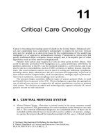
Geneticists estimate that approximately 30 percent of
mutations that cause human diseases are single base-pair
changes that create nonsense mutations. These mutations not only code for a prematurely terminated protein
product, but also trigger rapid decay of the mRNA. Many
more mutations are missense mutations that alter the
amino acid sequence of a protein and frameshift mutations that alter the protein sequence and create internal nonsense codons. Other common disease-associated
mutations affect the sequences of gene promoters, mRNA
splicing signals, and other noncoding sequences that
affect transcription, processing, and stability of mRNA or
protein. One recent study showed that about 15 percent
of all point mutations that cause human genetic diseases
result in abnormal mRNA splicing. Approximately 85
percent of these splicing mutations alter the sequence of
5′ and 3′ splice signals. The remainder create new splice
sites within the gene. Splicing defects often result in degradation of the abnormal mRNA or creation of abnormal
protein products.
Another type of single-gene mutation is caused by
expansions of trinucleotide repeat sequences—specific
short DNA sequences repeated many times. Normal individuals have a low number of repetitions of these sequences;
however, individuals with over 20 different human disorders appear to have abnormally large numbers of repeat
sequences—in some cases, over 200—within and surrounding specific genes.
Examples of diseases associated with these trinucleotide repeat expansions are fragile-X syndrome (discussed
in Chapter 6), myotonic dystrophy, and Huntington disease (discussed in Chapter 4). When trinucleotide repeats
such as (CAG)n occur within a coding region, they can be
translated into long tracks of glutamine. These glutamine
tracks may cause the proteins to aggregate abnormally.
When the repeats occur outside coding regions, but within
the mRNA, it is thought that the mRNAs may act as “toxic”
RNAs that bind to important regulatory proteins, sequestering them away from their normal functions in the
cell. Another possible consequence of long trinucleotide
repeats is that the regions of DNA containing the repeats
may become abnormally methylated, leading to silencing
of gene transcription.
The mechanisms by which the repeated sequences
expand from generation to generation are of great interest.
It is thought that expansion may result from errors during
either DNA replication or DNA damage repair. Whatever
the cause may be, the presence of these short and unstable
repeat sequences seems to be prevalent in humans and in
many other organisms.
14.5 Organisms Use DNA Repair
Systems to Detect and Correct
Mutations
Living systems have evolved a variety of elaborate repair
systems that counteract both spontaneous and induced
DNA damage. These DNA repair systems are absolutely
essential to the maintenance of the genetic integrity of
organisms and, as such, to the survival of organisms on
Earth. The balance between mutation and repair results in
the observed mutation rates of individual genes and organisms. In addition, DNA repair systems correct the genetic
damage that would otherwise result in human genetic diseases and cancer. The link between defective DNA repair
and cancer susceptibility is described in detail later in the
text (see Chapter 16).
We now embark on a review of some systems of DNA
repair, with the emphasis on the major approaches that
organisms use to counteract genetic damage.
14.5
O rganisms U se DNA R epair S y stems to D etect and Correct M utations
Proofreading and Mismatch Repair
Some of the most common types of mutations arise during
DNA replication when an incorrect nucleotide is inserted
by DNA polymerase. The major DNA synthesizing enzyme
in bacteria (DNA polymerase III) makes an error approximately once every 100,000 insertions, leading to an error
rate of 10-5. Fortunately, DNA polymerase proofreads each
step, catching 99 percent of those errors. If an incorrect
nucleotide is inserted during polymerization, the enzyme
can recognize the error and “reverse” its direction. It then
behaves as a 3′ to 5′ exonuclease, cutting out the incorrect nucleotide and replacing it with the correct one. This
improves the efficiency of replication 100-fold, creating
only 1 mismatch in every 107 insertions, for a final error
rate of 10-7.
To deal with errors such as base–base mismatches,
small insertions, and deletions that remain after proofreading, another mechanism, called mismatch repair, may
be activated. During mismatch repair, the mismatches are
detected, the incorrect nucleotide is removed, and the correct nucleotide is inserted in its place. But how does the
repair system recognize which nucleotide is correct (on the
template strand) and which nucleotide is incorrect (on the
newly synthesized strand)? If the mismatch is recognized
but no such discrimination occurs, the excision will be random, and the strand bearing the correct base will be clipped
out 50 percent of the time. Hence, strand discrimination is
a critical step.
The process of strand discrimination has been elucidated in some bacteria, including E. coli, and is based on
DNA methylation. These bacteria contain an enzyme,
adenine methylase, which recognizes the DNA sequence
5′¬GATC¬3′
3′¬CTAG¬5′
as a substrate, adding a methyl group to each of the adenine
residues during DNA replication.
Following replication, the newly synthesized DNA
strand remains temporarily unmethylated, as the adenine
methylase lags behind the DNA polymerase. Prior to methylation, the repair enzyme recognizes the mismatch and binds
to the unmethylated (newly synthesized) DNA strand. An
endonuclease enzyme creates a nick in the backbone of the
unmethylated DNA strand, either 5′ or 3′ to the mismatch.
An exonuclease unwinds and degrades the nicked DNA
strand, until the region of the mismatch is reached. Finally,
DNA polymerase fills in the gap created by the exonuclease,
using the correct DNA strand as a template. DNA ligase then
seals the gap.
A series of E. coli gene products, MutH, MutL, and
MutS, as well as exonucleases, DNA polymerase III and
ligase, are involved in mismatch repair. Mutations in the
283
MutH, MutL, and MutS genes result in bacterial strains deficient in mismatch repair. While the preceding mechanism
occurs in E. coli, similar mechanisms involving homologous
proteins exist in yeast and in mammals.
In humans, mutations in genes that code for DNA mismatch repair proteins (such as the hMSH2 and hMLH1,
which are the human equivalents of the MutS and MutL
genes of E. coli) are associated with the hereditary nonpolyposis colon cancer. Mismatch repair defects are commonly
found in other cancers, such as leukemias, lymphomas,
and tumors of the ovary, prostate, and endometrium. Cells
from these cancers show genome-wide increases in the rate
of spontaneous mutation. The link between defective mismatch repair and cancer is supported by experiments with
mice. Mice that are engineered to have deficiencies in mismatch repair genes accumulate large numbers of mutations
and are cancer-prone.
Postreplication Repair and the
SOS Repair System
Another DNA repair system, called postreplication repair,
responds after damaged DNA has escaped repair and
has failed to be completely replicated. As illustrated in
Figure 14–9, when DNA bearing a lesion of some sort
(such as a pyrimidine dimer) is being replicated, DNA
polymerase may stall at the lesion and then skip over
it, leaving an unreplicated gap on the newly synthesized strand. To correct the gap, the RecA protein directs
a recombinational exchange with the corresponding
region on the undamaged parental strand of the same
polarity (the “donor” strand). When the undamaged
segment of the donor strand DNA replaces the gapped
segment, a gap is created on the donor strand. The gap
can be filled by repair synthesis as replication proceeds.
Because a recombinational event is involved in this type
of DNA repair, it is considered to be a form of homologous recombination repair.
Still another repair pathway, the E. coli SOS repair
system, also responds to damaged DNA, but in a different way. In the presence of a large number of unrepaired DNA mismatches and gaps, bacteria can induce
the expression of about 20 genes (including lexA, recA,
and uvr) whose products allow DNA replication to occur
even in the presence of these lesions. This type of repair
is a last resort to minimize DNA damage, hence its name.
During SOS repair, DNA synthesis becomes error-prone,
inserting random and possibly incorrect nucleotides in
places that would normally stall DNA replication. As a
result, SOS repair itself becomes mutagenic—although
it may allow the cell to survive DNA damage that would
otherwise kill it.
284
14
GEN E MUTATION, D NA REPAIR , AND TRANS POS ITION
Postreplication repair
T T
AA
Lesion
Complementary region
DNA unwound prior
to replication
Base and Nucleotide Excision Repair
T T
AA
Replication skips over
lesion and continues
T T
Recombined
complement
AA
Undamaged complementary
region of parental strand
is recombined
New gap formed
photoreactivation repair is not absolutely essential in
E. coli; we know this because a mutation creating a null
allele in the gene coding for PRE is not lethal. Nonetheless,
the enzyme is detectable in many organisms, including bacteria, fungi, plants, and some vertebrates—though not in
humans. Humans and other organisms that lack photoreactivation repair must rely on other repair mechanisms to
reverse the effects of UV radiation.
A number of light-independent DNA repair systems exist
in all prokaryotes and eukaryotes. The basic mechanisms
involved in these types of repair—collectively referred to as
excision repair or cut-and-paste mechanisms—consist of
the following three steps.
1.The distortion or error present on one of the two strands
of the DNA helix is recognized and enzymatically clipped
out by an endonuclease. Excisions in the phosphodiester
backbone usually include a number of nucleotides adjacent to the error as well, leaving a gap on one strand of
the helix.
2.A DNA polymerase fills in the gap by inserting nucleotides complementary to those on the intact strand, which
T T
Photoreactivation repair
AA
AA
New gap is filled by DNA
polymerase and DNA ligase
Postreplication repair occurs if DNA replication
has skipped over a lesion such as a thymine dimer. Through the
process of recombination, the correct complementary sequence
is recruited from the parental strand and inserted into the gap
opposite the lesion. The new gap is filled by DNA polymerase
and DNA ligase.
TT
5‘
3‘
AA
F I G U RE 1 4 – 9
Photoreactivation Repair: Reversal
of UV Damage
As illustrated in Figure 14–8, UV light is mutagenic as a
result of the creation of pyrimidine dimers. UV-induced
damage to E. coli DNA can be partially reversed if, following
irradiation, the cells are exposed briefly to light in the blue
range of the visible spectrum. The process is dependent on
the activity of a protein called photoreactivation enzyme
(PRE). The enzyme’s mode of action is to cleave the bonds
between thymine dimers, thus directly reversing the effect
of UV radiation on DNA (Figure 14–10). Although the
enzyme will associate with a thymine dimer in the dark, it
must absorb a photon of light to cleave the dimer. In spite of
its ability to reduce the number of UV-induced mutations,
DNA is damaged
Dimer forms
T
T
A
PRE
Blue light
A
T
T
A
A
lesion
Dimer repaired
Normal pairing
restored
TT
AA
FIGUR E 14–10 Damaged DNA repaired by photoreactivation
repair. The bond creating the thymine dimer is cleaved by the
photoreactivation enzyme (PRE), which must be activated by
blue light in the visible spectrum.
14.5
O rganisms U se DNA R epair S y stems to D etect and Correct M utations
it uses as a replicative template. The enzyme adds these
nucleotides to the free 3′-OH end of the clipped DNA. In
E. coli, this step is usually performed by DNA polymerase I.
3.DNA ligase seals the final “nick” that remains at the
3′-OH end of the last nucleotide inserted, closing the gap.
There are two types of excision repair: base excision repair and nucleotide excision repair. Base excision
repair (BER) corrects DNA that contains a damaged DNA
base. The first step in the BER pathway in E. coli involves
the recognition of the altered base by an enzyme called
DNA glycosylase. There are a number of DNA glycosylases,
each of which recognizes a specific base (Figure 14–11). For
example, the enzyme uracil DNA glycosylase recognizes
the presence of uracil in DNA. DNA glycosylases first cut
the glycosidic bond between the base and the sugar, creating an apyrimidinic or apurinic site. The sugar with the
missing base is then recognized by an enzyme called AP
endonuclease. The AP endonuclease makes a cut in the
phosphodiester backbone at the apyrimidinic or apurinic
site. Endonucleases then remove the deoxyribose sugar,
and the gap is filled by DNA polymerase and DNA ligase.
Although much has been learned about the mechanisms of BER in E. coli, BER systems have also been
285
detected in eukaryotes from yeast to humans. Experimental evidence shows that both mouse and human cells that
are defective in BER activity are hypersensitive to the killing effects of gamma rays and oxidizing agents.
Nucleotide excision repair (NER) pathways repair
“bulky” lesions in DNA that alter or distort the double
helix. These lesions include the UV-induced pyrimidine
dimers and DNA adducts discussed previously.
The NER pathway (Figure 14–12) was first discovered
in E. coli by Paul Howard-Flanders and coworkers, who isolated several independent mutants that are sensitive to UV
radiation. One group of genes was designated uvr (ultraviolet repair) and included the uvrA, uvrB, and uvrC mutations.
In the NER pathway, the uvr gene products are involved in
recognizing and clipping out lesions in the DNA. Usually, a
specific number of nucleotides is clipped out around both
sides of the lesion. In E. coli, usually a total of 13 nucleotides
is removed, including the lesion. The repair is then completed by DNA polymerase I and DNA ligase, in a manner
similar to that occurring in BER. The undamaged strand
opposite the lesion is used as a template for the replication,
resulting in repair.
Nucleotide excision repair
5‘
Base excision repair
5‘
3‘
3‘
5‘
3‘
DNA is
damaged
ACUAGT
Duplex with
U–G mismatch
AC
Uracil DNA glycosylase
recognizes and excises
incorrect base
uvr gene
products
T GGT C A
AC
AGT
AP endonuclease
recognizes lesion and
nicks DNA strand
Gap is filled
T GGT C A
DNA
polymerase I
5‘
ACCAGT
Mismatch repaired
3‘
Nuclease
excises lesion
AGT
DNA polymerase and
DNA ligase fill gap
5‘
Lesion
T GGT C A
U
5‘
3‘
3‘
Gap is sealed;
normal pairing
is restored
DNA
ligase
T GGT C A
F I G U RE 1 4 – 1 1 Base excision repair (BER) accomplished by
uracil DNA glycosylase, AP endonuclease, DNA polymerase, and
DNA ligase. Uracil is recognized as a noncomplementary base,
excised, and replaced with the complementary base (C).
FIGUR E 14–12 Nucleotide excision repair (NER) of a UVinduced thymine dimer. During repair, 13 nucleotides are excised
in prokaryotes, and 28 nucleotides are excised in eukaryotes.
286
14
GEN E MUTATION, D NA REPAIR , AND TRANS POS ITION
Nucleotide Excision Repair and Xeroderma
Pigmentosum in Humans
The mechanism of NER in eukaryotes is much more complicated than that in prokaryotes and involves many more
proteins, encoded by about 30 genes. Much of what is
known about the system in humans has come from detailed
studies of individuals with xeroderma pigmentosum
(XP), a rare recessive genetic disorder that predisposes
individuals to severe skin abnormalities, skin cancers, and
a wide range of other symptoms including developmental
and neurological defects. Patients with XP are extremely
sensitive to UV radiation in sunlight. In addition, they have
a 2000-fold higher rate of cancer, particularly skin cancer,
than the general population. The condition is severe and
may be lethal, although early detection and protection from
sunlight can arrest it (Figure 14–13).
The repair of UV-induced lesions in XP has been investigated in vitro, using human fibroblast cell cultures derived
from normal individuals and those with XP. (Fibroblasts
are undifferentiated connective tissue cells.) The results
of these studies suggest that the XP phenotype is caused by
defects in NER pathways and by mutations in more than
one gene.
In 1968, James Cleaver showed that cells from XP
patients were deficient in DNA synthesis other than that
occurring during chromosome replication—a phenomenon
known as unscheduled DNA synthesis. Unscheduled
DNA synthesis is elicited in normal cells by UV radiation.
Because this type of synthesis is thought to represent the
activity of DNA polymerization during NER, the lack of
unscheduled DNA synthesis in XP patients suggested that
XP may be a deficiency in NER.
The involvement of multiple genes in NER and XP
was further investigated by studies using somatic cell
hybridization. Fibroblast cells from any two unrelated
XP patients, when grown together in tissue culture, can
fuse together, forming heterokaryons. A heterokaryon
is a single cell with two nuclei from different organisms
but a common cytoplasm. NER in the heterokaryon can be
measured by the level of unscheduled DNA synthesis. If
the mutation in each of the two XP cells occurs in the same
gene, the heterokaryon, like the cells that fused to form it,
will still be unable to undergo NER. This is because there is
no normal copy of the relevant gene present in the heterokaryon. However, if NER does occur in the heterokaryon,
the mutations in the two XP cells must have been present
in two different genes. Hence, the two mutants are said to
demonstrate complementation, a concept also discussed
earlier in the text (see Chapter 4). Complementation occurs
because the heterokaryon has at least one normal copy of
each gene in the fused cell. By fusing XP cells from a large
number of XP patients, researchers were able to determine
how many genes contribute to the XP phenotype.
Based on these and other studies, XP patients were
divided into seven complementation groups, indicating
that at least seven different genes are involved in nucleotide excision repair in humans. A gene representing each
of these complementation groups, XPA to XPG (Xeroderma Pigmentosum gene A to G), has now been identified, and a homologous gene for each has been identified
in yeast. Approximately 20 percent of XP patients do not
fall into any of the seven complementation groups. Cells
from most of these patients have mutations in the gene
coding for DNA polymerase h and are defective in repair
DNA synthesis.
As a result of the study of defective genes in XP, a
great deal is now known about how NER counteracts
DNA damage in normal cells. The first step in humans
is recognition of the damaged DNA by proteins encoded
by the XPC, XPE, and XPA genes. These proteins then
recruit the remainder of the repair proteins to the site
of DNA damage. The XPB and XPD genes encode helicases, and the XPF and XPG genes encode nucleases. The
excision repair complex containing these and other factors is responsible for the excision of an approximately
28-nucleotide-long fragment from the DNA strand that
contains the lesion.
Double-Strand Break Repair in Eukaryotes
F I G U RE 1 4 – 1 3 Two individuals with xeroderma pigmentosum.
These XP patients show characteristic XP skin lesions induced
by sunlight, as well as mottled redness (erythema) and irregular
pigment changes to the skin, in response to cellular injury.
Thus far, we have discussed repair pathways that deal with
damage or errors within one strand of DNA. We conclude
our discussion of DNA repair by considering what happens when both strands of the DNA helix are cleaved—as a
result of exposure to ionizing radiation, for example. These
14.5
O rganisms U se DNA R epair S y stems to D etect and Correct M utations
287
Double-stranded break
types of damage are extremely dangerous to cells,
leading to chromosome rearrangements, cancer, or
cell death. In this section, we will discuss double3‘
5‘
strand breaks in eukaryotic cells.
5‘
3‘
Specialized forms of DNA repair, the DNA
double-strand break repair (DSB repair) pathBreak detected and
5‘-ends digested
ways, are activated and are responsible for reat3‘
5‘
taching two broken DNA strands. Recently, interest
in DSB repair has grown because defects in these
5‘
3‘
pathways are associated with X-ray hypersensitiv3‘-end invades homologous
ity and immune deficiency. Such defects may also
region of sister chromatid
underlie familial disposition to breast and ovarian
3‘
5‘
cancer. Several human disease syndromes, such as
3‘
5‘
3‘
Fanconi anemia and ataxia telangiectasia, result
Sister
from defects in DSB repair.
chromatids
One pathway involved in double-strand break
3‘
5‘
3‘
repair is homologous recombination repair. The
5‘
3‘
first step in this process involves the activity of an
enzyme that recognizes the double-strand break,
DNA synthesis across
damaged region
and then digests back the 5′-ends of the broken DNA
3‘
5‘
helix, leaving overhanging 3′-ends (Figure 14–14).
One overhanging end searches for a region of
3‘
5‘
sequence complementarity on the sister chromatid and then invades the homologous DNA duplex,
5‘
3‘
aligning the complementary sequences. Once
5‘
3‘
aligned, DNA synthesis proceeds from the 3′ overHeteroduplex resolved and
hanging ends, using the undamaged DNA strands
gaps filled by DNA synthesis
as templates. The interaction of two sister chromaand ligation
tids is necessary because, when both strands of one
3‘
5‘
helix are broken, there is no undamaged parental
5‘
3‘
DNA strand available to use as a source of the com3‘
5‘
plementary template DNA sequence during repair.
After DNA repair synthesis, the resulting heterodu5‘
3‘
plex molecule is resolved and the two chromatids
FIGUR E 14–14 Steps in homologous recombination repair of doubleseparate.
stranded breaks.
DSB repair usually occurs during the late S or
early G2 phase of the cell cycle, after DNA replicadouble-strand break, the wrong ends could be joined together,
tion, a time when sister chromatids are available to be used
leading to abnormal chromosome structures, such as those
as repair templates. Because an undamaged template is used
discussed earlier in the text (see Chapter 6).
during repair synthesis, homologous recombination repair is
an accurate process.
A second pathway, called nonhomologous end joining,
also repairs double-strand breaks. However, as the name
14–4 Geneticists often use ethylmethane sulfonate (EMS)
implies, the mechanism does not recruit a homologous
to induce mutations in Drosophila. Why is EMS a mutagen
region of DNA during repair. This system is activated in G1,
of choice for genetic research? What would be the effects
prior to DNA replication. End joining involves a complex
of EMS in a strain of Drosophila lacking functional mismatch
of many proteins, and may include the DNA-dependent
repair systems?
protein kinase and the breast cancer susceptibility gene
HINT: This problem asks you to evaluate EMS as a useful
product, BRCA1. These and other proteins bind to the free
mutagen and to determine its effects in the absence of DNA repair.
ends of the broken DNA, trim the ends, and ligate them
The key to its solution is to consider the chemical effects of EMS
back together. Because some nucleotide sequences are
on DNA. Also, consider the types of DNA repair that may operate
lost in the process of end joining, it is an error-prone repair
on EMS-mutated DNA and the efficiency of these processes.
system. In addition, if more than one chromosome suffers a
288
14
GEN E MUTATION, D NA REPAIR , AND TRANS POS ITION
ESS EN T I A L P O IN T
Organisms counteract mutations by using a range of DNA repair
systems. Errors in DNA synthesis can be repaired by proofreading,
mismatch repair, and postreplication repair. DNA damage can
be repaired by photoreactivation repair, SOS repair, base excision
repair, nucleotide excision repair, and double-strand break repair.
14.6 The Ames Test Is Used to Assess
the Mutagenicity of Compounds
There is great concern about the possible mutagenic properties of any chemical that enters the human body, whether
through the skin, the digestive system, or the respiratory
tract. Examples of synthetic chemicals that concern us are
those found in air and water pollution, food preservatives,
artificial sweeteners, herbicides, pesticides, and pharmaceutical products. Mutagenicity can be tested in various
organisms, including fungi, plants, and cultured mammalian cells; however, one of the most common tests, which
we describe here, uses bacteria.
The Ames test uses a number of different strains of the
bacterium Salmonella typhimurium that have been selected
for their ability to reveal the presence of specific types of
mutations. For example, some strains are used to detect
base-pair substitutions, and other strains detect various
frameshift mutations. Each strain contains a mutation in
one of the genes of the histidine operon. The mutant strains
are unable to synthesize histidine (his− strains) and therefore require histidine for growth (Figure 14–15). The assay
measures the frequency of reverse mutations that occur
within the mutant gene, yielding wild-type bacteria (his+
revertants). These Salmonella strains also have an increased
sensitivity to mutagens due to the presence of mutations in
genes involved in both DNA damage repair and the synthesis
of the lipopolysaccharide barrier that coats bacteria and protects them from external substances.
Many substances entering the human body are relatively innocuous until activated metabolically, usually
in the liver, to more chemically reactive products. Thus,
the Ames test includes a step in which the test compound
is incubated in vitro in the presence of a mammalian liver
extract. Alternatively, test compounds may be injected
into a mouse where they are modified by liver enzymes and
then recovered for use in the Ames test.
In the initial use of Ames testing in the 1970s, a large
number of known carcinogens, or cancer-causing agents,
were examined, and more than 80 percent of these were
shown to be strong mutagens. This is not surprising, as
the transformation of cells to the malignant state occurs
as a result of mutations. For example, more than 60 compounds found in cigarette smoke test positive in the Ames
his- auxotrophs
plus liver enzymes
Potential mutagen
plus liver enzymes
Add mixture to
filter paper disk
Spread bacteria
on agar medium
without histidine
Place disk on
surface of medium
Incubate at 37°C
Spontaneous his+
his+ revertants
revertants (control) induced by mutagen
FIGUR E 14–15 The Ames test, which screens compounds for
potential mutagenicity.
test and cause cancer in animal tests. Although a positive
response in the Ames test does not prove that a compound
is carcinogenic, the Ames test is useful as a preliminary
screening device. The test is used extensively during the
development of industrial and pharmaceutical chemical
compounds.
14.7 Transposable Elements Move
within the Genome and May Create
Mutations
Transposable elements, also known as transposons or
“jumping genes,” can move or transpose within and between
chromosomes, inserting themselves into various locations
within the genome.
Transposable elements are present in the genomes of
all organisms from bacteria to humans. Not only are they
ubiquitous, but they also comprise large portions of some
eukaryotic genomes. For example, almost 50 percent of
the human genome is derived from transposable elements.
14.7
T ransposabl e E l ements M ove wit h in t h e G enome and M ay Create M utations
289
IS
Some organisms with unusually large genomes, such
as salamanders and barley, contain hundreds of
Terminal sequence
Internal sequence
thousands of copies of various types of transposable
A T C CG
CGG A T
elements. Although the function of these elements is
5‘
3‘
unknown, data from human genome sequencing sug3‘
5‘
gest that some genes may have evolved from transT A GGC
GC C T A
posable elements and that the presence of these eleInverted terminal sequence
ments may help to modify and reshape the genome.
Transposable elements are also valuable tools in FIGUR E 14–16 An insertion sequence (IS), shown in purple. The
genetic research. Geneticists harness transposons as terminal sequences are perfect inverted repeats of one another.
mutagens, as cloning tags, and as vehicles for introducing foreign DNA into model organisms.
In this section, we discuss transposable elements as
segments of DNA that have the same nucleotide sequence
naturally occurring mutagens. The movement of transas each other but are oriented in the opposite direction
posable elements from one place in the genome to another
(Figure 14–16). Although Figure 14–16 shows the ITRs to
has the capacity to disrupt genes and cause mutations,
consist of only a few nucleotides, IS ITRs usually contain
as well as to create chromosomal damage such as doubleabout 20 to 40 nucleotide pairs. ITRs are essential for transstrand breaks.
position and act as recognition sites for the binding of the
Insertion Sequences and Bacterial
Transposons
There are two types of transposable elements in bacteria:
insertion sequences and bacterial transposons. Insertion
sequences (IS elements) can move from one location to
another and, if they insert into a gene or gene-regulatory
region, may cause mutations.
IS elements were first identified during analyses of
mutations in the gal operon of E. coli. Researchers discovered that certain mutations in this operon were due to
the presence of several hundred base pairs of extra DNA
inserted into the beginning of the operon. Surprisingly,
the segment of mutagenic DNA could spontaneously
excise from this location, restoring wild-type function to
the gal operon. Subsequent research revealed that several other DNA elements could behave in a similar fashion, inserting into bacterial chromosomes and affecting
gene function.
IS elements are relatively short, not exceeding 2000 bp
(2 kb). The first insertion sequence to be characterized in
E. coli, IS1, is about 800 bp long. Other IS elements such as
IS2, 3, 4, and 5 are about 1250 to 1400 bp in length. IS elements are present in multiple copies in bacterial genomes.
For example, the E. coli chromosome contains five to eight
copies of IS1, five copies each of IS2 and IS3, as well as copies of IS elements on plasmids such as F factors.
All IS elements contain two features that are essential
for their movement. First, they contain a gene that encodes
an enzyme called transposase. This enzyme is responsible
for making staggered cuts in chromosomal DNA, into which
the IS element can insert. Second, the ends of IS elements
contain inverted terminal repeats (ITRs). ITRs are short
transposase enzyme.
Bacterial transposons (Tn elements) are larger than
IS elements and contain protein-coding genes that are
unrelated to their transposition. Some Tn elements, such
as Tn10, are composed of a drug-resistance gene flanked
by two IS elements present in opposite orientations. The
IS elements encode the transposase enzyme that is necessary for transposition of the Tn element. Other types of
Tn elements, such as Tn3, have shorter inverted repeat
sequences at their ends and encode their transposase
enzyme from a transposase gene located in the middle of
the Tn element. Like IS elements, Tn elements are mobile
in both bacterial chromosomes and in plasmids and can
cause mutations if they insert into genes or gene-regulatory
regions.
Tn elements are currently of interest because they can
introduce multiple drug resistance onto bacterial plasmids.
These plasmids, called R factors, may contain many Tn
elements conferring simultaneous resistance to heavy
metals, antibiotics, and other drugs. These elements can
move from plasmids onto bacterial chromosomes and can
spread multiple drug resistance between different strains
of bacteria.
The Ac–Ds System in Maize
About 20 years before the discovery of transposons in
bacteria, Barbara McClintock discovered mobile genetic
elements in corn plants (maize). She did this by analyzing the genetic behavior of two mutations, Dissociation
(Ds) and Activator (Ac), expressed in either the endosperm or aleurone layers. She then correlated her genetic
observations with cytological examinations of the maize
290
14
GEN E MUTATION, D NA REPAIR , AND TRANS POS ITION
chromosomes. Initially, McClintock determined that Ds
was located on chromosome 9. If Ac was also present in the
genome, Ds induced breakage at a point on the chromosome adjacent to its own location. If chromosome breakage
occurred in somatic cells during their development, progeny cells often lost part of the broken chromosome, causing a variety of phenotypic effects. The chapter-opening
photo illustrates the types of phenotypic effects caused by
Ds mutations in kernels of corn.
Subsequent analysis suggested to McClintock that
both Ds and Ac elements sometimes moved to new chromosomal locations. While Ds moved only if Ac was also
present, Ac was capable of autonomous movement. Where
Ds came to reside determined its genetic effects—that is,
it might cause chromosome breakage, or it might inhibit
expression of a certain gene. In cells in which Ds caused a
gene mutation, Ds might move again, restoring the gene
mutation to wild type.
Figure 14–17 illustrates the types of movements
and effects brought about by Ds and Ac elements. In
McClintock’s original observation, pigment synthesis
was restored in cells in which the Ds element jumped
out of chromosome 9. McClintock concluded that the Ds
and Ac genes were mobile controlling elements. We
now commonly refer to them as transposable elements,
a term coined by another great maize geneticist, Alexander Brink.
Several Ac and Ds elements have now been analyzed,
and the relationship between the two elements has been
clarified. The first Ds element studied (Ds9) is nearly identical to Ac except for a 194-bp deletion within the transposase gene. The deletion of part of the transposase gene
in the Ds9 element explains its dependence on the Ac element for transposition. Several other Ds elements have also
been sequenced, and each contains an even larger deletion
within the transposase gene. In each case, however, the
ITRs are retained.
Although the significance of Barbara McClintock’s
mobile controlling elements was not fully appreciated following her initial observations, molecular analysis has
since verified her conclusions. She was awarded the Nobel
Prize in Physiology or Medicine in 1983.
Copia and P Elements in Drosophila
There are more than 30 families of transposable elements in
Drosophila, each of which is present in 20 to 50 copies in the
genome. Together, these families constitute about 5 percent
of the Drosophila genome and over half of the middle repetitive DNA of this organism. One study suggests that 50 percent of all visible mutations in Drosophila are the result of
the insertion of transposons into otherwise wild-type genes.
(a) In absence of Ac, Ds is not transposable.
Wild-type expression of W occurs.
Ds
W
(b) When Ac is present, Ds may be transposed.
Ac
Ds
W
Ac is present.
Ds is transposed.
Ac
Ds W
Chromosome breaks and fragment is lost.
W expression ceases, producing mutant effect.
Ac
Ds W
(c) Ds can move into and out of another gene.
Ac
Ds
W
Ds is transposed into W gene.
W expression is inhibited,
producing mutant effect.
W
Ds
Ac
Ds “jumps” out of W gene.
Wild-type expression of W is restored.
Ac
Ds
W
FIGUR E 14–17 Effects of Ac and Ds elements on gene expression. (a) If Ds is present in the absence of Ac, there is normal
expression of a distantly located hypothetical gene W. (b) In the
presence of Ac, Ds may transpose to a region adjacent to W. Ds
can induce chromosome breakage, which may lead to loss of a
chromosome fragment bearing the W gene. (c) In the presence
of Ac, Ds may transpose into the W gene, disrupting W-gene
expression. If Ds subsequently transposes out of the W gene,
W-gene expression may return to normal.
In 1975, David Hogness and his colleagues David
Finnegan, Gerald Rubin, and Michael Young identified
a class of DNA elements in Drosophila melanogaster that
they designated as copia. These elements are transcribed
into “copious” amounts of RNA (hence their name). Copia
elements are present in 10 to 100 copies in the genomes of
Drosophila cells. Mapping studies show that they are transposable to different chromosomal locations and are dispersed throughout the genome.
Each copia element consists of approximately 5000 to
8000 bp of DNA, including a long direct terminal repeat
14.7
T ransposabl e E l ements M ove wit h in t h e G enome and M ay Create M utations
copia gene (5000 bp)
DTR
(267 bp)
DTR
ITR (17 bp)
ITR
F I G U RE 1 4 – 1 8 Structural organization of a copia transposable
element in Drosophila melanogaster, showing the terminal repeats.
(DTR) sequence of 267 bp at each end. Within each DTR is
an inverted terminal repeat (ITR) of 17 bp (Figure 14–18).
Insertion of copia is dependent on the presence of the
ITR sequences and seems to occur preferentially at specific
target sites in the genome. The copia-like elements demonstrate regulatory effects at the point of their insertion in the
chromosome. Certain mutations, including those affecting
eye color and segment formation, are due to copia insertions within genes. For example, the eye-color mutation
white-apricot is caused by an allele of the white gene, which
contains a copia element within the gene. Transposition of
the copia element out of the white-apricot allele can restore
the allele to wild type.
Perhaps the most significant Drosophila transposable elements are the P elements. These were discovered
while studying the phenomenon of hybrid dysgenesis,
a condition characterized by sterility, elevated mutation
rates, and chromosome rearrangements in the offspring of
crosses between certain strains of fruit flies. Hybrid dysgenesis is caused by high rates of P element transposition
in the germ line, in which transposons insert themselves
into or near genes, thereby causing mutations. P elements
range from 0.5 to 2.9 kb long, with 31-bp ITRs. Full-length
P elements encode at least two proteins, one of which is
the transposase enzyme that is required for transposition,
and another is a repressor protein that inhibits transposition. The transposase gene is expressed only in the germ
line, accounting for the tissue specificity of P element
transposition. Strains of flies that contain full-length P elements inserted into their genomes are resistant to further
transpositions due to the presence of the repressor protein
encoded by the P elements.
Mutations can arise from several kinds of insertional
events. If a P element inserts into the coding region of a
gene, it can terminate transcription of the gene and destroy
normal gene expression. If it inserts into the promoter
region of a gene, it can affect the level of expression of the
gene. Insertions into introns can affect splicing or cause the
premature termination of transcription.
Geneticists have harnessed P elements as tools for genetic
analysis. One of the most useful applications of P elements is
291
as vectors to introduce transgenes into Drosophila—a technique known as germ-line transformation. P elements are
also used to generate mutations and to clone mutant genes. In
addition, researchers are perfecting methods to target P element insertions to precise single-chromosomal sites, which
should increase the precision of germ-line transformation in
the analysis of gene activity.
Transposable Elements in Humans
The human genome, like that of other eukaryotes, is riddled with DNA derived from transposons. Recent genomic
sequencing data reveal that approximately half of the
human genome is composed of transposable element DNA.
As discussed earlier in the text (see Chapter 11), the major
families of human transposable elements are the long interspersed elements and short interspersed elements (LINEs
and SINEs, respectively). Together, they comprise over 30
percent of the human genome.
Although most human transposable elements appear
to be inactive, the potential mobility and mutagenic effects
of these elements have far-reaching implications for human
genetics, as can be seen in a recent example of a transposable element “caught in the act.” The case involves a male
child with hemophilia. One cause of hemophilia is a defect
in blood-clotting factor VIII, the product of an X-linked
gene. Haig Kazazian and his colleagues found LINEs
inserted at two points within the gene. Researchers were
interested in determining if one of the mother’s X chromosomes also contained this specific LINE. If so, the unaffected mother would be heterozygous and pass the LINEcontaining chromosome to her son. The surprising finding
was that the LINE sequence was not present on either of
her X chromosomes but was detected on chromosome 22 of
both parents. This suggests that this mobile element may
have transposed from one chromosome to another in the
gamete-forming cells of the mother, prior to being transmitted to the son.
LINE insertions into the human dystrophin gene have
resulted in at least two separate cases of Duchenne muscular
dystrophy. In one case, a LINE inserted into exon 48, and in
another case, it inserted into exon 44, both leading to frameshift mutations and premature termination of translation
of the dystrophin protein. There are also reports that LINEs
have inserted into the APC and c-myc genes, leading to mutations that may have contributed to the development of some
colon and breast cancers. In the latter cases, the transposition
had occurred within one or a few somatic cells. As of 2012,
researchers have determined that at least 25 LINE element
insertions have resulted in single-gene human diseases.
SINE insertions are also responsible for more than
30 cases of human disease. In one case, an Alu element
292
14
GEN E MUTATION, D NA REPAIR , AND TRANS POS ITION
integrated into the BRCA2 gene, inactivating this tumor
suppressor gene and leading to a familial case of breast
cancer. Other genes that have been mutated by Alu integrations are the factor IX gene (leading to hemophilia B),
the ChE gene (leading to acholinesterasemia), and the NF1
gene (leading to neurofibromatosis).
Transposons, Mutations, and Evolution
Transposons can have a wide range of effects on genes. The
insertion of a transposon into the coding region of a gene
may disrupt the gene’s normal translation reading frame
or may induce premature termination of translation of the
mRNA transcribed from the gene. Many transposable elements contain their own promoters and enhancers, as well
as splice sites and polyadenylation signals. The presence
of these regulatory sequences can have effects on nearby
genes. The insertion of a transposable element containing
polyadenylation or transcription termination signals into
a gene’s intron may bring about termination of the gene’s
transcription within the element. In addition, it can cause
aberrant splicing of an RNA transcribed from the gene.
Insertions of a transposon into a gene’s transcription regulatory region may disrupt the gene’s normal regulation or
may cause the gene to be expressed differently as a result of
the presence of the transposon’s own promoter or enhancer
sequences. The presence of two or more identical transposons in a genome creates the potential for recombination
between the transposons, leading to duplications, deletions, inversions, or chromosome translocations. Any of
these rearrangements may bring about phenotypic changes
or disease.
It is thought that about 0.2 percent of detectable
human mutations may be due to transposable element
insertions. Other organisms appear to suffer more damage due to transposition. About 10 percent of new mouse
mutations and 50 percent of Drosophila mutations are
CASE STUDY
S
caused by insertions of transposable elements in or
near genes.
Because of their ability to alter genes and chromosomes, transposons may contribute to the variability
that underlies evolution. For example, the Tn elements
of bacteria carry antibiotic resistance genes between
organisms, conferring a survival advantage to the bacteria under certain conditions. Another example of a
transposon’s contribution to evolution is provided by
Drosophila telomeres. LINE-like elements are present at
the ends of Drosophila chromosomes, and these elements
act as telomeres, maintaining the length of Drosophila
chromosomes over successive cell divisions. Other examples of evolved transposons are the RAG1 and RAG2 genes
in humans. These genes encode recombinase enzymes
that are essential to the development of the immune
system. These two genes appear to have evolved from
transposons.
Transposons may also affect the evolution of genomes
by altering gene-expression patterns in ways that are subsequently retained by the host. For example, the human
amylase gene contains an enhancer that causes the gene to
be expressed in the parotid gland. This enhancer evolved
from transposon sequences that were inserted into the
generegulatory region early in primate evolution. Other
examples of gene-expression patterns that were affected
by the presence of transposon sequences are T-cell-specific
expression of the CD8 gene and placenta-specific expression of the leptin and CYP19 genes.
ESSEN T IAL PO IN T
Transposable elements can move within a genome, creating mutations
and altering gene expression. Besides creating mutations, transposons
may contribute to evolution. Geneticists use transposons as research
tools to create mutations, clone genes, and introduce genes into
model organisms.
Genetic dwarfism
even months pregnant, an expectant mother was undergoing a routine ultrasound. While prior tests had been
normal, this one showed that the limbs of the fetus were
unusually short. The doctor suspected that the baby might
have a genetic form of dwarfism called achondroplasia. He
told her that the disorder was due to an autosomal dominant
mutation and occurred with a frequency of about 1 in 25,000
births. The expectant mother had studied genetics in college
and immediately raised several questions. How would you
answer them?
1.How could her baby have a dominantly inherited disorder if
there was no history of this condition on either side of the
family?
2.Is the mutation more likely to have come from the mother or
the father?
3.If this child has achondroplasia, is there an increased chance
that their next child would also have this disorder?
4.Could this disorder have been caused by X rays or ultrasounds she had earlier in pregnancy?
PROBLE MS A ND DIS C U S S IO N Q U ES T I O N S
293
INSIGHTS AND SOLUTIONS
1. The base analog 2-amino purine (2-AP) substitutes for
adenine during DNA replication, but it may base-pair with
cytosine. The base analog 5-bromouracil (5-BU) substitutes
for thymidine, but it may base-pair with guanine. Follow the
double-stranded trinucleotide sequence shown here through
three rounds of replication, assuming that, in the first round,
both analogs are present and become incorporated wherever
possible. Before the second and third round of replication,
any unincorporated base analogs are removed. What final
sequences occur?
Solution:
5-BU
substitutes
for T
A
T
G
C
T
A
A
5BU
2AP
T
G
C
G
C
T
2AP
5BU
A
Solution: Only four cases represent a new mutation. Because
each live birth represents two gametes, the sample size is
from 80,000 meiotic events. The rate is equal to
4>80,000 = 1>20,000 = 5 * 10-5
We have assumed that the mutant gene is fully penetrant and
is expressed in each individual bearing it. If it is not fully penetrant, our calculation may be an underestimate because one
or more mutations may have gone undetected. We have also
assumed that the screening was 100 percent accurate. One
or more mutant individuals may have been “missed,” again
leading to an underestimate. Finally, we assumed that the
viability of the mutant and nonmutant individuals is equivalent and that they survive equally in utero. Therefore, our
assumption is that the number of mutant individuals at birth
is equal to the number at conception. If this were not true, our
calculation would again be an underestimate.
2-AP
substitutes
for A
Round I
2. A rare dominant mutation expressed at birth was studied in
humans. Records showed that six cases were discovered in
40,000 live births. Family histories revealed that in two cases,
the mutation was already present in one of the parents. Calculate the spontaneous mutation rate for this mutation. What are
some underlying assumptions that may affect our conclusions?
3. Consider the following estimates:
a. There are 7 * 109 humans living on this planet.
b. Each individual has about 20,000 (0.2 * 105) genes.
c. The average mutation rate at each locus is 10-5.
Round II
A
T
G
5BU
2AP
C
A
T
G
C
G
C
G
C
G
C
T
A
C
2AP
5BU
G
T
A
C
G
C
C and
G
C
G
C
G
Round III
G
C
G
G
C
and G
C
G
C
5BU
2AP
C
G
2AP
5BU
Problems and Discussion Questions
HOW DO WE KNOW
?
1. In this chapter, we focused on how gene mutations arise and
how cells repair DNA damage. In particular, we discussed spontaneous and induced mutations, DNA repair methods, and
transposable elements. Based on your knowledge of these topics,
answer several fundamental questions:
(a) How do we know that mutations occur spontaneously?
(b) How do we know that certain chemicals and wavelengths of
radiation induce mutations in DNA?
How many spontaneous mutations are currently present in
the human population? Assuming that these mutations are
equally distributed among all genes, how many new mutations have arisen in each gene in the human population?
Solution: First, since each individual is diploid, there are
two copies of each gene per person, each arising from a separate gamete. Therefore, the total number of spontaneous
mutations is
(2 * 0.2 * 105 genes) * (7 * 109 humans) * (10-5 mutations)
= (0.4 * 105) * (7 * 109) * (10 - 5) mutations
= 2.8 * 109 mutations in the population
2.8 * 109 mutations>0.2 * 105 genes
= 14 * 104 mutations per gene in the population
Visit for
instructor-assigned tutorials and problems.
(c) How do we know that DNA repair mechanisms detect and
correct the majority of spontaneous and induced mutations?
CONCEPT QUESTION
2. Review the Chapter Concepts list on page 273. These concepts
relate to how gene mutations occur, their phenotypic effects,
and how mutations can be repaired. The first four concepts
focus on the effects of gene mutations in diploid organisms.
Write a short essay describing how these concepts would apply,
or not apply, to a haploid organism such as E. coli.
294
14
GEN E MUTATION, D NA REPAIR , AND TRANS POS ITION
3. Distinguish between spontaneous and induced mutations. Give
some examples of mutagens that cause induced mutations.
4. Why would a mutation in a somatic cell of a multicellular organism escape detection?
5. Why is a random mutation more likely to be deleterious than
beneficial?
6. Why are organisms that have a haploid life cycle valuable tools
for mutagenesis studies?
7. What is meant by a conditional mutation?
8. Describe a tautomeric shift and how it may lead to a mutation.
9. Contrast and compare the mutagenic effects of deaminating
agents, alkylating agents, and base analogs.
10. Why are frameshift mutations likely to be more detrimental
than point mutations, in which a single pyrimidine or purine
has been substituted?
11. In which phases of the cell cycle would you expect double-strand
break repair and nonhomologous end joining to occur and why?
12. DNA damage brought on by a variety of natural and artificial
agents elicits a wide variety of cellular responses. In addition to the activation of DNA repair mechanisms, there can
be activation of pathways leading to apoptosis (programmed
cell death) and cell-cycle arrest. Why would apoptosis and
cell-cycle arrest often be part of a cellular response to DNA
damage?
13. Distinguish between proofreading and mismatch repair.
14. How would you expect the misincorporation of bases by a DNA
polymerase to change if the relative ratios of the dNTPs were
A = T = G but a five-fold excess of C?
15. A chemist has synthesized a novel chemical, which he suspects
to be a potential mutagen. Name and explain a popular test that
can be used to test the mutagenicity of this product in bacteria.
16. What genetic defects result in the disorder xeroderma pigmentosum (XP) in humans? How do these defects create the phenotypes associated with the disorder?
17. In a bacterial culture in which all cells are unable to synthesize leucine (leu-), a potent mutagen is added, and the cells are
allowed to undergo one round of replication. At that point, samples
are taken, a series of dilutions is made, and the cells are plated
on either minimal medium or minimal medium containing
leucine. The first culture condition (minimal medium) allows
the growth of only leu+ cells, while the second culture condition
(minimum medium with leucine added) allows the growth of all
cells. The results of the experiment are as follows:
Culture Condition
Minimal medium
Minimal + leucine
Dilution
Colonies
10-1
10-7
18
6
What is the rate of mutation at the locus associated with leucine
biosynthesis?
18. DNA mismatch repair is a mechanism of DNA repair that has
been observed in E. coli. Give a list of genes in E. coli, mutations
in which can adversely affect DNA mismatch repair. Give a list
of equivalent genes in humans.
19. A number of different types of mutations in the HBB gene can
cause human b-thalassemia, a disease characterized by various
levels of anemia. Many of these mutations occur within introns
or in upstream noncoding sequences. Explain why mutations
in these regions often lead to severe disease, although they may
not directly alter the coding regions of the gene.
20. Some mutations that lead to diseases such as Huntington
disease are caused by the insertion of trinucleotide repeats.
Describe how the process of DNA replication could lead to
expansions of trinucleotide repeat regions.
21. In maize, a Ds or Ac transposon can cause mutations in genes at
or near the site of transposon insertion. It is possible for these
elements to transpose away from their original site, causing a
reversion of the mutant phenotype. In some cases, however,
even more severe phenotypes appear, due to events at or near
the mutant allele. What might be happening to the transposon
or the nearby gene to create more severe mutations?
22. Presented here are hypothetical findings from studies of heterokaryons formed from seven human xeroderma pigmentosum
cell strains:
XP1
XP2
XP3
XP4
XP5
XP6
XP1
-
XP2
-
-
XP3
-
-
-
XP4
+
+
+
-
XP5
+
+
+
+
-
XP6
+
+
+
+
-
-
XP7
+
+
+
+
-
-
XP7
-
Note: “ + ” = complementation; “ - ” = no complementation
These data are measurements of the occurrence or nonoccurrence of unscheduled DNA synthesis in the fused heterokaryon.
None of the strains alone shows any unscheduled DNA synthesis. What does unscheduled DNA synthesis represent? Which
strains fall into the same complementation groups? How many
different groups are revealed based on these data? What can we
conclude about the genetic basis of XP from these data?
23. Cystic fibrosis (CF) is a severe autosomal recessive disorder in
humans that results from a chloride ion channel defect in epithelial cells. More than 500 mutations have been identified in
the 24 exons of the responsible gene (CFTR, or cystic fibrosis
transmembrane regulator), including dozens of different missense mutations, frameshift mutations, and splice-site defects.
Although all affected CF individuals demonstrate chronic
obstructive lung disease, there is variation in whether or not
they exhibit pancreatic enzyme insufficiency (PI). Speculate
as to which types of mutations are likely to give rise to less
severe symptoms of CF, including only minor PI. Some of the
300 sequence alterations that have been detected within the
exon regions of the CFTR gene do not give rise to cystic fibrosis.
Taking into account your knowledge of the genetic code, gene
expression, protein function, and mutation, describe why this
might be so.
24. Electrophilic oxidants are known to create the modified base
named 7,8-dihydro-8-oxoguanine (oxoG) in DNA. Whereas guanine base-pairs with cytosine, oxoG base-pairs with either cytosine or adenine.
(a) What are the sources of reactive oxidants within cells that
cause this type of base alteration?
(b) Drawing on your knowledge of nucleotide chemistry, draw
the structure of oxoG, and, below it, draw guanine. Opposite
guanine, draw cytosine, including the hydrogen bonds that
allow these two molecules to base-pair. Does the structure of
oxoG, in contrast to guanine, provide any hint as to why it basepairs with adenine?
PROBLE MS AND DIS C U S S IO N Q U ES T I O N S
(c) Assume that an unrepaired oxoG lesion is present in the
helix of DNA opposite cytosine. Predict the type of mutation
that will occur following several rounds of replication.
(d) Which DNA repair mechanisms might work to counteract
an oxoG lesion? Which of these is likely to be most effective?
25. Skin cancer carries a lifetime risk nearly equal to that of all
other cancers combined. Following is a graph (modified from
Kraemer, 1997. Proc. Natl. Acad. Sci. (USA) 94: 11–14) depicting
the age of onset of skin cancers in patients with or without XP,
where cumulative percentage of skin cancer is plotted against
age. The non-XP curve is based on 29,757 cancers surveyed by
the National Cancer Institute, and the curve representing those
with XP is based on 63 skin cancers from the Xeroderma Pigmentosum Registry.
(a) Provide an overview of the information contained in the
graph.
(b) Explain why individuals with XP show such an early age of
onset.
Cumulative percentage
100
XP
50
Non-XP
0
0
20
40
60
Age in years
80
295
26. The initial discovery of IS elements in bacteria revealed the
presence of an element upstream (5′) of three genes controlling
galactose metabolism. All three genes were affected simultaneously, although there was only one IS insertion. Offer an explanation as to why this might occur.
27. Suppose you are studying a DNA repair system, such as the
nucleotide excision repair in vitro. By mistake, you add DNA
ligase from a tube that has already expired. What would be the
result?
28. It has been noted that most transposons in humans and other
organisms are located in noncoding regions of the genome—
regions such as introns, pseudogenes, and stretches of particular types of repetitive DNA. There are several ways to interpret
this observation. Describe two possible interpretations. Which
interpretation do you favor? Why?
29. Two related forms of muscular dystrophy—Duchenne muscular
dystrophy (DMD) and Becker muscular dystrophy (BMD)—are
both recessive, X-linked, single-gene conditions caused by point
mutations, deletions, and insertion in the dystrophin gene. Each
mutated form of dystrophin is one allele. Of the two diseases,
DMD is much more severe. Given your knowledge of mutations,
the genetic code, and translation, propose an explanation for
why the two disorders differ greatly in severity.
15
Regulation of Gene Expression
CHAPTER CONCEPTS
■■
Expression of genetic information
is regulated by intricate regulatory
mechanisms that control transcription,
mRNA stability, translation, and
posttranslational modifications.
■■
In prokaryotes, genes that encode
proteins with related functions tend
to be organized in clusters and are
often under coordinated control. Such
clusters, including their associated
regulatory sequences, are called operons.
■■
Transcription within operons is either
inducible or repressible and is often
regulated by the metabolic substrate or
end product of the pathway.
■■
Eukaryotic gene regulation is more
complex than prokaryotic gene
regulation.
■■
The organization of eukaryotic
chromatin in the nucleus plays a role in
regulating gene expression. Chromatin
must be remodeled to provide access to
regulatory DNA sequences within it.
■■
Eukaryotic transcription initiation
requires the presence of transcription
regulators at enhancer sites and general
transcription complexes at promoter
sites.
■■
Eukaryotic gene expression is also
regulated at posttranscriptional steps,
including splicing of pre-mRNA,
mRNA stability, translation, and
posttranslational processing.
296
Chromosome territories in an interphase chicken cell nucleus. Each
chromosome is stained with a different-colored probe.
I
n previous chapters, we described how DNA is organized into genes, how
genes store genetic information, and how this information is expressed
through the processes of transcription and translation. We now consider
one of the most fundamental questions in molecular genetics: How is gene
expression regulated?
It is clear that not all genes are expressed at all times in all situations. For
example, some proteins in the bacterium E. coli are present in as few as 5 to 10
molecules per cell, whereas others, such as ribosomal proteins and the many
proteins involved in the glycolytic pathway, are present in as many as 100,000
copies per cell. Although many prokaryotic gene products are present continuously at low levels, the level of these products can increase dramatically when
required. In multicellular eukaryotes, differential gene expression is also essential, not only to allow appropriate and rapid responses to their environments,
but also as the basis for embryonic development and adult organ function.
The activation and repression of gene expression are part of a delicate
balancing act for both prokaryotic and eukaryotic organisms. Expression of
a gene at the wrong time, in the wrong cell type, or in abnormal amounts
can lead to a deleterious phenotype, cancer, or cell death—even when the
gene itself is normal.
In this chapter, we will explore the ways in which prokaryotic and
eukaryotic organisms regulate gene expression. We will describe some
of the fundamental components of gene regulation, including the cisacting DNA elements and trans-acting factors that regulate transcription
15.2 L AC TOS E
ME TABOLIS M IN E . COLI IS RE G U L ATE D BY AN INDUCI BL E S Y S T EM
initiation. We will then explain how these components interact with each other and with other factors such as activators,
repressors, and chromatin proteins. We will also consider
the roles that posttranscriptional mechanisms play in the
regulation of eukaryotic gene expression. Please note that
some of the topics discussed in this chapter are explored in
greater depth later in the text (see Special Topic Chapter 1–
Epigenetics and Special Topic Chapter 2—Emerging Roles
of RNA.)
297
enzymes. In contrast to the inducible system controlling
lactose metabolism, the system governing tryptophan
expression is said to be repressible.
Regulation, whether it is inducible or repressible, may
be under either negative or positive control. Under negative control, gene expression occurs unless it is shut off by
some form of a regulator molecule. In contrast, under positive control, transcription occurs only if a regulator molecule
directly stimulates RNA production. In theory, either type of
control or a combination of the two can govern inducible or
repressible systems.
15.1 Prokaryotes Regulate Gene
Expression in Response to Both
External and Internal Conditions
Not only do bacteria respond metabolically to changes in
their environment, but they also regulate gene expression in
order to synthesize products required for a variety of normal
cellular activities, including DNA replication, recombination, repair, and cell division. In the following sections, we
will focus on prokaryotic gene regulation at the level of transcription, which is the predominant level of regulation in prokaryotes. Keep in mind, however, that posttranscriptional
regulation also occurs in bacteria. We will defer discussion
of posttranscriptional gene-regulatory mechanisms to subsequent sections dealing with eukaryotic gene expression.
The idea that microorganisms regulate the synthesis
of gene products is not a new one. As early as 1900, it was
shown that when lactose (a galactose and glucose-containing
disaccharide) is present in the growth medium of yeast,
the organisms synthesize enzymes required for lactose
metabolism. When lactose is absent, the enzymes are not
manufactured. Soon thereafter, investigators were able to
generalize that bacteria also adapt to their environment,
producing certain enzymes only when specific chemical
substrates are present. These enzymes are referred to as
inducible enzymes, reflecting the role of the substrate,
which serves as the inducer in enzyme production. In
contrast, those enzymes that are produced continuously,
regardless of the chemical makeup of the environment, are
called constitutive enzymes.
More recent investigation has revealed a contrasting
system whereby the presence of a specific molecule inhibits
gene expression. This is usually true for molecules that are
end products of anabolic biosynthetic pathways. For example, the amino acid tryptophan can be synthesized by bacterial cells. If a sufficient supply of tryptophan is present in
the environment or culture medium, it is energetically inefficient for the organism to synthesize the enzymes necessary for tryptophan production. A mechanism has evolved
whereby tryptophan plays a role in repressing transcription of genes that encode the appropriate biosynthetic
15.2 Lactose Metabolism in E. coli Is
Regulated by an Inducible System
Beginning in 1946, the studies of Jacques Monod (with later
contributions by Joshua Lederberg, François Jacob, and André
Lwoff ) revealed genetic and biochemical insights into the
mechanisms of lactose metabolism in bacteria. These studies explained how gene expression is repressed when lactose
is absent, but induced when it is available. In the presence of
lactose, concentrations of the enzymes responsible for lactose
metabolism increase rapidly from a few molecules to thousands per cell. The enzymes responsible for lactose metabolism are thus inducible, and lactose serves as the inducer.
In prokaryotes, genes that code for enzymes with
related functions (in this case, the genes involved with
lactose metabolism) tend to be organized in clusters on
the bacterial chromosome. In addition, transcription of
these genes is often under the coordinated control of a
single transcription regulatory region. The location of
this regulatory region is almost always on the same DNA
molecule and upstream of the gene cluster it controls.
We refer to this type of regulatory region as a cis-acting
site. Cis-acting regulatory regions bind molecules that
control transcription of the gene cluster. Such molecules
are called trans-acting molecules. Actions at the cisacting regulatory site determine whether the genes are
transcribed into RNA and thus whether the corresponding
enzymes or other protein products are synthesized from
the mRNA. Binding of a trans-acting molecule at a cisacting site can regulate the gene cluster either negatively
(by turning off transcription) or positively (by turning on
transcription of genes in the cluster). In this section, we
discuss how transcription of such bacterial gene clusters
is coordinately regulated.
ESSEN T IAL PO IN T
Research on the lac operon in E. coli pioneered our understanding of
gene regulation in bacteria.