Histology a text and atlas - With correlated cell and molecular biology (7th edition): Part 2
Bạn đang xem bản rút gọn của tài liệu. Xem và tải ngay bản đầy đủ của tài liệu tại đây (48.02 MB, 629 trang )
12
Nerve Tissue
OVERVIEW OF THE NERVOUS SYSTEM / 356
COMPOSITION OF NERVE TISSUE / 357
THE NEURON / 357
Cell Body / 358
Dendrites and Axons / 360
Synapses / 361
Axonal Transport Systems / 367
SUPPORTING CELLS OF THE NERVOUS
SYSTEM: THE NEUROGLIA / 368
Peripheral Neuroglia / 368
Schwann Cells and the Myelin Sheath / 368
Satellite Cells / 371
Central Neuroglia / 371
Impulse Conduction / 378
ORIGIN OF NERVE TISSUE CELLS / 378
ORGANIZATION OF THE PERIPHERAL
NERVOUS SYSTEM / 379
Peripheral Nerves / 379
Connective Tissue Components of a
Peripheral Nerve / 379
Afferent (Sensory) Receptors / 381
ORGANIZATION OF THE AUTONOMIC
NERVOUS SYSTEM / 381
Enteric Division of the Autonomic Nervous
System / 383
A Summarized View of Autonomic
Distribution / 384
ORGANIZATION OF THE CENTRAL NERVOUS
SYSTEM / 385
Cells of the Gray Matter / 385
Organization of the Spinal Cord / 385
Connective Tissue of the Central Nervous
System / 386
Blood–Brain Barrier / 388
RESPONSE OF NEURONS TO INJURY / 389
Degeneration / 389
Regeneration / 391
Folder 12.1 Clinical Correlation: Parkinson’s
Disease / 362
Folder 12.2 Clinical Correlation: Demyelinating
Diseases / 370
Folder 12.3 Clinical Correlation: Reactive Gliosis: Scar
Formation in the Central Nervous System / 391
HISTOLOGY 101 / 392
Sympathetic and Parasympathetic Divisions
of the Autonomic Nervous System / 382
outside the CNS called ganglia; and specialized nerve
endings (both motor and sensory). Interactions between
sensory (afferent) nerves that receive stimuli, the CNS that
interprets them, and motor (efferent) nerves that initiate
responses create neural pathways. These pathways mediate reflex actions called reflex arcs. In humans, most
sensory neurons do not pass directly into the brain but
instead communicate by specialized terminals (synapses)
with motor neurons in the spinal cord.
O V E R V I E W O F T H E NER V O U S
SYSTEM
The nervous system enables the body to respond to continuous changes in its external and internal environment.
It controls and integrates the functional activities of the organs and organ systems. Anatomically, the nervous system is
divided into the following:
•
•
The central nervous system (CNS) consists of the
brain and the spinal cord, which are located in the cranial
cavity and spinal canal, respectively.
The peripheral nervous system (PNS) consists of cranial, spinal, and peripheral nerves that conduct impulses
from (efferent or motor nerves) and to (the afferent or sensory nerves of ) the CNS; collections of nerve cell bodies
Functionally, the nervous system is divided into the
following:
•
The somatic nervous system (SNS) consists of
somatic [Gr. soma, body] parts of the CNS and PNS.
The SNS controls functions that are under conscious voluntary control with the exception of reflex arcs. It provides
356
Pawlina_CH12.indd 356
9/29/14 7:02 PM
•
Nerve tissue consists of two principal types of cells: neurons
and supporting cells.
•
•
•
•
•
•
physical support (protection) for neurons;
insulation for nerve cell bodies and processes, which facilitates rapid transmission of nerve impulses;
repair of neuronal injury;
regulation of the internal fluid environment of the CNS;
clearance of neurotransmitters from synaptic clefts; and
metabolic exchange between the vascular system and the
neurons of the nervous system.
In addition to neurons and supporting cells, an extensive vasculature is present in both the CNS and the PNS.
Pawlina_CH12.indd 357
The nervous system evolved from the simple neuroeffector
system of invertebrate animals. In primitive nervous systems,
only simple receptor–effector reflex loops exist to respond
to external stimuli. In higher animals and humans, the SNS
retains the ability to respond to stimuli from the external
environment through the action of effector cells (such as skeletal muscle), but the neuronal responses are infinitely more
varied. They range from simple reflexes that require only the
spinal cord to complex operations of the brain, including
memory and learning.
The autonomic part of the nervous system regulates the
function of internal organs.
The specific effectors in the internal organs that respond to the
information carried by autonomic neurons include the following:
•
•
•
Smooth muscle. Contraction of smooth muscle modi-
fies the diameter or shape of tubular or hollow viscera such
as the blood vessels, gut, gallbladder, and urinary bladder.
Cardiac conducting cells (Purkinje fibers) located
within the conductive system of the heart. The inherent
frequency of Purkinje fiber depolarization regulates the
rate of cardiac muscle contraction and can be modified by
autonomic impulses.
Glandular epithelium. The autonomic nervous system
regulates the synthesis, composition, and release of secretions.
THE NEURON
The neuron or nerve cell is the functional unit of the nervous system. It consists of a cell body, containing the nucleus,
and several processes of varying length. Nerve cells are specialized to receive stimuli from other cells and to conduct electrical impulses to other parts of the system via their processes.
Several neurons are typically involved in sending impulses
from one part of the system to another. These neurons are
arranged in a chain-like fashion as an integrated communications network. Specialized contacts between neurons that
provide for transmission of information from one neuron to
the next are called synapses.
Supporting cells are nonconducting cells that are located
close to the neurons. They are referred to as neuroglial cells
or simply glia. The CNS contains four types of glial cells:
oligodendrocytes, astrocytes, microglia, and ependymal cells
(see page 371). Collectively, these cells are called the central
neuroglia. In the PNS, supporting cells are called peripheral
neuroglia and include Schwann cells, satellite cells, and a variety of other cells associated with specific structures. Schwann
cells surround the processes of nerve cells and isolate them from
adjacent cells and extracellular matrix. Within the ganglia of the
PNS, peripheral neuroglial cells are called satellite cells. They
surround the nerve cell bodies, the part of the cell that contains
the nucleus, and are analogous to Schwann cells. The supporting cells of the ganglia in the wall of the alimentary canal are
called enteric neuroglial cells. They are morphologically and
functionally similar to central neuroglia (see page 371).
Functions of the various neuroglial cell types include:
The nervous system allows rapid response to external
stimuli.
357
Nerve Tissue
C O M P O S I T I O N O F NER V E
TISSUE
The blood vessels are separated from the nerve tissue by the
basal laminae and variable amounts of connective tissue,
depending on vessel size. The boundary between blood vessels
and nerve tissue in the CNS excludes many substances that
normally leave blood vessels to enter other tissues. This selective restriction of blood-borne substances in the CNS is called
the blood–brain barrier, which is discussed on page 388.
CHAPTER 12
sensory and motor innervation to all parts of the body
except viscera, smooth and cardiac muscle, and glands.
The autonomic nervous system (ANS) consists of
autonomic parts of the CNS and PNS. The ANS provides
efferent involuntary motor innervation to smooth muscle,
the conducting system of the heart, and glands. It also provides afferent sensory innervation from the viscera (pain
and autonomic reflexes). The ANS is further subdivided
into a sympathetic division and a parasympathetic
division. A third division of ANS, the enteric division,
serves the alimentary canal. It communicates with the
CNS through the parasympathetic and sympathetic nerve
fibers; however, it can also function independently of the
other two divisions of the ANS (see page 381).
The regulation of the function of internal organs involves
close cooperation between the nervous system and the endocrine system. Neurons in several parts of the brain and other sites
behave as secretory cells and are referred to as neuroendocrine
tissue. The varied roles of neurosecretions in regulating the
functions of the endocrine, digestive, respiratory, urinary, and
reproductive systems are described in subsequent chapters.
TH E NEU R O N
The neuron is the structural and functional unit of the
nervous system.
The human nervous system contains more than 10 billion
neurons. Although neurons show the greatest variation in
size and shape of any group of cells in the body, they can be
grouped into three general categories.
•
Sensory neurons convey impulses from receptors to
the CNS. Processes of these neurons are included in somatic afferent and visceral afferent nerve fibers. Somatic
afferent fibers convey sensations of pain, temperature,
touch, and pressure from the body surface. In addition,
these fibers convey pain and proprioception (nonconscious
sensation) from organs within the body (e.g., muscles,
tendons, and joints) to provide the brain with information
9/29/14 7:02 PM
related to the orientation of the body and limbs. Visceral
afferent fibers transmit pain impulses and other sensa-
THE NEURON
358
•
Neurons are classified on the basis of the number of
processes extending from the cell body.
Most neurons can be anatomically characterized as the
following:
•
The functional components of a neuron include the cell
body, axon, dendrites, and synaptic junctions.
The cell body (perikaryon) of a neuron contains the nucleus
and those organelles that maintain the cell. The processes
extending from the cell body constitute the single common structural characteristic of all neurons. Most neurons
have only one axon, usually the longest process extending
from the cell, which transmits impulses away from the cell
body to a specialized terminal (synapse). The synapse makes
CENTRAL NERVOUS SYSTEM
CHAPTER 12
Nerve Tissue
•
tions from internal organs, mucous membranes, glands,
and blood vessels.
Motor neurons convey impulses from the CNS or
ganglia to effector cells. Processes of these neurons are
included in somatic efferent and visceral efferent nerve
fibers. Somatic efferent neurons send voluntary
impulses to skeletal muscles. Visceral efferent neurons
transmit involuntary impulses to smooth muscle, cardiac
conducting cells (Purkinje fibers), and glands (Fig. 12.1).
Interneurons, also called intercalated neurons, form
a communicating and integrating network between the
sensory and motor neurons. It is estimated that more than
99.9% of all neurons belong to this integrating network.
contact with another neuron or an effector cell (e.g., a muscle
cell or glandular epithelial cell). A neuron usually has many
dendrites, shorter processes that transmit impulses from the
periphery (i.e., other neurons) toward the cell body.
•
cell body
dendrites
synapse
Nissl bodies
axon hillock
oligodendrocyte
initial segment
axon
myelin
PERIPHERAL NERVOUS SYSTEM
node of Ranvier
Schwann
cell
myelin
dendrites (Fig. 12.2). The direction of impulses is from
dendrite to cell body to axon or from cell body to axon.
Functionally, the dendrites and cell body of multipolar
neurons are the receptor portions of the cell, and their
plasma membrane is specialized for impulse generation.
The axon is the conducting portion of the cell, and its
plasma membrane is specialized for impulse conduction.
The terminal portion of the axon, the synaptic ending,
contains various neurotransmitters—that is, small molecules released at the synapse that affect other neurons
as well as muscle cells and glandular epithelium. Motor
neurons and interneurons constitute most of the
multipolar neurons in the nervous system.
Bipolar neurons have one axon and one dendrite (see
Fig. 12.2). Bipolar neurons are rare. They are most often
associated with the receptors for the special senses
(taste, smell, hearing, sight, and equilibrium). They are
generally found within the retina of the eye and the ganglia of the vestibulocochlear nerve (cranial nerve VIII) of
the ear. Some neurons in this group do not fit the above
generalizations. For example, amacrine cells of the retina
have no axons, and olfactory receptors resemble neurons
of primitive neural systems in that they retain a surface
location and regenerate at a much slower rate than other
neurons.
Pseudounipolar (unipolar) neurons have one process,
the axon that divides close to the cell body into two long
axonal branches. One branch extends to the periphery,
and the other extends to the CNS (see Fig. 12.2). The
two axonal branches are the conducting units. Impulses
are generated in the peripheral arborizations (branches)
of the neuron that are the receptor portions of the cell.
Each pseudounipolar neuron develops from a bipolar
neuron as its axon and dendrite migrate around the cell
body and fuse into a single process. The majority of pseudounipolar neurons are sensory neurons located close
to the CNS (Fig. 12.3). Cell bodies of sensory neurons
are situated in the dorsal root ganglia and cranial
nerve ganglia.
motor end plate
Cell Body
skeletal muscle
The cell body of a neuron has characteristics of a proteinproducing cell.
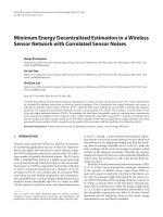
FIGURE 12.1 ▲ Diagram of a motor neuron. The nerve cell
body, dendrites, and proximal part of the axon are within the CNS. The
axon leaves the CNS and, while in the PNS, is part of a nerve (not shown)
as it courses to its effectors (striated muscle). In the CNS, the myelin for the
axon is produced by, and is part of, an oligodendrocyte; in the PNS, the
myelin is produced by, and is part of, a Schwann cell.
Pawlina_CH12.indd 358
•
Multipolar neurons have one axon and two or more
The cell body is the dilated region of the neuron that contains a large, euchromatic nucleus with a prominent nucleolus and surrounding perinuclear cytoplasm (Fig.12.4a,
Plate 27, page 394). The perinuclear cytoplasm reveals
abundant rough-surfaced endoplasmic reticulum (rER)
and free ribosomes when observed with the transmission
9/29/14 7:02 PM
359
Nissl bodies
MOTOR
large motor neuron
CHAPTER 12
striated
(skeletal)
muscle
pseudounipolar
neuron
THE NEURON
SENSORY
smooth muscle
of blood vessels
postsynaptic
autonomic
neuron
Nerve Tissue
presynaptic
autonomic
neuron
INTEGRATIVE
bipolar
neuron
pyramidal
cell
interneurons
Purkinje
cell
FIGURE 12.2 ▲ Diagram illustrating different types of neurons. The cell bodies of pseudounipolar (unipolar), bipolar, and postsynaptic
autonomic neurons are located outside the CNS. Purkinje and pyramidal cells are restricted to the CNS; many of them have elaborate dendritic arborizations that facilitate their identification. Central axonal branch and all axons in remaining cells are indicated in green.
electron microscope (TEM), a feature consistent with its
protein synthetic activity. In the light microscope, the ribosomal content appears as small bodies called Nissl bodies
that stain intensely with basic dyes and metachromatically with thionine dyes (see Fig. 12.4a). Each Nissl body
corresponds to a stack of rER. The perinuclear cytoplasm
also contains numerous mitochondria, a large perinuclear
Golgi apparatus, lysosomes, microtubules, neurofilaments
(intermediate filaments), transport vesicles, and inclusions
(Fig. 12.4b). Nissl bodies, free ribosomes, and occasionally
the Golgi apparatus extend into the dendrites but not into
the axon. This area of the cell body, called the axon hillock,
Pawlina_CH12.indd 359
lacks large cytoplasmic organelles and serves as a landmark
to distinguish between axons and dendrites in both light
microscope and TEM preparations.
The euchromatic nucleus, large nucleolus, prominent
Golgi apparatus, and Nissl bodies indicate the high level of
anabolic activity needed to maintain these large cells.
Neurons do not divide; however, in some areas of the brain,
neural stem cells are present and are able to differentiate
and replace damaged nerve cells.
Although neurons do not replicate, the subcellular components of the neurons turn over regularly and have life spans
9/29/14 7:02 PM
blood vessels
epineurium
360
perineurium
CHAPTER 12
Nerve Tissue
THE NEURON
endoneurium
node of Ranvier
Schwann cell
somatic
sensory
neuron
dorsal root
SPINAL CORD
somatic
motor
neuron
dorsal root
ganglion
cell bodies
of sensory
neurons
autonomic
unmyelinated
neurons
axons
myelin
axon
cell body of
motor neuron
ventral
root
myelin
nucleus of
Schwann cell
Pacinian
corpuscle
spinal nerve
cell body of
sympathetic neuron
striated
muscle
smooth muscle and
enteroceptors of ANS
FIGURE 12.3 ▲ Schematic diagram showing arrangement of motor and sensory neurons. The cell body of a motor neuron is
located in the ventral (anterior) horn of the gray matter of the spinal cord. Its axon, surrounded by myelin, leaves the spinal cord via a ventral
(anterior) root and becomes part of a spinal nerve that carries it to its destination on striated (skeletal) muscle fibers. The sensory neuron
originates in the skin within a receptor (here, a Pacinian corpuscle) and continues as a component of a spinal nerve, entering the spinal cord
via the dorsal (posterior) root. Note the location of its cell body in the dorsal root ganglion (sensory ganglion). A segment of the spinal nerve
is enlarged to show the relationship of the nerve fibers to the surrounding connective tissue (endoneurium, perineurium, and epineurium).
In addition, segments of the sensory, motor, and autonomic unmyelinated neurons have been enlarged to show the relationship of the axons
to the Schwann cells. ANS, autonomic nervous system.
measured in hours, days, and weeks. The constant need to
replace enzymes, neurotransmitter substances, membrane
components, and other complex molecules is consistent
with the morphologic features characteristic of a high level
of synthetic activity. Newly synthesized protein molecules are
transported to distant locations within a neuron in a process
referred to as axonal transport (pages 367–368).
It is generally accepted that nerve cells do not divide.
However, recently it has been shown that the adult brain
retains some cells that exhibit the potential to regenerate.
In certain regions of the brain such as olfactory bulb and dentate gyrus of the hippocampus, these neural stem cells are
able to divide and generate new neurons. They are characterized by prolonged expression of a 240 kDa intermediate
filament protein nestin, which is used to identify these cells
by histochemical methods. Neural stem cells are also able
to migrate to sites of injury and differentiate into new nerve
cells. Research studies on the animal model demonstrate that
newly generated cells mature into functional neurons in the
adult mammalian brain. These findings may lead to therapeutic strategies that use neural cells to replace nerve cells lost or
Pawlina_CH12.indd 360
damaged by neurodegenerative disorders such as Alzheimer
and Parkinson diseases.
Dendrites and Axons
Dendrites are receptor processes that receive stimuli from
other neurons or from the external environment.
The main function of dendrites is to receive information
from other neurons or from the external environment and
carry that information to the cell body. Generally, dendrites
are located in the vicinity of the cell body. They have a greater
diameter than axons, are unmyelinated, are usually tapered,
and form extensive arborizations called dendritic trees.
Dendritic trees significantly increase the receptor surface
area of a neuron. Many neuron types are characterized by
the extent and shape of their dendritic trees (see Fig. 12.2).
In general, the contents of the perinuclear cytoplasm of the
cell body and cytoplasm of dendrites are similar, with the
exception of the Golgi apparatus. Other organelles characteristic of the cell body, including ribosomes and rER, are found
in the dendrites, especially in the base of the dendrites.
9/29/14 7:02 PM
361
G
neuroglial
nuclei
CHAPTER 12
G
nucleolus
Nerve Tissue
L
nucleus
Nissl bodies
rER
b
a
FIGURE 12.4 ▲ Nerve cell bodies. a. This photomicrograph shows a region of the ventral (anterior) horn of a human spinal cord stained with
THE NEURON
M
toluidine blue. Typical features of the nerve cell bodies visible in this image include large, spherical, pale-stained nuclei with a single prominent nucleolus and abundant Nissl bodies within the cytoplasm of the nerve cell body. Most of the small nuclei belong to neuroglial cells. The remainder of the
field consists of nerve fibers and cytoplasm of central neuroglial cells. ϫ640. b. Electron micrograph of a nerve cell body. The cytoplasm is occupied by
aggregates of free ribosomes and profiles of rough-surfaced endoplasmic reticulum (rER) that constitute the Nissl bodies of light microscopy. The Golgi
apparatus (G) appears as isolated areas containing profiles of flattened sacs and vesicles. Other characteristic organelles include mitochondria (M) and
lysosomes (L). The neurofilaments and neurotubules are difficult to discern at this relatively low magnification. ϫ15,000.
Axons are effector processes that transmit stimuli to other
neurons or effector cells.
Some large axon terminals are capable of local protein
synthesis, which may be involved in memory processes.
The main function of the axon is to convey information away
from the cell body to another neuron or to an effector cell, such
as a muscle cell. Each neuron has only one axon, and it may be
extremely long. Axons that originate from neurons in the motor
nuclei of the CNS (Golgi type I neurons) may travel more
than a meter to reach their effector targets, skeletal muscle.
In contrast, interneurons of the CNS (Golgi type II neurons)
have very short axons. Although an axon may give rise to a recurrent branch near the cell body (i.e., one that turns back toward
the cell body) and to other collateral branches, the branching of
the axon is most extensive in the vicinity of its targets.
The axon originates from the axon hillock. The axon
hillock usually lacks large cytoplasmic organelles such as
Nissl bodies and Golgi cisternae. Microtubules, neurofilaments, mitochondria, and vesicles, however, pass through the
axon hillock into the axon. The region of the axon between
the apex of the axon hillock and the beginning of the myelin
sheath (see below) is called the initial segment. The initial
segment is the site at which an action potential is generated
in the axon. The action potential (described in more detail
below) is stimulated by impulses carried to the axon hillock
on the membrane of the cell body after other impulses are
received on the dendrites or the cell body itself.
Almost all of the structural and functional protein molecules are synthesized in the nerve cell body. These molecules are distributed to the axons and dendrites via axonal
transport systems (described on pages 367–368). However, contrary to the common view that the nerve cell body
is the only site of protein synthesis, recent studies indicate that local synthesis of axonal proteins takes place in
some large nerve terminals. Some vertebral axon terminals
(i.e., from the retina) contain polyribosomes with complete translational machinery for protein synthesis. These
discrete areas within the axon terminals, called periaxoplasmic plaques, possess biochemical and molecular
characteristics of active protein synthesis. Protein synthesis
within the periaxoplasmic plaques is modulated by neuronal activity. These proteins may be involved in the processes
of neuronal cell memory.
Pawlina_CH12.indd 361
Synapses
Neurons communicate with other neurons and with
effector cells by synapses.
Synapses are specialized junctions between neurons
that facilitate the transmission of impulses from one
9/29/14 7:02 PM
FOLDER 12.1 Clinical Correlation: Parkinson’s Disease
362
THE NEURON
Parkinson’s disease is a slowly progressive neurologic
disorder caused by the loss of dopamine (DA)-secreting cells
in the substantia nigra and basal ganglia of the brain. DA is a
neurotransmitter responsible for synaptic transmission in the
nerve pathways coordinating smooth and focused activity
of skeletal muscles. Loss of DA-secreting cells is associated
with a classic pattern of symptoms, including the following:
CHAPTER 12
Nerve Tissue
• Resting tremor in the limb, especially of the hand
when in a relaxed position; tremor usually increases
during stress and is often more severe on one side of
the body
• Rigidity or increased tone (stiffness) in all muscles
• Slowness of movement (bradykinesia) and inability to
initiate movement (akinesia)
• Lack of spontaneous movements
• Loss of postural reflexes, which leads to poor balance
and abnormal walking (festinating gait)
• Slurred speech, slowness of thought, and small,
cramped handwriting
The cause of idiopathic Parkinson’s disease, in
which DA-secreting neurons in the substantia nigra are
damaged and lost by degeneration or apoptosis, is not
known. However, some evidence suggests a hereditary
predisposition; about 20% of Parkinson’s patients have a
family member with similar symptoms.
Symptoms that resemble idiopathic Parkinson’s disease
may also result from infections (e.g., encephalitis), toxins
(presynaptic) neuron to another (postsynaptic) neuron.
Synapses also occur between axons and effector (target) cells, such as muscle and gland cells. Synapses between neurons may be classified morphologically as the
following.
•
•
•
(e.g., MPTP), drugs used in the treatment of neurologic
disorders (e.g., neuroleptics used to treat schizophrenia),
and repetitive trauma. Symptoms with these causes are
called secondary parkinsonism.
On the microscopic level, degeneration of neurons in
the substantia nigra is very evident. This region loses its
typical pigmentation, and an increase in the number of
glial cells is noticeable (gliosis). In addition, nerve cells
in this region display characteristic intracellular inclusions
called Lewy bodies, which represent accumulation of
intermediate neurofilaments in association with proteins
␣-synuclein and ubiquitin.
Treatment of Parkinson’s disease is primarily symptomatic and must strike a balance between relieving symptoms and minimizing psychotic side effects. L-Dopa is a
precursor of DA that can cross the blood–brain barrier and
is then converted to DA. It is often the primary agent used
to treat Parkinson’s disease. Other drugs that are used
include a group of cholinergic receptor blockers and amantadine, a drug that stimulates release of DA from neurons.
If drug therapies are not effective, several surgical options
can be considered. Stereotactic surgery, in which nuclei in
selective areas of the brain (globus pallidus, thalamus) are
destroyed by a thermocoagulative probe inserted into the
brain, can be effective in some cases. Several new surgical
procedures are being developed and are still in experimental
stages. These include transplantation of DA-secreting neurons into the substantia nigra to replace lost neurons.
or end bulb. The number of synapses on a neuron or its
processes, which may vary from a few to tens of thousands
per neuron (Fig. 12.6), appears to be directly related to
the number of impulses that a neuron is receiving and
processing.
Axodendritic. These synapses occur between axons and
dendrites. In the CNS, some axodendritic synapses possess dendritic spines (Fig. 12.5), a dynamic projection
containing actin filaments. Their function is associated
with long-term memory and learning.
Axosomatic. These synapses occur between axons and
the cell body.
Axoaxonic. These synapses occur between axons and
axons (see Fig. 12.5).
Synapses are not resolvable in routine hematoxylin and
eosin (H&E) preparations. However, silver precipitation
staining methods (e.g., Golgi method) not only demonstrate the overall shape of some neurons but also show synapses as oval bodies on the surface of the receptor neuron.
Typically, a presynaptic axon makes several of these button-like contacts with the receptor portion of the postsynaptic neuron. Often, the axon of the presynaptic neuron
travels along the surface of the postsynaptic neuron, making several synaptic contacts along the way that are called
boutons en passant [Fr. buttons in passing]. The axon
then continues, ending finally as a terminal branch with
an enlarged tip, a bouton terminal [Fr. terminal button],
Pawlina_CH12.indd 362
axodendritic
axoaxonic
axosomatic
dendritic spine
dendrites
FIGURE 12.5 ▲ Schematic diagram of different types of synapses. Axodendritic synapses represent the most common type of connection between presynaptic axon terminal and dendrites of the postsynaptic
neuron. Note that some axodendritic synapses possess dendritic spines, which
are linked to learning and memory; axosomatic synapses are formed between
presynaptic axon terminal and the postsynaptic nerve cell body, and axoaxonic synapses are formed between the axon terminal of presynaptic neuron
and the axon of a postsynaptic neuron. The axoaxonic synapse may enhance
or inhibit the axodendritic (or axosomatic) synaptic transmission.
9/29/14 7:02 PM
•
•
Chemical synapses. Conduction of impulses is achieved
by the release of chemical substances (neurotransmitters)
from the presynaptic neuron. Neurotransmitters then
diffuse across the narrow intercellular space that separates the presynaptic neuron from the postsynaptic neuron or target cell. A specialized type of chemical synapses
called ribbon synapses are found in the receptor hair
cells of the internal ear and photoreceptor cells of the
retina. Their structures and functions are described in
Chapter 25).
Electrical synapses. Common in invertebrates, these
synapses contain gap junctions that permit movement
of ions between cells and consequently permit the direct
spread of electrical current from one cell to another. These
synapses do not require neurotransmitters for their function. Mammalian equivalents of electrical synapses include
gap junctions in smooth muscle and cardiac muscle cells.
A typical chemical synapse contains a presynaptic element,
synaptic cleft, and postsynaptic membrane.
Components of a typical chemical synapse include the
following.
•
A presynaptic element (presynaptic knob, presynaptic component, or synaptic bouton) is the end
of the neuron process from which neurotransmitters
are released. The presynaptic element is characterized
by the presence of synaptic vesicles, membranebound structures that range from 30 to 100 nm in diameter and contain neurotransmitters (Fig. 12.7). The
binding and fusion of synaptic vesicles to the presynaptic plasma membrane is mediated by a family of
Pawlina_CH12.indd 363
•
THE NEURON
Classification depends on the mechanism of conduction
of the nerve impulses and the way the action potential is
generated in the target cells. Thus, synapses may also be classified as the following.
•
Nerve Tissue
Synapses are classified as chemical or electrical.
363
CHAPTER 12
FIGURE 12.6 ▲ Scanning electron micrograph of the nerve
cell body. This micrograph shows the cell body of a neuron. Axon endings forming axosomatic synapses are visible as are numerous oval
bodies with tail-like appendages. Each oval body represents presynaptic axon terminal from different neurons making contact with the large
postsynaptic nerve cell body. ϫ76,000. (Courtesy of Dr. George Johnson.)
transmembrane proteins called SNAREs (which stands
for “soluble NSF attachment receptors”; see page 35).
The specific SNARE proteins involved in this activity
are known as v-SNARE (vesicle-bound) and t-SNARE
(target-membrane–bound proteins found in specialized
areas of the presynaptic membrane). Another vesiclebound protein called synaptotagmin 1 then replaces
the SNARE complex, which is subsequently dismantled
and recycled by NSF/SNAP25 protein complexes. Dense
accumulations of proteins are present on the cytoplasmic
side of the presynaptic plasma membrane. These presynaptic densities represent specialized areas called active
zones where synaptic vesicles are docked and where
neurotransmitters are released. Active zones are rich
in Rab-GTPase docking complexes (see page 35),
t-SNAREs, and synaptotagmin binding proteins.
The vesicle membrane that is added to the presynaptic
membrane is retrieved by endocytosis and reprocessed
into synaptic vesicles by the smooth-surfaced endoplasmic reticulum (sER) located in the nerve ending.
Numerous small mitochondria are also present in the
presynaptic element.
The synaptic cleft is the 20- to 30-nm space that separates the presynaptic neuron from the postsynaptic neuron or target cell, which the neurotransmitter must cross.
The postsynaptic membrane (postsynaptic component) contains receptor sites with which the neurotransmitter interacts. This component is formed from a portion
of the plasma membrane of the postsynaptic neuron
(Fig. 12.8) and is characterized by an underlying layer of
dense material. This postsynaptic density represents
an elaborate complex of interlinked proteins that serve
numerous functions, such as translation of the neurotransmitter–receptor interaction into an intracellular signal,
anchoring of and trafficking neurotransmitter receptors
to the plasma membrane, and anchoring various proteins
that modulate receptor activity.
Synaptic Transmission
Voltage-gated Ca2ϩ channels in the presynaptic membrane
regulate transmitter release.
When a nerve impulse reaches the synaptic bouton, the voltage
reversal across the membrane produced by the impulse (called
depolarization) causes voltage-gated Ca2ϩ channels to
open in the plasma membrane of the bouton. The influx of
Ca2ϩ from the extracellular space causes the synaptic vesicles
to migrate, anchor, and fuse with the presynaptic membrane,
thereby releasing the neurotransmitter into the synaptic cleft
by exocytosis. Vesicle docking and fusion is mainly driven
by the actions of SNARE and synaptotagmin proteins.
Alternative to the massive release of neurotransmitter following vesicle fusion is the process of porocytosis, in which
vesicles anchored at the active zones release neurotransmitters
through a transient pore connecting the lumen of the vesicle
with the synaptic cleft. The neurotransmitter then diffuses
across the synaptic cleft. At the same time, the presynaptic
membrane of the synaptic bouton that released the neurotransmitter quickly forms endocytotic vesicles that return
to the endosomal compartment of the bouton for recycling
or reloading with neurotransmitter.
9/29/14 7:02 PM
364
CHAPTER 12
Nerve Tissue
THE NEURON
synaptic vesicle
with neurotransmitters
recycled vesicle
presynaptic
element of
axon
voltagegated Ca2ϩ
channel
active zone
a
Ca2ϩ
G-proteingated ion
channel
Ca2ϩ
synaptic cleft
postsynaptic
membrane
of dendrite
Naϩ
synaptotagmin
Naϩ
enzyme
transmittergated
channel
SNARE
complex
Naϩ
second
messengers
G-protein–
coupled
receptor
G-protein
Ca2ϩ
b
FIGURE 12.7 ▲ Diagram of a chemical axodendritic synapse. This diagram illustrates three components of a typical synapse. The presynaptic
knob is located at the distal end of the axon from which neurotransmitters are released. The presynaptic element of the axon is characterized by the presence
of numerous neurotransmitter-containing synaptic vesicles. The plasma membrane of the presynaptic knob is recycled by the formation of clathrin-coated
endocytotic vesicles. The synaptic cleft separates the presynaptic knob of the axon from the postsynaptic membrane of the dendrite. The postsynaptic
membrane of the dendrite is frequently characterized by a postsynaptic density and contains receptors with an affinity for the neurotransmitters. Note
two types of receptors: Green-colored molecules represent transmitter-gated channels, and the purple-colored structure represents a G-protein–coupled
receptor that, when bound to a neurotransmitter, may act on G-protein–gated ion channels or on enzymes producing a second messenger. a. Diagram
showing the current view of neurotransmitter release from a presynaptic knob by a fusion of the synaptic vesicles with presynaptic membrane. b. Diagram
showing a newly proposed model of the neurotransmitter release via porocytosis. In this model, the synaptic vesicle is anchored and juxtaposed to
calcium-selective channels in the presynaptic membrane. In the presence of Ca2ϩ, the bilayers of the vesicle and presynaptic membranes are reorganized
to create a 1-nm transient pore connecting the lumen of the vesicle, with the synaptic cleft allowing the release of a neurotransmitter. Note the presence
of the SNARE complex and the synaptotagmin that anchor the vesicle to the active zones within plasma membrane of the presynaptic element.
The neurotransmitter binds to either transmitter-gated
channels or G-protein–coupled receptors on the postsynaptic
membrane.
The released neurotransmitter molecules bind to the extracellular part of postsynaptic membrane receptors called
transmitter-gated channels. Binding of neurotransmitter
induces a conformational change in these channel proteins
that causes their pores to open. The response that is ultimately
generated depends on the identity of the ion that enters the
cell. For instance, influx of Naϩ causes local depolarization
in the postsynaptic membrane, which under favorable conditions (sufficient amount and duration of neurotransmitter release) prompts the opening of voltage-gated Naϩ
channels, thereby generating a nerve impulse.
Some amino acid and amine neurotransmitters may
bind to G-protein–coupled receptors to produce longer
lasting and more diverse postsynaptic responses. The neurotransmitter binds to a transmembrane receptor protein
on the postsynaptic membrane. Receptor binding activates
G-proteins, which move along the intracellular surface of
the postsynaptic membrane and eventually activate effector
proteins. These effector proteins may include transmembrane
Pawlina_CH12.indd 364
G-protein–gated ion channels or enzymes that syn-
thesize second-messenger molecules (page 365). Several
neurotransmitters (e.g., acetylcholine) can generate different
postsynaptic actions, depending on which receptor system
they act (see below).
Porocytosis describes the secretion of neurotransmitter
that does not involve the fusion of synaptic vesicles with
the presynaptic membrane.
Based on evaluation of physiologic data and the structural organization of nerve synapses, an alternate model of neurotransmitter secretion called porocytosis has recently been proposed
to explain the regulated release of neurotransmitters. In this
model, secretion from the vesicles occurs without fusion of the
vesicle membrane with the presynaptic membrane. Instead,
the synaptic vesicle is anchored to the presynaptic membrane
next to Ca2ϩ selective channels by SNARE and synaptotagmin
proteins. In the presence of Ca2ϩ, the vesicle and presynaptic
membranes are reorganized to create a 1-nm transient pore
connecting the lumen of the vesicle with the synaptic cleft.
Neurotransmitters can then be released in a controlled fashion
through these transient membrane pores (see Fig. 12.7).
9/29/14 7:02 PM
In these synapses, the generation of an action potential
then becomes more difficult.
Neurotransmitters
dendrite
axon
FIGURE 12.8 ▲ Electron micrograph of nerve processes in
the cerebral cortex. A synapse can be seen in the center of the micrograph, where an axon ending is apposed to a dendrite. The ending of the
axon exhibits numerous neurotransmitter-containing synaptic vesicles
that appear as circular profiles. The postsynaptic membrane of the dendrite shows a postsynaptic density. A substance of similar density is also
present in the synaptic cleft (intercellular space) at the synapse. ϫ76,000.
(Courtesy of Drs. George D. Pappas and Virginia Kriho.)
The chemical nature of the neurotransmitter determines
the type of response at that synapse in the generation of
neuronal impulses.
The release of neurotransmitter by the presynaptic component
can cause either excitation or inhibition at the postsynaptic membrane.
•
•
In excitatory synapses, release of neurotransmitters such as acetylcholine, glutamine, or serotonin
opens transmitter-gated Naϩ channels (or other
cation channels), prompting an influx of Naϩ that
causes local reversal of voltage of the postsynaptic membrane to a threshold level (depolarization). This leads
to initiation of an action potential and generation of a
nerve impulse.
In inhibitory synapses, release of neurotransmitters
such as ␥-aminobutyric acid (GABA) or glycine opens
transmitter-gated ClϪ channels (or other anion channels), causing ClϪ to enter the cell and hyperpolarize the
postsynaptic membrane, making it even more negative.
Pawlina_CH12.indd 365
Neurotransmitters act either on ionotropic receptors
to open membrane ion channels or on metabotropic
receptors to activate G-protein signaling cascade.
THE NEURON
Many molecules that serve as neurotransmitters have been
identified in various parts of the nervous system. A neurotransmitter that is released from the presynaptic element diffuses
through the synaptic cleft to the postsynaptic membrane, where
it interacts with a specific receptor. Action of the neurotransmitter depends on its chemical nature and on the characteristics of
the receptor present on the postsynaptic plate of the effector cell.
Nerve Tissue
axon
ending
365
CHAPTER 12
The ultimate generation of a nerve impulse in a postsynaptic
neuron (firing) depends on the summation of excitatory and
inhibitory impulses reaching that neuron. This allows precise
regulation of the reaction of a postsynaptic neuron (or muscle
fiber or gland cell). The function of synapses is not simply to
transmit impulses in an unchanged manner from one neuron
to another. Rather, synapses allow for the processing of neuronal input. Typically, the impulse passing from the presynaptic
to the postsynaptic neuron is modified at the synapse by other
neurons that, although not in the direct pathway, nevertheless
have access to the synapse (see Fig. 12.5). These other neurons
may influence the membrane of the presynaptic neuron or the
postsynaptic neuron and facilitate or inhibit the transmission of
impulses. The firing of impulses in the postsynaptic neuron is
caused by the summation of the actions of hundreds of synapses.
Almost all known neurotransmitters act on multiple receptors, which are integral membrane proteins. These receptors
can be divided into two major classes: ionotropic and metabotropic receptors. Ionotropic receptors contain integral
transmembrane ion channels, also referred to as transmitteror ligand-gated channels. Binding of neurotransmitter to
ionotropic receptors triggers a conformational change of the
receptor proteins that leads to the opening of the channel and
subsequent movement of selective ions in or out of the cell.
This generates action potential in the effector cell. In general,
signaling using ionotropic channels is very rapid and occurs
in the major neuronal pathways of the brain and in somatic
motor pathways in the PNS. Metabotropic channels are
responsible not only for binding a specific neurotransmitter
but also for interacting with G-protein at their intracellular
domain. G-protein is an important protein that is involved in
intracellular signaling. It conveys signals from the outside to
the inside of the cell by altering activities of enzymes involved
in synthesis of a second messenger. Activation of metabotropic receptors is mostly involved in the modulation of neuronal activity.
The most common neurotransmitters are described below.
A summary of selected neurotransmitters and their characteristics in both the PNS and CNS is provided in Table 12.1.
•
Acetylcholine (ACh). ACh is the neurotransmitter be-
tween axons and striated muscle at the neuromuscular
junction (see page 327) and serves as a neurotransmitter in
the ANS. ACh is released by the presynaptic sympathetic
and parasympathetic neurons and their effectors. ACh is
also secreted by postsynaptic parasympathetic neurons, as
9/29/14 7:02 PM
TAB LE 1 2.1
Characterizations of the Most Common Neurotransmitters
CHAPTER 12
Nerve Tissue
THE NEURON
366
Receptor Type and Action
Class of Molecule
Neurotransmitter
Ester
Monoamine
Amino acids
Small peptides
Free radical
Physiological Role
Ionotropic
Metabotropic
ACh
Nicotinic ACh receptors
(nAChR); activates Naϩ
channels
Muscarinic
ACh receptor
(mAChR); acts via
G protein
Fast excitatory synaptic transmission at the neuromuscular
junction (acting on nAChR);
also present in PNS (e.g.,
sympathetic ganglia, adrenal
medulla) and CNS; both excitatory and inhibitory action (acting on mAChR), e.g., decreasing
heart rate, smooth muscle relaxation of gastrointestinal tract
Epinephrine,
norepinephrine
N/A
␣ and  Adrenergic
receptors; acts
via G protein
Slow synaptic transmission in
CNS and in smooth muscles
Dopamine
N/A
D1 and D2 dopamine receptors;
acts via G protein
Slow synaptic transmission in
CNS
Serotonin
5-HT3 ligand-gated
Naϩ/Kϩ channel; activates
ion channels
5-HT1,2,4–7 receptors
Fast excitatory synaptic transmission (acting on 5-HT3);
both excitatory and inhibitory
depending on receptor; acts in
CNS and PNS (enteric system)
Glutamate
NMDA, kainite, and AMPA;
activates Naϩ, Kϩ, and Ca2ϩ
channels
mGluR receptor;
acts via G protein
Fast excitatory synaptic
transmission in CNS
GABA
GABAA receptor; activates
ClϪ channels
GABAB receptor;
acts via G protein
Both fast and slow inhibitory
synaptic transmission in CNS
Glycine
Glycine receptor (GlyR);
activates ClϪ channels
N/A
Fast inhibitory synaptic
transmission in CNS
Substance P
N/A
Neurokinin 1 (NK1)
receptor; acts via
G protein
Slow excitation of smooth
muscles and sensory neurons
in CNS, especially when
conveying pain sensation
Enkephalins
N/A
␦ (DOP) and
(MOP) opioid
receptors; acts
via G protein
Reduces synaptic excitability
(slow synaptic signaling);
relaxes smooth muscles in
gastrointestinal tract; causes
analgesia
-Endorphin
N/A
Opioid (KOP)
receptor; acts via
G protein
Slow synaptic signaling in
brain and spinal cord; causes
analgesia
NO
NO does not act on receptors; it activates guanylyl
cyclase and then via cGMP signaling increases
G protein synthesis in target cells
Influences neurotransmitter
release in CNS and PNS;
acts as potent vasodilator,
relaxes smooth muscles in
gastrointestinal tract
5-HT, 5-hydroxytryptamine; ACh, acetylcholine; AMPA, ␣-amino-3-hydroxy-5-methyl-4-isoxazolepropionic acid; cGMP, cyclic guanosine monophosphate;
CNS, central nervous system; GABA, ␥-aminobutyric acid; mGluR, metabotropic glutamate receptor; N/A, not applicable; NMDA, N-methyl D-aspartate
receptor; NO, nitric oxide; PNS, peripheral nervous system.
well as by a specific type of postsynaptic sympathetic neuron that innervates sweat glands. Neurons that use ACh as
their neurotransmitter are called cholinergic neurons.
The receptors for ACh in the postsynaptic membrane are
known as cholinergic receptors and are divided into
two classes. Metabotropic receptors interact with muscarine, a substance isolated from poisonous mushrooms
Pawlina_CH12.indd 366
(muscarinic ACh receptors), and ionotropic recep-
tors interact with nicotine isolated from tobacco plants
(nicotinic ACh receptors). The muscarinic ACh re-
ceptor in the heart is an example of a G-protein–coupled
receptor that is linked to Kϩ channels. Parasympathetic
stimulation of the heart releases ACh, which in turn opens
Kϩ channels, causing hyperpolarization of cardiac muscle
9/29/14 7:02 PM
•
•
Pawlina_CH12.indd 367
The degradation or recapture of neurotransmitters is necessary to limit the duration of stimulation or inhibition of the
postsynaptic membrane. The most common process of neurotransmitter removal after its release into the synaptic cleft
is called high-affinity reuptake. About 80% of released
neurotransmitters are removed by this mechanism, in which
they are bound into specific neurotransmitter transport
proteins located in the presynaptic membrane. Neurotransmitters that were transported into the cytoplasm of the
presynaptic bouton are either enzymatically destroyed or reloaded into empty synaptic vesicles. For example, the action of
catecholamines on postsynaptic receptors is terminated by
the reuptake of neurotransmitters into the presynaptic bouton
utilizing Naϩ dependent transporters. The efficiency of
this uptake can be regulated by several pharmacologic agents
such as amphetamine and cocaine, which block catecholamine
reuptake and prolong the actions of neurotransmitters on the
postsynaptic neurons. Once inside the presynaptic bouton,
catecholamines are reloaded into synaptic vesicles for future
use. The excess of catecholamines is inactivated by the enzyme
catechol O-methyltransferase (COMT) or is destroyed
by another enzyme found on the outer mitochondrial membrane, monoamine oxidase (MAO). Therapeutic substances that inhibit the action of MAO are frequently used
in the treatment of clinical depression; selective COMT
inhibitors have been also developed.
Enzymes associated with the postsynaptic membrane degrade the remaining 20% of neurotransmitters. For example,
acetylcholinesterase (AChE), which is secreted by the
muscle cell into the synaptic cleft, rapidly degrades ACh
into acetic acid and choline. Choline is then taken up by the
cholinergic presynaptic bouton and reused for ACh synthesis. The AChE action at the neuromuscular junction
can be inhibited by various pharmacological compounds,
nerve agents, and pesticides, resulting in prolonged muscle
contraction. Clinically, AChE inhibitors have been used in
the treatment of myasthenia gravis (see Folder 11.4 in
Chapter 11), a degenerative neuromuscular disorder; glaucoma; and more recently, Alzheimer’s disease.
THE NEURON
•
Neurotransmitters released into the synaptic cleft may be
degraded or recaptured.
367
Nerve Tissue
•
(e.g., -endorphin, enkephalins, dynorphins),
vasoactive intestinal peptide (VIP), cholecystokinin (CCK), and neurotensin. Many of these same
substances are synthesized and released by enteroendocrine cells of the intestinal tract. They may act immediately on neighboring cells (paracrine secretion) or be
carried in the bloodstream as hormones to act on distant
target cells (endocrine secretion). They are also synthesized
and released by endocrine organs and by the neurosecretory neurons of the hypothalamus.
CHAPTER 12
•
fibers. This hyperpolarization slows rhythmic contraction
of the heart. In contrast, the nicotinic ACh receptor in
skeletal muscles is an ionotropic ligand-gated Naϩ channel. Opening of this channel causes rapid depolarization
of skeletal muscle fibers and initiation of contraction.
Various drugs affect the release of ACh into the synaptic
cleft as well as its binding to its receptors. For instance,
curare, the South American arrow-tip poison, binds to
nicotinic ACh receptors, blocking their integral Naϩ channels and causing muscle paralysis. Atropine, an alkaloid
extracted from the belladonna plant (Atropa belladonna),
blocks the action of muscarinic ACh receptors.
Catecholamines such as norepinephrine (NE), epinephrine (EPI, adrenaline), and dopamine (DA).
These neurotransmitters are synthesized in a series of enzymatic reactions from the amino acid tyrosine. Neurons
that use catecholamines as their neurotransmitter are
called catecholaminergic neurons. Catecholamines
are secreted by cells in the CNS that are involved in the
regulation of movement, mood, and attention. Neurons that
utilize epinephrine (adrenaline) as their neurotransmitter are
called adrenergic neurons. They all contain an enzyme
that converts NE to adrenaline (EPI), which serves as a
transmitter between postsynaptic sympathetic axons and effectors in the ANS. EPI is also released into the bloodstream
by the endocrine cells (chromaffin cells) of the adrenal medulla during the fight-or-flight response.
Serotonin or 5-hydroxytryptamine (5-HT). Serotonin is formed by the hydroxylation and decarboxylation
of tryptophan. It functions as a neurotransmitter in neurons of the CNS and enteric nervous system. Neurons
that use serotonin as their neurotransmitter are called
serotonergic. After the release of serotonin, a portion
is recycled by reuptake into presynaptic serotonergic
neurons. Recent studies indicate serotonin as an important molecule in establishing asymmetrical right–left
development in embryos.
Amino acids such as ␥-aminobutyrate (GABA), glutamate (GLU), aspartate (ASP), and glycine (GLY) also act
as neurotransmitters, mainly in the CNS.
Nitric oxide (NO), a simple gas with free radical properties,
also has been identified as a neurotransmitter. At low concentrations, NO carries nerve impulses from one neuron to
another. Unlike other neurotransmitters, which are synthesized in the nerve cell body and stored in synaptic vesicles,
NO is synthesized within the synapse and used immediately.
It is postulated that excitatory neurotransmitter GLU induces a chain reaction in which NO synthase is activated
to produce NO, which in turn diffuses from the presynaptic
knob via the synaptic cleft and postsynaptic membrane to
the adjacent cell. Biological actions of NO are due to the
activation of guanylyl cyclase, which then produces cyclic
guanosine monophosphate (cGMP) in target cells. cGMP
in turn acts on G-protein synthesis, ultimately resulting in
generation/modulation of neuronal action potentials.
Small peptides have also been shown to act as synaptic
transmitters. Among these are substance P (so named
because it was originally found in a powder of acetone
extracts of brain and intestinal tissue), hypothalamic
releasing hormones, endogenous opioid peptides
Axonal Transport Systems
Substances needed in the axon and dendrites are synthesized
in the cell body and require transport to those sites.
Most neurons possess elaborate axonal and dendritic processes.
Because the synthetic activity of the neuron is concentrated in
the nerve cell body, axonal transport is required to convey
9/29/14 7:02 PM
CHAPTER 12
Nerve Tissue
S U P P O R T I N G C E LLS O F T H E N E R V O U S S Y S T E M : T H E N E U R O G L I A
368
newly synthesized material to the processes. Axonal transport
is a bidirectional mechanism. It serves as a mode of intracellular communication, carrying molecules and information
along the microtubules and intermediate filaments from the
axon terminal to the nerve cell body and from the nerve cell
body to the axon terminal. Axonal transport is described as
the following:
•
•
Anterograde transport carries material from the nerve
cell body to the periphery. Kinesin, a microtubuleassociated motor protein that uses ATP, is involved in
anterograde transport (see pages 57–58).
Retrograde transport carries material from the axon
terminal and the dendrites to the nerve cell body. This
transport is mediated by another microtubule-associated
motor protein, dynein (see pages 57–58).
The transport systems may also be distinguished by the
rate at which substances are transported:
•
•
A slow transport system conveys substances from
the cell body to the terminal bouton at the speed of 0.2
to 4 mm/day. It is only an anterograde transport system.
Structural elements such as tubulin molecules (microtubule precursors), actin molecules, and the proteins that
form neurofilaments are carried from the nerve cell body
by the slow transport system. So, too, are cytoplasmic
matrix proteins such as actin, calmodulin, and various
metabolic enzymes.
A fast transport system conveys substances in both
directions at a rate of 20 to 400 mm/day. Thus, it is
both an anterograde and a retrograde system. The fast
anterograde transport system carries to the axon terminal different membrane-limited organelles, such as sER
components, synaptic vesicles, and mitochondria, and
low-molecular-weight materials such as sugars, amino
acids, nucleotides, some neurotransmitters, and calcium.
The fast retrograde transport system carries to the nerve
cell body many of the same materials as well as proteins
and other molecules endocytosed at the axon terminal. Fast
transport in either direction requires ATP, which is used
by microtubule-associated motor proteins, and depends on
the microtubule arrangement that extends from the nerve
cell body to the termination of the axon. Retrograde transport is the pathway followed by toxins and viruses that
enter the CNS at nerve endings. Retrograde transport of
exogenous enzymes, such as horseradish peroxidase, and
of radiolabeled or immunolabeled tracer materials is now
used to trace neuronal pathways and to identify the nerve
cell bodies related to specific nerve endings.
Dendritic transport appears to have the same characteristics and to serve the same functions for the dendrite as
axonal transport does for the axon.
S U P P O R T I N G C E L LS O F TH E
NERVOUS SYSTEM:
THE NEUROGLIA
In the PNS, supporting cells are called peripheral neuroglia;
in the CNS, they are called central neuroglia.
Pawlina_CH12.indd 368
Peripheral Neuroglia
Peripheral neuroglia include Schwann cells, satellite
cells, and a variety of other cells associated with specific
organs or tissues. Examples of the latter include terminal
neuroglia (teloglia), which are associated with the motor
end plate; enteric neuroglia associated with the ganglia located in the wall of the alimentary canal; and Müller’s cells
in the retina.
Schwann Cells and the Myelin Sheath
In the PNS, Schwann cells produce the myelin sheath.
The main function of Schwann cells is to support myelinated and unmyelinated nerve cell fibers. Schwann cells
develop from neural crest cells and differentiate by expressing
transcription factor Sox-10. In the PNS, Schwann cells
produce a lipid-rich layer called the myelin sheath that surrounds the axons (Fig. 12.9). The myelin sheath isolates the
axon from the surrounding extracellular compartment of endoneurium. Its presence ensures the rapid conduction of nerve
impulses. The axon hillock and the terminal arborizations
where the axon synapses with its target cells are not covered by
myelin. Unmyelinated fibers are also enveloped and nurtured
by Schwann cell cytoplasm. In addition, Schwann cells aid in
cleaning up PNS debris and guide the regrowth of PNS axons.
Myelination begins when a Schwann cell surrounds the
axon and its cell membrane becomes polarized.
During formation of the myelin sheath (also called
myelination), the axon initially lies in a groove on the surface of the Schwann cell (Fig. 12.10a). A 0.08- to 0.1-mm
segment of the axon then becomes enclosed within each
Schwann cell that lies along the axon. The Schwann cell surface becomes polarized into two functionally distinct membrane domains. The part of the Schwann cell membrane that
is exposed to the external environment or endoneurium,
the abaxonal plasma membrane, represents one domain. The other domain is represented by the adaxonal or
periaxonal plasma membrane, which is in direct contact
with the axon. When the axon is completely enclosed by the
Schwann cell membrane, a third domain, the mesaxon, is
created (Fig. 12.10b). This third domain is a double membrane that connects the abaxonal and adaxonal membranes
and encloses the narrow extracellular space.
The myelin sheath develops from compacted layers of
Schwann cell mesaxon wrapped concentrically around
the axon.
Myelin sheath formation is initiated when the Schwann
cell mesaxon surrounds the axon. A sheet-like extension
of the mesaxon then wraps around the axon in a spiraling
motion. The first few layers or lamellae of the spiral are not
compactly arranged—that is, some cytoplasm is left in the
first few concentric layers (Fig. 12.10c). The TEM reveals the
presence of a 12- to 14-nm gap between the outer (extracellular) leaflets and the Schwann cell cytoplasm that separates
the inner (cytoplasmic) leaflets. As the wrapping progresses,
cytoplasm is squeezed out from between the membrane of the
concentric layers of the Schwann cell.
External to, and contiguous with, the developing myelin
sheath is a thin outer collar of perinuclear cytoplasm
9/29/14 7:02 PM
Epi
369
SL
CHAPTER 12
*
A
A
A
Nerve Tissue
NR
A
*
*
SL
A
SL
SL
*
*
A
A
a
b
FIGURE 12.9 ▲ Photomicrographs of a peripheral nerve in cross and longitudinal sections. a. Photomicrograph of an osmium-fixed,
toluidine blue–stained peripheral nerve cut in cross-section. The axons (A) appear clear. The myelin is represented by the dark ring surrounding the axons.
Note the variation in diameter of the individual axons. In some of the nerves, the myelin appears to consist of two separate rings (asterisks). This is caused by
the section passing through a Schmidt-Lanterman cleft. Epi, epineurium. ϫ640. b. Photomicrograph showing longitudinally sectioned myelinated nerve
axons (A) in the same preparation as above. A node of Ranvier (NR) is seen near the center of the micrograph. In the same axon, a Schmidt-Lanterman cleft
(SL) is seen on each side of the node. In addition, a number of Schmidt-Lanterman clefts can be seen in the adjacent axons. The perinodal cytoplasm of the
Schwann cell at the node of Ranvier and the Schwann cell cytoplasm at the Schmidt-Lanterman cleft appear virtually unstained. ϫ640.
called the sheath of Schwann. This part of the cell is enclosed
by an abaxonal plasma membrane and contains the nucleus and
most of the organelles of the Schwann cell. Surrounding the
Schwann cell is a basal or external lamina. The apposition of
the mesaxon of the last layer to itself as it closes the ring of the
spiral produces the outer mesaxon, the narrow intercellular
space adjacent to the external lamina. Internal to the concentric layers of the developing myelin sheath is a narrow inner
collar of Schwann cell cytoplasm surrounded by the adaxonal plasma membrane. The narrow intercellular space between
mesaxon membranes communicates with the adaxonal plasma
membrane to produce the inner mesaxon (Fig. 12.10d).
Once the mesaxon spirals on itself, the 12- to 14-nm gaps
disappear and the membranes form the compact myelin
sheath. Compaction of the sheath corresponds with the expression of transmembrane myelin-specific proteins such
as protein 0 (P0), a peripheral myelin protein of 22 kDa
(PMP22), and myelin basic protein (MBP). The inner
(cytoplasmic) leaflets of the plasma membrane come close together as a result of the positively charged cytoplasmic domains
of P0 and MBP. With the TEM, these closely aligned inner leaflets are electron opaque, appearing as the major dense lines
in the TEM image of myelin (Fig. 12.10d). The concentric
dense lamellae alternate with the slightly less dense intraperiod lines that are formed by closely apposed, but not fused,
outer (extracellular) membrane leaflets. The narrow 2.5-nm
Pawlina_CH12.indd 369
gap corresponds to the remaining extracellular space containing the extracellular domains of P0 protein (Fig. 12.10d). P0 is
a 30 kDa cell adhesion molecule expressed within the mesoaxial plasma membrane during myelination. This transmembrane glycoprotein mediates strong adhesions between the
two opposite membrane layers and represents a key structural
component of peripheral nerve myelin. Structural and genetic
studies indicate that mutations in human genes encoding P0
produce unstable myelin and may contribute to the development of demyelinating diseases (see Folder 12.2).
The thickness of the myelin sheath at myelination is
determined by axon diameter and not by the Schwann cell.
Myelination is an example of cell-to-cell communication in
which the axon interacts with the Schwann cell. Experimental
studies show that the number of layers of myelin is determined
by the axon and not by the Schwann cell. Myelin sheath thickness is regulated by a growth factor called neuregulin (Ngr1)
that acts on Schwann cells. Ngr1 is a transmembrane protein
expressed on the axolemma (cell membrane) of the axon.
S U P P O R T I N G C E LLS O F T H E N E R VO US S Y S T E M : T HE N E U R OG L I A
A
The node of Ranvier represents the junction between two
adjacent Schwann cells.
The myelin sheath is segmented because it is formed by
numerous Schwann cells arrayed sequentially along the axon.
The junction where two adjacent Schwann cells meet is devoid
9/29/14 7:02 PM
abaxonal
domain
CHAPTER 12
Nerve Tissue
S U P P O R T I N G C E LLS O F T H E N E R V O U S S Y S T E M : T H E N E U R O G L I A
370
a
Nrg1
mesaxon
adaxonal
domain
Major dense
line
b
+
+
+
+
+
+
+
+
+
cytoplasm
Intraperiod
line
extracellular
space
+
c
+
+
+
+
+
+
+
+
cytoplasm
d
P0
MBP
PMP 22
inner
mesaxon
outer
mesaxon
FIGURE 12.10 ▲ Diagram showing successive stages in the formation of myelin by a Schwann cell. a. The axon initially lies in a groove on
the surface of the Schwann cell. b. The axon is surrounded by a Schwann cell. Note the two domains of the Schwann cell, the adaxonal plasma-membrane
domain and abaxonal plasma-membrane domain. The mesaxon plasma membrane links these domains. The mesaxon membrane initiates myelination by
surrounding the embedded axon. c. A sheet-like extension of the mesaxon membrane then wraps around the axon, forming multiple membrane layers.
d. During the wrapping process, the cytoplasm is extruded from between the two apposing plasma membranes of the Schwann cell, which then become
compacted to form myelin. The outer mesaxon represents invaginated plasma membrane extending from the abaxonal surface of the Schwann cell to the
myelin. The inner mesaxon extends from the adaxonal surface of the Schwann cell (the part facing the axon) to the myelin. The inset shows the major proteins responsible for compaction of the myelin sheath. MBP, myelin basic protein; Nrg1, neuregulin; P0, protein 0; PM P22, peripheral myelin protein of 22 kDa.
of myelin. This site is called the node of Ranvier. Therefore,
the myelin between two sequential nodes of Ranvier is called
an internodal segment (Plate 28, page 396). The node of
Ranvier constitutes a region where the electrical impulse is
regenerated for high-speed propagation down the axon. The
highest density of voltage-gated Naϩ channels in the nervous
system occurs at the node of Ranvier; their expression is regulated by interactions with perinodal cytoplasm of Schwann cells.
Myelin is composed of about 80% lipids because, as
the Schwann cell membrane winds around the axon, the
FOLDER 12.2 Clinical Correlation: Demyelinating Diseases
In general, demyelinating diseases are characterized by
preferential damage to the myelin sheath. Clinical symptoms
of these diseases are related to decreased or lost ability
to transmit electrical impulses along nerve fibers. Several
immune-mediated diseases affect the myelin sheath.
Guillain-Barré syndrome, known also as acute
inflammatory demyelinating polyradiculoneuropathy, is one of the most common life-threatening diseases
of the PNS. Microscopic examination of nerve fibers
obtained from patients affected by this disease shows a
large accumulation of lymphocytes, macrophages, and
plasma cells around nerve fibers within nerve fascicles.
Large segments of the myelin sheath are damaged, leaving the axons exposed to the extracellular matrix. These
findings are consistent with a T cell–mediated immune response directed against myelin, which causes its destruction and slows or blocks nerve conduction. Patients exhibit
symptoms of ascending muscle paralysis, loss of muscle
coordination, and loss of cutaneous sensation.
Pawlina_CH12.indd 370
Multiple sclerosis (MS) is a disease that attacks myelin
in the CNS. MS is also characterized by preferential damage
to myelin, which becomes detached from the axon and is
eventually destroyed. In addition, destruction of oligodendroglia, which are responsible for the synthesis and maintenance
of myelin, occurs. The myelin basic protein appears to be the
major autoimmune target in this disease. Chemical changes in
the lipid and protein constituents of myelin produce irregular,
multiple plaques throughout the white matter of the brain.
Symptoms of MS depend on the area in the CNS in which
myelin is damaged. MS is usually characterized by distinct
episodes of neurologic deficits such as unilateral vision impairment, loss of cutaneous sensation, lack of muscle coordination and movement, and loss of bladder and bowel control.
Treatment of both diseases is related to diminishing
the causative immune response by immunomodulatory
therapy with interferon as well as by administrating adrenal steroids. For more severe, progressive forms, immunosuppressive drugs may be used.
9/29/14 7:02 PM
Schmidt-Lanterman clefts correlates with the diameter of the
axon; larger axons have more clefts.
Unmyelinated axons in the peripheral nervous system are
enveloped by Schwann cells and their external lamina.
Satellite Cells
M
OM
BL
SC
A
IM
Central Neuroglia
There are four types of central neuroglia:
•
•
•
•
FIGURE 12.11 ▲ Electron micrograph of an axon in the
process of myelination. At this stage of development, the myelin (M)
consists of about six membrane layers. The inner mesaxon (IM) and outer
mesaxon (OM) of the Schwann cell (SC) represent parts of the mesaxon
membrane. Another axon (see upper left A) is present that has not yet
been embedded within a Schwann cell mesaxon. Other notable features
include the Schwann cell basal (external) lamina (BL) and the considerable amount of Schwann cell cytoplasm associated with the myelination
process. ϫ50,000. (Courtesy of Dr. Stephen G. Waxman.)
Pawlina_CH12.indd 371
Astrocytes are morphologically heterogeneous cells that pro-
vide physical and metabolic support for neurons of the CNS.
Oligodendrocytes are small cells that are active in the
formation and maintenance of myelin in the CNS.
Microglia are inconspicuous cells with small, dark, elon-
gated nuclei that possess phagocytotic properties.
Ependymal cells are columnar cells that line the ventricles
of the brain and the central canal of the spinal cord.
S U P P O R T I N G C E LLS O F T H E N E R VO US S Y S T E M : T HE N E U R OG L I A
A
The neuronal cell bodies of ganglia are surrounded by a layer
of small cuboidal cells called satellite cells. Although they
form a complete layer around the cell body, only their nuclei
are typically visible in routine H&E preparations (Fig. 12.16a
and b). In paravertebral and peripheral ganglia, neural cell
processes must penetrate between the satellite cells to establish a synapse (there are no synapses in sensory ganglia). They
help to establish and maintain a controlled microenvironment
around the neuronal body in the ganglion, providing electrical insulation as well as a pathway for metabolic exchanges.
Thus, the functional role of the satellite cell is analogous to
that of the Schwann cell except that it does not make myelin.
Neurons and their processes located within ganglia of the
enteric division of the ANS are associated with enteric neuroglial cells. These cells are morphologically and functionally similar to astrocytes in the CNS (see below). Enteric
neuroglial cells share common functions with astrocytes, such
as structural, metabolic, and protective support of neurons.
However, recent studies indicate that enteric glial cells may
also participate in enteric neurotransmission and help coordinate activities of the nervous and immune systems of the gut.
Nerve Tissue
The nerves in the PNS that are described as unmyelinated
are nevertheless enveloped by Schwann cell cytoplasm as
shown in Figure 12.15. The Schwann cells are elongated in
parallel to the long axis of the axons, and the axons fit into
grooves in the surface of the cell. The lips of the groove may
be open, exposing a portion of the axolemma of the axon to
the adjacent external lamina of the Schwann cell, or the lips
may be closed, forming a mesaxon.
A single axon or a group of axons may be enclosed in a single invagination of the Schwann cell surface. Large Schwann
cells in the PNS may have 20 or more grooves, each containing one or more axons. In the ANS, it is common for bundles
of unmyelinated axons to occupy a single groove.
371
CHAPTER 12
cytoplasm of the Schwann cell, as noted, is extruded from
between the opposing layers of the plasma membranes. Electron micrographs, however, typically show small amounts of
cytoplasm in several locations (Figs. 12.11 and 12.12): the
inner collar of Schwann cell cytoplasm, between the axon
and the myelin; the Schmidt-Lanterman clefts, small
islands within successive lamellae of the myelin; perinodal
cytoplasm, at the node of Ranvier; and the outer collar of
perinuclear cytoplasm, around the myelin (Fig. 12.13). These
areas of cytoplasm are what light microscopists identified as
the Schwann sheath. If one conceptually unrolls the Schwann
cell process, as shown in Figure 12.14, its full extent can be
appreciated, and the inner collar of Schwann cell cytoplasm
can be seen to be continuous with the body of the Schwann
cell through the Schmidt-Lanterman clefts and through the
perinodal cytoplasm. Cytoplasm of the clefts contains lysosomes and occasional mitochondria and microtubules, as well
as cytoplasmic inclusions, or dense bodies. The number of
Only the nuclei of glial cells are seen in routine histologic
preparations of the CNS. Heavy metal staining or immunocytochemical methods are necessary to demonstrate the shape
of the entire glial cell.
Although glial cells have long been described as supporting cells of nerve tissue in the purely physical sense, current
concepts emphasize the functional interdependence of
neuroglial cells and neurons. The most obvious example of
9/29/14 7:02 PM
CHAPTER 12
Nerve Tissue
S U P P O R T I N G C E LLS O F T H E N E R V O U S S Y S T E M : T H E N E U R O G L I A
372
M
BL
C
ICS
MT
Mit
CV
OCS
FIGURE 12.12 ▲ Electron micrograph of a mature myelinated axon. The myelin sheath (M) shown here consists of 19 paired layers of
Schwann cell membrane. The pairing of membranes in each layer is caused by the extrusion of the Schwann cell cytoplasm. The axon displays an abundance of neurofilaments, most of which have been cross-sectioned, giving the axon a stippled appearance. Also evident in the axon are microtubules
(MT) and several mitochondria (Mit). The outer collar of Schwann cell cytoplasm (OCS) is relatively abundant compared with the inner collar of Schwann
cell cytoplasm (ICS). The collagen fibrils (C ) constitute the fibrillar component of the endoneurium. BL, basal (external) lamina. ϫ70,000. Inset. Higher
magnification of the myelin. The arrow points to cytoplasm within the myelin that would contribute to the appearance of the Schmidt-Lanterman
cleft as seen in the light microscope. It appears as an isolated region here because of the thinness of the section. The intercellular space between axon
and Schwann cell is indicated by the arrowhead. A coated vesicle (CV ) in an early stage of formation appears in the outer collar of the Schwann cell
cytoplasm. ϫ130,000. (Courtesy of Dr. George D. Pappas.)
nucleus of
Schwann cell
outer collar
of Schwann
cell cytoplasm
node of Ranvier
Na+
outer collar of
Schwann
cell cytoplasm
myelin
+ +
+ + + +
+ + +
+
+ + +
+
+
+
+ +
Na+
inner collar
of Schwann
cell cytoplasm
myelin
axon
Schmidt-Lanterman
cleft
Na+
voltage-gated Na+ channels
perinodal cytoplasm
of Schwann cell
Pawlina_CH12.indd 372
FIGURE 12.13 ▲ Diagram
of the node of Ranvier and associated Schwann cells. This diagram
shows a longitudinal section of the
axon and its relationships to the myelin, cytoplasm of the Schwann cell,
and node of Ranvier. Schwann cell
cytoplasm is present at four locations:
the inner and the outer cytoplasmic
collar of the Schwann cell, the nodes
of Ranvier, and the Schmidt-Lanterman clefts. Note that the cytoplasm
throughout the Schwann cell is
continuous (see Fig. 12.14); it is not a
series of cytoplasmic islands as it appears on the longitudinal section of
the myelin sheath. The node of Ranvier is the site at which successive
Schwann cells meet. The adjacent
plasma membranes of the Schwann
cells are not tightly apposed at the
node, and extracellular fluid has free
access to the neuronal plasma membrane. Also, the node of Ranvier is the
site of depolarization of the neuronal
plasma membrane during nerve
impulse transmission and contains
clusters of high-density, voltagegated Naϩ channels.
9/29/14 7:02 PM
inner collar of
Schwann cell
cytoplasm
embryonic glial cells extend through the entire thickness of
the neural tube in a radial manner. These radial glial cells
serve as the physical scaffolding that directs the migration of
neurons to their appropriate position in the brain.
perinodal
cytoplasm of
Schwann cell
axon
myelin
outer collar of
Schwann cell
cytoplasm
network of cells within the CNS and communicate with neurons to support and modulate many of their activities. Some
astrocytes span the entire thickness of the brain, providing a scaffold for migrating neurons during brain development. Other
astrocytes stretch their processes from blood vessels to neurons.
The ends of the processes expand, forming end feet that cover
large areas of the outer surface of the vessel or axolemma.
Astrocytes do not form myelin. Two kinds of astrocytes
are identified:
•
FIGURE 12.14 ▲ Three-dimensional diagrams conceptualizing the relationship of myelin and cytoplasm of a Schwann cell.
This diagram shows a hypothetically uncoiled Schwann cell. Note how
the inner collar of the Schwann cell cytoplasm is continuous with the
outer collar of Schwann cell cytoplasm via Schmidt-Lanterman clefts.
physical support occurs during development. The brain and
spinal cord develop from the embryonic neural tube.
In the head region, the neural tube undergoes remarkable
thickening and folding, leading ultimately to the final
structure, the brain. During the early stages of the process,
Both types of astrocytes contain prominent bundles of
intermediate filaments composed of glial fibrillary acidic
protein (GFAP). The filaments are much more numerous in
the fibrous astrocytes, however, hence the name. Antibodies
to GFAP are used as specific stains to identify astrocytes in
sections and tissue cultures (see Fig. 12.18b). Tumors arising
from fibrous astrocytes, fibrous astrocytomas, account
for about 80% of adult primary brain tumors. They can be
identified microscopically and by their GFAP specificity.
BL
A
M
G
A
N
A
A
S U P P O R T I N G C E LLS O F T H E N E R VO US S Y S T E M : T HE N E U R OG L I A
•
nucleus of
Schwann cell
Protoplasmic astrocytes are more prevalent in the
outermost covering of brain called gray matter. These
astrocytes have numerous, short, branching cytoplasmic
processes (Fig. 12.17).
Fibrous astrocytes are more common in the inner core
of the brain called white matter. These astrocytes have fewer
processes, and they are relatively straight (Fig. 12.18).
Nerve Tissue
SchmidtLanterman
clefts
Astrocytes are the largest of the neuroglial cells. They form a
CHAPTER 12
Astrocytes are closely associated with neurons to support
and modulate their activities.
373
A
A
FIGURE 12.15 ▲ Electron micrograph of unmyelinated nerve fibers. The individual fibers or axons (A) are engulfed by the cytoplasm of a Schwann
cell. The arrows indicate the site of mesaxons. In effect, each axon is enclosed by the Schwann cell cytoplasm, except for the intercellular space of the mesaxon.
Other features evident in the Schwann cell are its nucleus (N), the Golgi apparatus (G), and the surrounding basal (external) lamina (BL). In the upper part of the micrograph, myelin (M) of two myelinated nerves is also evident. ϫ27,000. Inset. Schematic diagram showing the relationship of axons engulfed by the Schwann cell.
Pawlina_CH12.indd 373
9/29/14 7:02 PM
374
CT
NF
NF
NF
a
b
FIGURE 12.16 ▲ Photomicrograph of a nerve ganglion. a. Photomicrograph showing a ganglion stained by the Mallory-Azan method.
Note the large nerve cell bodies (arrows) and nerve fibers (NF ) in the ganglion. Satellite cells are represented by the very small nuclei at the periphery
of the neuronal cell bodies. The ganglion is surrounded by a dense irregular connective tissue capsule (CT ) that is comparable to, and continuous with,
the epineurium of the nerve. ϫ200. b. Higher magnification of the ganglion, showing individual axons and a few neuronal cell bodies with their satellite
cells (arrows). The nuclei in the region of the axons are mostly Schwann cell nuclei. ϫ640.
perivascular
feet
axon
Nerve Tissue
S U P P O R T I N G C E LLS O F T H E N E R V O U S S Y S T E M : T H E N E U R O G L I A
CT
CHAPTER 12
myelin
sheath
perineural
feet
blood vessel
a
b
FIGURE 12.17 ▲ Protoplasmic astrocyte in the gray matter of the brain. a. This schematic drawing shows the foot processes of the protoplasmic astrocyte terminating on a blood vessel and the axonal process of a nerve cell. The foot processes terminating on the blood vessel contribute to the
blood–brain barrier. The bare regions of the vessel as shown in the drawing would be covered by processes of neighboring astrocytes, thus forming the overall
barrier. b. This laser-scanning confocal image of protoplasmic astrocyte in the gray matter of the dentate gyrus was visualized by intracellular labeling method.
In lightly fixed tissue slices, selected astrocytes were impaled and iontophoretically injected with fluorescent dye (Alexa Fluor 568) using pulses of negative
current. Note the density and spatial distribution of cell processes. ϫ480. (Reprinted with permission from Bushong EA, Martone ME, Ellisman MH. Examination
of the relationship between astrocyte morphology and laminar boundaries in the molecular layer of adult dentate gyrus. J Comp Neurol 2003;462:241–251.)
Pawlina_CH12.indd 374
9/29/14 7:02 PM
375
CHAPTER 12
b
Astrocytes play important roles in the movement of metabolites and wastes to and from neurons. They help maintain the tight junctions of the capillaries that form the
blood–brain barrier (see page 388). In addition, astrocytes
provide a covering for the “bare areas” of myelinated axons—
for example, at the nodes of Ranvier and at synapses. They
may confine neurotransmitters to the synaptic cleft and remove excess neurotransmitters by pinocytosis. Protoplasmic astrocytes on the brain and spinal cord surfaces extend
their processes (subpial feet) to the basal lamina of the pia
mater to form the glia limitans, a relatively impermeable
barrier surrounding the CNS (Fig. 12.19).
basement membrane
subpial foot process
glia limitans
pia mater
pericyte
ependyma
astrocyte
neuron
microglial cell
S U P P O R T I N G C E LLS O F T H E N E R VO US S Y S T E M : T HE N E U R OG L I A
FIGURE 12.18 ▲ Fibrous astrocytes in the white matter of the brain. a. Schematic drawing of a fibrous astrocyte in the white mater of
the brain. b. Photomicrograph of the white matter of the brain, showing the extensive radiating cytoplasmic processes for which astrocytes are named.
They are best visualized, as shown here, with immunostaining methods that use antibodies against GFAP. ϫ220. (Reprinted with permission from
Fuller GN, Burger PC. Central nervous system. In: Sternberg SS, ed. Histology for Pathologists. Philadelphia: Lippincott-Raven, 1997.)
Nerve Tissue
a
oligomyelin dendrocyte
FIGURE 12.19 ▲ Distribution of glial cells in the brain. This diagram shows the four types of glial cells—astrocytes, oligodendrocytes, microglial cells, and ependymal cells—interacting with several structures and cells found in the brain tissue. Note that the astrocytes and their processes
interact with the blood vessels as well as with axons and dendrites. Astrocytes also send their processes toward the brain surface, where they contact
the basement membrane of the pia mater, forming the glia limitans. In addition, processes of astrocytes extend toward the fluid-filled spaces in the
CNS, where they contact the ependymal lining cells. Oligodendrocytes are involved in myelination of the nerve fibers in the CNS. Microglia exhibit
phagocytotic functions.
Pawlina_CH12.indd 375
9/29/14 7:03 PM
It is now generally accepted that astrocytes regulate Kϩ
concentrations in the brain’s extracellular compartment,
thus maintaining the microenvironment and modulating
activities of the neurons. The astrocyte plasma membrane
contains an abundance of Kϩ pumps and Kϩ channels that
mediate the transfer Kϩ ions from areas of high to low concentration. Accumulation of large amounts of intracellular
Kϩ in astrocytes decreases local extracellular Kϩ gradients.
The astrocyte membrane becomes depolarized, and the
charge is dissipated over a large area by the extensive network
of astrocyte processes. The maintenance of the Kϩ concentration in the brain’s extracellular space by astrocytes is called
potassium spatial buffering.
Oligodendrocytes produce and maintain the myelin sheath
in the CNS.
The oligodendrocyte is the cell responsible for producing CNS myelin. The myelin sheath in the CNS is formed
by concentric layers of oligodendrocyte plasma membrane.
The formation of the sheath in the CNS is more complex,
however, than the simple wrapping of Schwann cell mesaxon
membranes that occurs in the PNS (pages 178–180).
Oligodendrocytes appear in specially stained light microscopic preparations as small cells with relatively few processes compared with astrocytes. They are often aligned in
rows between axons. Each oligodendrocyte gives off several
tongue-like processes that find their way to the axons, where
each process wraps itself around a portion of an axon, forming an internodal segment of myelin. The multiple processes of a single oligodendrocyte may myelinate one axon
or several nearby axons (Fig. 12.20). The nucleus-containing
region of the oligodendrocyte may be at some distance from
the axons it myelinates.
CHAPTER 12
Nerve Tissue
S U P P O R T I N G C E LLS O F T H E N E R V O U S S Y S T E M : T H E N E U R O G L I A
376
Astrocytes modulate neuronal activities by buffering the
Kϩ concentration in the extracellular space of the brain.
oligodendrocyte
Because a single oligodendrocyte may myelinate several
nearby axons simultaneously, the cell cannot embed multiple
axons in its cytoplasm and allow the mesaxon membrane to
spiral around each axon. Instead, each tongue-like process
appears to spiral around the axon, always staying in proximity to it, until the myelin sheath is formed.
The myelin sheath in the CNS differs from that in the PNS.
There are several other important differences between the
myelin sheaths in the CNS and those in the PNS. Oligodendrocytes in the CNS express different myelin-specific proteins
during myelination than those expressed by Schwann cells in
the PNS. Instead of P0 and PMP22, which are expressed only
in myelin of the PNS, other proteins, including proteolipid
protein (PLP), myelin oligodendrocyte glycoprotein
(MOG), and oligodendrocyte myelin glycoprotein
(OMgp), perform similar functions in CNS myelin.
Deficiencies in the expression of these proteins appear to
be important in the pathogenesis of several autoimmune
demyelinating diseases of the CNS.
On the microscopic level, myelin in the CNS exhibits fewer
Schmidt-Lanterman clefts because the astrocytes provide metabolic support for CNS neurons. Unlike Schwann cells of the
PNS, oligodendrocytes do not have an external lamina. Furthermore, because of the manner in which oligodendrocytes
form CNS myelin, little or no cytoplasm may be present in the
outermost layer of the myelin sheath, and with the absence of
external lamina, the myelin of adjacent axons may come into
contact. Thus, where myelin sheaths of adjacent axons touch,
they may share an intraperiod line. Finally, the nodes of Ranvier in the CNS are larger than those in the PNS. The larger
areas of exposed axolemma thus make saltatory conduction
(see below) even more efficient in the CNS.
Another difference between the CNS and the PNS in
regard to the relationships between supporting cells and
neurons is that unmyelinated neurons in the CNS are often
found to be bare—that is, they are not embedded in glial
cell processes. The lack of supporting cells around unmyelinated axons as well as the absence of basal lamina material and
connective tissue within the substance of the CNS helps to
distinguish the CNS from the PNS in histologic sections and
in TEM specimens.
Microglia possess phagocytotic properties.
nerve
fibers
axons
myelin
node of Ranvier
(showing axon
in contact with
extracellular space)
FIGURE 12.20 ▲ Three-dimensional view of an oligodendrocyte as it relates to several axons. Cytoplasmic processes from the
oligodendrocyte cell body form flattened cytoplasmic sheaths that wrap
around each of the axons. The relationship of cytoplasm and myelin is
essentially the same as that of Schwann cells.
Pawlina_CH12.indd 376
Microglia are phagocytotic cells. They normally account for
about 5% of all glial cells in the adult CNS but proliferate and
become actively phagocytotic (reactive microglial cells) in
regions of injury and disease. Microglial cells are considered
part of the mononuclear phagocytotic system (see Folder 6.4,
page 181) and originate from granulocyte/monocyte progenitor (GMP) cells. Microglia precursor cells enter the CNS parenchyma from the vascular system. Recent evidence suggests
that microglia play a critical role in defense against invading
microorganisms and neoplastic cells. They remove bacteria,
injured cells, and the debris of cells that undergo apoptosis.
They also mediate neuroimmune reactions, such as those occurring in chronic pain conditions.
Microglia are the smallest of the neuroglial cells and have
relatively small, elongated nuclei (Fig. 12.21). When stained
with heavy metals, microglia exhibit short, twisted processes.
Both the processes and the cell body are covered with numerous
9/29/14 7:03 PM
377
CHAPTER 12
b
FIGURE 12.21 ▲ Microglial cell in the gray matter of the brain. a. This diagram shows the shape and characteristics of a microglial cell.
Note the elongated nucleus and relatively few processes emanating from the body. b. Photomicrograph of microglial cells (arrows) showing their characteristic elongated nuclei. The specimen was obtained from an individual with diffuse microgliosis. In this condition, the microglial cells are present in
large numbers and are readily visible in a routine H&E preparation. ϫ420. (Reprinted with permission from Fuller GN, Burger PC. Central nervous system.
In: Sternberg SS, ed. Histology for Pathologists. Philadelphia: Lippincott-Raven, 1997.)
Ependymal cells form the epithelial-like lining of the
ventricles of the brain and spinal canal.
Ependymal cells form the epithelium-like lining of the
fluid-filled cavities of the CNS. They form a single layer
of cuboidal-to-columnar cells that have the morphologic and physiologic characteristics of fluid-transporting
cells (Fig. 12.22). They are tightly bound by junctional
complexes located at the apical surfaces. Unlike a typical
epithelium, ependymal cells lack an external lamina. At the
TEM level, the basal cell surface exhibits numerous infoldings that interdigitate with adjacent astrocyte processes.
The apical surface of the cell possesses cilia and microvilli.
The latter are involved in absorbing cerebrospinal fluid.
A specialized type of ependymal cells is called tanycytes.
They are most numerous in the floor of the third ventricle.
Tanycytes’ free surface is in direct contact with cerebrospinal
fluid, but in contrast to the ependymal cells, they do not possess
cilia. The cell body of tanycytes gives rise to a long process that
projects into the brain parenchyma. Their role remains unclear;
however, they are involved in the transport of substances from
the cerebrospinal fluid to the blood within the portal circulation
of the hypothalamus. Tanycytes are sensitive to glucose concentration; therefore, they may be involved in detecting and responding to changes in energy balance as well as in monitoring
other circulating metabolites in the cerebrospinal fluid.
Within the system of the brain ventricles, the
epithelium-like lining is further modified to produce the
C
M
JC
BB
S U P P O R T I N G C E LLS O F T H E N E R VO US S Y S T E M : T HE N E U R OG L I A
spikes. The spikes may be the equivalent of the ruffled border
seen on other phagocytotic cells. The TEM reveals numerous
lysosomes, inclusions, and vesicles. However, microglia contain little rER and few microtubules or actin filaments.
Nerve Tissue
a
G
a
b
c
FIGURE 12.22 ▲ Ependymal lining of the spinal canal. a. Photomicrograph of the central region of the spinal cord stained with toluidine
blue. The arrow points to the central canal. ϫ20. b. At higher magnification, ependymal cells, which line the central canal, can be seen to consist of a
single layer of columnar cells. ϫ340. (Courtesy of Dr. George D. Pappas.) c. Transmission electron micrograph showing a portion of the apical region of
two columnar ependymal cells. They are joined by a junctional complex (JC) that separates the lumen of the canal from the lateral intercellular space.
The apical surface of the ependymal cells has both cilia (C) and microvilli (M). Basal bodies (BB) and a Golgi apparatus (G) within the apical cytoplasm
are also visible. ϫ20,000. (Courtesy of Dr. Paul Reier.)
Pawlina_CH12.indd 377
9/29/14 7:03 PM
CHAPTER 12
Nerve Tissue
ORIGIN OF NERVE TISSUE CELLS
378
cerebrospinal fluid by transport and secretion of materials
derived from adjacent capillary loops. The modified ependymal cells and associated capillaries are called the choroid
plexus.
Impulse Conduction
An action potential is an electrochemical process triggered
by impulses carried to the axon hillock after other impulses
are received on the dendrites or the cell body itself.
A nerve impulse is conducted along an axon much as a
flame travels along the fuse of a firecracker. This electrochemical process involves the generation of an action potential,
a wave of membrane depolarization that is initiated at the
initial segment of the axon hillock. Its membrane contains
a large number of voltage-gated Naϩ and Kϩ channels.
In response to a stimulus, voltage-gated Naϩ channels in the
initial segment of the axon membrane open, causing an influx
of Naϩ into the axoplasm. This influx of Naϩ briefly reverses
(depolarizes) the negative membrane potential of the resting
membrane (ϳ70 mV) to positive (ϩ30 mV). After depolarization, the voltage-gated Naϩ channels close and voltagegated Kϩ channels open. Kϩ rapidly exits the axon, returning
the membrane to its resting potential. Depolarization of one
part of the membrane sends electrical current to neighboring
portions of unstimulated membrane, which is still positively
charged. This local current stimulates adjacent portions of the
axon’s membrane and repeats depolarization along the membrane. The entire process takes less than 1,000th of a second.
After a very brief (refractory) period, the neuron can repeat
the process of generating an action potential once again.
Rapid conduction of the action potential is attributable to
the nodes of Ranvier.
Myelinated axons conduct impulses more rapidly than
unmyelinated axons. Physiologists describe the nerve impulse
as “jumping” from node to node along the myelinated axon.
This process is called saltatory [L. saltus, to jump] or discontinuous conduction. In myelinated nerves, the myelin
sheath around the nerve does not conduct an electric current
and forms an insulating layer around the axon. However, the
voltage reversal can only occur at the nodes of Ranvier, where
the axolemma lacks a myelin sheath. Here, the axolemma is
exposed to extracellular fluids and possesses a high concentration of voltage-gated Naϩ and Kϩ channels (see Figs. 12.13
and 12.20). Because of this, the voltage reversal (and, thus, the
impulse) jumps as current flows from one node of Ranvier to
the next. The speed of saltatory conduction is related not only to
the thickness of the myelin but also to the diameter of the axon.
Conduction is more rapid along axons of greater diameter.
In unmyelinated axons, Naϩ and Kϩ channels are distributed uniformly along the length of the fiber. The nerve
impulse is conducted more slowly and moves as a continuous
wave of voltage reversal along the axon.
ORIGIN OF NERVE TISSUE CELLS
CNS neurons and central glia, except microglial cells, are
derived from neuroectodermal cells of the neural tube.
Neurons, oligodendrocytes, astrocytes, and ependymal cells
are derived from cells of the neural tube. After developing
Pawlina_CH12.indd 378
neurons have migrated to their predestined locations in the
neural tube and have differentiated into mature neurons, they
no longer divide. However, in the adult mammalian brain,
a very small number of cells left from development called
neural stem cells retain the ability to divide. These cells
migrate into sites of injury and differentiate into fully functional nerve cells.
Oligodendrocyte precursors are highly migratory cells.
They appear to share a developmental lineage with motor
neurons migrating from their site of origin to developing
axonal projections (tracts) in the white matter of the brain or
spinal cord. The precursors then proliferate in response to the
local expression of mitogenic signals. The matching of oligodendrocytes to axons is accomplished through a combination
of local regulation of cell proliferation, differentiation, and
apoptosis.
Astrocytes are also derived from cells of the neural tube.
During the embryonic and early postnatal stages, immature
astrocytes migrate into the cortex, where they differentiate
and become mature astrocytes. Ependymal cells are derived
from the proliferation of neuroepithelial cells that immediately
surround the canal of the developing neural tube.
In contrast to other central neuroglia, microglia cells
are derived from mesodermal macrophage precursors, specifically from granulocyte/monocyte progenitor (GMP)
cells in bone marrow. They infiltrate the neural tube in the
early stages of its development and under the influence of
growth factors such as colony stimulating factor-1 (CSF-1)
produced by developing neural cells undergo proliferation
and differentiation into motile ameboid cells. These motile
cells are commonly observed in the developing brain. As the
only glial cells of mesenchymal origin, microglia possess the
vimentin class of intermediate filaments, which can
be useful in identifying these cells with immunocytochemical methods.
PNS ganglion cells and peripheral glia are derived from the
neural crest.
The development of the ganglion cells of the PNS involves the proliferation and migration of ganglion precursor
cells from the neural crest to their future ganglionic sites,
where they undergo further proliferation. There, the cells develop processes that reach the cells’ target tissues (e.g., glandular tissue or smooth muscle cells) and sensory territories.
Initially, more cells are produced than are needed. Those that
do not make functional contact with a target tissue undergo
apoptosis.
Schwann cells also arise from migrating neural crest
cells that become associated with the axons of early embryonic nerves. Several genes have been implicated in Schwann
cell development. Sex-determining region Y (SRY) box 10
(Sox10) is required for the generation of all peripheral glia
from neural crest cells. Axon-derived neuregulin 1 (Nrg-1)
sustains the Schwann cell precursors that undergo differentiation and divide along the growing nerve processes.
The fate of all immature Schwann cells is determined by the
nerve processes with which they have immediate contact.
Immature Schwann cells that associate with large-diameter
axons mature into myelinating Schwann cells, while those
that associate with small-diameter axons mature into nonmyelinating cells.
9/29/14 7:03 PM
To understand the PNS, it is also necessary to describe
some parts of the CNS.
The peripheral nervous system (PNS) consists of peripheral
nerves with specialized nerve endings and ganglia containing
nerve cell bodies that reside outside the central nervous system.
The cell bodies of motor neurons that innervate skeletal muscle
(somatic efferents) are located in the brain, brain stem, and
spinal cord. The axons leave the CNS and travel in peripheral
nerves to the skeletal muscles that they innervate. A single neuron conveys impulses from the CNS to the effector organ.
Peripheral Nerves
A peripheral nerve is a bundle of nerve fibers held together
by connective tissue.
In the sensory system (both the somatic afferent and the
visceral afferent components), a single neuron connects
the receptor, through a sensory ganglion, to the spinal cord or
brain stem. Sensory ganglia are located in the dorsal roots
of the spinal nerves and in association with sensory components of cranial nerves V, VII, VIII, IX, and X (see Table 12.2).
Connective Tissue Components of a
Peripheral Nerve
The bulk of a peripheral nerve consists of nerve fibers and
their supporting Schwann cells. The individual nerve fibers
and their associated Schwann cells are held together by connective tissue organized into three distinctive components,
each with specific morphologic and functional characteristics
(Fig. 12.23; also, see Fig. 12.3).
•
•
The endoneurium includes loose connective tissue
surrounding each individual nerve fiber.
The perineurium includes specialized connective tissue
surrounding each nerve fascicle.
Peripheral Gangliaa
Ganglia that contain cell bodies of sensory neurons; these are not synaptic stations
• Dorsal root ganglia of all spinal nerves
• Sensory ganglia of cranial nerves
• Trigeminal (semilunar, gasserian) ganglion of the trigeminal (V) nerve
• Geniculate ganglion of the facial (VII) nerve
• Spiral ganglion (contains bipolar neurons) of the cochlear division of the vestibulocochlear (VIII) nerve
• Vestibular ganglion (contains bipolar neurons) of the vestibular division of the vestibulocochlear (VIII) nerve
• Superior and inferior ganglia of the glossopharyngeal (IX) nerve
• Superior and inferior ganglia of the vagus (X) nerve
Ganglia that contain cell bodies of autonomic (postsynaptic) neurons; these are synaptic stations
• Sympathetic ganglia
• Sympathetic trunk (paravertebral) ganglia (the highest of these is the superior cervical ganglion)
• Prevertebral ganglia (adjacent to origins of large unpaired branches of abdominal aorta), including celiac, superior mesenteric,
inferior mesenteric, and aorticorenal ganglia
• Adrenal medulla, which may be considered a modified sympathetic ganglion (each of the secretory cells of the medulla, as
well as the recognizable ganglion cells, is innervated by cholinergic presynaptic sympathetic nerve fibers)
• Parasympathetic ganglia
• Head ganglia
• Ciliary ganglion associated with the oculomotor (III) nerve
• Submandibular ganglion associated with the facial (VII) nerve
• Pterygopalatine (sphenopalatine) ganglion of the facial (VII) nerve
• Otic ganglion associated with the glossopharyngeal (IX) nerve
• Terminal ganglia (near or in wall of organs), including ganglia of the submucosal (Meissner’s) and myenteric (Auerbach’s) plexuses of
the gastrointestinal tract (these are also ganglia of the enteric division of the ANS) and isolated ganglion cells in a variety of organs
a
O R G A N I Z AT I O N O F T H E P E R I P H E R AL NE RV OU S S Y S T E M
TAB LE 1 2.2
Sensory neuron cell bodies are located in ganglia outside
of, but close to, the CNS.
379
Nerve Tissue
The nerves of the PNS are made up of many nerve fibers that
carry sensory and motor (effector) information between the
organs and tissues of the body and the brain and spinal cord.
The term nerve fiber is used in different ways that can be
confusing. It can connote the axon with all of its coverings
(myelin and Schwann cell), as used above, or it can connote the
axon alone. It is also used to refer to any process of a nerve cell,
either dendrite or axon, especially if insufficient information is
available to identify the process as either an axon or a dendrite.
The cell bodies of peripheral nerves may be located within the
CNS or outside the CNS in peripheral ganglia. Ganglia contain clusters of neuronal cell bodies and the nerve fibers leading to
and from them (see Fig. 12.16). The cell bodies in dorsal root ganglia as well as ganglia of cranial nerves belong to sensory neurons
(somatic afferents and visceral afferents that belong to the
autonomic nervous system discussed below), whose distribution
is restricted to specific locations (Table 12.2 and Fig. 12.3). The
cell bodies in the paravertebral, prevertebral, and terminal ganglia
belong to postsynaptic “motor” neurons (visceral efferents) of
the autonomic nervous system (see Table 12.1 and Fig. 12.16).
Motor neuron cell bodies of the PNS lie in the CNS.
CHAPTER 12
O R G A N I Z AT I O N O F TH E
P E R I P H E R A L N E R V O U S S Y S TEM
Practical note: Neuron cell bodies seen in tissue sections such as tongue, pancreas, urinary bladder, and heart are invariably terminal ganglia or
“ganglion cells” of the parasympathetic nervous system.
Pawlina_CH12.indd 379
9/29/14 7:03 PM
CHAPTER 12
Nerve Tissue
O R G A N I Z AT I O N O F T H E P E R I P H E R A L N E R V O U S S Y S T E M
380
MF
MF
rER
BV
*
*
M
A
P
*
A
R
A
*
CD
BL
a
rER
b
FIGURE 12.23 ▲ Electron micrograph of a peripheral nerve and its surrounding perineurium. a. Electron micrograph of unmyelinated
nerve fibers and a single myelinated fiber (MF). The perineurium (P), consisting of several cell layers, is seen at the left of the micrograph. Perineurial cell
processes (arrowheads) have also extended into the nerve to surround a group of axons (A) and their Schwann cell as well as a small blood vessel (BV).
The enclosure of this group of axons represents the root of a small nerve branch that is joining or leaving the larger fascicle. ϫ10,000. The area within
the circle encompassing the endothelium of the vessel and the adjacent perineurial cytoplasm is shown in the inset at higher magnification. Note
the basal (external) laminae of the vessel and the perineurial cell (arrows). The junction between endothelial cells of the blood vessel is also apparent
(arrowheads). ϫ46,000. b. Electron micrograph showing the perineurium of a nerve. Four cellular layers of the perineurium are present. Each layer has
a basal (external) lamina (BL) associated with it on both surfaces. Other features of the perineurial cell include an extensive population of actin microfilaments (MF), pinocytotic vesicles (arrows), and cytoplasmic densities (CD). These features are characteristic of smooth muscle cells. The innermost
perineurial cell layer (right) exhibits tight junctions (asterisks) where one cell is overlapping a second cell in forming the sheath. Other features seen in
the cytoplasm are mitochondria (M), rough-surfaced endoplasmic reticulum (rER), and free ribosomes (R). ϫ27,000.
•
The epineurium includes dense irregular connective tissue that surrounds a peripheral nerve and fills the spaces
between nerve fascicles.
Endoneurium constitutes the loose connective tissue
associated with individual nerve fibers.
The endoneurium is not conspicuous in routine light
microscope preparations, but special connective tissue
stains permit its demonstration. At the electron microscope level, collagen fibrils that constitute the endoneurium are readily apparent (see Figs. 12.11 and 12.12). The
fibrils run both parallel to, and around, the nerve fibers,
binding them together into a fascicle or bundle. Because
fibroblasts are relatively sparse in the interstices of the
nerve fibers, it is likely that most of the collagen fibrils
Pawlina_CH12.indd 380
are secreted by the Schwann cells. This conclusion is supported by tissue culture studies in which collagen fibrils
are formed in pure cultures of Schwann cells and dorsal
root neurons.
Other than occasional fibroblasts, the only other connective tissue cells normally found within the endoneurium
are mast cells and macrophages. Macrophages mediate immunologic surveillance and also participate in nerve
tissue repair. Following nerve injury, they proliferate and
actively phagocytose myelin debris. In general, most of the
nuclei (90%) found in cross-sections of peripheral nerves
belong to Schwann cells; the remaining 10% is equally distributed between the occasional fibroblasts and other cells
such as endothelial cells of capillaries, macrophages, and
mast cells.
9/29/14 7:03 PM