Ebook Catheter ablation of cardiac arrhythmias (2nd edition): Part 1
Bạn đang xem bản rút gọn của tài liệu. Xem và tải ngay bản đầy đủ của tài liệu tại đây (9.95 MB, 331 trang )
Catheter Ablation of
Cardiac Arrhythmias
Catheter Ablation of
Cardiac Arrhythmias
SECOND EDITION
Edited by
Shoei K. Stephen Huang, MD
Professor of Medicine
College of Medicine
Texas A&M University Health Science Center;
Section of Cardiac Electrophysiology and Pacing
Scott & White Heart and Vascular Institute
Scott & White Healthcare
Temple, Texas
Distinguished Chair, Professor of Medicine
College of Medicine
Tzu Chi University
Hualien, Taiwan
Mark A. Wood, MD
Professor of Medicine
Assistant Director
Cardiac Electrophysiology Laboratory
Virginia Commonwealth University Medical Center
Richmond, Virginia
1600 John F. Kennedy Blvd.
Ste 1800
Philadelphia, PA 19103-2899
CATHETER ABLATION OF CARDIAC ARRHYTHMIAS
ISBN: 978-1-4377-1368-8
Copyright © 2011, 2006 by Saunders, an imprint of Elsevier Inc. All rights reserved.
No part of this publication may be reproduced or transmitted in any form or by any means, electronic or
mechanical, including photocopying, recording, or any information storage and retrieval system, without
permission in writing from the publisher. Details on how to seek permission, further information about the
Publisher's permissions policies and our arrangements with organizations such as the Copyright Clearance
Center and the Copyright Licensing Agency, can be found at our website: www.elsevier.com/permissions.
This book and the individual contributions contained in it are protected under copyright by the Publisher (other
than as may be noted herein).
Notices
Knowledge and best practice in this field are constantly changing. As new research and experience broaden
our understanding, changes in research methods, professional practices, or medical treatment may become
necessary.
Practitioners and researchers must always rely on their own experience and knowledge in evaluating and
using any information, methods, compounds, or experiments described herein. In using such information
or methods they should be mindful of their own safety and the safety of others, including parties for whom
they have a professional responsibility.
With respect to any drug or pharmaceutical products identified, readers are advised to check the most
current information provided (i) on procedures featured or (ii) by the manufacturer of each product to be
administered, to verify the recommended dose or formula, the method and duration of administration, and
contraindications. It is the responsibility of practitioners, relying on their own experience and knowledge of
their patients, to make diagnoses, to determine dosages and the best treatment for each individual patient,
and to take all appropriate safety precautions.
To the fullest extent of the law, neither the Publisher nor the authors, contributors, or editors assume any
liability for any injury and/or damage to persons or property as a matter of products liability, negligence or
otherwise, or from any use or operation of any methods, products, instructions, or ideas contained in the
material herein.
Library of Congress Cataloging-in-Publication Data
Catheter ablation of cardiac arrhythmias / edited by Shoei K. Stephen Huang, Mark A. Wood. – 2nd ed.
p. ; cm.
Includes bibliographical references and index.
ISBN 978-1-4377-1368-8 (hardcover)
1. Catheter ablation. 2. Arrhythmia–Surgery. I. Huang, Shoei K. II. Wood, Mark A.
[DNLM: 1. Tachycardia–therapy. 2. Arrhythmias, Cardiac–therapy. 3. Catheter Ablation–methods. WG 330]
RD598.35.C39C36 2011
617.4'12–dc22
2010039806
Executive Publisher: Natasha Andjelkovic
Senior Developmental Editor: Mary Beth Murphy
Publishing Services Manager: Anne Altepeter
Team Manager: Radhika Pallamparthy
Senior Project Manager: Doug Turner
Project Manager: Preethi Kerala Varma
Designer: Steve Stave
Printed in Canada
Last digit is the print number: 9 8 7 6 5 4 3 2 1
Huang, 978-1-4377-1368-8
To all the physicians, electrophysiology fellows, and friends who are interested in
cardiac electrophysiology and catheter ablation as a means to treat patients with cardiac
arrhythmias.
To my dearest wife, Su-Mei Kuo, for her love, support, and encouragement; my grown-up
children, Priscilla, Melvin, and Jessica, for their love and inspiration; my late parents,
Yu-Shih (father) and Hsing-Tzu (mother) for spiritual support.
To Pablo Denes, MD, Robert G. Hauser, MD, and Joseph S. Alpert, MD, who, as my
respected mentors, have taught and inspired me.
Shoei K. Stephen Huang, MD
To my wife, Helen E. Wood, PhD, for all of her patience and love, and to our daughter, Lily
Anne Fuyan Wood, who fills my life with joy.
Mark A. Wood, MD
ctr0185
Contributors
<CN>
Amin Al-Ahmad, MD
Assistant Professor of Cardiovascular Medicine
Associate Director
Cardiac Arrhythmia Service;
Director
Cardiac Electrophysiology Laboratory
Stanford University Medical Center
Stanford, California
Eric Buch, MD
Assistant Professor of Medicine
Clinical Cardiac Electrophysiology;
Director
Specialized Program for Atrial Fibrillation
UCLA Cardiac Arrhythmia Center
David Geffen School of Medicine at UCLA
Los Angeles, California
Robert H. Anderson, MD, PhD, FRCPath, FESC
Emeritus Professor of Paediatric Cardiac Morphology
London Great Ormond Street Hospital
University College
London, United Kingdom
José A. Cabrera, MD, PhD
Chief of Cardiology
Department of Cardiology
Hospital Quirón Pozuelo de Alarcón
Madrid, Spain
Rishi Arora, MD
Assistant Professor of Medicine
Feinberg School of Medicine
Northwestern University
Chicago, Illinois
Hugh Calkins, MD
Professor of Medicine
Director of Electrophysiology
Johns Hopkins Medical Institutions
Johns Hopkins Hospital
Baltimore, Maryland
Nitish Badhwar, MD
Assistant Professor of Medicine
Division of Cardiology, Cardiac Electrophysiology
University of California, San Francisco
San Francisco, California
Javier E. Banchs, MD
Assistant Professor of Medicine
Penn State Hershey Heart & Vascular Institute
Penn State College of Medicine
Hershey, Pennsylvania
Juan Benezet-Mazuecos, MD
Arrhythmia Unit
Department of Cardiology
Fundación Jiménez Díaz-Capio
Universidad Autónoma de Madrid
Madrid, Spain
Deepak Bhakta, MD
Associate Professor of Clinical Medicine
Krannert Institute of Cardiology
School of Medicine
Indiana University
Indianapolis, Indiana
vi
David J. Callans, AB, MD
Professor of Medicine
Department of Cardiology;
Director
Electrophysiology Laboratory
Department of Cardiology
Hospital of the University of Pennsylvania
Philadelphia, Pennsylvania
Shih-Lin Chang, MD
Division of Cardiology
Department of Medicine
National Yang-Ming University School of Medicine
Taipei Veterans General Hospital
Taipei, Taiwan
Henry Chen, MD
Stanford Hospital and Clinics
East Bay Cardiology Medical Group
San Pablo, California
Contributors vii
Shih-Ann Chen, MD
Professor of Medicine
Division of Cardiology
Department of Medicine
National Yang-Ming University School of Medicine
Taipei Veterans General Hospital
Taipei, Taiwan
Thomas Crawford, MD
Lecturer
Division of Cardiovascular Medicine
University of Michigan
Ann Arbor, Michigan
Mithilesh K. Das, MBBS
Associate Professor of Clinical Medicine
Krannert Institute of Cardiology
School of Medicine
Indiana University
Indianapolis, Indiana
Sanjay Dixit, MD
Assistant Professor of Cardiovascular Division
Hospital of the University of Pennsylvania
Philadelphia, Pennsylvania
Shephal K. Doshi, MD
Director
Cardiac Electrophysiology
Pacific Heart Institute
St. Johns Health Center
Santa Monica, California
Marc Dubuc, MD, FRCPC, FACC
Staff Cardiologist and Clinical Electrophysiologist
Montreal Heart Institute;
Associate Professor of Medicine
Faculty of Medicine
University of Montreal
Montreal, Quebec, Canada
Srinivas Dukkipati, MD
Assistant Professor of Medicine
Mount Sinai School of Medicine
New York, New York
Sabine Ernst, MD, PhD
Consultant Cardiologist
Royal Brompton and Harefield NHS Foundation Trust;
Honorary Senior Lecturer
National Heart and Lung Institute
Imperial College
London, United Kingdom
Jerónimo Farré, MD, PhD, FESC
Professor of Cardiology and Chairman
Department of Cardiology
Fundación Jiménez Diaz-Capio
Universidad Autónoma de Madrid
Madrid, Spain
Gregory K. Feld, MD
Professor of Medicine
Department of Medicine;
Director
Electrophysiology Program
San Diego Medical Center
University of California, San Diego
San Diego, California
Westby G. Fisher, MD, FACC
Assistant Professor of Medicine
Feinberg School of Medicine;
Director
Cardiac Electrophysiology
Evanston Northwestern Healthcare
Northwestern University
Evanston, Illinois
Andrei Forclaz, MD
Physician
Hôpital Cardiologique du Haut Lévèque
Université Victor Segalen (Bordeaux II)
Bordeaux, France
Mario D. Gonzalez, MD, PhD
Professor of Medicine
Penn State Heart & Vascular Institute
Penn State University
Hershey, Pennsylvania
David E. Haines, MD
Professor
Oakland University-Beaumont Hospital
School of Medicine;
Chairman
Department of Cardiovascular Medicine;
Director
Heart Rhythm Center
William Beaumont Hospital
Royal Oak, Michigan
Michel Haïssaguerre, MD
Professor of Cardiology
Hôpital Cardiologique du Haut Lévèque
Université Victor Segalen (Bordeaux II)
Bordeaux, France
Haris M. Haqqani, PhD, MBBS(Hons)
Senior Electrophysiology Fellow
Section of Electrophysiology
Division of Cardiology
University of Pennsylvania Health System
Philadelphia, Pennsylvania
Satoshi Higa, MD, PhD
Second Department of Internal Medicine
Faculty of Medicine
University of the Ryukyus
Okinawa, Japan
viii Contributors
Mélèze Hocini, MD
Physician
Hôpital Cardiologique du Haut Lévèque
Université Victor Segalen (Bordeaux II)
Bordeaux, France
Bobbi Hoppe, MD
Cardiologist
Cardiovascular Consultants, Ltd
Minneapolis, Minnesota
Henry H. Hsia, MD
Associate Professor of Medicine
School of Medicine
Stanford University
Stanford, California
Lynne Hung, MD
Cardiac Electrophysiologist
Mission Internal Medical Group
Mission Viejo, California
Amir Jadidi, MD
Physician
Hôpital Cardiologique du Haut Lévèque
Université Victor Segalen (Bordeaux II)
Bordeaux, France
Pierre Jaïs, MD
Physician
Hôpital Cardiologique du Haut Lévèque
Université Victor Segalen (Bordeaux II)
Bordeaux, France
Alan Kadish, MD
Professor of Medicine
Northwestern University
Chicago, Illinois
Jonathan M. Kalman, MBBS, PhD
Professor of Medicine
Department of Cardiology
University of Melbourne;
Director of Cardiac Electrophysiology
The Royal Melbourne Hospital
Melbourne, Australia
David Keane, MD, PhD
Cardiac Electrophysiologist
Cardiac Arrhythmia Service
St. James's Hospital
Dublin, Ireland
Paul Khairy, MD, PhD
Research Director
Boston Adult Congenital Heart (BACH) Service
Harvard University
Boston, Massachusetts;
Associate Professor of Medicine
University of Montreal;
Director, Adult Congenital Heart Center
Canada Research Chair, Electrophysiology and Adult
Congenital Heart Disease
Montreal Heart Institute Montreal, Quebec, Canada
George J. Klein, MD, FRCP(C)
Professor of Medicine
Division of Cardiology
Department of Medicine
University of Western Ontario and University Hospital
London, Ontario, Canada
Sebastien Knecht, MD
Physician
Hôpital Cardiologique du Haut Lévèque
Université Victor Segalen (Bordeaux II)
Bordeaux, France
Andrew D. Krahn, MD
Professor
Division of Cardiology
Department of Medicine
University of Western Ontario
London, Ontario, Canada
Ling-Ping Lai, MD
Professor of Medicine
College of Medicine
National Taiwan University
Taipei, Taiwan
Byron K. Lee, MD
Assistant Professor of Medicine
Division of Cardiology, Cardiac Electrophysiology
University of California Medical Center
University of California School of Medicine
San Francisco, California
Bruce B. Lerman, MD
H. Altshul Professor of Medicine
Division of Cardiology;
Chief, Division of Cardiology
Director of the Cardiac Electrophysiology Laboratory
Cornell University Medical Center
New York Presbyterian Hospital
New York, New York
David Lin, MD
Assistant Professor of Medicine
Department of Medicine
Attending Physician;
Medicine/Cardiac Electrophysiology
Hospital of the University of Pennsylvania
Philadelphia, Pennsylvania
Kuo-Hung Lin, MD
Instructor of Medicine
College of Medicine
China Medical University
Taichung, Taiwan
Yenn-Jiang Lin, MD
Division of Cardiology
Department of Medicine
National Yang-Ming University School of Medicine
Taipei Veterans General Hospital
Taipei, Taiwan
Contributors ix
Nick Linton, MEng MRCP
Physician
Hôpital Cardiologique du Haut Lévèque
Université Victor Segalen (Bordeaux II)
Bordeaux, France
Li-Wei Lo, MD
Division of Cardiology
Department of Medicine
National Yang-Ming University School of Medicine
Taipei Veterans General Hospital
Taipei, Taiwan
Francis E. Marchlinski, MD
Professor of Medicine
School of Medicine
University of Pennsylvania;
Director of Electrophysiology
Hospital of the University of Pennsylvania
Philadelphia, Pennsylvania
Steven M. Markowitz, MD
Associate Professor of Medicine
Division of Cardiology
New York Presbyterian Hospital
Cornell University Medical Center
New York, New York
John M. Miller, MD
Professor of Medicine
Indiana University School of Medicine
Director, Clinical Cardiac Electrophysiology
Clarian Health Partners
Indianapolis, Indiana
Shinsuke Miyazaki, MD
Surgeon
Hôpital Cardiologique du Haut-Lévêque
Université Victor Segalen (Bordeaux II)
Bordeaux, France
Joseph B. Morton, PhD, MBBS, FRACP
Department of Cardiology
The Royal Melbourne Hospital
Melbourne, Australia
Isabelle Nault, MD
Cardiologist and Electrophysiologist
Hôpital Cardiologique du Haut Lévèque
Université Victor Segalen (Bordeaux II)
Bordeaux, France
Akihiko Nogami, MD, PhD
Clinical Professor
Department of Cardiology
Tokyo Medical and Dental University
Bunkyo, Tokyo;
Chief of Cardiac Electrophysiology Laboratory
Cardiology Division;
Director of Coronary Care Unit
Cardiology Division
Yokohama Rosai Hospital
Yokohama, Japan
Jeffrey E. Olgin, MD
Professor in Residence
Cardiac Electrophysiology
Division of Cardiology
Department of Medicine
Chief Cardiac Electrophysiology
University of California, San Francisco
San Francisco, California
Hakan Oral, MD
Associate Professor
Director, Cardiac Electrophysiology
University of Michigan
Ann Arbor, Michigan
Basilios Petrellis, MB, BS, FRACP
Consultant, Arrhythmia Service
University of Toronto
St. Michael's Hospital
Toronto, Ontario, Canada
Vivek Y. Reddy, MD
Professor of Medicine
Mount Sinai School of Medicine
New York, New York
Jaime Rivera, MD
Cardiac Electrophysiologist
Director of Cardiac Electrophysiology
Instituto Nacional de Ciencias Medicas y Nutricion
Hospital Médica Sur
Mexico City, Mexico
Alexander S. Ro, MD
Clinical Instructor, Electrophysiology
Northwestern University;
Director
Cardiac Device Therapies
Department of Electrophysiology
Evanston Northwestern Healthcare
Evanston, Illinois
Raphael Rosso, MD
Senior Electrophysiologist
Department of Cardiology
The Royal Melbourne Hospital
Melbourne, Australia
José M. Rubio, MD, PhD
Associate Professor of Cardiology
Director of the Arrhythmia Unit
Department of Cardiology
Fundación Jiménez Díaz-Capio
Universidad Autónoma de Madrid
Madrid, Spain
Damián Sánchez-Quintana, MD, PhD
Chair Professor of Anatomy
Department of Anatomy and Cell Biology
Universidad de Extremadura
Badajoz, Spain
x Contributors
Prashanthan Sanders, MD
Professor
Hôpital Cardiologique du Haut Lévèque
Université Victor Segalen (Bordeaux II)
Bordeaux, France
J. Philip Saul, MD, FACC
Professor of Pediatrics
Director, Pediatric Cardiology
Department of Pediatrics
Medical University of South Carolina
Charleston, South Carolina
Mauricio Scanavacca, MD, PhD
Assistant Professor
Department of Cardiology
Heart Institute (INCOR)
São Paulo Medical School
São Paulo, Brazil
Ashok Shah, MD
Physician
Hôpital Cardiologique du Haut Lévèque
Université Victor Segalen (Bordeaux II)
Bordeaux, France
Kalyanam Shivkumar, MD, PhD
Professor of Medicine & Radiology
Director, UCLA Cardiac Arrhythmia Center
and EP Programs
David Geffen School of Medicine at UCLA
Los Angeles, California
Allan C. Skanes, MD
Associate Professor
Division of Cardiology
Department of Medicine
University of Western Ontario
London, Ontario, Canada
Kyoko Soejima, MD
Assistant Professor
Department of Cardiology
St. Marianna University School of Medicine
Kawasaki Municipal Hospital
Kawasaki, Japan
Eduardo Sosa, MD, PhD
Associate Professor
Director of Clinical Arrythmia and Pacemaker Units
Heart Institute (INCOR)
São Paulo Medical School
São Paulo, Brazil
Uma Srivatsa, MD
Assistant Professor of Medicine
Division of Cardiology
University of California Davis Medical Center
Sacramento, California
Ching-Tai Tai, MD
Professor of Medicine
Division of Cardiology
Department of Medicine
National Yang-Ming University School of Medicine
Taipei Veterans General Hospital
Taipei, Taiwan
Taresh Taneja, MD
Assistant Professor of Medicine
Cardiology
Scott & White Healthcare
Texas A&M Health Sciences Center
Temple, Texas
Mintu Turakhia, MD, MAS
Director of Cardiac Electrophysiology
Palo Alto VA Health Care System;
Investigator
Center for Health Care Evaluation;
Instructor of Medicine (Cardiovascular Medicine)
School of Medicine
Stanford University
Stanford, California
George F. Van Hare, MD
Professor of Pediatrics
School of Medicine
Washington University;
Director of Pediatric Cardiology
St. Louis Children’s Hospital
St. Louis, Missouri
Edward P. Walsh, MD
Chief, Electrophysiology Division
Department of Cardiology
Children’s Hospital Boston;
Professor of Pediatrics
Harvard Medical School
Boston, Massachusetts
Paul J. Wang, MD
School of Medicine
Stanford University
Stanford, California
Matthew Wright, PhD, MRCP
Cardiac Electrophysiology
Academic Clinical Lecturer
Rayne Institute
Department of Cardiology
St. Thomas’ Hospital
London, United Kingdom;
EP Fellow
Hôpital Cardiologique du Haut Lévèque
Université Victor Segalen (Bordeaux II)
Bordeaux, France
Contributors xi
Anil V. Yadav, MD
Associate Professor of Clinical Medicine
Krannert Institute of Cardiology
Indiana University School of Medicine
Indianapolis, Indiana
Raymond Yee, MD
Professor
Department of Medicine
University of Western Ontario;
Director
Department of Cardiology, Arrhythmias Services
London Health Sciences Center
London, Ontario, Canada
Paul C. Zei, MD, PhD
Clinical Associate Professor
Cardiac Electrophysiology Service
School of Medicine
Stanford University
Stanford, California
Preface
pre0200
“Art is never finished, only abandoned.”
Leonardo da Vinci
So it is with textbooks as well. Textbooks are inherently
dated when they appear, especially in the era of electronic
media. No sooner are the latest revisions for a chapter sent
for typesetting than an important new article is published,
a more illustrative figure appears, or a better phrasing for
a passage is conceived. At some point and reluctantly, the
revisions must be abandoned and the pages printed. Further
amendments must await the next edition. Therefore, the
nature of a textbook is based less on being the most current source than on being a permanent record. To be useful,
the book's content should comprise enduring concepts and
involatile knowledge. This principle underlies the philosophy for this book.
The first edition of this book was a fusion of purposes
by the editors. Through his seminal work, Dr. Shoei K.
Stephen Huang first demonstrated the vast scope of cardiac catheter ablation by publishing the original textbook
on the subject in 1995. My own vision for the book began
with a binder of handwritten notes, sketches, and copies of
important publications that stayed “at bedside” within the
electrophysiology laboratory. This rough collection served
as a reference for critical values, algorithms, and information that always seemed beyond my memory. Conceived
from these two necessities—the need to organize the vast
literature on catheter ablation and the need for ready access
to specific information—the publication of this book continues with the second edition.
To serve these purposes, we have placed a premium on
organization and consistency throughout the book. The
content is selected to facilitate catheter ablation before and
during the procedure. The scope of the book is not intended
to include the global management of arrhythmia patients.
We have retained the unique chapter format of the first
edition. This includes the consistent organization and content among chapters. We have made liberal use of tables to
summarize key points, diagnostic criteria, differential diagnosis, targets for ablation, and troubleshooting of difficult
cases for each arrhythmia. In response to readers’ feedback
from the first edition, we have expanded the descriptions
of catheter manipulation techniques for mapping and ablation of most arrhythmias and have paid particular attention
to the completeness of the troubleshooting sections that
have been widely acclaimed. In addition to the revisions
and updates of each chapter, new chapters have been added
to reflect the latest approaches to atrial fibrillation ablation. An emphasis has been placed on illustrative figures
and their high quality reproduction.
We have striven to make the book useful to practitioners
of ablation at all levels of experience. For those in training,
the fundamentals of anatomy, pathophysiology, mapping,
and catheter manipulation are presented. For more seasoned practitioners, the concepts of advanced mapping and
troubleshooting are organized for easy access. We envision
practitioners consulting the book in preparation for a procedure and keeping the book at bedside in the electrophysiology laboratory for reference. Finally, new to this second
edition is online access to all the figures and tables in the
book, as well as videos that supplement the text.
It is our sincerest hope that this book will be a valuable
part of every electrophysiology laboratory. We have tried
to build on the success of the first edition and always value
reader comments, criticisms, and suggestions to improve
future editions.
Mark A. Wood, MD
Shoei K. Stephen Huang, MD
August 31, 2010
xiii
Acknowledgments
I offer my sincerest thanks to all the contributors to this
textbook. Each is recognized as a leading expert in the
field of catheter ablation. The vast time required to prepare each chapter is an act of dedication made by every
author. Special thanks go to my department chairmen, Drs. George Vetrovec and Kenneth Ellenbogen,
for providing the academic freedom to prepare the second edition of this textbook. I also thank Elsevier for
their commitment to produce a book true to the editors’ visions. Most importantly, I must recognize each
of my colleagues at Virginia Commonwealth University
Medical Center—Dr. Kenneth Ellenbogen, Dr. Richard
Shepard, Dr. Gauthum Kalahasty, Dr. Jordana Kron, Dr.
Jose Huizar, and Dr. Karoly Kaszala—for the support they
have given me through this endeavor and all my absences.
I can never repay their kindness.
I thank all the contributing authors for their efforts, allowing the second edition of this book to successfully publish
on time. Many of them contributed to the first edition
and kindly updated their chapters. I particularly thank
those new authors for their incredible accomplishment.
My special thanks go to Elsevier executive publisher,
Natasha Andjelkovic; senior developmental editor, Mary
Beth Murphy; senior project manager, Doug Turner; and
the many other co-workers at Elsevier who devoted their
efforts in such a professional manner to bring this book
to completion. Finally, I need to give my sincerest thanks
to my co-editor and dearest friend, Dr. Mark Wood, who
devoted invaluable time and effort to this book.
Shoei K. Stephen Huang, MD
Mark A. Wood, MD
xv
1
Biophysics of Radiofrequency
Lesion Formation
David E. Haines
Key Points
Radiofrequency (RF) energy induces thermal
lesion formation through resistive heating of
myocardial tissue. Tissue temperatures of 50˚C
or higher are necessary for irreversible injury.
Under controlled conditions, RF lesion size
is directly proportional to delivered power,
electrode-tissue interface temperature, electrode diameter, and contact pressure.
Power density declines with the square of distance from the source and tissue temperature
declines inversely with distance from the heat
source.
The ultimate RF lesion size is determined by the
zone of acute necrosis as well as the region of
microvascular injury.
Electrode cooling reduces the efficiency of tissue heating. For a fixed energy delivery, blood
flow over the electrode-tissue interface reduces
lesion size by convective tissue cooling. Cooled
ablation increases lesion size by increasing the
power that can be delivered before limiting
electrode temperatures are achieved.
When Huang and colleagues first introduced radiofrequency (RF) catheter ablation in 19851 as a potentially
useful modality for the management of cardiac arrhythmias,2 few would have predicted its meteoric rise. In the
past two decades, it has become one of the most useful and
widely employed therapies in the field of cardiac electrophysiology. RF catheter ablation has enjoyed a high efficacy and safety profile, and indications for its use continue
to expand. Improvements in catheter design have continued to enhance the operator’s ability to target the arrhythmogenic substrate, and modifications in RF energy delivery
and electrode design have resulted in more effective energy
coupling to the tissue. It is likely that most operators view
2
RF catheter ablation as a “black box” in that once the target is acquired, they need only push the button on the RF
generator. However, gaining insight into the biophysics of
RF energy delivery and the mechanisms of tissue injury in
response to this intervention will help the clinician optimize catheter ablation and ultimately may enhance its efficacy and safety.
Biophysics of Tissue Heating
Resistive Heating
RF energy is a form of alternating electrical current that
generates a lesion in the heart by electrical heating of the
myocardium. A common form of RF ablation found in
the medical environment is the electrocautery employed
for tissue cutting and coagulation during surgical procedures. The goal of catheter ablation with RF energy is to
effectively transform electromagnetic energy into thermal
energy in the tissue and destroy the arrhythmogenic tissues
by heating them to a lethal temperature. The mode of tissue heating by RF energy is resistive (electrical) heating.
As electrical current passes through a resistive medium, the
voltage drops, and heat is produced (similar to the heat that
is created in an incandescent light bulb). The RF electrical current is typically delivered in a unipolar fashion with
completion of the circuit through an indifferent electrode
placed on the skin. Typically, an oscillation frequency of
500 kHz is selected. Lower frequencies are more likely to
stimulate cardiac muscle and nerves, resulting in arrhythmia generation and pain sensation. Higher frequencies will
result in tissue heating, but in the megahertz range the
mode of energy transfer changes from electrical (resistive)
heating to dielectric heating (as observed with microwave
energy). With very high frequencies, conventional electrode
catheters become less effective at transferring the electromagnetic energy to the tissue, and complex and expensive
catheter “antenna” designs must be employed.3
Resistive heat production within the tissue is proportional to the RF power density and that, in turn, is proportional to the square of the current density (Table 1-1).
When RF energy is delivered in a unipolar fashion, the
current distributes radially from the source. The current density decreases in proportion to the square of the
1 n Biophysics of Radiofrequency Lesion Formation 3
TABLE 1-1
Equations Describing Biophysics
of Radiofrequency Ablation
V=IR
Ohm’s law: V, voltage; I, current;
R, resistance
Power = V I (cos ά)
Cos ά represents the phase shift
between voltage (V) and current
(I) in alternating current
Current density = I/4 π r2
I, total electrode current; r,
distance from electrode center
H ≈ p I2/16 π2 r4
H, heat production per unit
volume of tissue; p, tissue
resistivity; I, current; r, distance
from the electrode center
T (t) = Tss + (Tinitial – Tss)e−t/τ
Monoexponential relationship
between tissue temperature (T)
and duration of radiofrequency
energy delivery (t): Tinitial, starting
tissue temperature; Tss, tissue
temperature at steady state; τ, time
constant
r/ri = (to – T)/(t – T)
Relationship between tissue
temperature and distance from
heat source in ideal system: r,
distance from center of heat
source; ri, radius of heat source;
to, temperature at electrode
tissue interface; T, basal tissue
temperature; t, temperature at
radius r
istance from the RF electrode source. Thus, direct resisd
tive heating of the tissue decreases proportionally with the
distance from the electrode to the fourth power (Fig. 1-1).
As a result, only the narrow rim of tissue in close contact
with the catheter electrode (2 to 3 mm) is heated directly.
All heating of deeper tissue layers occurs passively through
heat conduction.4 If higher power levels are used, the depth
of direct resistive heating will increase, and the volume and
radius of the virtual heat source will increase as well.
Thermal Conduction
Most of the tissue heating resulting in lesion formation
during RF catheter ablation occurs as a result of thermal
conduction from the direct resistive heat source. Transfer of
heat through tissue follows basic thermodynamic principles and is represented by the bioheat transfer equation.5
The tissue temperature change with increasing distance
from the heat source is called the radial temperature gradient. At onset of RF energy delivery, the temperature is very
high at the source of heating and falls off rapidly over a
short distance (Fig 1.1 and Videos 1-1 and 1-2). As time
progresses, more thermal energy is transferred to deeper
tissue layers by means of thermal conduction. The rise of
tissue temperature at any given distance from the heat
source increases in a monoexponential fashion over time.
Sites close to the heat source have a rapid rise in temperature (a short half-time of temperature rise), whereas sites
remote from the source heat up more slowly.6 Eventually,
the entire electrode-tissue system reaches steady state,
meaning that the amount of energy entering the tissue at
the thermal source equals the amount of energy that is
being dissipated at the tissue margins beyond the lesion
border. At steady state, the radial temperature gradient
becomes constant. If RF power delivery is interrupted
before steady state is achieved, tissue temperature will continue to rise in deeper tissue planes as a result of thermal
conduction from more superficial layers heated to higher
temperatures. In one study, the duration of continued temperature rise at the lesion border zone after a 10-second RF
energy delivery was 6 seconds. The temperature rose an
additional 3.4°C and remained above the temperature
recorded at the termination of energy delivery for more
than 18 seconds. This phenomenon, termed thermal latency,
has important clinical implications because active ablation,
with beneficial or adverse effects, will continue for a period
of time despite cessation of RF current flow.7
Because the mechanism of tissue injury in response to
RF ablation is thermal, the final peak temperature at the
border zone of the ablative lesion should be relatively constant. Experimental studies predict this temperature with
hyperthermic ablation to be about 50°C.3 This is called the
isotherm of irreversible tissue injury. The point at which the
radial temperature gradient crosses the 50°C isothermal
line defines the lesion radius in that dimension. One may
predict the three-dimensional temperature gradients with
thermodynamic modeling and finite element analysis and
by doing so can predict the anticipated lesion dimensions
and geometry with the 50°C isotherm. In an idealized
medium of uniform thermal conduction without convective heat loss, a number of relationships can be defined
using boundary conditions when a steady-state radial temperature gradient is achieved. In this theoretical model, it is
predicted that radial temperature gradient is inversely proportional to the distance from the heat source. The 50°C
isotherm boundary (lesion radius) increases in distance
from the source in direct proportion to the temperature at
that source. It was predicted, then demonstrated experimentally, that in the absence of significant heat loss due
to convective cooling, the lesion depth and diameter are
best predicted by the electrode-tissue interface temperature.4 In the clinical setting, however, the opposing effects
of convective cooling by circulating blood flow diminish
the value of electrode-tip temperature monitoring to assess
lesion size.
The idealized thermodynamic model of catheter ablation by tissue heating predicted, then demonstrated, that
the radius of the lesion is directly proportional to the radius
of the heat source (Fig. 1-2).8 When one considers the virtual heat source radius as the shell of direct resistive heating
in tissue contiguous to the electrode, it is not surprising that
larger electrode diameter, length, and contact area all result
in a larger source radius and larger lesion size, and that this
may result in enhanced procedural success. Higher power
delivery not only increases the source temperature but also
increases the radius of the heat source, thereby increasing
lesion size in two ways. These theoretical means of increasing efficacy of RF catheter ablation have been realized in
the clinical setting with large-tip catheters and cooled-tip
catheters.9–11
The relationship of ablation catheter distance from the
ablation target to the power requirements for clinical effect
4 I n Fundamental Concepts of Transcatheter Energy Applications
A
B
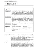
FIGURE 1-1. Infrared thermal imaging of tissue heating during radiofrequency ablation with a closed irrigation catheter. Power is delivered at 30 W to
blocks of porcine myocardium in a tissue bath. The surface of the tissue is just above the fluid level to permit thermal imaging of tissue and not the fluid.
Temperature scale (right) and a millimeter scale (top) are shown in each panel. A, Viewed from the surface, there is radial heating of the tissue from the
electrode. B, Tissue heating visualized in cross section. The electrode is partially submerged in the fluid bath and perpendicular to the upper edge of the
tissue. In both cases, very high tissue temperatures (>96°C) are achieved at 60 seconds because of the absence of fluid flow over the tissue surface.
were tested in a Langendorff-perfused canine heart preparation. Catheter ablation of the right bundle branch was
attempted at varying distances, and while delivered, power
was increased in a stepwise fashion. The RF power required
to block right bundle branch conduction increased exponentially with increasing distance from the catheter. At a
distance of 4 mm, most RF energy deliveries reached the
threshold of impedance rise before block was achieved.
When pulsatile flow was streamed past the ablation electrode, the power requirements to cause block increased
fourfold.12 Thus, the efficiency of heating diminished with
cooling from circulating blood, and small increases in distances from the ablation target corresponded with large
increases in ablation power requirements, emphasizing
the importance of optimal targeting for successful catheter
ablation.
1 n Biophysics of Radiofrequency Lesion Formation 5
50
P 0.0001
r 0.85
16
12
8
4
0.00
2.50
5.00
7.50
0
10.00
Distance (mm)
P 0.0001
r 0.89
8
Depth (mm)
60
40
A
10
20
70
Diameter (mm)
Temperature (°C)
80
6
4
2
0
B
0.5 1.0 1.5 2.0 2.5
0
0
0.5 1.0 1.5 2.0 2.5
Electrode radius (mm)
FIGURE 1-2. A, Radial temperature gradients measured during in vitro catheter ablation with source temperatures varying from 50° to 80°C. The tissue
temperature falls in an inverse proportion to distance. The dashed line represents the 50°C isothermal line. The point at which the radial temperature gradient crosses the 50°C isotherm determines the boundary of the lesion. A higher source temperature results in a greater lesion depth. B, Lesion depth and
diameter are compared to the electrode radius in temperature feedback power controlled radiofrequency ablation. A larger-diameter ablation electrode
results in higher power delivery and a proportional increase in lesion dimension. (From Haines DE, Watson DD, Verow AF. Electrode radius predicts lesion
radius during radiofrequency energy heating: validation of a proposed thermodynamic model. Circ Res. 1990;67:124–129. With permission.)
Sudden Impedance Rise
160
140
Temperature (°C)
In a uniform medium, the steady-state radial temperature gradient should continue to shift deeper into the
medium as the source temperature increases. A very high
source temperature, therefore, should theoretically yield a
very deep 50°C isotherm temperature and, in turn, very
large ablative lesions. Unfortunately, this process is limited in the biologic setting by the formation of coagulum
and char at the electrode-tissue interface if temperatures
exceed 100°C. At 100°C, blood literally begins to boil.
This can be observed in the clinical setting with generation of showers of microbubbles if tissue heating is
excessive.13 As the blood and tissue in contact with the
electrode catheter desiccate, the residue of denatured proteins adheres to the electrode surface. These substances
are electrically insulating and result in a smaller electrode
surface area available for electrical conduction. In turn, the
same magnitude of power is concentrated over a smaller
surface area, and the power density increases. With higher
power density, the heat production increases, and more
coagulum forms. Thus, in a positive-feedback fashion, the
electrode becomes completely encased in coagulum within
1 to 2 seconds. In a study testing ablation with a 2-mm-tip
electrode in vitro and in vivo, a measured temperature of
at least 100°C correlated closely with a sudden rise in electrical impedance (Fig. 1-3).14 Modern RF energy ablation
systems all have an automatic energy cutoff if a rapid rise
in electrical impedance is observed. Some experimenters
have described soft thrombus that accumulates when temperatures exceed 80°C.15 This is likely due to blood protein
denaturation and accumulation, but fortunately appears to
be more of a laboratory phenomenon than one observed in
the clinical setting. When high temperatures and sudden
rises in electrical impedance are observed, there is concern
about the accumulation of char and coagulum, with the
subsequent risk for char embolism. Anticoagulation and
antiplatelet therapies have been proposed as preventative
measures,16 but avoidance of excessive heating at the electrode-tissue interface remains the best strategy to avoid
this risk.
120
100
80
60
40
No impedance
rise
Impedance
rise
FIGURE 1-3. The association of measured electrode-tip temperature
and sudden rise in electrical impedance is shown in this study of radio
frequency catheter ablation with a 2-mm-tip ablation electrode in vitro
(blue circles) and in vivo (yellow squares). The peak temperature recorded
at the electrode-tissue interface is shown. Almost all ablations without a
sudden rise in electrical impedance had a peak temperature of 100°C or
less, whereas all but one ablation manifesting a sudden rise in electrical
impedance had peak temperatures of 100°C or more. (From Haines DE,
Verow AF. Observations on electrode-tissue interface temperature and effect on
electrical impedance during radiofrequency ablation of ventricular myocardium.
Circulation. 1990;82:1034–1038. With permission.)
Convective Cooling
The major thermodynamic factor opposing the transfer of
thermal energy to deeper tissue layers is convective cooling. Convection is the process whereby heat is distributed
through a medium rapidly by active mixing of that medium.
With the case of RF catheter ablation, the heat is produced
by resistive heating and transferred to deeper layers by
thermal conduction. Simultaneously, the heat is conducted
back into the circulating blood pool and metal electrode
tip. Because the blood is moving rapidly past the electrode
and over the endocardial surface, and because water (the
main constituent of blood) has a high heat capacity, a large
amount of the heat produced at the site of ablation can
6 I n Fundamental Concepts of Transcatheter Energy Applications
0.83 Amp
0.90 Amp
exceeded 100°C, resulting in sudden steam formation and
a steam pop. The clinical concern about “pop lesions” is
that sudden steam venting to the endocardial or epicardial surface (or both) can potentially cause perforation and
tamponade.20
The observation of increasing lesion size with ablationtip cooling holds true only so long as the ablation is not
power limited. If a level of power is used that is insufficient to overcome the heat lost by convection, the resulting
tissue heating may be inadequate. In this case, convective
cooling will dissipate a greater proportion of energy, and
less of the available RF energy will be converted into tissue
heat. The resulting lesion may be smaller than it would be
if there were no convective cooling. As power is increased
to a higher level, more energy will be converted to tissue
heat, and larger lesions will result. If power is unlimited
and temperature feedback power control is employed,
greater magnitudes of convective cooling will allow for
higher power levels and very large lesions. Thus, paradoxically in this situation, lesion size may be inversely related to
the electrode-tissue interface temperature if the ablation is
not power limited.21 However, if power level is fixed (most
commercial RF generators limit power delivery to 50 W
for use with these catheters), lesion size increases in proportion to electrode-tissue interface temperature even in
the setting of significant convective cooling (Fig. 1-5).22
1200
3
Lesion volume (mm )
be carried away by the blood. Convective cooling is such
an important factor that it dominates the thermodynamics of catheter ablation.17 Efficiency of energy coupling to
the tissue can be as low as 10%, depending on electrode
size, catheter stability, and position relative to intracavitary blood flow.18 Unstable, sliding catheter contact results
in significant tip cooling and decreased efficiency of tissue
heating.19 This is most often observed with ablation along
the tricuspid or mitral valve annuli.
Paradoxically, the convective cooling phenomenon has
been used to increase lesion size. As noted earlier, maximal
power delivery during RF ablation is limited by the occurrence of boiling and coagulum formation at the electrode
tip. However, if the tip is cooled, a higher magnitude of
power may be delivered without a sudden rise in electrical
impedance. The higher magnitude of power increases the
depth of direct resistive heating and, in turn, increases the
radius of the effective heat source. In addition, higher temperatures are achieved 3 to 4 mm below the surface, and the
entire radial temperature curve is shifted to a higher temperature over greater tissue depths. The result is a greater
50°C isotherm radius and a greater depth and diameter of
the lesion. Nakagawa demonstrated this phenomenon in
a blood-superfused exposed thigh muscle preparation. In
this study, intramural tissue temperatures 3.5 mm from
the surface averaged 95°C with an irrigated-tip catheter despite a mean electrode-tissue interface temperature
of 69°C. Lesion depths were 9.9 mm compared with 6.1
mm in a comparison group of temperature-feedback power
control delivery and no electrode irrigation (Fig. 1-4). An
important finding of this study was that 6 of 75 lesions
had a sudden rise in electrical impedance associated with
an audible pop. In these cases, the intramural temperature
1000
Group 1
Group 2
800
600
400
200
0
Current
40
50
66 volts
36
44
27
38 Tissue temp
(3.5 mm depth)
Interface temp
36
30°C
102
100°C
73 67
53
27
50°C
41
80
Tissue temp
(7.0 mm depth)
Irrigation
30°C
10 sec
FIGURE 1-4. Current, voltage, and temperatures measured during radio
frequency catheter ablation with a perfused-tip electrode catheter in a canine
exposed thigh muscle preparation are shown. Temperatures were recorded
within the electrode, at the electrode-tissue interface, and within the muscle
below the ablation catheter at depths of 3.5 and 7 mm. Because the electrodetissue interface is actively cooled, high current and voltage levels can be
employed. This results in an increased depth of direct resistive heating and
superheating of the tissue below the surface of ablation. The peak temperature
in this example at a depth of 3.5 mm was 102°C, and at 7 mm was 67°C, indicating that the 50°C isotherm defining the lesion border was significantly deeper
than 7 mm. (From Nakagawa H, Yamanashi WS, Pitha JV, et al. Comparison of
in vivo tissue temperature profile and lesion geometry for radiofrequency ablation
with a saline-irrigated electrode versus temperature control in a canine thigh muscle
preparation. Circulation. 1995;91:2264–2273. With permission.)
1200
3
Electrode
temp
70
Tip temperature (°C)
Lesion volume (mm )
Voltage
60
1000
Group 1
Group 2
800
600
400
200
0
0
10
20
30
40
50
60
70
80
Power (W)
FIGURE 1-5. Temperatures measured at the tip of the electrode dur-
ing experimental radiofrequency ablation and power are compared to
the resulting lesion volume in this study. A maximal power of 70 W was
employed. If lesion creation was not power limited (group 1), the lesion
volume was a function of the delivered power. But if lesion production
was limited by the 70-W available power maximum (group 2), the temperature measured at the electrode tip correlated with lesion size. (From
Petersen HH, Chen X, Pietersen A, et al. Lesion dimensions during temperature-controlled radiofrequency catheter ablation of left ventricular porcine
myocardium: impact of ablation site, electrode size, and convective cooling.
Circulation. 1999;99:319–325. With permission.)
1 n Biophysics of Radiofrequency Lesion Formation 7
Electrode-tip cooling can be achieved passively or
actively. Passive tip cooling occurs when the circulating
blood flow cools the mass of the ablation electrode and
cools the electrode-tissue interface. This can be enhanced
by use of a large ablation electrode.23 Active tip cooling
can be realized with a closed or open perfused-tip system. In each case, circulating saline from an infusion pump
actively cools the electrode tip. One design recirculates
the saline through a return port, and the opposing design
infuses the saline through weep holes in the electrode into
the bloodstream. Both designs are effective and result in
larger lesions and greater procedure efficacy than standard
RF catheter ablation. Theoretical advantages and disadvantages of open perfusion versus closed perfusion catheter designs are claimed by device manufacturers and their
spokespeople, but the lesions produced and the clinical
efficacy and safety profiles of these competing designs are
very comparable.24–27 The tip cooling or perfusion has the
apparent advantage of reducing the prevalence of coagulum and char formation. However, because the peak tissue temperature is shifted from the endocardial surface to
deeper intramyocardial layers, there is the risk for excessive
intramural heating and pop lesions. The challenge for the
clinician lies with the fact that with varying degrees of convective cooling, there is no reliable method for monitoring
whether tissue heating is inadequate, optimal, or excessive.
Cooling at the electrode-tissue interface limits the value of
temperature monitoring to prevent excess power delivery
and steam pops. With closed irrigation catheters, there is
some value in the use of temperature feedback power control. In this case, target temperatures of 42° to 45°C have
been empirically determined to optimize energy delivery.27,28 If the ablation is power limited and the target temperature has not been reached, one may assume that the
combination of passive cooling (from sliding or bouncing
catheter-tissue contact) and active cooling is dissipating
too much energy to allow for adequate tissue heating. In
this situation, active electrode cooling can be held, and the
operator can depend on passive cooling alone.
Catheter orientation will affect lesion size and geometry.
Perpendicular catheter orientation results in less electrode
surface area in contact with the tissue and more surface area
in contact with the circulating blood pool. Parallel catheter
orientation provides more electrode-tissue contact. With
unrestricted power delivery, the parallel orientation should
produce the larger lesion. In perfused-tip catheters, parallel orientation also results in more active tissue cooling and
smaller lesion sizes than a perpendicular orientation.29 The
resultant interplay among active cooling, passive cooling,
and power availability or limitation determines whether
the lesions will be larger or smaller in these varying conditions. If perfused-tip catheters are positioned in a parallel orientation with greater tissue cooling, the lesions are
smaller in vitro because of diminished efficiency of energy
delivery. The effects of catheter orientation are less important with 4- or 5-mm-tip catheters but become more dominant when 8- or 10-mm tips are employed.
Since its inception, conventional RF ablation has been
characterized by its excellent safety profile. This undoubtedly has been due to the relatively small size of the lesions. As
new catheter technologies designed to increase the depth of
the ablative lesion have been employed, it is not surprising
that complications due to collateral injury have increased.
For example, left atrial ablation with cooled ablation catheters and high-intensity, focused ultrasound has resulted in
cases of esophageal injury, perforation, and death. Despite
the routine positioning of ablation catheters in close proximity to coronary arteries, there has been a dearth of coronary arterial complications with this procedure. The blood
flow within the coronary artery is rapid, and the zone of
tissue around the artery is convectively cooled by this blood
flow. Fuller and Wood tested the effect of flow rate through
a marginal artery of Langendorff perfused rabbit hearts. 30
RF ablation with an electrode-tissue interface temperature
of 60° or 80°C was performed on the right ventricular free
wall with two lesions straddling the artery, and conduction
through this region was monitored. They observed that arterial flow rates as low as 1 mL/minute through these small
(0.34 ± 0.1 mm diameter) arteries prevented complete transmural ablation and conduction block. This heat-sink effect
is especially protective of the vascular endothelium. With
higher power output of new ablation technologies, however, the convective cooling of the arterial flow may be overwhelmed, and there may be increased risk for vascular injury.
With greater destructive power possible, operators need to
be mindful to use only enough power to achieve complete
ablation of the targeted tissue in order to safely accomplish
the goal of arrhythmia ablation.
Electrical Current Distribution
Catheter ablation depends on the passage of RF electrical current through tissue. Tissue contact can be assessed
by measuring baseline system impedance. In one clinical
study, a very small (10 μA) current was passed through the
ablation catheter, and the efficiency of heating was measured to assess tissue contact. A significant positive correlation between preablation impedance and heating efficiency
was observed. As tissue is heated, there is a temperaturedependent fall in the electrical impedance.31,32 A significant
correlation is also observed between heating efficiency and
the maximal drop in impedance during energy delivery.
When electrode-tissue interface temperature monitoring is
unreliable because of high-magnitude convective cooling,
the slow impedance drop is a useful indicator that tissue
heating is occurring. With the progressive fall in impedance during ablation, the delivered current increases along
with tissue heating. If no impedance drop is observed,
catheter repositioning is warranted.33,34
Because the magnitude of tissue heating is determined by
the current density, the distribution of RF field around the
electrodes in unipolar, bipolar, or phased RF energy delivery
will determine the distribution of tissue heating. If energy is
delivered in a unipolar fashion in a uniform medium from a
spherical electrode to an indifferent electrode with infinite
surface area, current density around the electrode should
be entirely uniform. As geometries and tissue properties
change, heating becomes nonuniform. Standard 4-mm
electrode tips are small enough so that heating around the
tip is fairly evenly distributed, even with varying tip contact angle to the tissue. One study showed that temperature monitoring with a thermistor located at the tip of a
4-mm electrode underestimated the peak electrode-tissue
interface temperature recorded from multiple temperature
8 I n Fundamental Concepts of Transcatheter Energy Applications
sensors distributed around the electrode in only 4% of
the applications. In RF applications where high power
was employed and a sudden rise in electrical impedance
occurred, the peak temperature recorded from the electrode
tip was below 95°C in only one of 17 cases.35 However,
present-day electrode geometries vary considerably. The
presence of fat will alter both electrical and thermal conductivity. Epicardial ablation over fat will result in minimal
ablation of the underlying myocardium. Conversely, ablation of tissue insulated by fat outside of the ablation target will produce an “oven” effect, with higher temperatures
for longer durations after cessation of energy delivery.36
Also, tissue characteristics and placements of indifferent
electrodes will affect tissue heating. Surface temperature
recordings routinely underestimate peak subendocardial
tissue temperatures. For that reason, most operators limit
ablation temperatures to 60° or 70°C during ablation with
noncooled catheters.
Dispersive Electrode
The power dissipated in the complete circuit is proportional to the voltage drop and impedance for each part of
the series circuit. The impedance of the ablation system
and transmission lines is low, so there is little energy dissipation outside the body. The site of greatest impedance,
voltage drop, and power dissipation is at the electrodetissue interface (Fig. 1-6). However, most power is consumed with electrical conduction through the body and
blood pool and into the dispersive electrode. In fact, only a
fraction of the total delivered power actually is deposited in
A
Blood pool
RF generator
Myocardial tissue
Cables and
catheter
Body and skin
electrode
50 W
50 W
RF generator
RF generator
5 watts
delivered
5.6 watts
delivered
82 Ω
82 Ω
165 Ω
165 Ω
45 Ω
Skin patch
B
Total 100 Ω
C
Skin
patch
45 Ω
45 Ω
Skin patch
Total 88 Ω
FIGURE 1-6. “Circuit diagrams” for radiofrequency (RF) ablation. A, From the RF generator, the cables and catheter present minimal resistance. The
myocardial tissue and blood pool represent resistance circuits in parallel from the distal electrode. The return path from the ablation electrode to the generator comprises the patient’s body and dispersive electrode in series. B, Hypothetical resistances for RF ablation circuit path. The resistance of the blood
pool is about half that of the myocardial tissue. In this situation, for 50 W of energy delivered to the catheter, only 5 W is deposited in the myocardial tissue because of shunting of current through the lower resistance blood pool and power loss in the return path. C, Effect of adding a second dispersive skin
electrode to the circuit. Assuming that the impedance of each dispersive electrode is 45 ohms and the generator voltage is constant, the total ablation circuit
impedance is decreased by 12%. This allows for greater current delivery through the circuit and a proportional increase in power delivered to the tissue.
1 n Biophysics of Radiofrequency Lesion Formation 9
the myocardial tissue (Fig. 1-6). The return path of current
to the indifferent electrode will certainly affect the current
density close to that indifferent electrode, but its placement
anterior versus posterior, and high versus low on the torso,
has only a small effect on the distribution of RF current
field lines within millimeters of the electrode. Therefore,
lesion geometry should not be affected greatly by dispersive electrode placement. However, the proportion of RF
energy contributing to lesion formation will be reduced if
a greater proportion of that energy is dissipated in a long
return pathway to the dispersive electrode. When the ablation is power limited, it is advantageous to minimize the
proportion of energy that is dissipated along the current
pathway at sites other than the electrode-tissue interface
to achieve the greatest magnitude of tissue heating and the
largest lesion. In an experiment that tested placement of the
dispersive electrode directly opposite the ablation electrode
versus at a more remote site, lesion depth was increased
26% with optimal placement.37 Vigorous skin preparation
to minimize impedance at the skin interface with the dispersive electrode, closer placement of the dispersive electrode to the heart, and use of multiple dispersive electrodes
to increase skin contact area will all increase tissue heating in a power-limited energy delivery. Nath and associates reported that in the setting of a system impedance
higher than 100 ohms, adding a second dispersive electrode
increased the peak electrode-tip temperature during clinical catheter ablation (Fig. 1-7).38
electrodes. The less symmetrical the electrode design (such
as if found with long electrodes), the greater the degree of
nonuniform heating. McRury and coworkers tested ablation
with electrodes with 12.5-mm length.39 They found that a
centrally placed temperature sensor significantly underestimated the peak electrode-tissue interface temperature. Finite
element analysis demonstrated a concentration of electrical
current at the each of the electrode edges (Fig. 1-8). When
dual thermocouples were placed on the edge of the electrode,
the risk for coagulum formation and impedance rise was significantly reduced during ablation testing in vivo.
150
P0.001
Current
(I)
Voltage
(V)
Impedance
()
80
P0.001
0.80
P0.001
Temperature
(°C)
80
P0.05
60
100
60
0.60
40
0.40
20
0.20
20
0
0.00
0
40
50
0
Single dispersive electrode
Double dispersive electrode
FIGURE 1-7. Impedance, voltage, current, and catheter-tip temperature
Edge Effect
Electrical field lines are not entirely uniform around the tip
of a unipolar ablation electrode. The distribution of field
lines from an electrode source is affected by changes in electrode geometry. At points of geometric transition, the field
lines become more concentrated. This so-called edge effect
can result in significant nonuniformity of heating around
Catheter
body
Insulating UV
adhesive
readings during radiofrequency catheter ablation in a subset of patients
with a baseline system impedance of more than 100 ohms. Ablations using
a single dispersive electrode were compared with those using a double dispersive electrode. A lower system impedance was observed with addition
of the second dispersive patch. This resulted in a greater current delivery
and higher temperatures measured at the electrode-tissue interface. (From
Nath S, DiMarco JP, Gallop RG, et al. Effects of dispersive electrode position
and surface area on electrical parameters and temperature during radiofrequency
catheter ablation. Am J Cardiol. 1996;77:765–767. With permission.)
Ablation
electrode
coil
Insulating UV
adhesive
Catheter
body
161
145
Blood
12.5 mm
130
114
99.0
83.5
Tissue
68.0
52.5
37.0
FIGURE 1-8. Steady-state temperature distribution derived from a finite element analysis of radiofrequency ablation with a 12-mm long coil electrode.
In this analysis, the electrode temperature at the center of the electrode was maintained at 71°C. The legend of temperatures is shown at the right of the
graph and ranges from the physiologic normal (violet = 37°C) to the maximal tissue temperature (red = 161°C) located below the electrode edges. There
is a significant gradient of heating between the peak temperatures at the electrode edges and the center of the electrode. UV, ultraviolet. (From McRury
ID, Panescu D, Mitchell MA, Haines DE. Nonuniform heating during radiofrequency catheter ablation with long electrodes: monitoring the edge effect. Circulation.
1997;96:4057–4064. With permission.)